Isolation, Characterization, and Differentiation Potential of Canine Adipose-Derived Stem Cells
←
→
Page content transcription
If your browser does not render page correctly, please read the page content below
Cell Transplantation, Vol. 19, pp. 279–289, 2010 0963-6897/10 $90.00 + .00
Printed in the USA. All rights reserved. DOI: 10.3727/096368909X481764
Copyright 2010 Cognizant Comm. Corp. E-ISSN 1555-3892
www.cognizantcommunication.com
Isolation, Characterization, and Differentiation Potential
of Canine Adipose-Derived Stem Cells
N. M. Vieira,* V. Brandalise,* E. Zucconi,* M. Secco,* B. E. Strauss,† and M. Zatz*
*Human Genome Research Center, Biosciences Institute, University of São Paulo, São Paulo, Brazil
†Viral Vector Group, Heart Institute, InCor, University of São Paulo, São Paulo, Brazil
Adipose tissue may represent a potential source of adult stem cells for tissue engineering applications in
veterinary medicine. It can be obtained in large quantities, under local anesthesia, and with minimal discom-
fort. In this study, canine adipose tissue was obtained by biopsy from subcutaneous adipose tissue or by
suction-assisted lipectomy (i.e., liposuction). Adipose tissue was processed to obtain a fibroblast-like popula-
tion of cells similar to human adipose-derived stem cells (hASCs). These canine adipose-derived stem cells
(cASCs) can be maintained in vitro for extended periods with stable population doubling and low levels of
senescence. Immunofluorescence and flow cytometry show that the majority of cASCs are of mesodermal
or mesenchymal origin. cASCs are able to differentiate in vitro into adipogenic, chondrogenic, myogenic,
and osteogenic cells in the presence of lineage-specific induction factors. In conclusion, like human lipoaspir-
ate, canine adipose tissue may also contain multipotent cells and represent an important stem cell source
both for veterinary cell therapy as well as preclinical studies.
Key words: Canine adipose-derived stem cells; Tissue engineering; Veterinary cell therapy
INTRODUCTION Successful transplantation of canine adipose-derived
stem cells (cACSs) in dogs was reported by Li et al.
A promising application in the emerging field of vet- (12) and Black et al. (1). However, these manuscripts
erinary regenerative medicine and surgery is cell ther- lacked the full characterization of the administered cell
apy, rendering the isolation and characterization of stem population. Here we report the isolation, characteriza-
cells from a variety of sources areas of great interest. tion, and multilineage differentiation potential of cASCs
An abundant and accessible source of stem cells is from subcutaneous adipose tissue by liposuction and bi-
adipose tissue. These cells, called adipose-derived stro- opsy procedures.
mal cells (ASCs), are fibroblast-like cells capable of
multipotential differentiation, which have been found in MATERIALS AND METHODS
different species (4,27,29, 35). Several groups have All experimental protocols were approved by the eth-
demonstrated that human mesenchymal cells within the ics committee on animal use from the Institute of Biosci-
stromal-vascular fraction (SVF) of subcutaneous adipose ences, University of São Paulo. For this study, adipose
tissue [processed lipoaspirate (PLA) cells] are capable tissue was collected from normal golden retriever dogs
of differentiation in multiple lineages, including myo- from the Brazilian Colony of Golden Retriever Muscular
cytes, in the presence of lineage-specific inductive me- Dystrophy, Faculty of Veterinary Medicine and Zoo-
dia (2,5,8–10,15,16,19,20,22,23,25,34,35). tecny, University of São Paulo. Subcutaneous adipose
In humans, ASCs for autologous transplantation are tissue was collected from the area over the dorsal gluteal
isolated relatively quickly from adipose tissue by colla- muscles of 10 dogs (aged 4 months to 4 years).
genase digestion (6). We have recently shown that ASCs
from human subcutaneous fat were able to differentiate Adipose Tissue Harvesting
in adipogenic, osteogenic, chondrogenic, and myogenic Dogs were sedated upon intramuscular (IM) injection
lineages and produce human muscle proteins in vitro with meperidine (2 mg/kg) and acetylpromazine (0.05
and in vivo (30,31). mg/kg). The area over the dorsal gluteal muscles was
Received March 27, 2009; final acceptance December 2, 2009. Online prepub date: December 8, 2009.
Address correspondence to Dr. Mayana Zatz, Ph.D., Human Genome Research Center, Institute of Biosciences, University of São Paulo, Rua do
Matão, n.106 Cidade Universitária, São Paulo-SP, Brasil-CEP: 05508-090. Tel/Fax: (55) (11) 3091-7966; E-mail: mayazatz@usp.br
279
280                                                                                                               VIEIRA ET AL.

asceptically prepared, and skin and subcutaneous tissues           debris and seeded in tissue culture plates (NUNC) at
were desensitized by local infiltration of 2% lidocaine            1,000–3,500 cells/cm2 in DMEM-HG 10% FBS. Cul-
(Fig. 1A). A 0.5–1.0-cm incision was made parallel to              tures were washed with PBS 24–48 h after plating to
the vertebral column. The liposuction procedure was                remove unattached cells and fed with fresh media.
performed by injecting infiltrate containing the vasocon-             The cultures were maintained at 37°C with 5% CO2
strictor epinephrine. Then adipose tissue was removed              in growth media (GM-DMEM-HG 10% FBS). When
from the subcutaneous space by means of blunt-tip hol-             they achieved about 70% confluence, the cells were
low cannula attached to a syringe at negative pressure             trypsinised (0.025%, Invitrogen) and plated at a density
(Fig. 1B). About 15 ml of adipose tissue was harvested             of 5,000/cm2. Cultures were passaged repeatedly after
over the superficial gluteal fascia for immediate cASC             achieving a density of 70–80%. The remaining cells
isolation and the skin incision apposed with nylon su-             were cryopreserved in cryopreservation media (10% di-
tures (Fig. 1C). Adipose tissue biopsies were performed            methylsulfoxide, 10% DMEM-HG, 80% FBS), frozen at
under local anesthesia. A 1–2-cm incision was made and             −80°C in an isopropanol-jacketed closed container, and
the subcutaneous adipose tissue was collected (Fig. 1D)            stored in liquid nitrogen the next day.
and the incision was closed with nylon sutures (Fig .1E).
                                                                   Multilineage Differentiation
cASC Isolation and Expansion                                           Cells were analyzed for their capacity to differentiate
   Cells were isolated using modified methods pre-                 into adipogenic, chondrogenic, osteogenic, and myo-
viously described (7). Briefly, the adipose tissue was             genic lineages as described in Zuk et al. (35).
washed extensively with equal volumes of PBS contain-                  Adipogenic Differentiation. Subconfluent cells were
ing antibiotics (100 U/ml of penicillin and 100 g/ml of            cultured in GM supplemented with 1 µM dexametha-
streptomycin). The infranatant containing hemopoietic              sone (Sigma), 500 µM 3-isobutyl-1-methyl-xanthine
cells suspended in PBS was removed. Then the tissue                (IBMX, Sigma), 60 µM indomethacin (Sigma), and 5
was dissociated with 0.075% collagenase (Sigma) for 15             µg/ml insulin (Sigma). Adipogenic differentiation was
min. Enzyme activity was neutralized with Dulbecco’s               confirmed on day 21 by intracellular accumulation of
modified Eagle’s media- high glucose (DMEM-HG;                     lipid-rich vacuoles stainable with Oil Red O (Sigma).
Gibco) containing 10% FBS (Gibco). The infranatant                 For the Oil Red O stain, cells were fixed with 4% para-
was centrifuged at 1200 × g for 5 min to pellet the cells.         formaldehyde for 30 min, washed, and stained with a
The cells from the pellet SVF were filtered to remove              working solution of 0.16% Oil Red O for 20 min.

Figure 1. Adipose tissue harvest. (A) The area over the dorsal gluteal muscles prepared for the procedure. (B) Liposuction proce-
dure. (C) Skin incision after the liposuction. (D) Subcutaneous adipose tissue biopsy. (E) Skin incision after the biopsy.
CANINE ADIPOSE-DERIVED STEM CELLS 281
Chondrogenic Differentiation. Subconfluent cells were temperature. After incubation, cells were washed three
cultured in chondrogenic differentiation medium con- times with PBS and resuspended in 0.25 ml of cold PBS.
sisting of DMEM-low glucose supplemented with 100 Cell viability was accessed with Guava ViaCount re-
nM dexamethasone, 50 µM ascorbic acid-2 phosphate agent (Guava Technologies).
(Sigma), 1 mM sodium pyruvate (Gibco), 10 ng/ml cASCs were incubated with the following primary
TGF-β1 (R&D Systems), and 1% ITS-Premix (Becton antibodies: CD13-PE, CD29-PECy5, CD31-PE, CD34-
Dickinson). Medium was changed every 3–4 days, and PE, CD44-FITC, CD45, CD73, CD90-PE, CD105 e
cells were fixed on day 21 with 4% paraformaldehyde CD117-PECy5 (Becton Dickinson). The following anti-
(PFA). Chondrogenesis was demonstrated by staining bodies have been raised against human cells: CD13,
with toluidine blue and immunofluorescence using anti- CD71, and CD105. Unconjugated markers were treated
collagen type II antibody (1:100, Abcam). with anti-mouse PE secondary antibody (Guava Tech-
Osteogenic Differentiation. To promote osteogenic nologies).
differentiation, subconfluent cells were treated with GM Flow cytometer settings were established using un-
supplemented with 50 µM ascorbate-2 phosphate, 10 stained cells. Cells were gated by forward scatter to
mM β-glycerophosphate (Sigma), and 0.1 µM dexa- eliminate debris. To eliminate the possible autofluores-
methasone for 21 days. Osteogenesis was demonstrated cence of cASCs, we removed the contribution of uns-
by accumulation of mineralized calcium phosphate as- tained cells in the measurement channel. A minimum of
sessed by von Kossa stain. Briefly, cells were stained 10,000 events was counted for each analysis.
with 1% silver nitrate (Sigma) for 45 min under ultravio-
RNA Isolation and Reverse Transcriptase-Polymerase
let light, followed by 3% sodium thiosulphate (Sigma)
Chain Reaction (RT-PCR)
for 5 min, and then counterstained with van Gieson.
Total RNA was harvested from cultured cells using
Myogenic Differentiation. For myogenic differentia- Tryzol (Invitrogen) following manufacturer’s instruc-
tion, cASCs cells were cultured in GM supplemented tions. The RNA was treated with DNase (Invitrogen). A
with 0.1 µM dexamethasone (Sigma), 50 µM hydrocor- total of 1 µg of total RNA was reverse-transcribed with
tisone (Sigma), and 5% horse serum (Gibco) for 45 SuperScriptTM III First-Strand Synthesis System (In-
days. After that cells were labeled with anti-myosin (1: vitrogen). All amplifications were performed in an MJ
100, Sigma). Research PTC-200 thermocycler (MJ Research) for 24
cycles after the initial 2-min denaturation at 94°C. The
Immunofluorescence
PCR primers are listed in Table 1. The PCR products
Cells were fixed in 4% paraformaldehyde in PBS for were separated on 6% polyacrylamide gel by electropho-
20 min at 4°C, permeabilized in 0.05% Triton X-100 in resis, stained with ethidium bromide, and visualized un-
PBS for 5 min. Nonspecific binding was blocked with der UV light. Digital images were captured with Image-
10% FBS in PBS for 1 h at room temperature. Cells Quant (GE Healthcare).
were incubated with primary antibody (1:100) overnight
at 4°C. After several washes, cells were incubated with cASCs Transduction With Lentivirus Vector
secondary (1:100, Sigma) antibodies against mouse IgG The visualization of cASCs cells for in vitro and in
tagged with Cyanine 3 (Cy3; red) for 2 h at room tem- vivo studies can be improved if done with GFP-positive
perature. Slides were counterstained with DAPI (4′-6- cells. For this purpose we transducted cASCs with GFP
diamidino-2-phenylindole, Sigma). All images in the lentivirus.
same set (samples and controls) were obtained using the Supernatant containing the FUGW lentivirus (13)
same photographic parameters of exposition and speed. was produced as described previously by Strauss et al.
Images were captured using the Axiovision 3.0 image (26) and concentrated by ultracentrifugation. Undiffer-
analysis system (Carl Zeiss). entiated cASCs at passage 2 were incubated at 37°C, in
a six-well plate (Nunc), using a minimal volume of GM
Flow Cytometry in the presence of vector particles (20 PFU/cell) and 8
Cells were evaluated for cell surface protein expres- µg/ml Polybrene (Sigma). After 4 h, 2 ml of GM was
sion using flow cytometry. The flow cytometry was per- added and the media was changed the next day.
formed on Guava EasyCyte System (Guava Technolo-
gies) using a blue laser (488 nm). Cells were pelleted, Karyotype Analysis
resuspended in PBS at a concentration of 1 × 10⁵ cells/ For evaluation of any chromosomal abnormality at
µl, and stained with saturating concentration of antibod- latter passages, chromosome preparations were per-
ies. Cells were incubated in the dark for 45 min at room formed in cASC cultures. Briefly, metaphase cells were282 VIEIRA ET AL.
Table 1. PCR Primers
Amplicon Ann.
Markers/Gene Primer Sequence (5′–3′) Size Temp. (°C) Reference
Myogenic
MyoD Forward GACGGCATGATGGACTACAG 118 60
Reverse ACACCGCAGCACTCTTCC
Dystrophin Forward AAACACAGTGGTAGCCCACAAGAT 116 60
Reverse TGGTGACAGCCTGTGAAATC
Myogenin Forward GACGGCATGATGGACTACAG 102 60
Reverse ACACCGCAGCACTCTTCC
Adipocytes
FABP4 Forward ATCAGTGTAAACGGGGATGTG 117 60 17
Reverse GACTTTTCTGTCATCCGCAGTA
Leptin Forward CTATCTGTCCTGTGTTGAAGCTG 102 60 17
Reverse GTGTGTGAAATGTCATTGATCCTG
LPL Forward ACACATTCACAAGAGGGTCAC 132 60 17
Reverse CTCTGCAATCACACGGATG
Chondrocytes
COL2A Forward GAAACTCTGCCACCCTGAATG 156 64 17
Reverse GCTCCACCAGTTCTTCTTGG
SOX9 Forward GCTCGCAGTACGACTACACTGAC 101 60 17
Reverse GTTCATGTAGGTGAAGGTGGAG
Aggrecan Forward ATCAACAGTGCTTACCAAGACA 122 58 17
Reverse ATAACCTCACAGCGATAGATCC
Osteocytes
Osteopontin Forward CATATGATGGCCGAGGTGATAG 114 60
Reverse CAAGTGATGTGAAGTCCTCCTC
COL1A1 Forward GTAGACACCACCCTCAAGAGC 118 62
Reverse CCAGTCGGAGTGGCACAT
BSP Forward TTGCTCAGCATTTTGGGAAT 295 60
Reverse AACGTGGCCGATACTTAAAGAC
Housekeeping
GAPDH Forward CCATCTTCCAGGAGCGAGAT 97 60
Reverse TTCTCCATGGTGGTGAAGAC
arrested with 0.1 µg/ml colchicine (Sigma) for 20 min. are easy to expand in vitro and show a fibroblast-like
Then, cASCs were detached from cultures flasks using morphology, consistent with that of human ASCs (Fig.
TrypLE (Gibco), resuspended in a hypotonic solution 2A–D). At both early and late passages, cells main-
(0.075M KCl), and incubated for 20 min at 37°C. Cells tained a diploid karyotype of 78 chromosomes (Fig. 2E).
were pelleted at 1000 rpm for 10 min and fixed by wash- cASCs from four unrelated dogs were characterized
ing three times in methanol/glacial acetic acid (3:1). by flow cytometry for the expression of 10 cell surface
Chromosome spreads were obtained by pipetting sus- proteins (CD13, CD29, CD31, CD34, CD44, CD45,
pension drops onto clean glass slides and air dried. The CD73, CD90, CD105, and CD117). Cell viability was
best metaphases were captured with Axioplan 2 micro- above 96% by Guava ViaCount reagent (Guava Tech-
scope (Zeiss) and analyzed with Ikaros 3 software nologies).
(Zeiss). At passage 4, the majority of cASCs expressed
CD44, CD29 (β1 integrin) and CD90 (Thy1) adhesion
RESULTS
molecules. Other markers, including CD14, CD34,
Characterization of cASCs CD45, and CD117, were consistently absent or ex-
cASC cultures were maintained in DMEM supple- pressed in few cells (Fig. 3). Interestingly, CD13,
mented with 10% FBS. Supplementation with FBS has CD105, and CD73, known to be positively expressed in
been shown to be important for human ASC attachment human ASCs, were negative in the canine ASC popula-
and proliferation in vitro (35). We observed that cASCs tion, which might be explained by the nonspecific stain-CANINE ADIPOSE-DERIVED STEM CELLS 283
ing of human antibodies in canine cells. As surface the mesenchymal nature of the isolated cells and their
markers are not sufficient for the identification or defini- multipotent potential.
tion of mesenchymal stem cell (MSC), cASCs were sub-
jected to differentiation studies for further confirmation Adipogenesis. cASCs showed a rounder shape after
of their MSC property. 7 days in adipogenic medium. Two weeks after initial
The plasticity of cASCs was assessed after lineage induction, the adipogenic differentiation was confirmed
induction. Myogenic, adipogenic, chondrogenic, and os- by Oil Red O staining of lipid droplets present through-
teogenic differentiation was demonstrated by the expres- out the cytoplasm (Fig. 4A). Expression of FABP4 and
sion of myogenic markers (myosin), lipid vacuoles, mu- LPL was seen only in adipo-induced cells (Fig. 5B). On
copolysaccharide-rich extracellular matrix, and calcium the other hand, basal level of leptin mRNA was ob-
deposits, respectively (Fig. 4) and by the expression of served in noninduced control cells, and the expression
tissue-specific mRNAs (Fig. 5). These results confirmed level was increased following adipogenic induction.
Figure 2. Typical morphology of cASCs. (A) Forty-five minutes after the establishment of the
culture. Some cells remain in the supernatant. Scale bar: 200 µm. (B) cASCs at passage 3. Scale
bar: 200 µm. (C) cASCs at passage 4: cASCs morphology is similar to that found in human ASCs.
Scale bar: 100 µm. (D) High-density ASCs culture at passage 4. Scale bar: 200 µm. (E) Karyotype
of cASC cell lineage after 10 passages, showing an euploid number of chromosomes.284 VIEIRA ET AL. Figure 3. Immunophenotyping of ASCs at passage 4. Values represent the mean percentage of positively stained cells as analyzed by flow cytometry. Graphs show forward scatter versus fluorescence intensity of the indicated antigen. Osteogenesis. cASCs exposed to osteogenic me- Chondrogenic Differentiation. After 21 days cul- dium exhibited changes in cell morphology after 5 tured in chondrogenic medium, cASCs cells were days in culture, showing a polygonal form. Mineral- stained with toluidine blue, showing the typical metach- ized nodular structures appeared in 1 or 2 weeks and romasia of cartilage. Chondrogenic differentiation was were assessed by von Kossa Stain, which localized demonstrated by the mucopolyssaccharide-rich extracel- the calcium deposits (Fig. 4C). Expression of osteo- lular matrix (Fig. 4E, G). Chondrogenic treatment re- pontin, COL1A1, and BSP was observed only in in- sulted in specific expression of COL2A, SOX9, and ag- duced cells, with no basal expression in control cells grecan, all of which were undetected or had basal (Fig. 5C). expression in noninduced cells (Fig. 5D).
CANINE ADIPOSE-DERIVED STEM CELLS                                                                                 285

            Figure 4. Differentiation potential of cASCs at passage 4. (A) The adipogenic differentiation was
            detected by the formation of intracytoplasmic lipid droplets stained with Oil Red O. Scale bar: 200
            µm. (B) Undifferentiated ASCs stained with Oil Red O. Scale bar: 200 µm. (C) cASCs after
            induction with adipogenic media still show GFP expression. Scale bar: 200 µm. (D) Osteogenic
            differentiation was demonstrated by calcium deposition shown by von Kossa stain. Scale bar: 200
            µm. (E) Undifferentiated ASCs stained with Von Kossa. Scale bar: 200 µm. (F) cASCs after
            induction with osteogenic media still show GFP expression. Scale bar: 200 µm. (G) Chondrogenic
            differentiation in monolayer culture was demonstrated by staining with toluidine blue. Scale bar:
            200 µm. (H) Undifferentiated ASCs stained with toluidine blue. Scale bar: 200 µm. (I) cASCs
            after induction with chondrogenic media still show GFP expression. Scale bar: 200 µm. (J) Chon-
            drogenic differentiated cells labeled with anti-collagen type II antibody. Scale bar, 200 µm. (K)
            Undifferentiated ASCs labeled with anti-collagen type II antibody. Scale bar: 200 µm. (L) Myo-
            genic differentiation was assessed by immunofluorescence. Induced cells were labeled with anti-
            myosin monoclonal antibody. Scale bar: 50 µm. (M) Undifferentiated ASCs labeled with anti-
            human myosin monoclonal antibody. Scale bar: 50 µm. (N) cASCs after induction with myogenic
            media still show GFP expression. Scale bar: 50 µm.
286 VIEIRA ET AL.
Figure 5. mRNA expression of specific differentiation markers. (A) Myogenic markers: myogenin
(Myog), dystrophin (Dyst) and MyoD. (B) Adipogenic markers; FABP4, leptil, and LPL. (C)
Osteogenic markers: osteopontin (OPN), COL1A1, bone sialoprotein (BSP). (D) Chondrogenic
markers; COL2A, SOX9, and aggrecan (AGC). The expression of glyceraldehyde-3-phosphate
dehydrogenase (GAPDH) was used as reference for evaluating the quality of mRNA.
Myogenesis. After 10 days in myogenic medium, GFP Transduction of cASCs
cASCs formed multinucleated structures. Controls main-
tained only with GM did not contain any multinucleated Transgene expression was examined by flow cy-
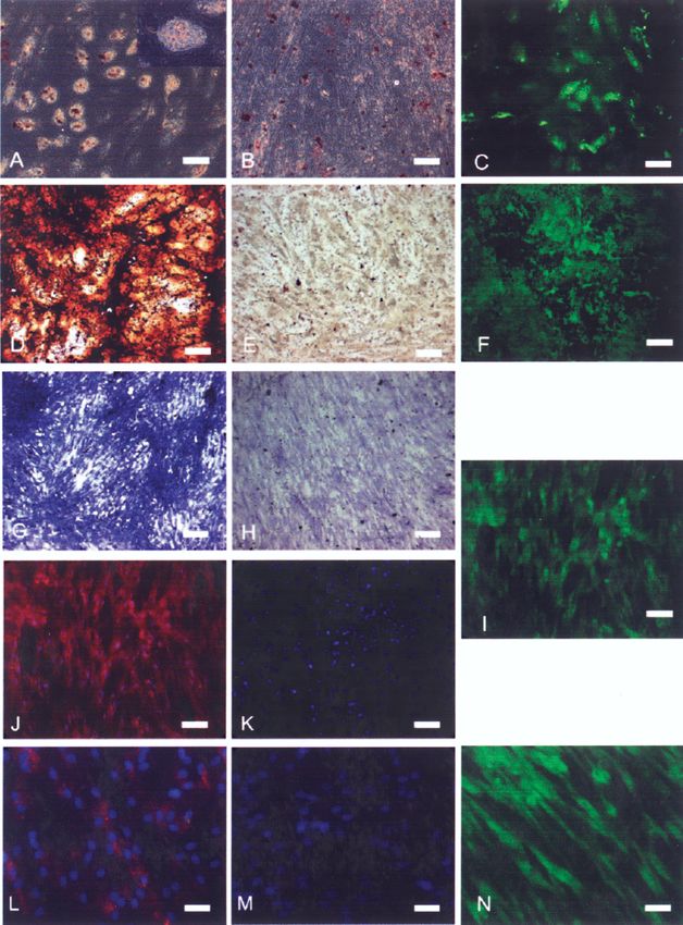
structures. To confirm the myogenic differentiation, the tometry 72 h posttransduction. About 75% of cells
expression of myosin by immunofluorescence was as- were GFP positive and GFP expression did not de-
sessed after 45 days (Fig. 4H). The specificity of this cline during culture passages (Fig. 6). To evaluate if
assay was corroborated by the absence of staining in GFP interfered with the multipotent capacity of
cASCs. Expression of myogenin, dystrophin, and MyoD cASCs, both GFP-positive and -negative cells at suc-
was observed only in induced cells, with no basal ex- cessive passages were analyzed by flow cytometry
pression in control cells (Fig. 5A). and multilineage differentiation, revealing no influ-CANINE ADIPOSE-DERIVED STEM CELLS 287
Figure 6. GFP-positive cASCs. (A) GFP-positive cASCs at passage 8. (B) Percentage of positively GFP cells as analyzed by flow
cytometry.
ence of GFP on the cellular response to inductive me- isolation of adipose stem cells from other mammals,
dia (Fig. 4). such as rabbit, mice, horse, and pig (27,29,32,33).
We observed that the plastic adherent cells obtained
DISCUSSION after isolation can be expanded in vitro, reaching num-
Zuk et al. (35) were the first to describe the isolation bers that would be sufficient for a therapeutic assay,
and characterization of human stem cells derived from without any numeric chromosome alteration. In addi-
adipose tissue. These cells were able to differentiate into tion, cASCs can be stored frozen in liquid nitrogen with-
adipogenic, osteogenic, chondrogenic, and myogenic out cell death.
lineages when exposed to inductive media. During the first days in culture, endothelial cell popu-
Human ASCs are usually obtained from fat tissue lations were found in the plates; however, these cells
that is discarded after liposuction cosmetic surgery (35). were not seen after passage 4. These data are in accor-
Adipose tissue can be harvested in large quantities with dance with Rodriguez et al. (21), where the isolation of
minimal morbidity in several regions of the body and, hASCs by adherence properties was reported. At pas-
on average, 100 ml of human adipose tissue yields about sage 4, cASCs show a fibroblast-like morphology com-
1 × 10⁶ stem cells (14). In dogs, the adipose tissue can monly found in mesenchymal stem cell (MSCs). The
be collected by a simple adapted liposuction surgery, analysis of the cell surface markers showed that the
through biopsies or in routine veterinary surgery proce- cASCs cell population expresses the known immnophe-
dures because we could isolate cASCs from just 100 µl notype of MSCs (35). At passage 4 the majority of the
of adipose tissue. cells are positive for CD29, CD44, and CD90. cASCs
In the present study we show the isolation and char- do not express the hematopoietic marker CD45, but 10%
acterization of the canine adipose-derived stromal cell of the cells are CD34 positive. Traktuev et al. (28) de-
(cASC) population. While this manuscript was in prepa- scribed that the population of CD34-positive cells that
ration, Neupane et al. (17) published an article reporting are found in human adipose stromal-vascular fraction
the isolation of canine adipose stem cells. However, are reside in a periendothelial location. The authors
their article lacked important characteristics of the iso- showed that these cells are CD31 negative. This result
lated cell population such as the immunophenotype, the is in accordance with our finding with cASCs, because
myogenic and chondrogenic potential, and karyotype we found CD34-positive cells but not CD31-positive
analysis at late passages. These characteristics are im- cells. This adherent cASCs cell population (CD34+/
portant for veterinary cell therapy and preclinical studies. CD31−) may also interact with endothelial cells at the
Our results show that cASCs can be harvested by a perivascular niche. However, further studies will be es-
rapid process, an important step towards preclinical sential to identify the localization of these cells at the
studies of cell therapy. Using this methodology, we were canine adipose tissue. In order to evaluate the MSC
able to harvest cells from 10 canine subcutaneous fat property of cASCs, we subjected them to differentiation
samples (2 from liposuction and 8 from biopsy) with a studies.
100% rate of success. Other groups reported successful MSCs are defined by their ability to self-renew and288 VIEIRA ET AL.
their capacity to generate committed cells in vitro and potential relevance to future canine veterinary tissue en-
in vivo. Human ACSs can be induced to differentiate gineering and regenerative veterinary medical therapies.
along the adipogenic, chondrogenic, and osteogenic lin- ACKNOWLEDGMENTS: We gratefully acknowledge our col-
eages using specific culture medium (32). Even plated leagues Tatiana Jazedje, Marcos Valadares, Maria Denise
into scaffolds they survive long-term culture and could Carvalho, Amanda Assoni, Camila Almeida, Mayra Pellati,
be terminally differentiated into adipocytes and osteo- Bruno Lima, Heloisa Caetano, Constancia Urbani, Dr. Mariz
Vainzof, and Dr. Maria Rita Passos-Bueno for helpful sugges-
blasts (18). In all 3 of our 10 lineages obtained we dem-
tions as well as the earlier support of the veterinarians. This
onstrated the multipotency and plasticity of cASCs by research was supported by FAPESP-CEPID (Fundação de
their differentiation in adipogenic, chondrogenic, myo- Amparo à Pesquisa do Estado de São Paulo-Centro de Pes-
genic, and osteogenic lineages. The differentiation was quisa, Inovação e Difusão), CNPq (Conselho Nacional de De-
confirmed by the appearance of lipid vacuoles, muco- senvolvimento Cientı́fico e Tecnológico), and INCT (Instituto
Nacional de Ciência e Tecnologia: células-tronco em doenças
polysaccharide-rich extracellular matrix, myosin label-
genéticas).
ing, and calcium deposits, respectively. Although we ob-
served a morphological change in cells submitted to
adipogenic differentiation, we found poor lipid vacuoles REFERENCES
in cASCs when compared to hASCs submitted to the
1. Black, L. L.; Gaynor, J.; Gahring, D.; Adams, C.; Aron,
same conditions. D.; Harman, S.; Gingerich, D. A.; Harman, R. Effect of
The stable GFP expression after cASC transduction adipose-derived mesenchymal stem and regenerative cells
represents a great advantage in preclinical studies. GFP- on lameness in dogs with chronic osteoarthritis of the cox-
positive cells could be screened though fluorescence ofemoral joints: A randomized, double-blinded, multicen-
ter, controlled trial. Vet. Ther. 8:272–284; 2007.
microscopic imaging and flow cytometry, and no signif- 2. Brzoska, M.; Geiger, H.; Gauer, S.; Baer, P. Epithelial
icant decline of GFP expression during cASC differenti- differentiation of human adipose tissue-derived adult stem
ation was found. This property allows their potential use cells. Biochem. Biophys. Res. Commun. 330:142–150;
for in vitro (29) and in vivo assays (3), where cell mark- 2005.
ing with GFP was used to track stem cells seeded on a 3. Darabi, R.; Gehlbach, K.; Bachoo, R. M.; Kamath, S.;
Osawa, M.; Kamm, K. E.; Kyba, M.; Perlingeiro, R. C.
scaffold, fusing to other cells and engraftment into dif-
Functional skeletal muscle regeneration from differentiat-
ferent tissues. ing embryonic stem cells. Nat. Med. 14:134–143; 2008.
Successful canine stem cell transplantation was re- 4. Di Rocco, G.; Iachininoto, M. G.; Tritarelli, A.; Straino,
ported by Sampaolesi et al. (24). According to these S.; Zacheo, A.; Germani, A.; Crea, F.; Capogrossi, M. C.
authors, the intra-arterial delivery of canine mesoangi- Myogenic potential of adipose-tissue-derived cells. J. Cell
Sci. 119:2945–2952; 2006.
oblasts (vessel-associated stem cells) resulted in an ex-
5. Erickson, G. R.; Gimble, J. M.; Franklin, D. M.; Rice,
tensive recovery of muscle morphology and function in H. E.; Awad, H.; Guilak, F. Chondrogenic potential of
golden retriever muscular dystrophy (GRMD) dogs. Be- adipose tissue-derived stromal cells in vitro and in vivo.
cause ASCs are much easier to be obtained and can be Biochem. Biophys. Res. Commun. 290:763–769; 2002.
injected without immunosuppression (11,31), they might 6. Garcia-Olmo, D.; Garcia-Arranz, M.; Herreros, D.;
Pascual, I.; Peiro, C.; Rodriguez-Montes, J. A. A phase I
represent a promising alternative to canine mesoangi- clinical trial of the treatment of Crohn’s fistula by adipose
oblasts for GRMD stem cell therapeutic trials. mesenchymal stem cell transplantation. Dis. Colon Rec-
In short, results from the present study demonstrate tum 48:1416–1423; 2005.
that adherent cells isolated from canine adipose tissue 7. Gimble, J.; Guilak, F. Adipose-derived adult stem cells:
can be defined as multipotent MSC with the ability to Isolation; characterization; and differentiation potential.
Cytotherapy 5:362–369; 2003.
differentiate into at least four mesodermal lineages.
8. Halvorsen, Y. D.; Bond, A.; Sen, A.; Franklin, D. M.;
Mouse models of human diseases have been extensively Lea-Currie, Y. R.; Sujkowski, D.; Ellis, P. N.; Wilkison,
used in preclinical studies. However, mouse models W. O.; Gimble, J. M. Thiazolidinediones and glucocorti-
have several limitations when extrapolating to humans: coids synergistically induce differentiation of human adi-
the small size of the mouse limits the proliferative de- pose tissue stromal cells: Biochemical, cellular, and mo-
lecular analysis. Metabolism 50:407–413; 2001.
mand placed on transplanted tissue and the short life 9. Halvorsen, Y. D.; Bond, A.; Sen, A.; Franklin, D. M.;
span of the mouse also prevents long-term follow-up. Lea-Currie, Y. R.; Sujkowski, D.; Ellis, P. N.; Wilkison,
Large-animal models, such as the dog, more faithfully W. O.; Gimble, J. M. Extracellular matrix mineralization
mimic human pathologies. There are many canine ani- and osteoblast gene expression by human adipose tissue-
mal models of human genetic diseases that are well derived stromal cells. Tissue Eng. 7:729–741; 2001.
10. Justesen, J.; Pedersen, S. B.; Stenderup, K.; Kassem, M.
characterized, making them ideal for in vivo studies. Subcutaneous adipocytes can differentiate into bone-form-
The characterization of cASCs represents a valuable tool ing cells in vitro and in vivo. Tissue Eng. 10:381–391;
for in vitro and in vivo preclinical evaluation and for the 2004.
screening of therapeutic drugs. These findings also have 11. Keyser, K. A.; Beagles, K. E.; Kiem, H. P. ComparisonCANINE ADIPOSE-DERIVED STEM CELLS 289
of mesenchymal stem cells from different tissues to sup- Tonlorenzi, R.; Innocenzi, A.; Mognol, P.; Thibaud, J.;
press T-cell activation. Cell Transplant. 16:555–562; Galvez, B. G.; Barthelemy, I.; Perani, L.; Mantero, S.;
2007. Guttinger, M.; Pansarasa, O.; Rinaldi, C.; Angelis, M. G. C.;
12. Li, H.; Dai, K.; Tang, T.; Zhang, X.; Yan, M.; Lou, J. Torrente, Y.; Bordignon, C.; Bottinelli, R.; Cossu, G.
Bone regeneration by implantation of adipose-derived Mesoangioblast stem cells ameliorate muscle function in
stromal cells expressing BMP-2. Biochem. Biophys. Res. dystrophic dogs. Nature 444:574–579; 2006.
Commun. 356:836–842; 2007. 25. Seo, M. J.; Suh, S. Y.; Bae, Y. C.; Jung, J. S. Differentia-
13. Lois, C.; Hong, E. J.; Pease, S.; Brown, E. J.; Baltimore, tion of human adipose stromal cells into hepatic lineage
D. Germline transmission and tissue-specific expression in vitro and in vivo. Biochem. Biophys. Res. Commun.
of transgenes delivered by lentiviral vectors. Science 95: 328:258–264; 2005.
868–872; 2002. 26. Strauss, B. E.; Patricio, J. R.; de Carvalho, A. C.; Bajgel-
14. Meliga, E.; Strem, B. M.; Duckers, H. J.; Serruys, P. W. man, M. C. A. A lentiviral vector with expression con-
Adipose-derived cells. Cell Transplant. 16:963–970; trolled by E2F-1: A potential tool for the study and treat-
2007. ment of proliferative diseases. Biochem. Biophys. Res.
15. Miranville, A.; Heeschen, C.; Sengenes, C.; Curat, C. A.; Commun. 348:1411–1418; 2006.
Busse, R.; Bouloumie, A. Improvement of postnatal neo- 27. Torres, F. C.; Rodrigues, C. J.; Stocchero, I. N.; Ferreira,
vascularization by human adipose tissue-derived stem M. C. Stem cells from the fat tissue of rabbits: An easy-
cells. Circulation 110:349–355; 2002. to-find experimental source. Aesthetic Plast. Surg. 31:
16. Mizuno, H.; Zuk, P. A.; Zhu, M.; Lorenz, H. P.; Benhaim, 574–578; 2007.
P.; Hedrick, M. H. Myogenic differentiation by human 28. Traktuev, D. O.; Merfeld-Clauss, S.; Li, J.; Kolonin, M.;
processed lipoaspirate cells. Plast. Reconstr. Surg. 109: Arap, W.; Pasqualini, R.; Johnstone, B. H.; March, K. L.
199–209; 2002. A population of multipotent CD34-positive adipose stro-
17. Neupane, M.; Chang, C. C.; Kiupel, M.; Yuzbasiyan-Gur- mal cells share pericyte and mesenchymal surface mark-
kan, V. Isolation and characterization of canine adipose- ers, reside in a periendothelial location, and stabilize en-
derived mesenchymal stem cells. Tissue Eng. Part A 14: dothelial networks. Circ. Res. 102:77–85; 2008.
1007–1015; 2008. 29. Vidal, M. A.; Kilroy, G. E.; Lopez, M. J.; Johnson, J. R.;
18. Neuss, S.; Stainforth, R.; Salber, J.; Schenck, P.; Bovi, Moore, R. M.; Gimble, J. M. Characterization of equine
M.; Knüchel, R.; Perez-Bouza, A. Long-term survival and adipose tissue-derived stromal cells: Adipogenic and os-
bipotent terminal differentiation of human mesenchymal teogenic capacity and comparison with bone marrow-
stem cells (hMSC) in combination with a commercially derived mesenchymal stromal cells. Vet. Surg. 36:613–
available three-dimensional collagen scaffold. Cell Trans- 622; 2007.
plant. 17:977–986; 2008. 30. Vieira, N. M.; Brandalise, V.; Zucconi, E.; Jazedje, T.;
19. Planat-Benard, V.; Silvestre, J. S.; Cousin, B.; Andre, M.; Secco, M.; Nunes, V. A.; Strauss, B. E.; Vainzof, M.;
Nibbelink, M.; Tamarat, R.; Clergue, M.; Manneville, C.; Zatz, M. Human multipotent adipose-derived stem cells
Saillan-Barreau, C.; Duriez, M.; Tedgui, A.; Levy, B.; restore dystrophin expression of Duchenne skeletal-
Penicaud, L.; Casteilla, L. Plasticity of human adipose lin- muscle cells in vitro. Biol. Cell 100:231–241; 2008.
eage cells toward endothelial cells: Physiological and ther- 31. Vieira, N. M.; Bueno, Jr., C. R.; Brandalise, V.; Moraes,
apeutic perspectives. Circulation 109:656–663; 2004. L. V.; Zucconi, E.; Secco, M.; Suzuki, M. F.; Camargo,
20. Rehman, J.; Traktuev, D.; Li, J.; Merfeld-Clauss, S.; M. M.; Bartolini, P.; Brum, P. C.; Vainzof, M.; Zatz, M.
Temm-Grove, C. J.; Bovenkerk, J. E.; Pell, C. L.; John- SJL dystrophic mice express a significant amount of hu-
stone, B. H.; Considine, R. V.; March, K. L. Secretion man muscle proteins following systemic delivery of hu-
of angiogenic and antiapoptotic factors by human adipose man adipose-derived stromal cells without immunosup-
stromal cells. Circulation 109:1292–1298; 2004. pression. Stem Cells 26:2391–2398; 2008.
21. Rodriguez, A. M.; Pisani, D.; Dechesne, C. A.; Turc- 32. Williams, K. J.; Picou, A. A.; Kish, S. L.; Giraldo, A. M.;
Carel, C.; Kurzenne, J. Y.; Wdziekonski, B.; Villageois, Godke, R. A.; Bondioli, K. R. Isolation and characteriza-
A.; Bagnis, C.; Breittmayer, J. P.; Groux, H.; Ailhaud, G.; tion of porcine adipose tissue-derived adult stem cells.
Dani, C. Transplantation of a multipotent cell population Cells Tissues Organs 188:251–258; 2008.
from human adipose tissue induces dystrophin expression 33. Yamamoto, N.; Akamatsu, H.; Hasegawa, S.; Yamada, T.;
in the immunocompetent mdx mouse. J. Exp. Med. 201: Nakata, S.; Ohkuma, M.; Miyachi, E.; Marunouchi, T.;
1397–1405; 2005. Matsunaga, K. Isolation of multipotent stem cells from
22. Rodriguez, L. V.; Alfonso, Z.; Zhang, R.; Leung, J.; Wu, mouse adipose tissue. J. Dermatol. Sci. 48:43–52; 2007.
B.; Ignarro, L. J. Clonogenic multipotent stem cells in hu- 34. Zuk, P. A.; Zhu, M.; Ashjian, P.; De Ugarte, D. A.;
man adipose tissue differentiate into functional smooth Huang, J. I.; Mizuno, H.; Alfonso, Z. C.; Fraser, J. K.;
muscle cells. Proc. Natl. Acad. Sci. USA 103:12167– Benhaim, P.; Hedrick, M. H. Human adipose tissue is a
12172; 2006. source of multipotent stem cells. Mol. Biol. Cell 13:4279–
23. Safford, K. M.; Hicok, K. C.; Safford, S. D.; Halvorsen, 4295; 2002.
Y. D.; Wilkison, W. O.; Gimble, J. M.; Rice, H. E. Neuro- 35. Zuk, P. A.; Zhu, M.; Mizuno, H.; Huang, J.; Futrell,
genic differentiation of murine and human adipose-derived J. W.; Katz, A. J.; Benhaim, P.; Lorenz, H. P.; Hedrick,
stromal cells. Biochem. Biophys. Res. Commun. 294: M. H. Multilineage cells from human adipose tissue: Im-
371–379; 2002. plications for cell-based therapies. Tissue Eng. 7:211–
24. Sampaolesi, M.; Blot, S.; D’Antona, G.; Granger, N.; 228; 2001.You can also read