Epidermal Fatty Acid-Binding Protein 5 (FABP5) Involvement in Alpha-Synuclein-Induced Mitochondrial Injury under Oxidative Stress - MDPI
←
→
Page content transcription
If your browser does not render page correctly, please read the page content below
biomedicines
Article
Epidermal Fatty Acid-Binding Protein 5 (FABP5) Involvement
in Alpha-Synuclein-Induced Mitochondrial Injury under
Oxidative Stress
Yifei Wang            , Yasuharu Shinoda          , An Cheng       , Ichiro Kawahata         and Kohji Fukunaga *

                                          Department of Pharmacology, Graduate School of Pharmaceutical Sciences, Tohoku University,
                                          6–3 Aramaki-Aoba, Aoba-ku, Sendai 980-8578, Japan; yifei.wang.q4@dc.tohoku.ac.jp (Y.W.);
                                          yshinoda@tohoku.ac.jp (Y.S.); cheng.an.q6@dc.tohoku.ac.jp (A.C.); kawahata@tohoku.ac.jp (I.K.)
                                          * Correspondence: kfukunaga@tohoku.ac.jp; Tel.: +81-22-795-6836

                                          Abstract: The accumulation of α-synuclein (αSyn) has been implicated as a causal factor in the
                                          pathogenesis of Parkinson’s disease (PD). There is growing evidence that supports mitochondrial
                                          dysfunction as a potential primary cause of dopaminergic neuronal death in PD. Here, we focused
                                          on reciprocal interactions between αSyn aggregation and mitochondrial injury induced by oxidative
                                          stress. We further investigated whether epidermal fatty acid-binding protein 5 (FABP5) is related
                                          to αSyn oligomerization/aggregation and subsequent disturbances in mitochondrial function in
                                          neuronal cells. In the presence of rotenone, a mitochondrial respiratory chain complex I inhibitor, co-
                                          overexpression of FABP5 with αSyn significantly decreased the viability of Neuro-2A cells compared
                                          to that of αSyn alone. Under these conditions, FABP5 co-localized with αSyn in the mitochondria,
                                          thereby reducing mitochondrial membrane potential. Furthermore, we confirmed that pharmacologi-

                                   cal inhibition of FABP5 by its ligand prevented αSyn accumulation in mitochondria, which led to cell
                                          death rescue. These results suggested that FABP5 is crucial for mitochondrial dysfunction related to
Citation: Wang, Y.; Shinoda, Y.;
                                          αSyn oligomerization/aggregation in the mitochondria induced by oxidative stress in neurons.
Cheng, A.; Kawahata, I.; Fukunaga, K.
Epidermal Fatty Acid-Binding
                                          Keywords: α-Synuclein; FABP5; aggregation; mitochondria; Parkinson’s disease
Protein 5 (FABP5) Involvement in
Alpha-Synuclein-Induced
Mitochondrial Injury under
Oxidative Stress. Biomedicines 2021, 9,
110. https://doi.org/10.3390/             1. Introduction
biomedicines9020110                             Parkinson’s disease (PD) is the second-most common neurodegenerative disease,
                                          and is caused by the loss of dopaminergic neurons in the substantia nigra pars compacta
Received: 28 December 2020                (SNpc) [1]. The neuropathological hallmark of PD is the accumulation of intracellular
Accepted: 20 January 2021                 protein inclusions composed primarily of α-synuclein (αSyn), which are termed Lewy
Published: 22 January 2021                bodies [2,3]. Moreover, αSyn aggregation has been widely reported to be a harbinger of
                                          subsequent pathology that ultimately leads to neurodegeneration [4]. αSyn is expressed pre-
Publisher’s Note: MDPI stays neutral      synaptically as a native unfolded protein that can form oligomers, protofibrils, or amyloid
with regard to jurisdictional claims in
                                          fibrils [5,6]. Among the different species of αSyn, pre-fibrillar oligomers are considered to
published maps and institutional affil-
                                          be toxic to neurons [7]. The development of treatments aimed at reducing αSyn synthesis,
iations.
                                          secretion, aggregation, and increasing the clearance of pathogenic formations has become
                                          the most promising means to overcome Parkinson’s disease [8,9].
                                                Several lines of evidence support mitochondrial dysfunction as a primary pathogenic
                                          mechanism in PD. The most convincing evidence first appeared from accidental human
Copyright: © 2021 by the authors.         exposure to 1-methyl-4-phenyl-1,2,3,6-tetrahydrodropyridine, a metabolite that induces
Licensee MDPI, Basel, Switzerland.        Parkinsonian syndrome by inhibiting mitochondrial respiratory complex I activity [10,11].
This article is an open access article
                                          The genetic causes of PD, including PINK1, parkin, and DJ-1, particularly highlight the
distributed under the terms and
                                          importance of mitochondrial dysfunction in PD pathology [12]. Accumulating studies
conditions of the Creative Commons
                                          have demonstrated that αSyn itself is involved in mitochondrial injury in PD [13–15]. In
Attribution (CC BY) license (https://
                                          postmortem brains of PD patients, αSyn is especially localized to mitochondrial outer mem-
creativecommons.org/licenses/by/
                                          branes in nigrostriatal neurons, which was replicated in rodents treated with rotenone, a
4.0/).

Biomedicines 2021, 9, 110. https://doi.org/10.3390/biomedicines9020110                                 https://www.mdpi.com/journal/biomedicines
Biomedicines 2021, 9, 110                                                                                        2 of 15

                            respiratory chain complex I inhibitor [14]. Treatment of αSyn oligomers, but not monomers,
                            disturbed mitochondrial maintenance, leading to fission, reduction in mitochondrial mem-
                            brane potential, and upregulation of reactive oxidative stress in neuroblastoma cells. Pre-
                            formed fibril treatment also elicited the translocation of phosphorylated αSyn aggregates
                            to mitochondria, resulting in increased oxidative stress and mitochondrial dysfunction in
                            mouse primary hippocampal neurons [13]. These results indicate the bilateral links that
                            oligomeric αSyn localization in mitochondria induces reactive oxidative species produc-
                            tion/mitochondrial dysfunction, and vice versa.
                                  Fatty acid-binding proteins (FABPs) are a set of 14–15 kDa cytoplasmic proteins that
                            are traditionally considered as lipid chaperones that coordinate lipid responses inside
                            cells. FABPs perform pleiotropic functions to maintain healthy tissue homeostasis, and
                            participate in the pathogenesis of diseases [16]. To date, at least 10 genes encoding FABPs
                            (FABP1-9 and FABP12) have been identified in the human genome [17,18]. FABP5 localizes
                            to the cytosol, mitochondria, and nucleus, suggesting a role for fatty acid metabolism and
                            nuclear transcriptional regulation [19,20]. The central nervous system contains three types
                            of FABP, that is, epidermal FABP (E-FABP, FABP5), heart FABP (H-FABP, FABP3), and brain
                            FABP (B-FABP, FABP7) [21]. FABP5 is expressed in neurons and glial cells in broad areas
                            of the brain, including the cortex, hippocampus, and caudate putamen [22]. Moreover,
                            it has been reported that FABP5 mRNA expression levels, as well as αSyn and tyrosine
                            hydroxylase gene expression levels, are enriched in dopaminergic neurons of the SNpc,
                            and decrease after 6-hydroxydopamine injection in rats [23]. However, little is known
                            regarding whether FABP5 is involved in αSyn toxicity in dopaminergic neurons, or in the
                            pathogenesis of PD.
                                  To investigate whether FABP5 is related to αSyn oligomerization and mitochondrial
                            impairment, we transfected FABP5 with αSyn into neuroblastoma Neuro-2A cells, and
                            evaluated cell viability, αSyn oligomerization, cellular localization, and mitochondrial
                            membrane potential. Here, we report that FABP5 co-expression with αSyn reduced cell
                            viability, upregulated αSyn oligomerization, and aggregation under oxidative stress. FABP5
                            and αSyn co-localized in mitochondria in the presence of rotenone, which was abolished
                            by a ligand with high affinity for FABP5.

                            2. Materials and Methods
                            2.1. Materials
                                 Reagents and antibodies were obtained from the following sources: anti-αSyn anti-
                            body (1:1000; 4B12, GeneTex, Irvine, CA, USA); anti-FABP5 antibody (1:1000; AF3077, R&D
                            Systems, Minneapolis, MN, USA); anti-β-tubulin antibody (1:3000; T0198, Sigma-Aldrich,
                            St Louis, MO, USA); anti-VDAC antibody (1:1000; 4866S, Cell Signaling Technology, Dan-
                            vers, MA, USA); anti-rabbit or mouse IgG antibody conjugated with horseradish peroxidase
                            (1:5000; Southern Biotech, Birmingham, AL, USA); anti-goat IgG antibody conjugated with
                            peroxidase (1:5000; Rockland Immunochemical, Limerick, PA, USA); Alexa 594-labeled
                            anti-mouse IgG, Alexa 488-labeled anti-Goat IgG and Alexa 594-labeled anti-rabbit IgG
                            antibody (1:500; Invitrogen, Waltham, MA, USA); Biotin-SP anti-Goat IgG antibody (1:500;
                            Jackson ImmunoResearch, West Grove, PA, USA); and AMCA streptavidin (1:500; Jackson
                            ImmunoResearch). These materials were used according to the manufacturers’ datasheets.
                            Rotenone was purchased from MP Biomedicals (150154; Santa Ana, CA, USA). All other
                            reagents were obtained from FUJIFILM Wako Pure Chemicals (Osaka, Japan) unless other-
                            wise noted. The FABP5 ligand (ligand 7) was described in a previous report [24].

                            2.2. Cell Culture and Transfection
                                 Neuro-2A cells were obtained from the Human Science Research Resources Bank
                            (IFO50081) (Osaka, Japan). Cells were maintained in Dulbecco’s modified Eagle medium
                            (DMEM) supplemented with 10% fetal bovine serum (FBS) and 100 U/mL penicillin-
                            streptomycin at 37 ◦ C in a humidified atmosphere of 5% CO2 . For stimulation, rotenone
                            and FABP ligands were prepared in dimethyl sulfoxide (DMSO). Mouse FABP5 plasmid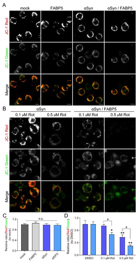
Biomedicines 2021, 9, 110                                                                                          3 of 15

                            was produced as described previously [24], and inserted into the multiple cloning site of
                            plasmid pcDNA3.1 (Invitrogen). Human αSyn plasmid encoding the protein with myc
                            and 6×His tag at the C-terminus was purchased from Abgent (San Diego, CA, USA).
                            A non-coding plasmid (pcDNA3.1) was used for a mock group. Transfection was carried
                            out using Lipofectamine LTX and Plus Reagent (Invitrogen) by incubating for 6 h. The
                            medium was then changed to DMEM supplemented with 5% FBS to maintain cell viability.
                            The next day, cells were treated with rotenone, ligand 7, or DMSO for 48 h at a dilution of
                            1:1000, and then used for the following experiments.

                            2.3. Evaluation of Cell Viability
                                Cells were cultured in 96-well microplates, and 1:10 dilution of CCK-8 solution was
                            added after transfection and drug treatment (Cell Counting Kit-8; Dojindo, Kumamoto,
                            Japan). Plates were incubated for 2 h at 37 ◦ C, and the optical absorbance at 450 nm was
                            measured using a microplate reader (Flex Station 3, Molecular Devices, San Jose, CA, USA).

                            2.4. Protein Extraction and Immunoblotting Assay
                                 Cells were cultured in a 35 mm dish, collected by scraping, and homogenized in
                            80 µL buffer containing 0.5% Triton X-100, 50 mM Tris-HCl, pH 7.4, 4 mM EGTA, 10 mM
                            EDTA, 1 mM Na3 VO4 , 40 mM Na4 P2 O7 ·10H2 O, 50 mM NaF, 0.15 M NaCl, and protease
                            inhibitor cocktail (50 µg/mL leupeptin, 25 µg/mL pepstatin A, 50 µg/mL trypsin inhibitor,
                            100 nM calyculin A, and 1 mM dithiothreitol). Then, lysates were centrifuged at 15,000 rpm
                            (10,000× g) for 10 min at 4 ◦ C, the supernatants (Triton-soluble fraction) collected, and
                            protein concentrations were measured using the Bradford assay. Insoluble materials were
                            washed three times and centrifuged at 15,000 rpm for 3 min at 4 ◦ C, and the supernatant
                            was discarded. The pellets were homogenized in 2% SDS solution (SDS-soluble fraction).
                            In order to ensure equivalent protein loading, the volume of the 2% SDS solution was
                            proportional to the concentration of Triton-soluble protein. Mixtures with Laemmli’s
                            sample buffer without β-mercaptoethanol were subjected to heating before immunoblotting
                            to evaluate αSyn oligomerization.
                                 For immunoblotting assays, equal amounts of protein were electrophoresed on ready-
                            made gels (414886; Cosmo Bio, Tokyo, Japan) and transferred onto PVDF membranes
                            (Millipore, Billerica, MA, USA). After blocking with 5% skim milk in Tris-buffered saline
                            with Tween 20, the membrane was incubated on a horizontal shaker overnight with primary
                            antibody at 4 ◦ C, followed by probing with secondary antibody for 2 h at 20 ◦ C. Signals
                            were detected by chemiluminescence using an imaging analyzer (LAS4000 Mini, Fuji
                            Film, Tokyo, Japan). Densitometry analysis was performed using Multi Gauge Software
                            (FUJIFILM). Briefly, we framed each target band and also framed the part without bands as
                            the background to get their densities. The difference between the two is the intensity of the
                            target band and is standardized with the internal reference protein (β-tubulin). Finally, the
                            values were normalized with the mock group before statistical analysis.

                            2.5. Immunofluorescent Staining
                                  Neuro-2A cells were cultured on glass slips in 12-well plates (Corning, Glendale, AZ,
                            USA) and fixed in 4% paraformaldehyde for 10 min at room temperature. For mitochon-
                            drial staining, cells were incubated with 0.1mM MitoTracker Red CMXRos dye (M7512;
                            Invitrogen) in DMEM without FBS for 30 min at 37 ◦ C before fixation. Cells were washed in
                            phosphate-buffered saline (PBS) and permeabilized with 0.1% Triton X-100/PBS for 10 min,
                            then blocked in 1% BSA/PBS at room temperature for 30 min. Cells were incubated with
                            primary antibodies at 4 ◦ C overnight. After washing in 1% BSA/PBS for 5 min three times,
                            cells were incubated with secondary antibodies for 2 h at 4 ◦ C. Cells were washed in 1%
                            BSA/PBS for 5 min three times, followed by staining with 40 ,6-diamidino-2-phenylindole
                            (DAPI) (Thermo Fisher Scientific, Waltham, MA, USA). Glass slips were mounted with
                            Vectashield (Vector Laboratories, Burlingame, CA, USA). Immunofluorescent images were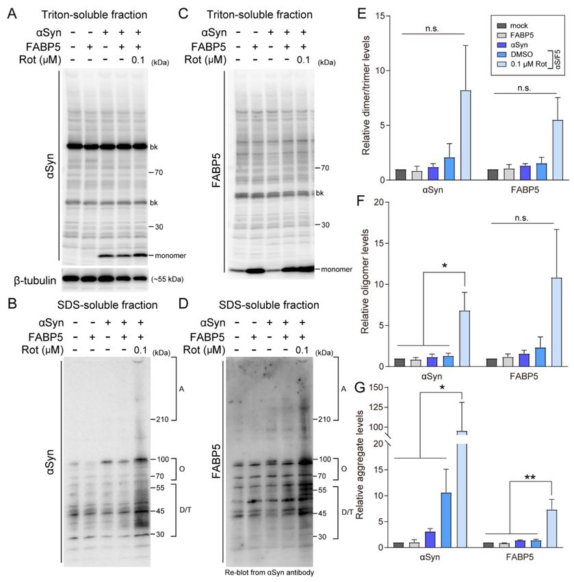
Biomedicines 2021, 9, 110                                                                                           4 of 15

                            captured with a confocal laser scanning microscope (TCS SP8, Leica Microsystems, Wetzlar,
                            Germany). The visible aggregates (diameter 0.5–2 µm approximately) were quantitated.

                            2.6. Measurement of Mitochondrial Membrane Potential
                                 Cells were cultured in 96-well plates (Corning) or glass-bottom dishes (Matsunami
                            Glass Ind, Osaka, Japan). Mitochondrial activity was visualized using JC-1 MitoMP
                            Detection Kit (Dojindo) staining, according to the manufacturer’s instructions. JC-1 is a
                            cationic dye that exhibits membrane potential-dependent accumulation in mitochondria,
                            which is indicated by a fluorescence emission shift from green (~530 nm) to red (~590 nm).
                            The fluorescence intensity was measured with Flex Station 3, or using ImageJ software
                            following acquisition of images by confocal microscopy [25]. The ratio of red/green
                            fluorescence intensity was calculated and converted into a value relative to the control
                            group for the identification of mitochondrial membrane potential.

                            2.7. Mitochondrial Purification
                                 Mitochondria and cytoplasm were isolated from Neuro-2A cells as described pre-
                            viously [26] with slight modifications. Briefly, cells were suspended in mitochondrial
                            isolation buffer containing 250 mM sucrose, 1 mM dithiothreitol, 10 mM KCl, 1 mM
                            EDTA, 1.5 mM MgCl2 , protease inhibitors, and 20 mM Tris-HCl, pH 7.4, and then homoge-
                            nized with a glass homogenizer using approximately 30 strokes, on ice. The homogenate
                            was centrifuged twice at 800× g for 10 min. The supernatant was further centrifuged
                            at 15,000× g for 10 min. After that, supernatants were collected as cytosolic fractions
                            (without mitochondria). Next, the mitochondrial pellets were immediately washed three
                            times in mitochondrial isolation buffer. Subsequently, the pellets were homogenized with
                            Triton X-100 buffer as described above, and supernatants were collected as mitochondrial
                            fractions. All steps were carried out at 4 ◦ C. Protein concentrations were determined by
                            using Bradford assays. All fractions were boiled at 100 ◦ C for 3 min with six × Laemmli
                            sample buffer containing β-mercaptoethanol. The quantity of cytosolic fractions and mito-
                            chondria were, respectively, estimated by quantification of β-tubulin and VDAC using an
                            immunoblotting assay.

                            2.8. Statistical Analysis
                                 Values are presented as means ± standard error of the mean (SEM), and were evalu-
                            ated with one- or two-way analysis of variance (ANOVA) followed by Bonferroni’s multiple
                            comparisons test using GraphPad Prism 7.04 (GraphPad Software, San Diego, CA, USA).
                            p < 0.05 was considered statistically significant.

                            3. Results
                            3.1. Co-Overexpression of FABP5 and α-Synuclein Increases Neurotoxicity in the Presence of
                            Rotenone
                                 To address the role of FABP5 in the toxicity of αSyn and oxidative stress, Neuro-
                            2A cells were transfected with plasmids and then stimulated with 0.01–10 µM rotenone
                            (Figure 1). Immunoblotting analysis showed moderate levels of endogenous FABP5 protein
                            and higher levels of FABP5 and αSyn in transfected cells (Figure 1A). There is no FABP3
                            expression and a very little FABP7 expression in Neuro-2A cells (Figure S1). CCK-8 assays
                            revealed that rotenone at concentrations > 1 µM significantly decreased cell viability in
                            mock- and FABP5-transfected cells (Figure 1B). Similar toxicity of oxidative stress was
                            demonstrated in αSyn-transfected cells, whereas above 0.5 µM, rotenone treatment further
                            decreased cell viability in αSyn/FABP5 (αS/F5)-expressing cells (Figure 1C). This effect
                            of rotenone was evident when treated at a moderate concentration (0.5 µM), which failed
                            to decrease cell survival in αSyn-transfected cells. These results suggested that FABP5
                            enhances oxidative stress toxicity in the presence of αSyn.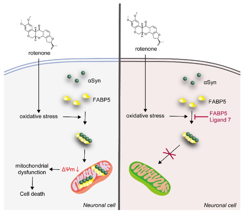
Biomedicines 2021, 9, 110                                                                                                     5 of 15

      Figure 1. Rotenone induced neurotoxicity, especially with co-overexpression of FABP5 and α-Synuclein. (A) Representative
      images of immunoblots probed with antibodies against αSyn (~19 kDa) and FABP5 (~12 kDa). Blots with anti-β-tubulin
      antibody showed that a similar amount of protein was loaded. (B,C) Viability of Neuro-2A cells treated with indicated
      concentrations of rotenone for 48 h was assessed using CCK-8 assays. The transfection conditions were pcDNA 3.1-
      transfected cells (mock) and FABP5-transfected cells in (B); αSyn-transfected cells and αSyn/FABP5-transfected cells
      (αS/F5) in (C). Results are presented as means ± SEM (n = 4 parallel cell experiments). * p < 0.05, ** p < 0.01, vs. 0 µM
      rotenone groups; ## p < 0.01 vs. αSyn-transfected cells. CCK-8: cell counting kit-8.

                                 3.2. Rotenone Induces α-Synuclein and FABP5 Oligomerization and Aggregation
                                      We next asked whether the toxicity of rotenone is associated with αSyn oligomeriza-
                                 tion or aggregation. We separated cell lysates into Triton-soluble and SDS-soluble fractions,
                                 as evaluated by immunoblotting under non-denatured conditions (Figure 2). αSyn and
                                 FABP5 monomers remained in the Triton-soluble fractions (Figure 2A,C). Meanwhile, the
                                 levels of αSyn oligomers (70–140 kDa) and aggregates (>210 kDa) significantly increased
                                 following 0.1 µM rotenone treatment (Figure 2B,F,G). αSyn dimer/trimer levels were also
                                 upregulated, but this difference was not statistically significant (Figure 2E). Furthermore,
                                 immunoblotting analysis with αSyn antibody and re-blotting with FABP5 antibody showed
                                 that there were also oligomers and aggregates at the same molecular weights as αSyn
                                 (Figure 2D). Quantitative analysis revealed upregulation of dimer/trimer (30–55 kDa),
                                 oligomers (70–140 kDa), and aggregates (>210 kDa) in rotenone-treated cells, but there
                                 was a statistically significant difference only in term of aggregate levels (Figure 2E–G). The
                                 negative control of immunoblotting was shown in Figure S2, and there is no target band
                                 detected. These results suggested that FABP5 cooperates with αSyn oligomerization and
                                 aggregation under oxidative stress.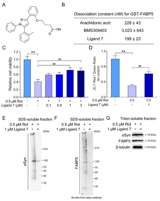
Biomedicines 2021, 9, 110                                                                                                          6 of 15

      Figure 2. Effects of rotenone on αSyn oligomerization and aggregation in FABP5/αSyn-transfected Neuro-2A cells.
      (A–D) Representative images of immunoblots probed with antibodies against αSyn and FABP5 under non-denaturing
      conditions of Triton-soluble and SDS-soluble fractions. Detection with anti-β-tubulin antibody was used as a protein-loading
      control. (E–G) Quantitative analyses of protein contents of different molecular weights, which indicate protein complexes
      of dimer/trimers, oligomers, and aggregates. Results are presented as means ± SEM (n = 3 respective cell experiments).
      * p < 0.05, ** p < 0.01, vs. 0.1 µM rotenone-treated αS/F5 cells. n.s., not statistically significant. D/T: dimers/trimers; O:
      oligomers; A: aggregates. bk besides blots indicate non-specific bands.

                                  3.3. FABP5 Co-Localizes with αSyn Aggregates Induced by Oxidative Stress
                                       Next, cells were subjected to immunofluorescent staining to analyze the subcellular
                                  localization of αSyn and FABP5 (Figure 3). The signals detected for both αSyn and FABP5
                                  showed a diffused cellular pattern of localization (Figure 3A). However, when treated with
                                  0.1 or 0.5 µM rotenone, immunolabeling revealed visible significant αSyn accumulation
                                  in FABP5/αSyn co-overexpressing cells (Figure 3B, arrow). FABP5 immunoreactivity
                                  co-localized with αSyn aggregates in rotenone-treated cells (Figure 3B), suggesting that
                                  FABP5 forms complexes with αSyn aggregates. Taken together, these results suggested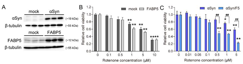
Biomedicines 2021, 9, 110                                                                                                        7 of 15

                                  that rotenone promotes FABP5 and αSyn interactions, thereby causing αSyn oligomer and
                                  aggregate formation.

      Figure 3. FABP5 co-localized with rotenone-induced α-Synuclein aggregates. (A,B) Representative images of immunoflu-
      orescent staining showing localization of αSyn (red), FABP5 (green), and DAPI (blue) in Neuro-2A cells. (A) Cells were
      transfected with pcDNA 3.1, FABP5, αSyn, and αS/F5 respectively, then exposed to DMSO. Scale bars: 10 µm. (B) αS/F5-
      transfected cells were exposed to 0.1 or 0.5 µM rotenone. Arrows indicate αSyn aggregates in cells treated with rotenone.
      Scale bars: 10 µm. The region of interest is indicated with a white square and magnified in insets. Scale bars in the higher
      magnifying images: 2 µm. (C,D) Quantitative analyses of the numbers of αSyn aggregates and αSyn/FABP5 positive
      aggregates. Results are presented as means ± SEM (n > 40 cells from three respective cell experiments). ** p < 0.01, vs.
      0.1 µM rotenone-treated αS/F5 cells; ## p < 0.01, vs. 0.5 µM rotenone-treated αS/F5 cells.

                                  3.4. FABP5 is Involved in Mitochondrial Membrane Potential Reduction by Rotenone
                                        We further employed the JC-1 assay to detect mitochondrial membrane potentials
                                  (Figure 4). Reductions in mitochondrial membrane potentials are indicated by a decrease
                                  in the ratio of red to green fluorescence of the JC-1 dye. First, we evaluated the effects of
                                  transfection on cells and found that there were no obvious effects (Figure 4A,C). Meanwhile,
                                  treatment with rotenone at 0.1 and 0.5 µM significantly decreased JC-1 red fluorescence
                                  and the ratio (red/green), especially in αSyn/F5 cells (Figure 4B,D). The ratio at 0.1 µM
                                  rotenone was not altered in αSyn-expressing cells, but was significantly reduced in αSyn/F5
                                  cells (Figure 4D).
Biomedicines 2021, 9, 110 8 of 15
Figure 4. FABP5 involvement in rotenone-induced reduction of mitochondrial activity. (A,B) Repre-
sentative images of JC-1-stained Neuro-2A cells. The decrease in the ratio of red to green fluorescence
of JC-1 dye was indicated by a reduction in mitochondrial membrane potential. (A) Cells were
transfected with pcDNA 3.1, FABP5, αSyn, and αS/F5, respectively, then exposed to DMSO. (B) αSyn
and αS/F5 transfected cells were exposed to 0.1 or 0.5 µM rotenone. (C,D) Graphs demonstrating
the quantification of the ratio of JC-1 red to green fluorescent intensity in Neuro-2A cells. Results are
presented as means ± SEM (n > 300 cells in C and n = 5 wells in D from parallel cell experiments).
* p < 0.05, ** p < 0.01, vs. DMSO-treated groups; # p < 0.05, vs. αSyn-transfected groups. n.s., not
statistically significant. Scale bars: 10 µm.Biomedicines 2021, 9, 110 9 of 15
3.5. FABP5 Ligand Rescues Rotenone-Induced Cell Death and Impedes αSyn Oligomerization and
Aggregation
Next, we evaluated whether FABP5 ligand can abolish oxidative stress toxicity and
αSyn oligomerization. BMS309403 was identified as a small-molecule FABP4-selective
inhibitor, with a particularly high affinity for FABP4 [27]. Previously, we developed several
BMS309403 derivatives and reported their affinity for GST-FABP5 [24,28]. Based on these
studies, we used ligand 7, which has high affinity for FABP5, with a dissociation constant
(Kd) of 199 ± 23 nM, which is almost identical to that of the intrinsic fatty acid, arachidonic
acid (AA) at 228 ± 43 nM (Figure 5A,B) [24]. Treatment of ligand 7 increased cell viability
after 0.5 µM rotenone treatment, and there was a statistically significant difference at 1 and
3 µM ligand 7 (Figure 5C). The reduction in the JC-1 ratio (red/green) by rotenone was also
significantly improved by treatment with 1 µM ligand 7 (Figure 5D). We further analyzed
oligomerization and aggregation of αSyn and FABP5 in the SDS-soluble fraction, which was
apparently suppressed by ligand 7 (Figure 5E,F). At the same time, the monomers of αSyn
and FABP5 remained in Triton-soluble fractions, which is consistent with Figure 2A,C and
Figure 5G. These results suggested that FABP5 inhibitor (ligand 7) effectively attenuated
αSyn oligomerization and toxicity by conserving mitochondrial function.Biomedicines 2021, 9, 110 10 of 15
Figure 5. Effects of FABP5 ligand 7 on cell death and αSyn oligomerization and aggregation. All experiments in Figure 5
were carried in αS/F5-transfected cells. (A) Chemical structure of ligand 7. (B) Dissociation constants (Kd values, nM)
of arachidonic acid, BMS309403, and ligand 7 for GST-FABP5 [24]. (C) αS/F5-transfected cells were exposed to 0.5 µM
rotenone and the indicated concentrations of ligand 7 for 48 h. Cell viability was measured using CCK-8 assays. Results
are presented as means ± SEM (n = 6 parallel cell experiments). ** p < 0.01, vs. non-rotenone-treated cells; ## p < 0.01,
vs. rotenone-treated cells without ligand. (D) Graphs demonstrating quantification of the JC-1 ratio (red/green). Results
are presented as means ± SEM (n = 5 parallel cell experiments). ** p < 0.01, vs. non-rotenone-treated cells; ## p < 0.01,
vs. rotenone-treated cells without ligand. (E,F) Representative images of immunoblots developed with antibodies against
αSyn and FABP5 in αS/F5 cells treated with ligand 7. Three parallel cell experiments were performed with similar results.
(G) The monomers of αSyn and FABP5 remain in Triton-soluble fractions. Incubation with β-tubulin antibody was used as
a protein-loading control.
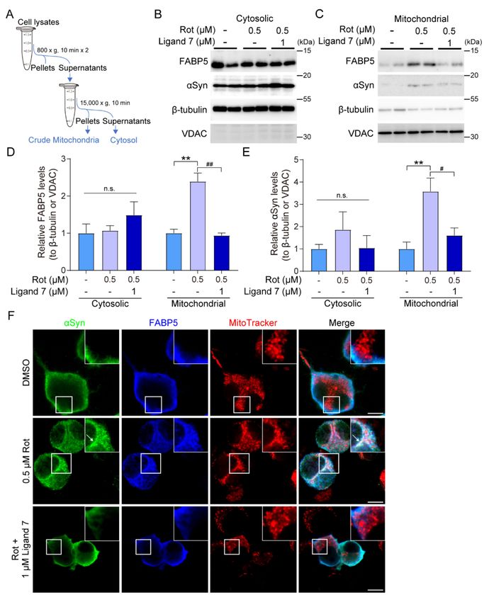
3.6. FABP5/αSyn Aggregates Targeting Mitochondria and Ligand 7 Prevent Abnormal
Accumulation
To further evaluate the mechanisms involved in protection, we isolated mitochondria
from αSyn/F5 cells (Figure 6A). Crude mitochondrial and cytosolic fractions were separated
and subjected to immunoblotting assays (Figure 6B,C). As a result, FABP5 and αSyn
abnormally accumulated in mitochondria in 0.5 µM rotenone-treated cells, which wasBiomedicines 2021, 9, 110 11 of 15
prevented by 1 µM ligand 7 treatment (Figure 6D,E). Consistent with these immunoblotting
results, αSyn and FABP5 were co-localized with the mitochondrial marker MitoTracker
in rotenone-treated cells (Figure 6F, arrow), which was blocked by ligand 7 treatment
(Figure 6F).
Figure 6. Abnormal accumulation of FABP5 and αSyn in mitochondria was prevented by ligand 7 treatment. All experiments
were carried in αS/F5-transfected cells. (A) Simple diagram of the mitochondrial isolation process in αS/F5-transfected cells.
(B,C) Representative images of immunoblots probed with antibodies against FABP5 and αSyn. Incubation with anti-β-
tubulin or VDAC antibodies were used as protein-loading controls for cytosolic or mitochondrial fractions, respectively.
(D,E) Quantitative densitometric analyses of αSyn and FABP5 in different fractions. Results are presented as means ± SEM
(n = 4, two respective cell experiments, each with 2 parallel dishes). ** p < 0.01, vs. non-rotenone-treated cells; # p < 0.05
and ## p < 0.01, 1 µM vs. rotenone-treated cells without ligand. (F) Confocal images of αSyn (green), FABP5 (blue), and
MitoTracker (red) in αS/F5-transfected cells. The region of interest is indicated with a white square and magnified in insets.
Arrow indicates αSyn aggregates which approach and gather in mitochondria with FABP5 in rotenone-treated cells. There
were no significant αSyn- and FABP5-positive aggregates in mitochondria of ligand 7-treated cells. Scale bars: 10 µm.Biomedicines 2021, 9, 110 12 of 15
4. Discussion
In this study, we demonstrated that FABP5 plays a key role in oligomerization and
aggregation of αSyn under oxidative stress in Neuro-2A cells. In the absence of rotenone,
both αSyn and FABP5 localized with diffuse patterns in these cells (Figure 3A); however,
their aggregates appeared concentration-dependent on rotenone (Figure 3B–D). Rotenone
also led to aggregate localization in mitochondria, and decreased mitochondrial membrane
potential, which might have resulted in cell death (Figure 7). We also found that FABP5
ligand 7 treatment abolished the toxicity, αSyn aggregation, and mitochondrial impairment
in Neuro-2A cells.
Figure 7. Schematic diagram of the mechanism by which αSyn/FABP5 aggregates aggravate mitochondrial dysfunction and
induce neuronal cell apoptosis. FABP5 ligand 7 binds with FABP5 and blocks aggregate formation to attenuate neurotoxicity.
(Left) when exposed to rotenone, αSyn accumulated with FABP5 to form oligomers and aggregates, which were further
targeted to mitochondria. The reduced mitochondrial membrane potential indicated mitochondrial dysfunction which
induced cell death. (Right) pharmacological inhibition of FABP5 by ligand 7 prevented αSyn/FABP5 aggregate formation,
thereby rescuing neuronal cells.
The most basic function of FABPs is to participate in the utilization of lipids in food-
stuffs, and to mediate fatty acid transport to various organelles, including mitochondria.
A recent study indicated that both acute shRNA knockdown of FABP5 and pharmacologic
inhibition results in decreased mitochondrial mass and respiration in regulatory T cells [29].
FABP5 induces mitochondrial macropore formation through BAX and voltage-dependent
anion channels (VDAC-1), which mediate mitochondrial outer membrane permeabiliza-Biomedicines 2021, 9, 110 13 of 15
tion and ultimately accelerates mitochondria-induced oligodendrocyte death. However,
FABP7 does not localize in mitochondria under mitochondrial damage [30]. In FABP3-
overexpressing embryonic cancer cells (P19 cells), lower cellular ATP production was
accompanied by a dramatic decrease in mitochondrial membrane potential, and FABP3
overexpression also led to an imbalance in mitochondrial dynamics and excess intracellular
reactive oxygen species production [31]. These studies suggest that FABP proteins regulate
mitochondrial function by transporting fatty acids and reactive oxygen species produc-
tion in cells. In this study, however, we failed to confirm the changes to mitochondrial
membrane potential by FABP5 overexpression alone (Figure 4).
Various stimuli, including oxidative stress, can trigger translocation of αSyn to the
mitochondria. Transient or stable transfection of αSyn particles exhibit localization in the
mitochondria of neuroblastoma SHSY or HEK293 cells [32,33]. Expression of αSyn induces
cytochrome C release from mitochondria, or vulnerability to rotenone in these cultured cells.
Cellular acidification is also reported to enhance mitochondrial αSyn accumulation [34].
In this study, we could not confirm αSyn localization to mitochondria, or upregulated
toxicity of rotenone by overexpression of αSyn alone. Oxidative stress induced by rotenone
triggered toxicity and αSyn oligomerization and mitochondrial injury was aggravated by
FABP5 co-expressed in Neuro-2A cells (Figure 6).
Aggregation and mitochondrial localization of αSyn and FABP5 may underlie the toxic
mechanisms induced by rotenone. Reactive oxygen species produce oxidized aldehydes of
polyunsaturated fatty acids such as 4-hydroxy-2-nonenal (4-HNE), 4-hydroxy-2-hexenal,
and malondialdehyde [35]. FABP4, another FABP family protein, binds not only with
fatty acids, but also with metabolites such as 4-HNE [36]. Fatty acids are usually located
in the internal pocket of FABPs; however, 4-HNE can undergo covalent binding with
a cysteine residue at amino acid 117. This suggests that strong modifications of FABP
protein leads to prolonged regulation of its function. Such modification may underlie
mechanisms involving FABP5 localization together with αSyn in mitochondria, because this
cysteine residue is also conserved in FABP5, and ligand 7 treatment inhibited aggregation
and mitochondrial localization (Figure 6) [24]. However, further studies are required to
evaluate the underlying mechanisms whereby FABP5 and αSyn aggregations localize
to mitochondria.
Finally, we investigated whether FABP5 ligand treatment prevented its aggregation
and localization with αSyn induced by rotenone. FABP5 is highly expressed in the dopamin-
ergic neurons of SNpc [23]; however, little is known regarding the physiological relevance
in the neurons or pathological roles of FABP5 in PD. Our study provides evidence that
FABP5 protein is a potential therapeutic target in PD therapy.
Supplementary Materials: The following are available online at https://www.mdpi.com/2227-905
9/9/2/110/s1, Figure S1: There is no FABP3 and a very little FABP7 expression in Neuro-2A cells
(mock cells), Figure S2: The negative control of immunoblotting assay.
Author Contributions: Y.W.: investigation and original draft writing; Y.S. and A.C.: investigation;
I.K.: methodology, K.F.: supervision, review/editing, project administration, and funding. All
authors have read and agreed to the published version of the manuscript.
Funding: This work was supported in part by the Strategic Research Program for Brain Sciences
from the Japan Agency for Medical Research and Development (JP19dm0107071 and JP20dm0107071;
awarded to K.F.
Data Availability Statement: The data presented in this study are available on request from the
corresponding author.
Conflicts of Interest: The authors declare no conflict of interest.Biomedicines 2021, 9, 110 14 of 15
References
1. Fields, C.R.; Bengoa-Vergniory, N.; Wade-Martins, R. Targeting Alpha-Synuclein as a Therapy for Parkinson’s Disease. Front. Mol.
Neurosci. 2019, 12, 299. [CrossRef] [PubMed]
2. Mhyre, T.R.; Boyd, J.T.; Hamill, R.W.; Maguire-Zeiss, K.A. Parkinson’s disease. Subcell. Biochem. 2012, 65, 389–455. [CrossRef]
[PubMed]
3. Spillantini, M.G.; Schmidt, M.L.; Lee, V.M.Y.; Trojanowski, J.Q.; Jakes, R.; Goedert, M. α-synuclein in Lewy bodies. Nature 1997,
388, 839–840. [CrossRef]
4. Gilmozzi, V.; Gentile, G.; Castelo Rueda, M.P.; Hicks, A.A.; Pramstaller, P.P.; Zanon, A.; Lévesque, M.; Pichler, I. Interaction of
Alpha-Synuclein With Lipids: Mitochondrial Cardiolipin as a Critical Player in the Pathogenesis of Parkinson’s Disease. Front.
Neurosci. 2020, 14, 1051. [CrossRef]
5. Villar-Piqué, A.; Lopes da Fonseca, T.; Outeiro, T.F. Structure, function and toxicity of alpha-synuclein: The Bermuda triangle in
synucleinopathies. J. Neurochem. 2016, 139, 240–255. [CrossRef]
6. Bengoa-Vergniory, N.; Roberts, R.F.; Wade-Martins, R.; Alegre-Abarrategui, J. Alpha-synuclein oligomers: A new hope. Acta
Neuropathol. 2017, 134, 819–838. [CrossRef]
7. Delenclos, M.; Burgess, J.D.; Lamprokostopoulou, A.; Outeiro, T.F.; Vekrellis, K.; McLean, P.J. Cellular models of alpha-synuclein
toxicity and aggregation. J. Neurochem. 2019, 150, 566–576. [CrossRef]
8. Kalia, L.V.; Kalia, S.K.; Lang, A.E. Disease-modifying strategies for Parkinson’s disease. Mov. Disord. 2015, 30, 1442–1450.
[CrossRef] [PubMed]
9. Rodríguez-Losada, N.; de la Rosa, J.; Larriva, M.; Wendelbo, R.; Aguirre, J.A.; Castresana, J.S.; Ballaz, S.J. Overexpression of
alpha-synuclein promotes both cell proliferation and cell toxicity in human SH-SY5Y neuroblastoma cells. J. Adv. Res. 2020, 23, 37–45.
[CrossRef]
10. Davis, G.C.; Williams, A.C.; Markey, S.P.; Ebert, M.H.; Caine, E.D.; Reichert, C.M.; Kopin, I.J. Chronic parkinsonism secondary to
intravenous injection of meperidine analogues. Psychiatry Res. 1979, 1, 249–254. [CrossRef]
11. Ramsay, R.R.; Salach, J.I.; Singer, T.P. Uptake of the neurotoxin 1-methyl-4-phenylpyridine (MPP+ ) by mitochondria and its
relation to the inhibition of the mitochondrial oxidation of NAD+ -linked substrates by MPP+ . Biochem. Biophys. Res. Commun.
1986, 134, 743–748. [CrossRef]
12. Schapira, A.H.V.; Gegg, M. Mitochondrial contribution to parkinson’s disease pathogenesis. Parkinsons. Dis. 2011, 2011, 159160.
[CrossRef] [PubMed]
13. Grassi, D.; Howard, S.; Zhou, M.; Diaz-Perez, N.; Urban, N.T.; Guerrero-Given, D.; Kamasawa, N.; Volpicelli-Daley, L.A.;
LoGrasso, P.; Lasmézas, C.I. Identification of a highly neurotoxic α-synuclein species inducing mitochondrial damage and
mitophagy in Parkinson’s disease. Proc. Natl. Acad. Sci. USA 2018, 115, E2634–E2643. [CrossRef] [PubMed]
14. Di Maio, R.; Barrett, P.J.; Hoffman, E.K.; Barrett, C.W.; Zharikov, A.; Borah, A.; Hu, X.; McCoy, J.; Chu, C.T.; Burton, E.A.; et al.
α-synuclein binds to TOM20 and inhibits mitochondrial protein import in Parkinson’s disease. Sci. Transl. Med. 2016, 8, 1–15.
[CrossRef] [PubMed]
15. Nakamura, K.; Nemani, V.M.; Azarbal, F.; Skibinski, G.; Levy, J.M.; Egami, K.; Munishkina, L.; Zhang, J.; Gardner, B.;
Wakabayashi, J.; et al. Direct membrane association drives mitochondrial fission by the Parkinson disease-associated protein
α-synuclein. J. Biol. Chem. 2011, 286, 20710–20726. [CrossRef] [PubMed]
16. Li, B.; Hao, J.; Zeng, J.; Sauter, E.R. SnapShot: FABP Functions. Cell 2020, 182, 1066. [CrossRef] [PubMed]
17. Furuhashi, M.; Hotamisligil, G.S. Fatty acid-binding proteins: Role in metabolic diseases and potential as drug targets. Nat. Rev.
Drug Discov. 2008, 7, 489–503. [CrossRef]
18. D’Anneo, A.; Bavisotto, C.C.; Gammazza, A.M.; Paladino, L.; Carlisi, D.; Cappello, F.; de Macario, E.C.; Macario, A.J.L.; Lauricella,
M. Lipid chaperones and associated diseases: A group of chaperonopathies defining a new nosological entity with implications
for medical research and practice. Cell Stress Chaperones 2020, 25, 805–820. [CrossRef]
19. Bando, Y.; Yamamoto, M.; Sakiyama, K.; Inoue, K.; Takizawa, S.; Owada, Y.; Iseki, S.; Kondo, H.; Amano, O. Expression of
epidermal fatty acid binding protein (E-FABP) in septoclasts in the growth plate cartilage of mice. J. Mol. Histol. 2014, 45, 507–518.
[CrossRef]
20. Yu, S.; Levi, L.; Casadesus, G.; Kunos, G.; Noy, N. Fatty acid-binding protein 5 (fabp5) regulates cognitive function both by
decreasing anandamide levels and by activating the nuclear receptor peroxisome proliferatoractivated receptor α/β (pparα/β)
in the brain. J. Biol. Chem. 2014, 289, 12748–12758. [CrossRef]
21. Owada, Y.; Yoshimoto, T.; Kondo, H. Spatio-temporally differential expression of genes for three members of fatty acid binding
proteins in developing and mature rat brains. J. Chem. Neuroanat. 1996, 12, 113–122. [CrossRef]
22. Liu, R.Z.; Mita, R.; Beaulieu, M.; Gao, Z.; Godbout, R. Fatty acid binding proteins in brain development and disease. Int. J. Dev.
Biol. 2010, 54, 1229–1239. [CrossRef] [PubMed]
23. Zhou, Q.; Li, J.; Wang, H.; Yin, Y.; Zhou, J. Identification of nigral dopaminergic neuron-enriched genes in adult rats. Neurobiol.
Aging 2011, 32, 313–326. [CrossRef] [PubMed]
24. Shinoda, Y.; Wang, Y.; Yamamoto, T.; Miyachi, H.; Fukunaga, K. Analysis of binding affinity and docking of novel fatty
acid-binding protein (FABP) ligands. J. Pharmacol. Sci. 2020, 143, 264–271. [CrossRef]
25. Schneider, C.A.; Rasband, W.S.; Eliceiri, K.W. NIH Image to ImageJ: 25 years of image analysis. Nat. Methods 2012, 9, 671–675.
[CrossRef]Biomedicines 2021, 9, 110 15 of 15
26. Carré, M.; André, N.; Carles, G.; Borghi, H.; Brichese, L.; Briand, C.; Braguer, D.; Medicine, U.F.R.; La, U.O. Tubulin is an inherent
component of mitochondrial membranes that interacts with the voltage-dependent anion channel. J. Biol. Chem. 2002, 277, 33664–33669.
[CrossRef]
27. Sulsky, R.; Magnin, D.R.; Huang, Y.; Simpkins, L.; Taunk, P.; Patel, M.; Zhu, Y.; Stouch, T.R.; Bassolino-Klimas, D.;
Parker, R.; et al. Potent and selective biphenyl azole inhibitors of adipocyte fatty acid binding protein (aFABP). Bioorg. Med.
Chem. Lett. 2007, 17, 3511–3515. [CrossRef]
28. Cheng, A.; Shinoda, Y.; Yamamoto, T.; Miyachi, H.; Fukunaga, K. Development of FABP3 ligands that inhibit arachidonic
acid-induced α-synuclein oligomerization. Brain Res. 2019, 1707, 190–197. [CrossRef]
29. Field, C.S.; Baixauli, F.; Kyle, R.L.; Puleston, D.J.; Cameron, A.M.; Sanin, D.E.; Hippen, K.L.; Loschi, M.; Thangavelu, G.;
Corrado, M.; et al. Mitochondrial Integrity Regulated by Lipid Metabolism Is a Cell-Intrinsic Checkpoint for Treg Suppressive
Function. Cell Metab. 2020, 31, 422–437. [CrossRef]
30. Cheng, A.; Kawahata, I.; Fukunaga, K. Fatty acid binding protein 5 mediates cell death by psychosine exposure through
mitochondrial macropores formation in oligodendrocytes. Biomedicines 2020, 8, 635. [CrossRef]
31. Song, G.X.; Shen, Y.H.; Liu, Y.Q.; Sun, W.; Miao, L.P.; Zhou, L.J.; Liu, H.L.; Yang, R.; Kong, X.Q.; Cao, K.J.; et al.
Overexpression of FABP3 promotes apoptosis through inducing mitochondrial impairment in embryonic cancer cells.
J. Cell. Biochem. 2012, 113, 3701–3708. [CrossRef] [PubMed]
32. Parihar, M.S.; Parihar, A.; Fujita, M.; Hashimoto, M.; Ghafourifar, P. Mitochondrial association of alpha-synuclein causes oxidative
stress. Cell. Mol. Life Sci. 2008, 65, 1272–1284. [CrossRef] [PubMed]
33. Shavali, S.; Brown-Borg, H.M.; Ebadi, M.; Porter, J. Mitochondrial localization of alpha-synuclein protein in alpha-synuclein
overexpressing cells. Neurosci. Lett. 2008, 439, 125–128. [CrossRef] [PubMed]
34. Cole, N.B.; DiEuliis, D.; Leo, P.; Mitchell, D.C.; Nussbaum, R.L. Mitochondrial translocation of α-synuclein is promoted by
intracellular acidification. Exp. Cell Res. 2008, 314, 2076–2089. [CrossRef]
35. Assies, J.; Mocking, R.J.T.; Lok, A.; Ruhé, H.G.; Pouwer, F.; Schene, A.H. Effects of oxidative stress on fatty acid- and one-carbon-
metabolism in psychiatric and cardiovascular disease comorbidity. Acta Psychiatr. Scand. 2014, 130, 163–180. [CrossRef]
36. Hellberg, K.; Grimsrud, P.A.; Kruse, A.C.; Banaszak, L.J.; Ohlendorf, D.H.; Bernlohr, D.A. X-ray crystallographic analysis of
adipocyte fatty acid binding protein (aP2) modified with 4-hydroxy-2-nonenal. Protein Sci. 2010, 19, 1480–1489. [CrossRef]You can also read