Imaging features of mycotic aortic aneurysms
←
→
Page content transcription
If your browser does not render page correctly, please read the page content below
Review Article

Imaging features of mycotic aortic aneurysms
Nan Zhang1#, Wei Xiong2#, Yu Li1, Qinxiang Mao3, Shangdong Xu4, Junming Zhu4, Zhonghua Sun5,
Lizhong Sun4
1
    Department of Radiology, Beijing Anzhen Hospital, Capital Medical University, Beijing, China; 2Department of Respiration, First Teaching
Hospital of Tianjin University of Traditional Chinese Medicine, Tianjin, China; 3Department of Radiology, Longtan Hospital of Guangxi Zhuang
Autonomous Region, Liuzhou, China; 4Department of Cardiovascular Surgery, Beijing Aortic Disease Centre, Beijing Anzhen Hospital, Capital
Medical University, Beijing Institute of Heart Lung and Blood Vessel Diseases, Beijing Engineering Research Centre for Vascular Prostheses,
Beijing, China; 5Discipline of Medical Radiation Science, Curtin Medical School, Curtin University, Perth, Australia

#
These authors contributed equally to this work as co-first author.

Correspondence to: Professor Yu Li. Department of Radiology, Beijing Anzhen Hospital, Capital Medical University, Chaoyang District, Anzhen Road
2nd, Beijing 100029, China. Email: 1523115105@qq.com; Prof Zhonghua Sun, Discipline of Medical Radiation Science, Curtin Medical School,
Curtin University, Perth, 6102, Australia. Email: z.sun@curtin.edu.au.

                  Abstract: Infectious aortitis (IA) is a rare and life-threatening cardiovascular disease. Early diagnosis and
                  timely intervention are crucial for reducing mortality associated with mycotic aortic aneurysms (MAAs);
                  however, early diagnosis is challenging due to the nonspecific symptoms. Some cases are diagnosed at an
                  advanced stage or after developing complications, such as rupture or aortic fistula. Current state-of-the-
                  art imaging modalities—including computed tomography (CT), magnetic resonance imaging (MRI), and
                  18F-fluorodeoxyglucose (FDG) positron emission tomography (PET)/CT—can detect infected aneurysms in
                  clinically suspicious cases. MAA features on imaging include lobulated pseudoaneurysm, indistinct irregular
                  arterial wall, perianeurysmal gas, perianeurysmal edema, perianeurysmal soft tissue mass, aneurysmal
                  thrombosis, and high metabolic activity with increased uptake of FDG. Enlarged lymph nodes are often
                  found adjacent to the aneurysm, while iliopsoas abscess (IPA), spondylitis, and aortic fistulas are commonly
                  associated complications. After surgery or endovascular repair, radiological features—including ectopic
                  gas, peri-graft fluid, thickening of adjacent bowel, pseudoaneurysm formed at the graft anastomosis, and
                  increased uptake of FDG—may indicate an infection of aortic graft. This article provides an overview of the
                  clinical and imaging features of MAAs. Thus, familiarity with the imaging appearances of MAAs may assist
                  radiologists in the diagnosis and facilitation of timely treatment.

                  Keywords: Infection; aorta; mycotic aneurysm; computed tomography; imaging

                  Submitted Aug 03, 2020. Accepted for publication Dec 23, 2020.
                  doi: 10.21037/qims-20-941
                  View this article at: http://dx.doi.org/10.21037/qims-20-941

Introduction                                                                     cause, such as a fungal infection (1); rather, MAAs is an
                                                                                 acute inflammatory response to pathogenic infection, which
Infectious aortitis (IA) is a rare, life-threatening cardiovascular              induces neutrophilic infiltration at the arterial wall. During
disease for which early diagnosis can be missed due to a lack                    this process, the collagenolytic and elastolytic enzymes are
of specific clinical, radiological, and laboratory features. IA’s                activated, which is concomitant with saccular lumen dilation
common sign is a mycotic aortic aneurysm (MAA), which                            and rupture (2-4).
manifests as a mushroom-shaped structure on a blood vessel.                         MAAs are associated with high mortality due to their
This manifestation does not refer to a specific pathogenic                       increased risk of rupture, which is especially common in the

© Quantitative Imaging in Medicine and Surgery. All rights reserved.     Quant Imaging Med Surg 2021;11(6):2861-2878 | http://dx.doi.org/10.21037/qims-20-941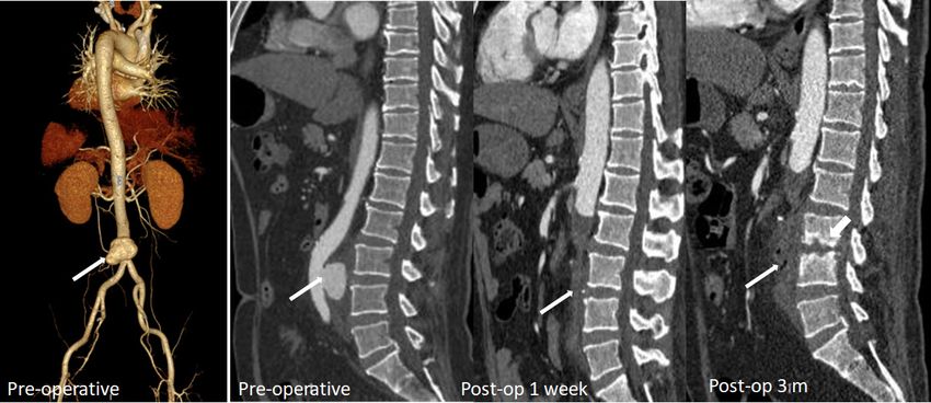
2862                                                                                          Zhang et al. Clinical and radiological features of IA

abdominal aorta compared with peripheral arteries (5). Early                   Salmonella
diagnosis and timely intervention are critical in reducing the
                                                                               Non-typhoidal Salmonella has been reported as the most
mortality of MAAs; however, early diagnosis is challenging
                                                                               common organism in Eastern countries, especially in the
due to nonspecific symptoms and low sensitivity of blood
                                                                               atherosclerotic abdominal aorta (8,16,18-20). Salmonella
cultures (6-9). Furthermore, the incidence of adverse
                                                                               usually resides in the phagosomes of host macrophages and
events in patients with MAAs after invasive treatment is
                                                                               other antigen-presenting cells, such as dendritic cells, which
higher than those with pseudoaneurysms associated with
                                                                               participate in the formation of atherosclerotic plaques.
other causes, such as trauma and atherosclerosis. The
                                                                               The immunocompromised condition contributes to its
endovascular stenting of MAAs may be associated with
                                                                               reproduction and invasiveness from atherosclerotic plaques
a high risk of stent infection, endoleak, reinfection, and
                                                                               (21-23) (Figure 1).
potential rupture (10,11).
    The diagnosis of MAAs requires awareness of the spectra
of computed tomography (CT), magnetic resonance                                Klebsiella pneumoniae (KP)
imaging (MRI), and 18 F-fluorodeoxyglucose (FDG)
positron emission tomography (PET)/CT. The use of CT                           Klebsiella infection can occur in almost all organs but
angiography (CTA) in the assessment of aortic disease—                         is most commonly observed in the liver and lungs.
including MAAs—is increasing, owing to its non-invasive,                       Approximately 60–93% of patients with KP have comorbid
efficient, broad coverage and its isotropic voxel capabilities.                diabetes mellitus (15,24,25). KP can invade the aortic wall
With CT technology development and advanced dose                               and induce MAAs from the location of damaged vascular
reduction techniques, CTA is a fast and high-quality                           endothelial walls (16). In patients suffering from aortic
method with minimal contrast medium and radiation dose.                        pseudoaneurysm and pyogenic liver abscess and who have a
The high tissue resolution of MRI can provide valuable                         history of diabetes mellitus, KP should be considered as the
anatomical and physiological information, especially in                        causative organism (Figure 2).
assessing abscess and tissue edema. In the latest studies,
18
   F-FDG-PET/CT has shown higher sensitivity and                               Mycobacterium tuberculosis
diagnostic accuracy in infected aortic aneurysms and aortic
prosthetic graft infection compared to CTA (12,13).                            About 75% of MAAs caused by Mycobacterium tuberculosis
    This review provides an overview of the clinical,                          present as a contiguous lesion on the surrounding tissue,
pathological, and radiological presentations of MAAs. The                      such as lymph node enlargement or paraspinal abscess
imaging findings during the period following medication,                       (26,27). Constant surveillance imaging can indicate
interventional, or surgical management are also described.                     pathogenesis (Figure 3).
The imaging characteristics in other infectious pathogens
are highlighted for differential diagnosis, especially in                      Staphylococcus aureus
patients with negative blood culture results.
                                                                               Pseudoaneurysm caused by Staphylococcus aureus has been
                                                                               reported in intravenous drug users, as well as iatrogenic
Pathogenesis
                                                                               or traumatic arterial wall injury patients (28,29) (Figure 4).
MAAs usually occur in the elderly, predominantly affecting                     In these patients, MAAs develop from direct infectious
immunocompromised patients, such as diabetes mellitus,                         inoculation at the time of vascular trauma.
liver cirrhosis, end-stage renal disease, alcoholism,
chronic glucocorticoid therapy, post-transplantation
                                                                               Brucella melitensis
immunosuppression, human immunodeficiency virus
infection, drug abuse, and malignancy (14-17). The                             Brucella melitensis is a zoonotic intracellular Gram-negative
known causative organisms of MAAs are Salmonella,                              coccobacillus responsible for multisystem infections,
Staphylococcus aureus, Klebsiella pneumoniae (KP), Escherichia                 including the aorta—especially in immunocompromised
coli, Mycobacterium, and Brucella melitensis. Fungi, such                      patients. The common transmission route is through
as Candida albicans and Aspergillus, are also rare causes of                   direct contact with infected cattle via milk or other body
infected aneurysms.                                                            fluids (30).

© Quantitative Imaging in Medicine and Surgery. All rights reserved.   Quant Imaging Med Surg 2021;11(6):2861-2878 | http://dx.doi.org/10.21037/qims-20-941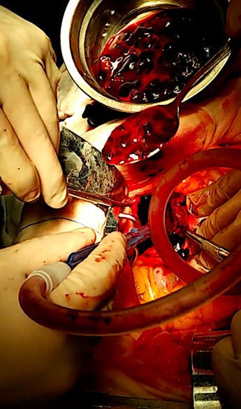
Quantitative Imaging in Medicine and Surgery, Vol 11, No 6 June 2021                                                                                 2863

                           A                                                 B

                           C                                                 D

Figure 1 MAAs caused by Salmonella. A 58-year-old man with a history of the human immunodeficiency virus (HIV) and cirrhosis presented
with fever and increasing abdominal pain. Salmonella was captured in hemocultures. (A) Three-dimensional (3D) CT angiography of
abdominal aorta shows saccular pseudoaneurysm formation (arrow). (B) Coronal CT view shows mycotic aneurysm with periaortic soft
tissue (long arrow) and mural thrombus (short arrow). (C,D) Transverse view (arterial and venous phases) shows disruption in the aortic
wall. Periaortic soft tissue can be enhanced in the venous phase (long arrow). Lymph nodes are enlarged (short arrow). MAAs, mycotic aortic
aneurysms.

Clinical features                                                                 Life-threatening hemorrhage—such as hemoptysis,
                                                                               gastrointestinal hemorrhage, and sequentially shock—could
Due to the risk of rapid expansion and consequent rupture,
                                                                               result from fistulae. The poor prognosis of these patients
timely diagnosis and treatment of MAAs are paramount.
                                                                               emphasizes the importance of early diagnosis. Aortoenteric
Unfortunately, some patients could be clinically silent
                                                                               fistula is caused by aneurysm infection spreading to the
until an aneurysm rupture. The most frequent presenting                        enteron, which usually begins at the duodenum, adjacent to
symptoms of MAAs are fever and pain (16,19). MAAs                              the aorta (Figure 5). Thoracic MAAs with surrounding lung
localized to the thoracic aorta usually manifest as chest                      parenchyma infection or compaction cause aortobronchial
pain, whereas infected abdominal aortic aneurysms usually                      fistula (Figure 6), a less recognized complication of
manifest as abdominal pain with or without a pulsatile mass.                   abdominal MAAs. A dramatic increase in mortality occurs
The laboratory abnormalities are often nonspecific and may                     in patients without clear preoperative diagnosis compared
include elevated erythrocyte sedimentation rate (ECR),                         to those with clear preoperative diagnosis (100% in patients
C-reactive protein (CRP), and leukocytosis. Blood cultures                     without a clear diagnosis vs. 15% in patients with clear
fail to detect bacteria in approximately 25% of cases, which                   diagnosis).
might contribute to broad-spectrum antibiotic therapy                             Iliopsoas abscess (IPA) is a common complication in
administration.                                                                abdominal MAAs with or without endograft infection,

© Quantitative Imaging in Medicine and Surgery. All rights reserved.   Quant Imaging Med Surg 2021;11(6):2861-2878 | http://dx.doi.org/10.21037/qims-20-941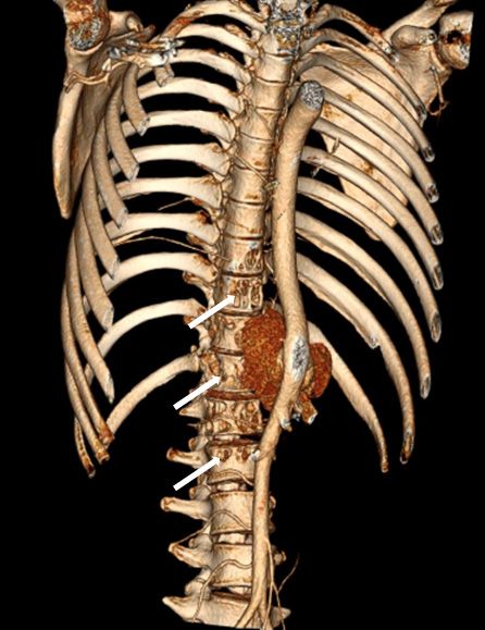
2864                                                                                          Zhang et al. Clinical and radiological features of IA

Figure 2 MAAs caused by Klebsiella pneumoniae. A 52-year-old male with a history of diabetes mellitus presented with fever (39 ℃), chills,
nausea, and abdominal pain. Blood culture of KP. Top row images: contrast-enhanced CT images obtained at 15 days after symptom onset.
Multiple cystic lesions (in S4 and S6 of the liver; arrows) with contrast-enhanced septum and wall demonstrate liver abscess. No signs
of aneurysm or inflammation around the abdominal aorta. Middle row images: contrast-enhanced CT images obtained 4 days after the
drainage of liver abscess. The abscess became smaller after drainage and antibiotic treatment. Bottom row images: contrast-enhanced CT
images obtained 35 days after the drainage of liver abscess (the patient relapsed 15 days after discharge). An aneurysm was found at the
original abscess focus (long arrow in the middle image). The saccular MAA showed surrounding soft tissue (short arrow). MAA, mycotic
aortic aneurysm.

presenting as a direct invasive infection with purulent                        with MAAs; thus, clinicians should be cautious about
materials occurring within the iliopsoas muscle. The                           this potential complication. In tuberculous spondylitis
causative organisms include Mycobacterium, Salmonella,                         patients, MAAs can involve secondary spread from spine
KP, other Gram-negative bacilli, and mixed bacteria. The                       lesions (Figure 7). The rate of mortality is high among
mortality rate is as high as 100% if IPA is left untreated                     MAA patients with combined pyogenic spondylitis
(31-33). IPA is reported as a major risk factor for patients                   (34,35).

© Quantitative Imaging in Medicine and Surgery. All rights reserved.   Quant Imaging Med Surg 2021;11(6):2861-2878 | http://dx.doi.org/10.21037/qims-20-941
Quantitative Imaging in Medicine and Surgery, Vol 11, No 6 June 2021                                                                                 2865

Figure 3 MAAs caused by Mycobacterium tuberculosis. A 72-year-old male repeatedly presented over 9 months with fevers. First-row images:
CT images in January 2018 showed multiple miliary nodules and peripheral reticulation in bilateral lungs. Right pleural effusion and pleural
thickness (black arrow), coronary artery calcification, and small lymph nodes adjacent to the posterior wall of the thoracic aorta are marked
(white arrow). Second-arrow images: contrast-enhanced CT images in March 2018 demonstrated a small pseudoaneurysm adjacent to
the enlarged lymph nodes (arrow). Third-row images: CT images in April 2018 showed stent graft and lymph nodes enlarged more than
previously, with hypodense center indicating necrosis (arrow). Fourth-row images: CT images in October 2018 (standard antitubercular
drug treatment for 6 months) showed partially absorbed lung nodules and pleural effusion. The size of lymph nodes decreased significantly
(arrow). MAAs, mycotic aortic aneurysms.

© Quantitative Imaging in Medicine and Surgery. All rights reserved.   Quant Imaging Med Surg 2021;11(6):2861-2878 | http://dx.doi.org/10.21037/qims-20-941
2866                                                                                          Zhang et al. Clinical and radiological features of IA

                           A                                                B

                           C                                                D

Figure 4 MAAs caused by Staphylococcus aureus. A 66-year-old woman presented with fever and increasing chest pain with a history of
coronary artery bypass grafting performed before 6 months. Blood culture of Staphylococcus aureus. (A) CTA demonstrates the patency of the
left descending artery bypass graft (arrow), (B,C) Irregular and lobular aneurysm arises from the ascending aorta (arrow). (D) The aneurysm
ruptures into the anterior chest wall (arrow) with extensive soft tissue thickening (short arrows). CTA, computed tomography angiography;
MAAs, mycotic aortic aneurysms.

Clinical management                                                             component for successfully treating MAAs.

Open surgical repair has been regarded as an effective
treatment for MAA but is associated with a mortality                            Imaging features
rate of over 20% (17,29). The endovascular repair of
MAAs—an expeditious temporization of MAA rupture                                CT, MRI, and PET/CT are the most commonly used
in hemodynamic instability cases—is well-established.                           imaging modalities in the detection and assessment of
Kan et al. demonstrated that there was no significant                           clinically suspected infected aneurysms. Due to the higher
difference in overall survival rate between open surgical                       quality spatial resolution of contrast-enhanced CT and
repair and endovascular repair (20); however, the insertion                     MRI, these modalities can provide valuable information
of an endovascular graft in an infected field remains a                         regarding the morphology of aortic aneurysm, aortic wall
major concern. Stent graft implantation is a significant                        enhancement, and the relationship between the aneurysm
independent predictor of persistent infection (20).                             and adjacent tissue; however, PET/CT is the most sensitive
Prolonged culture-specific antibiotic therapy, combined                         of these modalities in detecting infection, which is depicted
with open surgery or endovascular techniques, is a key                          by increased uptake of FDG. Like PET/CT, diffusion-

© Quantitative Imaging in Medicine and Surgery. All rights reserved.   Quant Imaging Med Surg 2021;11(6):2861-2878 | http://dx.doi.org/10.21037/qims-20-941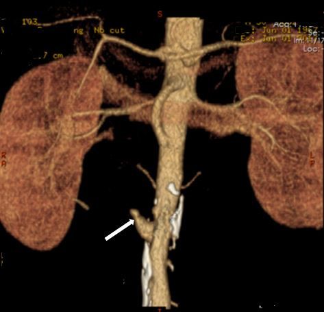
Quantitative Imaging in Medicine and Surgery, Vol 11, No 6 June 2021                                                                                 2867

A                                             B

Figure 5 Aortoenteric fistula. A 50-year-old male with a history of diabetes mellitus presented with fever, abdominal pain, and melena.
Blood culture showed the growth of Salmonella. (A) 3D CTA shows irregular MAA (arrow). (B) Transverse view demonstrates the
interruption of the aortic wall and saccular pseudoaneurysm formed with communication between the aorta and the duodenum (arrow).
MAA, mycotic aortic aneurysm.

A                                                       B                                             C

Figure 6 Aortobronchial fistula. An 84-year-old male with a history of diabetes mellitus and tuberculosis presented with fever, chest pain,
and hemoptysis. (A) Maximum-intensity projection image shows multiple small nodules with sharp edges and upper lobe distribution, cavity
(arrowhead), saccular pseudoaneurysm of the thoracic aorta with surrounding soft tissue (arrow), and patchy shadow in the right lower
lobe. (B) Minimum-intensity projection image shows the lumen of right endobasal segmental bronchus (adjacent to the aneurysm wall)
obstruction filled with high density material. (C) Patchy ground glass opacity and consolidation consistent with alveolar hemorrhage.

weighted imaging (DWI) MRI is also sensitive in detecting                      most sensitive imaging modality for detecting calcification
infection with restricted diffusion manifestation. DWI and                     and gas bubbles. The calcification interruption indicates
T2-weighted MRI can assist in detecting soft tissue edema                      the location of the disrupted aortic wall. Gas bubbles that
and adjacent organ involvement.                                                appear in and around MAAs have high diagnostic reliability
                                                                               of etiology (Figure 9). Rapidly progressive growth of true
                                                                               or false aneurysms (>5 mm in 2 weeks) is also suggestive of
Infected aortic aneurysm
                                                                               an infectious etiology (Figure 10). The thickened MAA wall
An infected aortic aneurysm appears on CT and MRI as a                         usually appears as a high signal intensity on T2-weighted
focal, contrast-enhancing, lobulated, saccular lumen, with                     MRI and DWI (Figure 11), with increased uptake of FDG
an indistinct, irregular aortic wall (Figure 8). CT is the                     on PET/CT.

© Quantitative Imaging in Medicine and Surgery. All rights reserved.   Quant Imaging Med Surg 2021;11(6):2861-2878 | http://dx.doi.org/10.21037/qims-20-941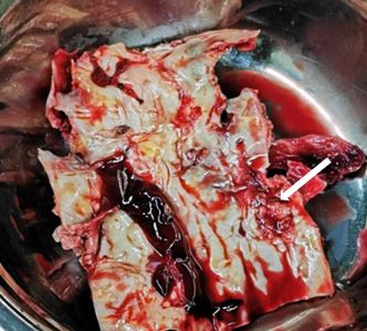
2868                                                                                          Zhang et al. Clinical and radiological features of IA

           A                                           B                                                C

           D                                                                                            E

Figure 7 Tuberculous spondylitis involving the aorta, with IPA and MAA formation. A 20-year-old male with a three-month history of back
pain, low fever (37.5–37.9 ℃), and a further acute episode of back pain aggravated in recent days. (A) 3D CTA shows giant irregular MAA
(arrow). (B,C) Right iliopsoas muscle is enlarged and swollen with a relatively low-density area and contrast-enhanced rim of the abscess wall
(arrows). (D) Initial spinal CT imaging shows erosion of the vertebral body and endplate at T11 (arrow) with soft tissue swelling or abscess
around the vertebral body. The outline of the aorta is distinct. CTA after 3 months shows the progression in the vertebral body’s destructive
changes (arrow), obliteration of fat planes between the vertebral body and aorta, and lobular pseudoaneurysm formed adjacent to the eroded
vertebral body. (E) 3D CTA shows multiple lesions in the vertebral body at T9–L2 and pseudoaneurysm. CTA, computed tomography
angiography; IPA, iliopsoas abscess; IPM, iliopsoas muscle; MAA, mycotic aortic aneurysm.

Periaortic tissue                                                              Adjacent organs

Eccentric periaortic inflammatory soft tissue usually                          CT provides a definitive diagnosis of IPA and other features
manifests as rim or septum enhancement following the                           that involve adjacent structures. IPA's typical features on
administration of contrast material (venous phase) on                          CT include enlarged iliopsoas muscle, single or multiple
contrast-enhanced CT and MRI (Figure 8). Periaortic                            relatively low-density abscess cavities with rim-like contrast-
edema appears as a distinctive fat stranding on CT; lymph                      enhanced walls (Figure 7), and in some cases, gas within
nodes adjacent to MAAs might also appear swollen and                           lesions. Interestingly, MRI and PET/CT are sensitive
enhanced (Figure 1). The edema of periaortic tissue and                        modalities in detecting IPA with typical restricted-diffusion
lymph nodes have the same MRI and PET/CT findings as                           manifestation on MRI and increased uptake of FDG on
the thickened MAAs wall described above (Figure 11).                           PET/CT (Figure 11).

© Quantitative Imaging in Medicine and Surgery. All rights reserved.   Quant Imaging Med Surg 2021;11(6):2861-2878 | http://dx.doi.org/10.21037/qims-20-941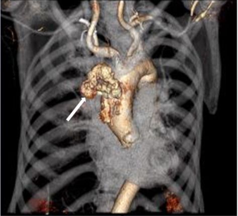
Quantitative Imaging in Medicine and Surgery, Vol 11, No 6 June 2021 2869
A
B C
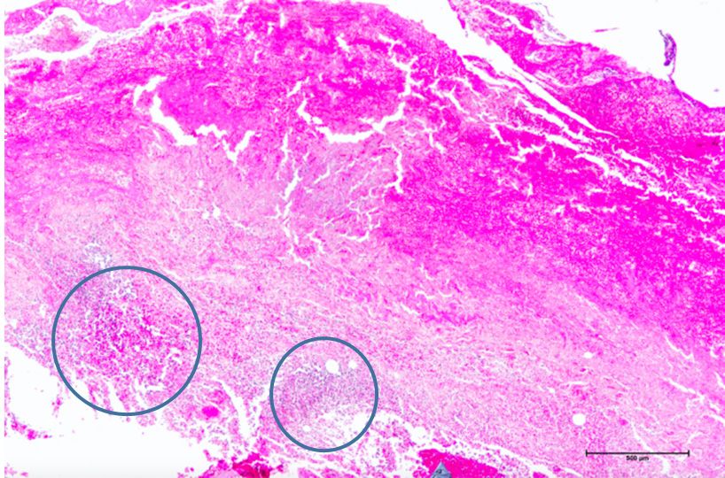
Figure 8 Morphology of MAAs. (A) Top row images: contrast-enhanced CT images show disruption to the aortic wall and saccular MAA
with rim enhancement of periaortic thickened soft tissue (arrows). (A) Bottom row images: Low-density periaortic soft tissue indicates
necrosis and abscess (arrows). The left posterior wall is intact, and a mural thrombus is observed in the lumen. (B) Intraoperative findings
identified stagnant abscess around the infected aorta. (C) The hematoxylin-eosin (H&E)-stained section of the infected aortic wall exhibits
inflammatory infiltration with microabscess formation (circles) and loss of normal media architecture. MAAs, mycotic aortic aneurysms.
Primary and secondary pyogenic spondylitis manifests early enhancement of the inferior vena cava well before
intervertebral disc and/or vertebral body destruction on CT it appears in the renal and hepatic parenchyma, with a
and MRI (Figure 12). The diagnosis of pyogenic spondylitis density of the adjacent digestive tract or bronchus similar
can also be confirmed on PET/CT with increased metabolic to that of the adjacent aorta. When aortacaval fistula
manifestations. MRI and PET/CT enable spondylitis occurs, dilated and retrograded enhanced renal or iliac
visualization without morphological changes in the early veins can be seen on enhanced CT and MRI, with direct
stage (Figure 11). communication between the aorta and inferior vena cava
Contrast medium shunt from the aorta to the inferior (Figure 13). Aortocutaneous fistulas are extremely rare and
vena cava, digestive tract, or bronchus, is a diagnostic sign are usually seen in cases involving prior vascular prosthetic
for aortic fistula (36,37). The shunt usually manifests as graft insertion (Figure 14) (38). Abnormal accumulation
© Quantitative Imaging in Medicine and Surgery. All rights reserved. Quant Imaging Med Surg 2021;11(6):2861-2878 | http://dx.doi.org/10.21037/qims-20-941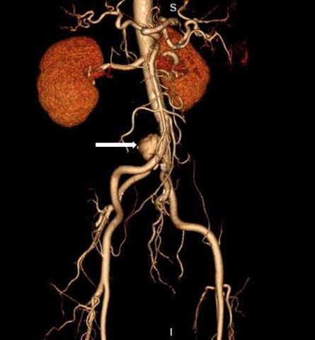
2870 Zhang et al. Clinical and radiological features of IA
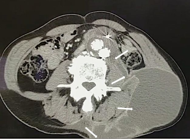
A B C
Figure 9 Air bubble sign. A 63-year-old male with a history of diabetes mellitus presented with fever and abdominal pain. Blood cultures consisted
of Salmonella. (A) 3D CTA shows saccular MAA at the abdominal aorta (arrow). (B,C) Transverse contrast-enhanced CT images show an eccentric
saccular MAA with posterior circumferential gas around the abdominal aorta. CTA, computed tomography angiography; MAA, mycotic aortic
aneurysm.
A B C
D E
Figure 10 The rapid progress of MAAs. A 56-year-old male with a history of diabetes mellitus presented with fever and chest pain. Salmonella was
detected in blood cultures. (A) 3D CTA of the aorta shows irregular MAA (arrow). (B) Transverse CTA image demonstrates the saccular aneurysm
(4.0×3.9 cm2). (C) The maximal caliber of the aneurysm increased to 7.6×6.1 cm2 on the 15-day follow-up. The left atrium is compressed (arrow).
Bilateral pleural effusion indicates the fragility of the aneurysm wall. (D) The macroscopic finding of the MAAs shows severe atherosclerosis and
mural thrombus in the lumen. The arrow shows the aortic destruction. (E) H&E-stained samples show diffuse inflammatory infiltrate (arrows) and
loss of normal media architecture. CTA, computed tomography angiography; MAAs, mycotic aortic aneurysms.
© Quantitative Imaging in Medicine and Surgery. All rights reserved. Quant Imaging Med Surg 2021;11(6):2861-2878 | http://dx.doi.org/10.21037/qims-20-941Quantitative Imaging in Medicine and Surgery, Vol 11, No 6 June 2021 2871
A B C
D E F
G H
Figure 11 A 69-year-old male with a history of diabetes mellitus presented with fever and abdominal pain. Blood cultures showed
Salmonella. (A) 3D MR angiography shows saccular MAA (arrow) at the abdominal aorta. The high signal intensity of the aneurysm wall and
anterior left psoas on T2-weighted image (B) was consistent with edema. The central part of the left psoas (arrow) showed significantly high
signal intensity on T2-weighted image (B) and DWI (C), low signal intensity on ADC (D), consistent with abscess. High signal intensity of
the vertebrae on T2-weighted image indicates edema on the edge of lumbar vertebrae (B) without bone destruction, but with obliteration of
fat planes between the vertebral body and left psoas (E). (F-H) During follow-up, the inflammation area was expanded on CT and MR after
4 months (arrow). DWI, diffusion weighted imaging; ADC, apparent diffusion coefficient; MAA, mycotic aortic aneurysm.
of FDG has diagnostic significance for aortoenteric and gas, peri-graft inflammation, and fluid, thickening of
aortobronchial fistulae. adjacent bowel, pseudoaneurysm formation, and increased
18
F-FDG uptake at the graft anastomosis (Figure 15)
(13,39). The peri-graft fluid within the first 3 months after
Follow-up imaging
surgery may contribute to postoperative changes (39).
Recognition of infection-related complications should Considering the white blood cell count and clinical
be considered during the analysis of follow-up images. symptoms, persistent existence, or gradual increase in the
Radiological signs are important diagnostic criteria in the peri-graft fluid should be suspected as an infection-related
detection of aortic graft infection (39), including ectopic complication. Extraanatomic bypass procedure avoids
© Quantitative Imaging in Medicine and Surgery. All rights reserved. Quant Imaging Med Surg 2021;11(6):2861-2878 | http://dx.doi.org/10.21037/qims-20-9412872 Zhang et al. Clinical and radiological features of IA
Figure 12 Spondylitis combined with MAAs. A 65-year-old male with a history of diabetes mellitus presented with fever and chest pain.
Blood cultures showed KP. Preoperative CTA shows lobular MAA (arrow in 3D view) with normal centrum (arrow in sagittal view).
Postoperative CTA shows normal vertebral body with prevertebral soft tissue (arrow in postoperative 1 week sagittal view). CTA after 3
months showed spondylitis short arrow with destruction of adjacent vertebral body endplate, lumbar vertebral space narrow at L3-4 level,
and air bubbles in the prevertebral soft tissue (long arrow). CTA, computed tomography angiography; MAAs, mycotic aortic aneurysms.
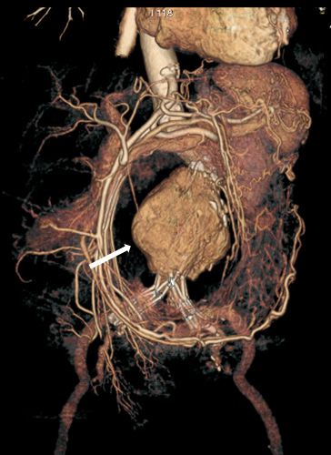
A B
C D
Figure 13 Aortocaval fistula. An 84-year-old female with a history of diabetes mellitus presented with fever, chest pain, and hemoptysis.
Blood cultures showed KP. (A) Coronal view of contrast-enhanced CT image shows eccentric pseudoaneurysm (arrow) with adjacent air
bubbles and surrounding soft tissue. (B,C) Air bubbles and low-density filling defect (thrombus) (arrow) are shown in the inferior vena
cava. Normal anatomical space between the aorta and inferior vena cava has disappeared. (D) Air bubbles and thrombus shown in the right
common iliac vein (arrow) with contrast enhancement earlier than that of the left indicates aortocaval fistula. The aortocaval fistula and
thrombus in the right common iliac vein and inferior vena cava was confirmed during surgery.
© Quantitative Imaging in Medicine and Surgery. All rights reserved. Quant Imaging Med Surg 2021;11(6):2861-2878 | http://dx.doi.org/10.21037/qims-20-941Quantitative Imaging in Medicine and Surgery, Vol 11, No 6 June 2021 2873
graft placement within an infected field, lowering the risk
of graft reinfection compared to in situ reconstruction
with a prosthetic graft (17). The endovascular repair of
MAAs should be regarded as a temporizing treatment
mainly performed on hemodynamically unstable patients.
Compared to surgery, the challenges of wide debridement
and effective drainage of suppuration brings risks of stent
infection (Figure 16), endoleak (Figure 17), aortic fistulas,
and potential rupture (8) (Figure 18). Spondylitis and IPA
secondary to reinfection occur after both open surgery and
endovascular repair (Figures 10,19). A prospective study—
Figure 14 Arteriocutaneous fistulas. A 75-year-old male with including 35 patients with suspected aortic graft infection—
a history of diabetes mellitus and infrarenal pseudoaneurysm, showed high concordance between 18F-FDG-PET/CT and
who had undergone endovascular aortic repair 6 months prior, expert consensus criteria from the Management of Aortic
presented with a left hip mass. Contrast-enhanced CT image Graft Infection Collaboration (MAGIC) for detecting aortic
shows an irregular subcutaneous cyst with rim enhancement graft infection (13). Further, 18F-FDG-PET/CT can also
connecting with the periaortic fluid. be used to monitor the response of MAAs and aortic graft
A B C
D E
Figure 15 Post-surgical reinfection. A 65-year-old male was diagnosed with mycotic aneurysm caused by Salmonella as shown on 3D CTA (arrow
in A). (B,C) The patient underwent open surgery, involving surgical resection of the aneurysm, extensive debridement of the surrounding soft
tissue, and extra-anatomic bypass which remained patent after 1 month. (D,E) Transverse and coronal views of follow-up CTA shows soft tissue
and air bubbles (arrows) in the native position of the mycotic aneurysm, indicating reinfection. CTA, computed tomography angiography.
© Quantitative Imaging in Medicine and Surgery. All rights reserved. Quant Imaging Med Surg 2021;11(6):2861-2878 | http://dx.doi.org/10.21037/qims-20-9412874 Zhang et al. Clinical and radiological features of IA
A B C
Figure 16 Stent graft infection. (A,B) A 61-year-old male diagnosed with MAAs (arrow) caused by Salmonella, who had undergone EVAR
2 months prior, presented with recurrent fever. (C) Transverse view of follow-up CTA shows an air bubble around the stent graft (arrow).
EVAR, endovascular aortic repair; CTA, computed tomography angiography; MAAs, mycotic aortic aneurysms.
A B C
Figure 17 Endoleak after EVAR. A 62-year-old male diagnosed with MAAs caused by Salmonella, who had undergone EVAR, presented
with recurrent fever and chest pain. (A) Preoperative CTA image. Curved planar reconstruction (CPR) shows MAA surrounded by soft
tissue. (B) CTA image 1 month post-EVAR shows that MAA is successfully occluded but periaortic soft tissue exists. (C) CPR 2 months
post-EVAR shows thickened periaortic soft tissue with endoleak at the caudal side of the stent graft (arrow). CTA, computed tomography
angiography; EVAR, endovascular aortic aneurysm repair; MAAs, mycotic aortic aneurysms.
infection to antibiotic treatment. PET/CT allow early identification of MAAs, which is
crucial for improving patient outcomes. Surveillance
imaging permits assessment and promotes treatment
Conclusions
efficacy. Figure 20 summarizes both clinical and radiological
Diagnosis of life-threatening MAAs is challenging due to features that can assist in the diagnosis of MAAs.
the non-specific symptoms and negative blood cultures Clinicians—especially cardiologists, vascular surgeons, and
associated with prior antibiotic use. CT, MRI, and FDG- radiologists—should be familiar with the clinical features
© Quantitative Imaging in Medicine and Surgery. All rights reserved. Quant Imaging Med Surg 2021;11(6):2861-2878 | http://dx.doi.org/10.21037/qims-20-941Quantitative Imaging in Medicine and Surgery, Vol 11, No 6 June 2021 2875
A B C
Figure 18 Aneurysm rupture. A 61-year-old male diagnosed with MAAs caused by Mycobacterium tuberculosis, who had undergone EVAR
2 years prior, presented with sudden-onset abdominal pain. (A) 3D CTA shows marked perivascular contrast media extravasation with
occupied effect (arrow). (B) Sagittal view of CTA shows destruction of vertebral bodies from L3 to L5 (arrows). (C) Transverse view of CTA
shows definite lobular perivascular contrast media extravasation, indicating rupture. A heterogenous, predominantly hypodense tissue existed
around the pseudoaneurysm, likely representing subacute hematoma and thrombus (arrow). No fat space is detected between the eroded
vertebral body and the pseudoaneurysm. CTA, computed tomography angiography; MAAs, mycotic aortic aneurysms.
A B C
D E
Figure 19 Spondylitis and iliopsoas abscess secondary to reinfection. A 60-year-old male with history of Sweet’s syndrome presented
with fever and blunt abdominal pain. (A-C) An infrarenal MAA (arrow) is shown on 3D volume rendering image and coronal multiplanar
reformatted (MPR) image with normal contour of iliopsoas. Hyperosteogeny was observed at the L5 vertebral body. (D,E) Follow-up CTA
at 5 months shows successful exclusion of the MAA. The stent graft in the left common iliac artery is occluded (short arrow in E). Bilateral
iliopsoai are swollen with a relatively low-density area with a small bubble (arrows) and contrast-enhanced rim, which support the diagnosis
of abscess. The abscess extends to the left erector spinae (arrow in D). Obliteration of fat planes between the vertebrae and iliopsoas, and
destruction of the vertebrae indicate spondylitis, is observed. MAA, mycotic aortic aneurysm.
© Quantitative Imaging in Medicine and Surgery. All rights reserved. Quant Imaging Med Surg 2021;11(6):2861-2878 | http://dx.doi.org/10.21037/qims-20-9412876 Zhang et al. Clinical and radiological features of IA
Diagnosis of mycotic aortic aneurysms
Clinical features Imaging features
Focal, saccular or lobular dilated lumen, with indistinct and
Most common symptoms
irregular aortic wall
Fever Laboratory CT MRI PET/CT
and paln abnormalities
• Contrast-enbancing
ECR, CRP, and Blood • Contrast-enhancing wall • Enhancement of
wall and periaortic
leukocytosis cultures (+) and periaortic inflammatory metabolism with increased
inflammatory tissue
elevated tissue FDG uptake
• Calcification interruption
• Edema with high SI on • Calcification interruption
• Gas bubbles and fat
T2W1 • Gas bubbles and fat
stranding
• Restricted diffusion on DWI stranding
• Lymph node
• Lymph node enlargement • Lymph node enlargement
enlargement
• IPA and aortic fistula • IPA and aortic fistula
• IPA and aortic fistula
Figure 20 Flow chart showing clinical and radiological features that assist with the diagnosis of mycotic aortic aneurysms. ECR, erythrocyte
sedimentation rate; CRP, C-reactive protein; DWI, diffusion weighted imaging; IPA, iliopsoas abscess; SI, signal intensity.
and common manifestations of MAAs to ensure appropriate Attribution-NonCommercial-NoDerivs 4.0 International
evaluation and management of the disease. License (CC BY-NC-ND 4.0), which permits the non-
commercial replication and distribution of the article with
the strict proviso that no changes or edits are made and the
Acknowledgments
original work is properly cited (including links to both the
The authors would like to thank Drs. Fengqiang Wang, formal publication through the relevant DOI and the license).
Huaiping Yuan and Jin Cheng for their assistance in See: https://creativecommons.org/licenses/by-nc-nd/4.0/.
providing some cases that were used in this study.
Funding: None.
References
1. Osler W. The Gulstonian Lectures, on Malignant
Footnote
Endocarditis. Br Med J 1885;1:522-6.
Conflicts of Interest: All authors have completed the 2. Maleszewski JJ. Inflammatory ascending aortic disease:
ICMJE uniform disclosure form (available at http://dx.doi. perspectives from pathology. J Thorac Cardiovasc Surg
org/10.21037/qims-20-941). Dr. ZS serves as an unpaid 2015;149:S176-83.
associate editor of Quantitative Imaging in Medicine and 3. Stone JR, Bruneval P, Angelini A, Bartoloni G, Basso C,
Surgery. The other authors have no conflicts of interest to Batoroeva L, Buja LM, Butany J, d’Amati G, Falloon JT,
declare. Gittenberger-de Groot AC, Gouveia RH, Halushka MK,
Kelly KL, Kholova I, Leone O, Litovsku SH, Maleszewski
Open Access Statement: This is an Open Access article JJ, Miller DV, Mitchell RN, Preston SD, Pucci A, Radio
distributed in accordance with the Creative Commons SJ, Rodriguez ER, Sheppard MN, Suvarna SK, Tan
© Quantitative Imaging in Medicine and Surgery. All rights reserved. Quant Imaging Med Surg 2021;11(6):2861-2878 | http://dx.doi.org/10.21037/qims-20-941Quantitative Imaging in Medicine and Surgery, Vol 11, No 6 June 2021 2877
CD, Thiene G, van der Wal AC, Veinot JP. Consensus s12350-020-02227-9.
statement on surgical pathology of the aorta from the 14. Silvestri V, Ettorre GD, Borrazzo C, Mele R. Many
Society for Cardiovascular Pathology and the Association Different Patterns under a Common Flag: Aortic
for European Cardiovascular Pathology: I. Inflammatory Pathology in HIV-A Review of Case Reports in Literature.
diseases. Cardiovasc Pathol 2015;24:267-78. Ann Vasc Surg 2019;59:268-84.
4. Buckmaster MJ, Curci JA, Murray PR, Liao S, Allen BT, 15. Pillsbury MM, Geha RM, Edson RS. Sticky Business:
Sicard GA, Thompson RW. Source of elastin-degrading a syndrome of mucoid bacterial spread. BMJ Case Rep
enzymes in mycotic aortic aneurysms: bacteria or host 2019;12:e226956.
inflammatory response? Cardiovasc Surg 1999;7:16-26. 16. Luo Y, Zhu J, Dai X, Fan H, Feng Z, Zhang Y, Hu
5. McCready RA, Bryant MA, Divelbiss JL, Chess BA, F. Endovascular treatment of primary mycotic aortic
Chitwood RW, Paget DS. Arterial infections in the new aneurysms: a 7-year single-center experience. J Int Med
millenium: an old problem revisited. Ann Vasc Surg Res 2018;46:3903-9.
2006;20:590-5. 17. Kyriakides C, Kan Y, Kerle M, Cheshire NJ, Mansfield
6. Carreras M, Larena JA, Tabernero G, Langara E, Pena AO, Wolfe JH. 11-year experience with anatomical and
JM. Evolution of Salmonella aortitis towards the formation extra-anatomical repair of mycotic aortic aneurysms. Eur J
of abdominal aneurysm. Eur Radiol 1997;7:54-6. Vasc Endovasc Surg 2004;27:585-9.
7. Maeda H, Umezawa H, Goshima M, Hattori T, Nakamura 18. Sorelius K, Mani K, Bjorck M, Sedivy P, Wahlgren CM,
T, Umeda T, Shiono M. Primary infected abdominal aortic Taylor P, Clough RE, Lyons O, Thompson M, Brownrigg
aneurysm: surgical procedures, early mortality rates, and a J, Ivancev K, Davis M, Jenkins MP, Jaffer U, Bown M,
survey of the prevalence of infectious organisms over a 30- Rancic Z, Mayer D, Brunkwall J, Gawenda M, Kolbel T,
year period. Surg Today 2011;41:346-51. Jean-Baptiste E, Moll F, Berger P, Liapos CD, Moulakakis
8. Plotkin A, Magee GA, Elsayed RS, Byerly S, Ham SW, KG, Langenskiold M, Roos H, Larzon T, Pirouzram
Han SM, Manzur MF, Rowe VL, Weaver FA. Methicillin- A. Wanhainen A for the European MAA collaborators.
resistant Staphylococcus aureus portends a poor prognosis Endovascular treatment of mycotic aortic aneurysms: a
after endovascular repair of mycotic aortic aneurysms and European multicenter study. Circulation 2014;130:2136-42.
aortic graft infections. J Vasc Surg 2020;72:276-85. 19. Huang YK, Chen CL, Lu MS, Tsai FC, Lin PL, Wu CH,
9. Sörelius K, di Summa PG. On the Diagnosis of Chiu CH. Clinical, microbiologic, and outcome analysis of
mycotic aortic aneurysms. Clin Med Insights Cardiol mycotic aortic aneurysm: the role of endovascular repair.
2018;12:1179546818759678. Surg Infect (Larchmt) 2014;15:290-8.
10. Jones KG, Bell RE, Sabharwal T, Aukett M, Reidy JF, 20. Kan CD, Lee HL, Yang YJ. Outcome after endovascular
Taylor PR. Treatment of mycotic aortic aneurysms stent graft treatment for mycotic aortic aneurysm: a
with endoluminal grafts. Eur J Vasc Endovasc Surg systematic review. J Vasc Surg 2007;46:906-12.
2005;29:139-44. 21. Chiu CH, Su LH, Chu C. Salmonella enterica serotype
11. Patel HJ, Williams DM, Upchurch GR Jr, Dasika NL, Choleraesuis: Epidemiology, pathogenesis, clinical disease,
Eliason JL, Deeb GM. Thoracic aortic endovascular and treatment. Clin Microbiol Rev 2004;17:311-22.
repair for mycotic aneurysms and fistulas. J Vasc Surg 22. Guiney DG. The role of host cell death in Salmonella
2010;52:37S-40S. infections. Curr Top Microbiol Immunol 2005;289:131-50.
12. Husmann L, Huellner MW, Ledergerber B, Eberhard 23. Moore KJ, Sheedy FJ, Fisher EA. Macrophages in
N, Kaelin MB, Anagnostopoulos A, Kudura K, Burger atherosclerosis: a dynamic balance. Nat Rev Immunol
IA, Mestres CA, Rancic Z, Hasse B; Vasgra Cohort. 2013;13:709-21.
Diagnostic accuracy of PET/CT and contrast enhanced 24. Siu LK, Yeh KM, Lin JC, Fung CP, Chang FY. Klebsiella
CT in patients with suspected infected aortic aneurysms. pneumoniae liver abscess: a new invasive syndrome. Lancet
Eur J Vasc Endovasc Surg 2020;59:972-81. Infect Dis 2012;12:881-7.
13. Dong W, Li Y, Zhu J, Xia J, He L, Yun M, Jiao J, Zhu 25. Shon AS, Bajwa RP, Russo TA. Hypervirulent
G, Hacker M, Wei Y, Zhang X, Li X. Detection of (hypermucoviscous) Klebsiella pneumoniae: a new and
aortic prosthetic graft infection with 18F-FDG PET/ dangerous breed. Virulence 2013;4:107-18.
CT imaging, concordance with consensus MAGIC graft 26. Long R, Guzman R, Greenberg H, Safneck J, Hershfield
infection criteria. J Nucl Cardiol 2020. doi: 10.1007/ E. Tuberculous mycotic aneurysm of the aorta: review
© Quantitative Imaging in Medicine and Surgery. All rights reserved. Quant Imaging Med Surg 2021;11(6):2861-2878 | http://dx.doi.org/10.21037/qims-20-9412878 Zhang et al. Clinical and radiological features of IA
of published medical and surgical experience. Chest Tsai CL, Hsu CY, Shen CH, Chang YZ. Clinical
1999;115:522-31. features, management, and outcome of iliopsoas abscess
27. Shigemitsu O, Hadama T, Miyamoto S, Anai H, Sako associated with cardiovascular disorders: a hospital- based
H. Tuberculous pseudoaneurysm of the ascending aorta observational case series study. BMC Musculoskelet
with intracranial tuberculoma. J Cardiovasc Surg (Torino) Disord 2019;20:474.
2002;43:59-62. 35. Nakamura T, Morimoto T, Katsube K, Yamamori Y,
28. Ting AC, Cheng SW. Femoral pseudoaneurysms in drug Mashino J, Kikuchi K. Clinical characteristics of pyogenic
addicts. World J Surg 1997;21:783-6. spondylitis and psoas abscess at a tertiary care hospital: a
29. Lee WK, Mossop PJ, Little AF, Fitt GJ, Vrazas JI, Hoang retrospective cohort study. J Orthop Surg Res 2018;13:302.
JK, Hennessy OF. Infected (mycotic) aneurysms: Spectrum 36. Saers SJ, Scheltinga MR. Primary aortoenteric fistulae. Br
of imaging appearances and management. Radiographics J Surg 2005;92:143-52.
2008;28:1853-68. 37. Bergqvist D, Björck M. Secondary arterioenterc
30. Ramachandran Nair H, Goura P, Pitchai S, Madathipat U. fistulation-a systematic literature analysis. Eur J Endovasc
Brucella-induced ruptured infrarenal dissecting abdominal Surg 2009;37:31-42.
aortic aneurysm. Aorta (Stamford) 2019;7:56-8. 38. Karhof S, van Roeden SE, Oosterheert JJ, Bleeker-Rovers
31. Huang JJ, Ruaan MK, Lan RR, Wang MC. Acute pyogenic CP, Renders NHM, de Borst GJ, Kampschreur LM,
iliopsoas abscess in Taiwan: clinical features, diagnosis, Hoepelman AIM, Koning OHJ, Wever PC. Primary and
treatments and outcome. J Inf Secur 2000;40:248-55. secondary arterial fistulas during chronic Q fever. J Vasc
32. Hsieh MS, Huang SC, Loh EW, Tsai CA, Hung YY, Surg 2018;68:1906-13.
Tsan YT, Huang JA, Wang LM, Hu WY. Features and 39. Lyons OT, Baguneid M, Barwick TD, Bell RE, Foster
treatment modality of iliopsoas abscess and its outcome: a N, Homer-Vanniasinkam S, Hopkins S, Hussain A,
6-year hospital-based study. BMC Infect Dis 2013;13:578. Katsanos K, Modarai B, Sandoe JA, Thomas S, Price NM.
33. Lai YC, Lin PC, Wang WS, Lai JI. An update on psoas Diagnosis of Aortic Graft Infection: A Case Definition by
muscle abscess: an 8- year experience and review of the Management of Aortic Graft Infection Collaboration
literature. Int J Gerontol 2011;5:75-9. (MAGIC). Eur J Vasc Endovasc Surg 2016;52:758-63.
34. Hu SY, Hsieh MS, Chang YT, Huang CC, Tsai CA,
Cite this article as: Zhang N, Xiong W, Li Y, Mao Q,
Xu S, Zhu J, Sun Z, Sun L. Imaging features of mycotic aortic
aneurysms. Quant Imaging Med Surg 2021;11(6):2861-2878.
doi: 10.21037/qims-20-941
© Quantitative Imaging in Medicine and Surgery. All rights reserved. Quant Imaging Med Surg 2021;11(6):2861-2878 | http://dx.doi.org/10.21037/qims-20-941You can also read