Inhibition of chaperone mediated autophagy reduces tumor growth and metastasis and promotes drug sensitivity in colorectal cancer
←
→
Page content transcription
If your browser does not render page correctly, please read the page content below
MOLECULAR MEDICINE REPORTS 23: 360, 2021

             Inhibition of chaperone‑mediated autophagy reduces
                 tumor growth and metastasis and promotes
                      drug sensitivity in colorectal cancer
             YING XUAN, SHUANG ZHAO, XINGJUN XIAO, LIWEI XIANG and HUA‑CHUAN ZHENG

                                  Department of Experimental Oncology, Shengjing Hospital of
                                China Medical University, Shenyang, Liaoning 110004, P.R. China

                                   Received September 16, 2020; Accepted February 18, 2021

                                                   DOI: 10.3892/mmr.2021.11999

Abstract. Chaperone‑mediated autophagy (CMA) is a selec‑              development and metastasis remain elusive (2). Although some
tive type of autophagy whereby a specific subset of intracellular     improvements have been made in early diagnosis and systemic
proteins is targeted to the lysosome for degradation. The present     therapies, only ~50% of patients with CRC survive for at
study investigated the mechanisms underlying the response and         ≥5 years following diagnosis (3). Chemotherapeutic drugs
resistance to 5‑fluorouracil (5‑FU) in colorectal cancer (CRC)        can significantly inhibit the progression of CRC, but drug
cell lines. In engineered 5‑FU‑resistant CRC cell lines, a signif‑    resistance remains a major cause of failure in chemotherapy
icant elevation of lysosome‑associated membrane protein 2A            regimens (4,5).
(LAMP2A), which is the key molecule in the CMA pathway,                   Chaperone‑mediated autophagy (CMA) is a highly selec‑
was identified. High expression of LAMP2A was found to be              tive form of autophagy for cellular quality control (6). During
responsible for 5‑FU resistance and to enhance PLD2 expres‑           CMA, the heat shock cognate protein 70 (HSC70) chaperone
sion through the activation of NF‑κ B pathway. Accordingly,           carries target proteins endowed with a certain KFERQ‑like
loss or gain of function of LAMP2A in 5‑FU‑resistant CRC              motif to the lysosome‑associated membrane protein 2A
cells rendered them sensitive or resistant to 5‑FU, respectively.     (LAMP2A), which then translocates them into lysosomes for
Taken together, the results of the present study suggested            degradation (6,7). LAMP2A is the best criterion to determine
that chemoresistance in patients with CRC may be mediated             whether protein degradation occurs via CMA (8). CMA is
by enhancing CMA. Thus, CMA is a promising predictor of               activated in multiple types of cancers such as breast (9,10),
chemosensitivity to 5‑FU treatment and anti‑CMA therapy               colorectal (11), gastric (12) and liver (13) cancer which demon‑
may be a novel therapeutic option for patients with CRC.              strates that LAMP2A overexpression contributes to tumor
                                                                      growth and metastasis. In addition, CMA is reported to be
Introduction                                                          associated with the resistance to anticancer therapy (11,14).
                                                                          Phospholipase D (PLD) is an enzyme that catalyzes the
Colorectal cancer (CRC) is one of the most frequent causes            hydrolysis of phosphatidylcholine, the most abundant phospho‑
of cancer‑related deaths worldwide (1). Its carcinogenesis            lipid in eukaryotic cell membranes, to produce phosphatidic
is a multistep process and the molecular mechanisms of its            acid (PA) and choline (15). PA is the second messenger that
                                                                      can be metabolized to other lipid metabolites, such as lyso‑PA
                                                                      and diacylglycerol (16). A total of two mammalian isoforms
                                                                      of PLD have been described; PLD1 and PLD2, which are
Correspondence to: Professor Hua‑Chuan Zheng, Department              almost ubiquitous and share ~50% homology (17,18). PLD2
of Experimental Oncology, Shengjing Hospital of China Medical         overexpression increases proliferation, adhesion, invasion
University, 36 Sanhao Street, Heping, Shenyang, Liaoning 110004,      and metastasis in a wide variety of cancers including gastric,
P.R. China                                                            colorectal, renal, stomach, lung and breast cancers (16,19‑22).
E‑mail: zheng_huachuan@hotmail.com                                    In addition, it has been reported that PLD2 is associated with
                                                                      multidrug resistance in human cancer cells (23).
Abbreviations: CMA, chaperone‑mediated autophagy; CRC,
                                                                          To obtain insights into the mechanism of 5‑FU
colorectal cancer; 5‑FU, 5‑fluorouracil; CQ, clioquinol; PDTC,
pyrrolidine dithiocarbamate; OXA, oxaliplatin; DMSO, dimethyl         resistance in CRC cells, the present study constructed
sulfoxide; shRNA, short hairpin RNA                                   5‑FU‑resistant HCT116 and DLD‑1 cells from parental
                                                                      HCT116 and DLD‑1 cell lines. It demonstrated that HCT116‑
Key words: chaperone‑mediated autophagy, lysosome‑associated          R/DLD‑1‑R cells exhibited a drastic elevation of LAMP2A
membrane protein 2A, colorectal        cancer,   drug   resistance,   and this resulted in enhanced PLD2 expression through the
NF‑κ B p65 pathway, 5‑fluorouracil                                    activation of the NF‑κ B‑p65 pathway. Accordingly, loss or
                                                                      gain of function of LAMP2A in 5‑FU resistant CRC cells
                                                                      rendered them sensitive or resistant to 5‑FU, respectively.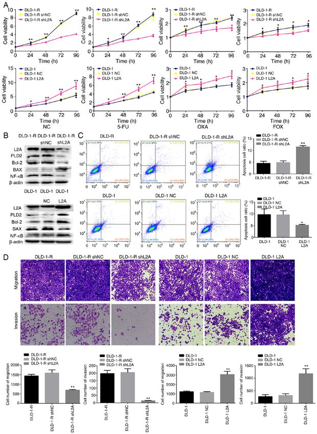
2                     XUAN et al: EFFECT OF CHAPERONE-MEDIATED AUTOPHAGY IN COLORECTAL CANCER

Materials and methods                                               and stained with 10% Giemsa dye for 10‑15 min at room
                                                                    temperature. For the invasive assay, the procedures were the
Cell culture and reagents. HCT‑116, DLD‑1 and NCM460 cell           same as above with a control‑membrane insert (BD Biosciences).
lines were obtained from Professor Miyagi Yohei (Clinical           A total of five random fields were selected to count and images
Research Institute, Kanagawa Cancer Center, Yokohama,               were captured under an inverted fluorescence microscope
Japan). The cell lines were cultured as monolayers in               (x10 magnification; Nikon Corporation).
RPMI‑1640 medium (Thermo Fisher Scientific, Inc.) supple‑
mented with 10% fetal bovine serum (FBS; Thermo Fisher              Quantification of apoptosis by flow cytometry. Flow cytometry
Scientific, Inc.) with 100 U/ml penicillin and 100 µg/ml            was performed with 7‑aminoactinomycin and phycoerythrin
streptomycin at 37˚C in a humidified atmosphere containing          labeled Annexin V (BD Pharmingen; BD Biosciences) to
5% CO2. All cells were harvested by centrifugation (1,500 x g       detect phosphatidylserine externalization as an endpoint
for 10 min at 4˚C) and rinsed with phosphate‑buffered saline        indicator of apoptosis. Cell apoptosis was detected by staining
(PBS). Anti‑LAMP2A (cat. no. ab18528) and anti‑PLD2                 with Annexin V‑PE/7AAD (KeyGen Biotech Co., Ltd.;
(cat. no. ab78907) antibodies were purchased from Abcam.            cat. no. KGA214) according to the manufacturer's instructions.
NF‑κ B Pathway Sampler kit (cat. no. 9936T) was purchased           Apoptotic rate was calculated by the percentage of early + late
from Cell Signaling Technology, Inc. Anti‑cluster of differen‑      apoptotic cells. After washing with PBS and centrifuging twice
tiation (CD)147/extracellular matrix metalloproteinase inducer      (both 1,000 x g, 5 min), cells were resuspended in Annexin‑V
(EMMPRIN; cat. no. SC‑21746) antibody was purchased from            binding buffer. They were stained with Annexin V‑PE/7AAD
Santa Cruz Biotechnology, Inc. Anti‑lung resistance‑related         and incubated for another 15 min at room temperature in a
protein 1 (LRP1; cat. no. ab92544), anti‑P‑glycoprotein 1           dark room. Cell samples were analyzed by flow cytometry
(PGP)/multidrug resistance (MDR; cat. no. ab242104),                (FACScan; BD Biosciences) to acquire the apoptotic fractions.
anti‑glutathione S‑transferase π (GST‑π; cat. no. ab138491),        Apoptotic fractions were investigated by Cell Quest 3.0 soft‑
anti‑MutL homologue 1 (MLH1; cat. no. ab92312), Anti‑F-box          ware (BD Biosciences).
and WD40 domain protein 7 (FBXW7) antibody (cat.
no. ab12292) and anti‑multidrug resistance‑associated               Cell transfection. The short hairpin (sh)RNA and LAMP2A
protein 1 (MRP1; cat. no. ab233383) antibodies were purchased       expression plasmids were designed, constructed and purified
from Abcam. Anti‑Bcl‑2 (cat. no. 15071) and anti‑BAX (cat.          by Shanghai GenePharma Co., Ltd. The shRNA and LAMP2A
no. 14796) antibodies were purchased from Cell Signaling            expression plasmids were transfected using Attractene
Technology, Inc. LysoTracker‑LysoGreen was purchased from           Transfection Reagent (Qiagen GmbH) in accordance with
Nanjing KeyGen Biotech Co., Ltd. (cat. no. KGMP006‑2).              the manufacturer's instructions. Cells cultured in 6‑well
Pyrrolidine dithiocarbamate (PDTC; cat. no. P8765.) and             plates were transfected with 1.2 ug/well shRNA or LAMP2A
chloroquine (CQ; cat. no. C6628.) were purchased from               expression plasmids. At 24 h after transfection, the cells were
Sigma‑Aldrich (Merck KGaA) and 5‑fluorouracil (5‑FU) was            collected to extract RNA and total protein for subsequent
purchased from Shanghai Xudong Haipu Pharmaceutical                 experimentation.
Co., Ltd. Oxaliplatin (OXA) was purchased from Nanjing
Pharmaceutical Factory, Co. Ltd.                                    RNA extraction. Total RNA was extracted from cultivated
                                                                    cells using TRIzol® reagent (Thermo Fisher Scientific, Inc.)
Cell viability assay. Cell viability was measured using the         in accordance with the manufacturer's protocols. RNA
MTT assay. CRC and 5‑FU‑resistant CRC cells were seeded             purification was performed using the RNeasy Mini kit
in triplicate in 96‑well plates at 6,000 cells/well and incubated   (Qiagen GmbH) according to the manufacturer's instructions.
at 37˚C for 24 h in RPMI‑1640 medium supplemented with
10% FBS. Subsequently, the cells were treated with the indi‑        Reverse transcription‑quantitative (RT‑q) PCR. Total RNA
cated doses of 5‑FU (10 mM), OXA (10 mM), or FOX (5‑FU:             was extracted from CRC cells (1x10 6 ) using the Qiagen
10 mM + OXA: 10 mM) for 96 h at 37˚C. The same volume of            RNeasy Mini kit (Qiagen GmbH), according to the manufac‑
dimethyl sulfoxide (DMSO) was used as the negative control.         turer's protocols. Total RNA was subjected to cDNA synthesis
Following this, 25 µl MTT solution (5 mg/ml) was added to           using avian myeloblastosis virus reverse transcriptase and
each well for 4 h at 37˚C, then the cell culture supernatants       random primer (Takara Bio, Inc.). General‑ and RT‑qPCR
were carefully removed and 200 µl DMSO was added to                 amplification was performed using TaKaRa Hot Start Taq
dissolve the purple formazan. Finally, the optical density was      Polymerase (Takara Bio, Inc.) and SYBR Premix Ex Taq TM II
measured at a wavelength of 570 nm using a microplate reader        kit (Takara Bio, Inc.), respectively, all according to the
(Model 550; Bio‑Rad Laboratories, Inc.).                            manufacturer's protocols. All experiments were performed
                                                                    three times. mRNA expression analysis was performed using
Cell migration and invasion assays. For the migra‑                  SYBR Green Master Mix (Life Technologies; Thermo Fisher
tion assay, 2x10⁵ cells were resuspended in serum‑free              Scientific, Inc.) on a LightCycler 96 Detection system (Roche
RPMI‑1640 and seeded in the Matrigel‑coated insert                  Diagnostics) using β ‑actin for normalization. The cycling
(BD Biosciences) insert on the top portion of the chamber           parameters were: 95˚C for 5 min, followed by 59 cycles at 95˚C
(BD Biosciences). The lower compartment of the chamber              for 30 sec, annealing at 55˚C for 30 sec and extension at 72˚C for
contained 10% FBS as a chemoattractant. After incubation            30 sec. Expression levels were normalized to endogenous
for 24 h, cells on the membrane were scrubbed, washed with          controls and relative quantification (2 ‑ΔΔCq) was used for
PBS, fixed in 100% methanol for 10 min at room temperature          fold‑change calculation (24). Using GenBank, oligonucleotide
MOLECULAR MEDICINE REPORTS 23: 360, 2021 3
primers were designed as follows: LAMP2A: Forward primer: Santa Cruz Biotechnology, Inc.; cat. no. sc‑47778) control
5'‑GCCGTTCTCACACTGCTCTA‑3'; reverse primer: 5'‑CCG using ImageJ software (version 1.52a; National Institutes of
CTATGGGCACAAGGAA‑3'. PLD2: Forward primer: 5'‑CCA Health).
CAAACAGGGGTGGTGTT‑3'; reverse primer: 5'‑GAATGG
CCTGGATGGAGTTG‑3'; β‑actin: Forward primer: 5'‑GGT Statistical analysis. All statistical analyses were performed by
GGC T TT TAG GAT G GCA AG‑3'; reverse primer: 5'‑ACT SPSS v24.0 (IBM Corp.) with the Student's t‑test (two tailed)
GGAACGGTGAAGGTGACAG‑3'. and by one‑way analysis of variance with Tukey's post hoc
test for multiple groups. The results are representative of
Western blot analysis. Cells were lysed in SDS cell lysis buffer three different experiments and data are expressed as mean ±
containing 50 mM Tris (pH, 8.1) and 1% SDS, supplemented standard deviation. P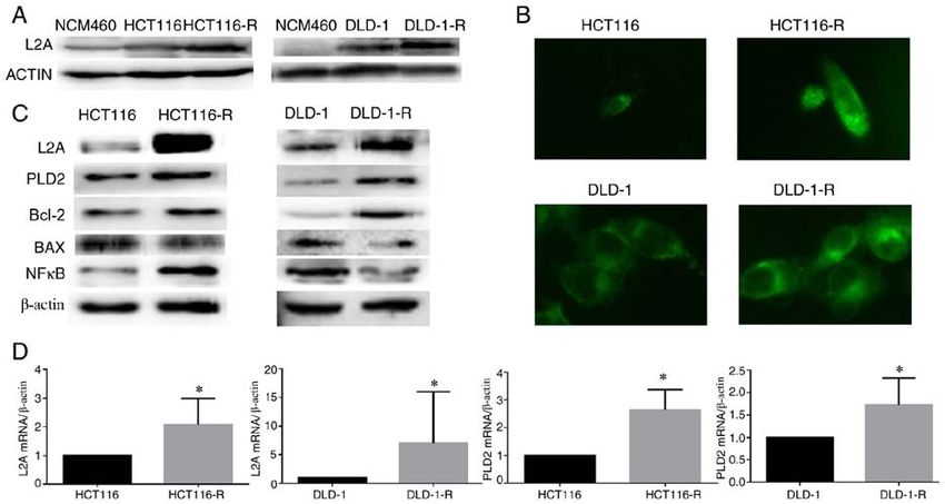
4                         XUAN et al: EFFECT OF CHAPERONE-MEDIATED AUTOPHAGY IN COLORECTAL CANCER

Figure 1. Generation and characterization of 5‑FU‑resistant cells from HCT116 and DLD‑1 cells. (A) Cell proliferation curve of HCT116/DLD‑1 and
HCT116‑R/DLD‑1‑R cells. (B) Cell survival curve of HCT116/DLD‑1 and HCT116‑R/DLD‑1‑R cells. (C) RI and IC50 of 5‑FU in HCT116/DLD‑1 and
HCT116‑R/DLD‑1‑R cells. (D) Protein expression of chemoresistance‑related genes by western blotting. (E) Cell proliferation curve of HCT116/DLD‑1 and
HCT116‑R/DLD‑1‑R cells after chemotherapy drug treatment. The data represent the mean ± standard deviation from three independent experiments. *P
MOLECULAR MEDICINE REPORTS 23: 360, 2021                                                               5

Figure 3. Effect of LAMP2A expression on the sensitivity to 5‑FU in human CRC cell line. (A) Cellular viability of different drug treatments in
shLAMP2A‑modified HCT116‑R cells or LAMP2A‑modified HCT116 cells. (B) Expression of associated proteins was visualized by western blotting in
shLAMP2A‑modified HCT116‑R cells or in LAMP2A‑modified HCT116 cells. (C) Apoptosis and (D) migration and invasion and in shLAMP2A‑modified
HCT116‑R cells or in LAMP2A‑modified HCT116 cells (x10 magnification). The data represent the mean ± standard deviation from three independent experi‑
ments. *P
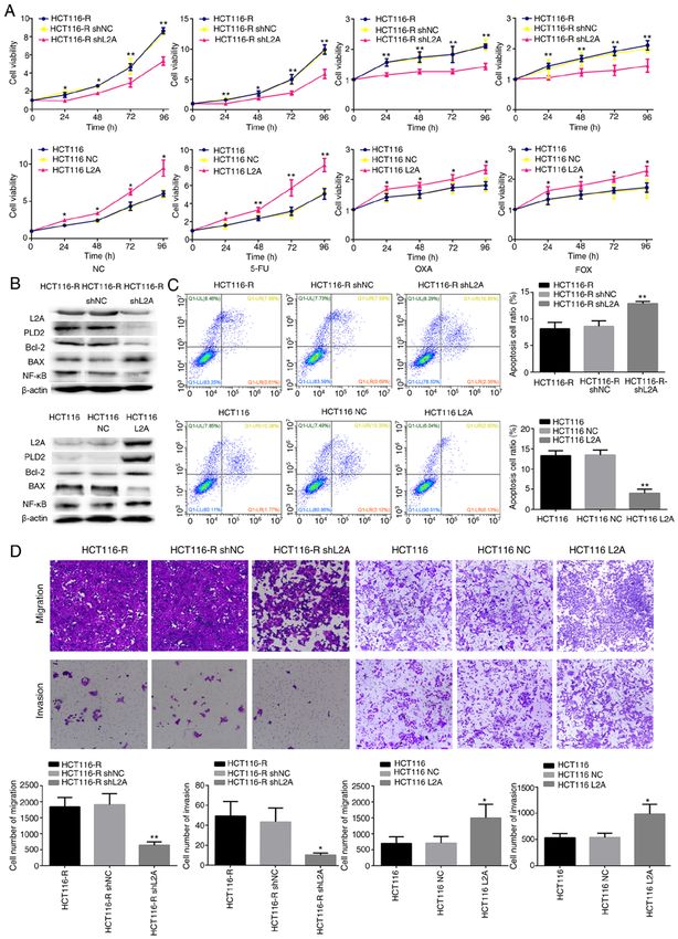
6                        XUAN et al: EFFECT OF CHAPERONE-MEDIATED AUTOPHAGY IN COLORECTAL CANCER

Figure 4. Effect of LAMP2A expression on the sensitivity to 5‑FU in human CRC cell line. (A) Cellular viability of different drug treatments in
shLAMP2A‑modified DLD‑1‑R or LAMP2A‑modified DLD‑1 cells. (B) Expression of associated proteins was visualized by western blotting in
shLAMP2A‑modified DLD‑1‑R cells or in LAMP2A‑modified DLD‑1 cells. (C) Apoptosis and (D) migration and invasion in shLAMP2A‑modified DLD‑1‑R
cells or in LAMP2A‑modified DLD‑1 cells (x10 magnification). The data represent the mean ± standard deviation from three independent experiments.
*
 P
MOLECULAR MEDICINE REPORTS 23: 360, 2021                                                                7

Figure 5. Inhibition of lysosomal and NF‑κ B p65 signaling pathways led to proliferation and decrease in expression level of PLD2 in CRC cell lines.
(A) Inhibition of the lysosomal pathway with CQ (50 mM); PLD2 first declined and then increased with the lowest point observed at 4 h. (B and C) Inhibition
of the lysosomal pathway (CQ; 50 Mm, 4 h) and NF‑κ B signaling pathway (PDTC; 10 mM; 2 h) led to a significant reduction in the expression level of PLD2
compared with the control group. (D) Co‑immunoprecipitation of NF‑κ B with PLD2, CD147, GST3, MLH1 in HCT116/DLD‑1, HCT116‑R/DLD‑1‑R and
HCT116L2A/DLD‑1L2A cells. The data represent the mean ± standard deviation from three independent experiments. *P
8                     XUAN et al: EFFECT OF CHAPERONE-MEDIATED AUTOPHAGY IN COLORECTAL CANCER

leukemia‑1 (38). CMA is also involved in the mechanism of               A significant elevation of LAMP2A was found in
CRC chemotherapy resistance. A previous study confirmed             CRC cell lines and the increase was even higher in
that 5‑FU undergoes extensive deacetylation in a variety of         HCT116‑R/DLD‑1‑R cells. The high expression of LAMP2A
CRC cell lines (11). It reduces the ability of histone acetyl‑      promoted proliferation, invasion and anti‑apoptotic function
transferase p300 and CBP to bind to chromatin and induces           of CRC cells.
lysosomal degradation. The lysosomal degradation is achieved            In conclusion, upregulated LAMP2A expression might
through CMA and inhibition of the degradation of p300 and           serve a role in tumorigenesis and growth and may be involved
CBP can enhance the effects of 5‑FU and reverse drug resis‑         in drug resistance in CRC. It is a potential biomarker that
tance (11). This suggests that the efficacy of chemotherapy can     indicates the aggressiveness of CRCs and, thus, may be a
be increased with anti‑CMA therapy administered at the same         therapeutic target.
time as 5‑FU chemotherapy.
    The present study constructed and analyzed two                  Acknowledgements
5‑FU‑resistant cell lines from HCT‑116 and DLD‑1 cell lines,
providing a tool to investigate the molecular pathways and          Not applicable.
detailed mechanisms that are associated with drug resistance
in CRC. In the engineered 5‑FU‑resistant CRC cell lines,            Funding
significant elevation of LAMP2A was identified; this was
responsible for loss of function of LAMP2A in 5‑FU‑resistant        The present study was supported by Liaoning BaiQianWan
CRC cells or gain of function in deficient cells, which rendered     Talents Program, Award for Liaoning Distinguished
them sensitive or resistant to 5‑FU, respectively.                  Professor, a Key Scientific and Technological Project of
    Concurrently, a positive association was identified between     Liaoning Province (grant no. 2015408001) and National
LAMP2A and PLD2. PLD2 is one of the two isoforms of PLD             Natural Scientific Foundation of China (grant nos. 81472544
in mammals, which catalyzes the hydrolysis of the diester           and 81672700).
bond of phospholipids to generate PA and the free lipid head‑
group (39). PLD2 expression is elevated in a number of human        Availability of data and materials
tumors and is highly variable (16,19‑22). In CRC, the expres‑
sion level of PLD2 was significantly associated with tumor          The datasets used and/or analyzed are available from the
size and survival of patients, which suggests that PLD2 may         corresponding author on reasonable request.
be a target for therapy in CRC (40).
    The present study verified whether PLD2 variation               Authors' contributions
exists along with LAMP2A through activation of the NF‑κ B
pathway. PDTC was used to block the NF‑κ B pathway and              YX, SZ, XX and LX performed the experiments. YX
resulted in decreased PLD2 levels. The relation between PLD2        analyzed the data and contributed to the manuscript writing.
and NF‑κ B was demonstrated by co‑immunoprecipitation.              YX and HCZ designed the experiments and contributed to
PDTC, a metal chelator and antioxidant, can inhibit the acti‑       data analysis. YX and HCZ were responsible for confirming
vation of the NF‑κ B pathway specifically by suppressing the        the authenticity of the data. All authors read and approved the
release of the inhibitory subunit, Iκ B (41). Cuervo et al (42)     final manuscript and agree to be accountable for all aspects
identified that Iκ B can be degraded by CMA during starva‑          of the research in ensuring that the accuracy or integrity
tion. The present study found that the decrease of Iκ B in the      of any part of the work are appropriately investigated
cytoplasm was associated with an increase of LAMP2A levels.         and resolved.
Co‑immunoprecipitation experiments further confirmed the
interaction of Iκ B with Hsc70. IkB was identified as a substrate   Ethics approval and consent to participate
of CMA. Therefore, when CMA was high in 5‑FU‑resistant
CRC cell lines, the Iκ B level in cytoplasm decreased, resulting    Not applicable.
in increased NF‑κ B activity. Thereafter, the combination
forms of PLD2 and NF‑κ B increased, which facilitated the           Patient consent for publication
grown of CRC cells.
    CQ was used to inhibit the lysosomal pathway and it             Not applicable.
was observed that the levels of PLD2 declined early on and
then increased. This might because Iκ B degraded though             Competing interests
two pathways; CMA and proteasome (42). When lysosome
pathway was blocked, the proteasome pathway was activated           The authors declare that they have no competing interests.
in compensation later. PLD contributes to signaling pools of
PA and is under tight regulation by elaborate mechanisms            References
and so may increase reactivity in cells (15). In addition, the
present study found the interaction of NF‑κ B with some drug         1. Siegel RL, Miller KD, Goding Sauer A, Fedewa SA,
resistance genes, such as CD147, GST3 and MLH1, through                 Butterly LF, Cercek A, Smith RA and Jemal A: Colorectal
                                                                        cancer statistics, 2020. CA Cancer J Clin 70: 145‑164, 2020.
co‑immunoprecipitation. This may be another important piece          2. Takayama T, Miyanishi K, Hayashi T, Sato Y and Niitsu Y:
of evidence that the NF‑κ B pathway is associated with drug             Colorectal cancer: Genetics of development and metastasis.
resistance in CRC.                                                      J Gastroenterol 41: 185‑192, 2006.
MOLECULAR MEDICINE REPORTS 23: 360, 2021 9
3. De Angelis R, Sant M, Coleman MP, Francisci S, Baili P, 24. Livak KJ and Schmittgen TD: Analysis of relative gene
Pierannunzio D, Trama A, Visser O, Brenner H, Ardanaz E, et al: expression data using real‑time quantitative PCR and the
Cancer survival in Europe 1999‑2007 by country and age: 2(‑Delta Delta C(T)) method. Methods 25: 402‑408, 2001.
Results of EUROCARE-5 ‑ a population‑based study. 25. Kuang YH, Chen X, Su J, Wu LS, Liao LQ, Li D, Chen ZS
Lancet Oncol 15: 23‑34, 2014. and Kanekura T: RNA interference targeting the CD147 induces
4. Peng SL, Thomas M, Ruszkiewicz A, Hunter A, Lawrence M apoptosis of multi‑drug resistant cancer cells related to XIAP
and Moore J: Conventional adverse features do not predict depletion. Cancer Lett 276: 189‑195, 2009.
response to adjuvant chemotherapy in stage II colon cancer. 26. Nakayama K, Kanzaki A, Ogawa K, Miyazaki K, Neamati N
ANZ J Surg 84: 837‑841, 2014. and Takebayashi Y: Copper‑transporting P‑type adenosine
5. Prados J, Melguizo C, Ortiz R, Perazzoli G, Cabeza L, triphosphatase (ATP7B) as a cisplatin based chemo‑
Alvarez PJ, Rodriguez‑Serrano F and Aranega A: Colon cancer resistance marker in ovarian carcinoma: Comparative
therapy: Recent developments in nanomedicine to improve the analysis with expression of MDR1, MRP1, MRP2, LRP and BCRP.
efficacy of conventional chemotherapeutic drugs. Anticancer Int J Cancer 101: 488‑495, 2002.
Agents Med Chem 13: 1204‑1216, 2013. 27. Ściskalska M and Milnerowicz H: The role of GSTπ isoform in the
6. Kaushik S and Cuervo AM: Chaperone‑mediated autophagy: cells signalling and anticancer therapy. Eur Rev Med Pharmacol
A unique way to enter the lysosome world. Trends Cell Biol 22: Sci 24: 8537‑8550, 2020.
407‑417, 2012. 28. He J, He J, Min L, He Y, Guan H, Wang J and Peng X:
7. Catarino S, Pereira P and Girão H: Molecular control of Extracellular vesicles transmitted miR‑31‑5p promotes
chaperone‑mediated autophagy. Essays Biochem 61: 663‑674, sorafenib resistance by targeting MLH1 in renal cell carcinoma.
2017. Int J Cancer 146: 1052‑1063, 2020.
8. Kaushik S and Cuervo AM: The coming of age of chaperone‑ 29. Germano G, Lamba S, Rospo G, Barault L, Magrì A, Maione F,
mediated autophagy. Nat Rev Mol Cell Biol 19: 365‑381, 2018. Russo M, Crisafulli G, Bartolini A, Lerda G, et al: Inactivation
9. Saha T: LAMP2A overexpression in breast tumors promotes of DNA repair triggers neoantigen generation and impairs tumour
cancer cell survival via chaperone‑mediated autophagy. growth. Nature 552: 116‑120, 2017.
Autophagy 8: 1643‑1656, 2012. 30. Xiao G, Li Y, Wang M, Li X, Qin S, Sun X, Liang R,
10. Zhang Y, Xu YY, Yao CB, Li JT, Zhao XN, Yang HB, Zhang B, Du N, Xu C, et al: FBXW7 suppresses epithelial‑
Zhang M, Yin M, Chen J and Lei QY: Acetylation targets mesenchymal transition and chemo‑resistance of non‑small‑cell
HSD17B4 for degradation via the CMA pathway in response to lung cancer cells by targeting snai1 for ubiquitin‑dependent degra‑
estrone. Autophagy 13: 538‑553, 2017. dation. Prolif 51: e12473, 2018.
11. Du C, Huang D, Peng Y, Yao Y, Zhao Y, Yang Y, Wang H, 31. Rezasoltani S, Asadzadeh‑Aghdaei H, Nazemalhosseini‑Mojarad E,
Cao L, Zhu WG and Gu J: 5‑Fluorouracil targets histone acet‑ Dabiri H, Ghanbari R and Zali MR: Gut microbiota, epigenetic
yltransferases p300/CBP in the treatment of colorectal cancer. modification and colorectal cancer. Iran J Microbiol 9: 55‑63, 2017.
Cancer Lett 400: 183‑193, 2017. 32. Skarkova V, Kralova V, Vitovcova B and Rudolf E: Selected aspects
12. Zhou J, Yang J, Fan X, Hu S, Zhou F, Dong J, Zhang S, of chemoresistance mechanisms in colorectal carcinoma‑A focus
Shang Y, Jiang X, Guo H, et al: Chaperone‑mediated autophagy on epithelial‑to‑mesenchymal transition, autophagy, and apoptosis.
regulates proliferation by targeting RND3 in gastric cancer. Cells 8: 234, 2019.
Autophagy 12: 515‑528, 2016. 33. Kon M, Kiffin R, Koga H, Chapochnick J, Macian F,
13. Ding ZB, Fu XT, Shi YH, Zhou J, Peng YF, Liu WR, Varticovski L and Cuervo AM: Chaperone‑mediated autophagy is
Shi GM, Gao Q, Wang XY, Song K, et al: Lamp2a is required for required for tumor growth. Sci Transl Med 3: 109ra117, 2011.
tumor growth and promotes tumor recurrence of hepatocellular 34. Li L, Fang R, Liu B, Shi H, Wang Y, Zhang W, Zhang X and
carcinoma. Int J Oncol 49: 2367‑2376, 2016. Ye L: Deacetylation of tumor‑suppressor MST1 in Hippo
14. Dubois A, Furstoss N, Calleja A, erhouni M, Cluzeau T, Savy C, pathway induces its degradation through HBXIP‑elevated HDAC6
Marchetti S, Hamouda MA, Boulakirba S, Orange F, et al: in promotion of breast cancer growth. Oncogene 35: 4048‑4057,
LAMP2 expression dictates azacytidine response and prognosis 2016.
in MDS/AML. Leukemia 33: 1501‑1513, 2019. 35. Quintavalle C, Di Costanzo S, Zanca C, Tasset I, Fraldi A,
15. Selvy PE, Lavieri RR, Lindsley CW and Brown HA: Incoronato M, Mirabelli P, Monti M, Ballabio A, Pucci P, et al:
Phospholipase D: Enzymology, functionality and chemical Phosphorylation‑regulated degradation of the tumor‑suppressor
modulation. Chem Rev 111: 6064‑6119, 2011. form of PED by chaperone‑mediated autophagy in lung cancer
16. Foster DA and Xu L: Phospholipase D in cell proliferation and cells. J Cell. Physiol 229: 1359‑1368, 2014.
cancer. Mol Cancer Res 1: 789‑800, 2003. 36. Vakifahmetoglu‑Norberg H, Kim M, Xia HG, Iwanicki MP,
17. Hammond SM, Altshuller YM, Sung TC, Rudge SA, Rose K, Ofengeim D, Coloff JL, Pan L, Ince TA, Kroemer G, Brugge JS
Engebrecht J, Morris AJ and Frohman MA: Human ADP and Yuan J: Chaperone‑mediated autophagy degrades mutant p53.
ribosylation factor‑activated phosphatidylcholine‑specific phos‑ Genes Dev 27: 1718‑1730, 2013.
pholipase D defines a new and highly conserved gene family. 37. Xie W, Zhang L, Jiao H, Guan L, Zha J, Li X, Wu M,
J Biol Chem 270: 29640‑29643, 1995. Wang Z, Han J and You H: Chaperone‑mediated autophagy
18. Colley WC, Sung TC, Roll R, Jenco J, Hammond SM, prevents apoptosis by degrading BBC3/PUMA. Autophagy 11:
Altshuller Y, Bar‑Sagi D, Morris AJ and Frohman MA: 1623‑1635, 2015.
Phospholipase D2, a distinct phospholipase D isoform with novel 38. Suzuki J, Nakajima W, Suzuki H, Asano Y and Tanaka N:
regulatory properties that provokes cytoskeletal reorganization. Chaperone‑mediated autophagy promotes lung cancer cell survival
Curr Biol 7: 191‑201, 1997. through selective stabilization of the pro‑survival protein, MCL1.
19. Henkels KM, Boivin GP, Dudley ES, Berberich SJ and Biochem Biophys Res Commun 482: 1334‑1340, 2017.
Gomez‑Cambronero J: Phospholipase D (PLD) drives cell 39. Yao Y, Wang X, Li H, Fan J, Qian X, Li H and Xu Y:
invasion, tumorgrowth and metastasis in a human breast cancer Phospholipase D as a key modulator of cancer progression.
xenograph model. Oncogene 32: 5551‑5562, 2013. Biol Rev Camb Philos Soc 95: 911‑935, 2018.
20. Uchida N, Okamura S and Kuwano H: Phospholipase D activity 40. Saito M, Iwadate M, Higashimoto M, Ono K, Takebayashi Y
in human gastric carcinoma. Anticancer Res 19: 671‑675, 1999. and Takenoshita S: Expression of phospholipase D2 in human
21. Diaz‑Aragon R, Ramirez‑Ricardo J, Cortes‑Reynosa P, colorectal carcinoma. Oncol. Rep 18: 1329‑1334, 2017.
Simoni‑Nieves A, Gomez‑Quiroz LE and Perez Salazar E: 41. Li H, Sun Y, Liang J, Fan Y and Zhang XD: pH‑Sensitive
Role of phospholipase D in migration and invasion induced pullulan‑DOX conjugate nanoparticles for co‑loading PDTC
by linoleic acid in breast cancer cells. Mol Cell Biochem 457: to suppress growth and chemoresistance of hepatocellular
119‑132, 2019. carcinoma. J Mater Chem B 3: 8070‑8078, 2015.
22. Liu M, Du K, Jiang B and Wu X: High expression of 42. Cuervo AM, Hu W, Lim B and Dice JF: IkappaB is a substrate
phospholipaseD2 induced by hypoxia promotes proliferation for a selective pathway of lysosomal proteolysis. Mol Biol Cell 9:
of colon cancer cells through activating NF‑κ Bp65 signaling 1995‑2010, 1998.
pathway. Pathol Oncol Res 26: 281‑290, 2018.
23. Fiucci G, Czarny M, Lavie Y, Zhao D, Berse B, Blusztajn JK This work is licensed under a Creative Commons
and Liscovitch M: Changes in phospholipaseD isoform activity Attribution-NonCommercial-NoDerivatives 4.0
and expression in multidrug‑resistant human cancer cells. International (CC BY-NC-ND 4.0) License.
Int J Cancer 85: 882‑888, 2000.You can also read