UBA6 and NDFIP1 regulate the degradation of ferroportin
←
→
Page content transcription
If your browser does not render page correctly, please read the page content below
UBA6 and NDFIP1 regulate the degradation of
ferroportin

by Lisa Traeger, Steffen B. Wiegand, Andrew J. Sauer, Benjamin H.P. Corman, Kathryn M.
Peneyra, Florian Wunderer, Anna Fischbach, Aranya Bagchi, Rajeev Malhotra,
Warren M. Zapol, and Donald B. Bloch

Received: February 6, 2021.
Accepted: July 22, 2021.

Citation: Lisa Traeger, Steffen B. Wiegand, Andrew J. Sauer, Benjamin H.P. Corman,
Kathryn M. Peneyra, Florian Wunderer, Anna Fischbach, Aranya Bagchi, Rajeev Malhotra,
Warren M. Zapol, and Donald B. Bloch. UBA6 and NDFIP1 regulate the degradation of ferroportin.
Haematologica. 2021 Jul 29. doi: 10.3324/haematol.2021.278530. [Epub ahead of print]

Publisher's Disclaimer.
E-publishing ahead of print is increasingly important for the rapid dissemination of science.
Haematologica is, therefore, E-publishing PDF files of an early version of manuscripts that
have completed a regular peer review and have been accepted for publication. E-publishing
of this PDF file has been approved by the authors. After having E-published Ahead of Print,
manuscripts will then undergo technical and English editing, typesetting, proof correction and
be presented for the authors' final approval; the final version of the manuscript will then
appear in a regular issue of the journal. All legal disclaimers that apply to the
journal also pertain to this production process.
UBA6 and NDFIP1 regulate the degradation of ferroportin

Lisa Traeger1, Steffen B. Wiegand1, Andrew J. Sauer1, Benjamin H.P. Corman1,
Kathryn M. Peneyra1, Florian Wunderer1,2, Anna Fischbach1, Aranya Bagchi1, Rajeev
Malhotra3, Warren M. Zapol1 and Donald B. Bloch1,4,
1
  Anesthesia Center for Critical Care Research of the Department of Anesthesia,
Critical Care and Pain Medicine, Massachusetts General Hospital and Harvard
Medical School, Boston, United States;
2
  Department of Anesthesiology, Intensive Care Medicine and Pain Therapy,
University Hospital Frankfurt, Goethe University, Frankfurt, Germany;
3
  Cardiovascular Research Center and the Cardiology Division of the Department of
Medicine, Massachusetts General Hospital and Harvard Medical School, Boston,
United States and
4
  Division of Rheumatology, Allergy and Immunology of the Department of Medicine,
Massachusetts General Hospital and Harvard Medical School, Boston, United States

Running Title: Regulation of ferroportin degradation

Correspondence: Donald B. Bloch, dbloch@mgh.harvard.edu; and Lisa Traeger,
email@lisatraeger.de

Data sharing statement: For original data, please contact email@lisatraeger.de or
dbloch@mgh.harvard.edu

Word count: Abstract: 228; Main Text: 3912
Acknowledgements: This study was supported by Luisa Hunnewell and Larry
Newman (DBB), the German Research Foundation (TR 1642/1-1 to LT, WI 5162/2-1
to SBW, Wu 841/1-1 to FW, FI 2429/1-1 to AF), NIH (R01HL142809 to RM,
R01DK125786 to AB) and the American Heart Association (18TPA34230025 to RM,
20IOA35360009 to AB).

Authorship and conflict-of-interest statement
LT, FW and DBB designed and conceived the study. LT and DBB wrote and edited
the manuscript. LT, SBW, BHPC, KP, AJS and AF performed experiments. LT, RM,
AB and DBB analyzed and interpreted the data. DBB and WMZ supervised the study.
All authors approved the final version for submission.

Conflict of Interest disclosure
All authors declare no competing financial interest related to the study.

                                           1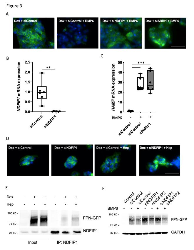
Abstract

Hepcidin regulates iron homeostasis by controlling the level of ferroportin, the only
membrane channel that facilitates export of iron from within cells. Binding of hepcidin
to ferroportin induces the ubiquitination of ferroportin at multiple lysine residues and
subsequently causes the internalization and degradation of the ligand-channel
complex within lysosomes. The objective of this study was to identify components of
the ubiquitin system that are involved in ferroportin degradation. A HepG2 cell line,
which inducibly expresses ferroportin-GFP (FPN-GFP), was established to test the
ability of siRNAs directed against components of the ubiquitin system to prevent
BMP6- and exogenous hepcidin-induced ferroportin degradation. Of the 88 siRNAs
directed against components of the ubiquitin pathway that were tested, siRNA-
mediated depletion of the alternative E1 enzyme UBA6 as well as the adaptor protein
NDFIP1 prevented BMP6- and hepcidin- induced degradation of ferroportin in vitro. A
third component of the ubiquitin pathway, ARIH1, indirectly inhibited ferroportin
degradation by impairing BMP6 mediated induction of hepcidin. In mice, the AAV-
mediated silencing of Ndfip1 in the murine liver increased the level of hepatic
ferroportin and increased circulating iron. The results suggest that the E1 enzyme
UBA6 and the adaptor protein NDFIP1 are involved in iron homeostasis by regulating
the degradation of ferroportin. These specific components of the ubiquitin system
may be promising targets for the treatment of iron related diseases, including iron
overload and anemia of inflammation.

                                           2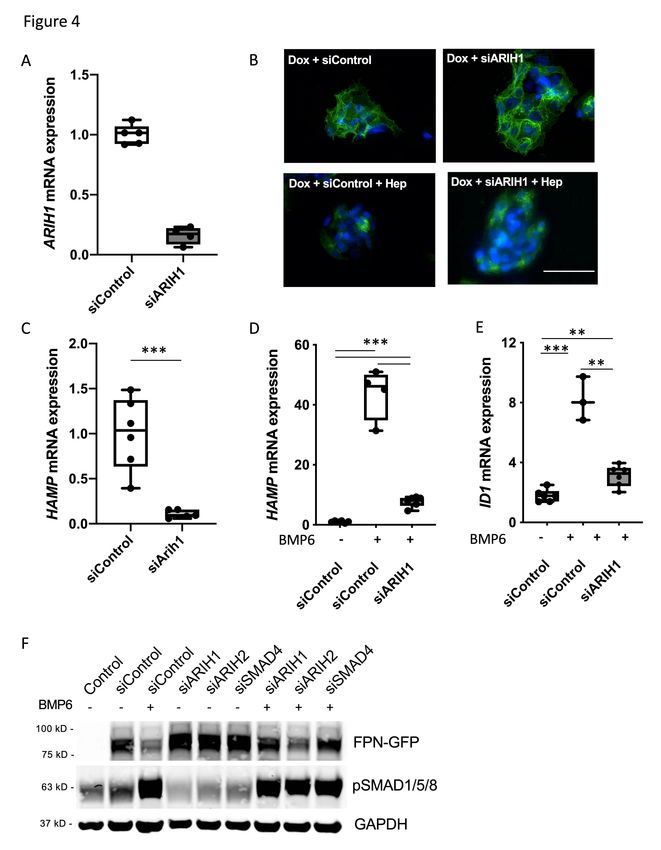
Introduction

       Iron is an essential element that is required for a spectrum of cellular and
biological processes including oxygen transport, DNA synthesis and the production of
energy. High levels of iron, in the presence of oxygen, may catalyze the production of
reactive oxygen species (ROS), which are free radicals that may damage cellular
proteins and membranes. The level of iron in the body must be tightly regulated to
provide sufficient levels to permit fundamental biological processes while preventing
the damaging effects of excess iron1,2.

       The hepatic hormone hepcidin is a critical regulator of systemic iron
homeostasis3–5. Hepcidin expression is controlled by at least three stimuli: 1)
increased serum and liver iron, which induce hepcidin via the bone morphogenetic
protein (BMP) signaling pathway; 2) increased mediators of inflammation (IL-1β and
IL-6), which increase hepcidin via the Jak/Stat pathway; and 3) the hormone
erythroferrone, which inhibits BMP signaling by sequestering BMP6 in response to
increased erythropoietic demand6–9. Hepcidin regulates iron homeostasis by
controlling the cell surface level of ferroportin, which is the only known membrane
channel that facilitates export of iron from within cells10. Ferroportin is a member of
the superfamily of transporters of small molecules, which includes more than 300
membrane-bound proteins11,12. Ferroportin is predominantly expressed in tissues
associated with iron transport including enterocytes, hepatocytes, macrophages and
erythrocytes10,11,13.    The protein has twelve membrane-spanning domains, which
create a channel through which iron is transported. Binding of hepcidin to the main
extracellular cavity of ferroportin causes ligation of ubiquitin molecules to multiple
intracellular   lysine   residues14.   Polyubiquitination   of   ferroportin   induces   the
internalization of the hepcidin-ferroportin complex followed by degradation within
lysosomes10,14,15. Degradation of ferroportin results in decreased serum iron,
because enterocytes, hepatocytes and macrophages are no longer able to transfer
intracellular iron to the circulation16. In addition to the ability of hepcidin to induce
degradation of ferroportin, the hormone is also able to inhibit iron export by directly
occluding the iron channel17. Occlusion of the channel may be especially important
for cells, such as mature red blood cells, which lack the endocytic machinery required
to degrade ferroportin17.

                                              3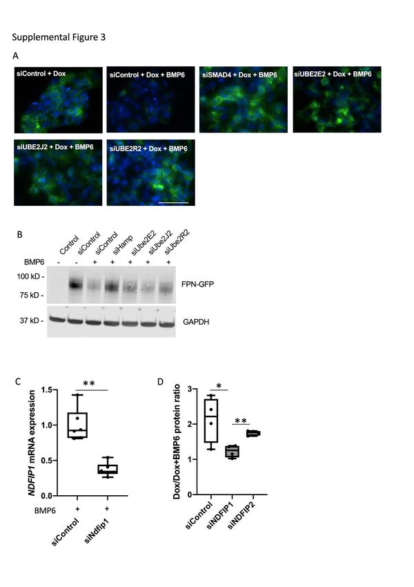
Ubiquitin is a 76 amino acid polypeptide that can be attached to lysine
residues in proteins. The attachment of ubiquitin to a protein regulates the protein’s
localization, stability and/or activity18. The process of ubiquitination involves the
activation and transient conjugation of ubiquitin to a carrier protein, with subsequent
final ligation of the ubiquitin molecule to a substrate. In general, ubiquitination
requires three different kinds of enzymes: a ubiquitin activating enzyme (E1), a
ubiquitin conjugating enzyme (E2), and a ubiquitin ligase (E3). The human ubiquitin
system encodes two different E1 enzymes (UBA1 and UBA6), approximately 50
different E2 enzymes, and more than 600 E3 enzymes19–21. The ubiquitin E3 ligases
are important for substrate recognition and are divided into three different classes22.
Depending on the class of ligase, the E3 enzyme either directly transfers ubiquitin to
a substrate (“HECT” and “RBR” ligases) or acts as an adaptor to facilitate the transfer
of ubiquitin from an E2 enzyme directly to the substrate (“RING” E3 ligases)22,23.
Binding of the E3 enzyme to the substrate may also require an adaptor protein that
acts as a scaffold between the E3 enzyme and the target protein.

       In this study, an in vitro siRNA screen was performed to determine which
proteins in the ubiquitin pathway are involved in ferroportin degradation. A previous
study used a modified HEK293 cell line, in which expression of ferroportin was
induced by the addition of ponasterone24. Exogenous hepcidin and putative inhibitors
of ferroportin degradation were added to this cell line and the level of ferroportin was
then measured. To permit screening for specific enzymes involved in ferroportin
ubiquitination without using exogenous hepcidin, we established a HepG2 cell line
that expresses the ferroportin-GFP (FPN-GFP) fusion protein in response to
doxycycline. In this cell line, BMP6 can be used to gradually induce the expression of
endogenous hepcidin. The HepG2-FPN-GFP cell line was used to show that the
alternative E1 enzyme UBA6 as well as the NEDD4 family binding protein NDFIP1
are able regulate the degradation of ferroportin in response to BMP6, as well as
exogenous hepcidin. Depletion of the E3 ligase ARIH1 indirectly inhibited ferroportin
degradation by impairing BMP6 mediated hepcidin induction. In vivo, depletion of
Ndfip1 in the murine liver increased the level of hepatic ferroportin and increased
circulating iron.

                                           4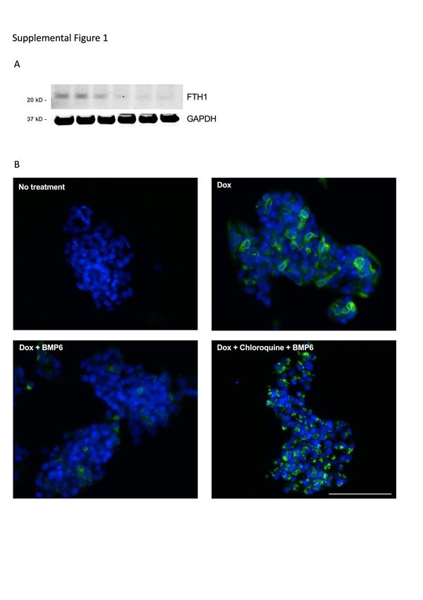
Methods

HepG2-FPN-GFP cell line

       A plasmid encoding human ferroportin (NM_016917) fused to green
fluorescent protein (GFP) was a gift from Tomas Ganz (David Geffen School of
Medicine, UCLA)14. DNA encoding the fusion protein was ligated into the NheI and
NotI sites of plasmid pTRE2hyg (Clontech cat#631014). The plasmid was transfected
into the HepG2 “tet-on advanced” cell line (Clonetech cat#630932) using Effectene
transfection   reagents   (Qiagen,   Germantown,    MD,   USA)    according   to   the
manufacturer’s instructions and successfully transfected cells were selected using
hygromycin (0.4mg/ml). Individual cell lines were established and were confirmed to
express FPN-GFP in the presence of doxycycline (2µg/ml). One colony, which
expressed a high level of FPN-GFP after treatment with doxycycline, was selected
for further experiments. HepG2-FPN-GFP cells were maintained in Eagle Minimum
Essential Medium, 10% FBS, L-glutamine (2mM), G418 (100µg/ml), hygromycin
(0.4mg/ml), penicillin (100units/ml), and streptomycin (100µg) at 37°C in 5% CO2 and
95% humidity.

Adeno-associated virus (AAV) administration

       All experiments using mice were approved by the Partners Subcommittee on
Research Animal Care at Massachusetts General Hospital, Boston, USA (Protocol #
2007N000052). Wild type mice on a C57BL/6J background were purchased from
Jackson Laboratories (Bar Harbor, ME, USA). Animals were fed a standard diet (380
ppm iron). AAV2/8-GFP-U6-m-Ndfip1-shRNA (AAV2/8-shNdfip1) and AAV2/8-GFP-
U6-scrmb-shRNA (AAV2/8-shControl) were obtained from Vector BioLabs (Malvern,
PA, USA). Eight-week-old male mice were injected intravenously with 1x10¹¹
particles of AAV2/8-shNdfip1 or AAV2/8-shControl via the tail vein. Six weeks later,
mice were anesthetized with 4% isoflurane and whole blood was collected by cardiac
puncture. Liver and spleen were harvested for further analysis.

Statistical analysis

       All statistical analyses were performed using GraphPad Prism 8.3.0
(GraphPad Software, San Diego, CA, USA). Data are expressed as mean ± SD. The

                                          5
Shapiro-Wilk test was performed to test for normality. Correlation analysis were performed using Pearson correlation. Comparison of two groups was performed using the Student’s t-test for parametric data and the Mann-Whitney-U test for non- parametric data. Comparison of more than two groups was performed using one-way ANOVA with Tukey post hoc test (for parametric data) or the Kruskal-Wallis test with Dunn’s post-hoc test (for non-parametric data). After adjusting for multiple comparisons, a P-value
Results
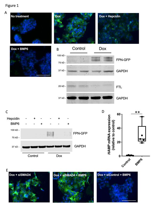
Preparation and characterization of the HepG2-FPN-GFP cell line
Binding of hepcidin to ferroportin induces the polyubiquitination, internalization
and lysosomal degradation of the ligand-channel complex10. To identify the specific
enzymes that mediate ubiquitination of ferroportin, we established a stable HepG2
cell line that inducibly expresses FPN-GFP (HepG2-FPN-GFP) in the presence of
doxycycline. Treatment of HepG2-FPN-GFP cells with 2µg/ml of doxycycline for 18
hours induced expression of the fusion protein, which was detected at the cell
surface (Figure 1A). The FPN-GFP fusion protein was able to export iron, as
indirectly indicated by decreased levels of intracellular FTL and FTH1 after FPN-GFP
induction (Figure 1B, Supplemental Figure 1A). Treatment with hepcidin (50ng/ml) for
90min caused FPN-GFP to localize to punctate structures in the cytoplasm (Figure
1A), and treatment with hepcidin for 18h caused degradation of the fusion protein
(Figure1C). Because BMP6 induces HepG2 cells to express hepcidin25 (Figure 1D),
we were able to investigate the effect of gradual induction of endogenous hepcidin on
ferroportin degradation. Treatment with BMP6 (10ng/ml) for 18h caused degradation
of the FPN-GFP fusion protein as detected by indirect fluorescence and
immunoblotting (Figure 1A, C). Pretreatment with chloroquine (100µM for 2h), an
inhibitor of lysosomal degradation, prevented BMP6-mediated FPN-GFP degradation
and caused FPN-GFP to localize to lysosomes in the cytoplasm (Supplemental
Figure 1B).
BMP6 induces expression of hepcidin through the BMP receptor-SMAD 1/5/8
pathway. After binding to the BMP receptor complex, activated BMP type I receptors
phosphorylate SMAD 1/5/8 proteins, which translocate together with SMAD4 to the
nucleus, and induce hepcidin expression26. The siRNA-mediated inhibition of SMAD4
in HepG2-FPN-GFP cells prevented BMP6 mediated degradation of FPN-GFP
(Figure 1E). Taken together, these results show that the HepG2-FPN-GFP cell line
expresses inducible, functional FPN-GFP. Both BMP6-induced endogenous hepcidin
and exogenous hepcidin cause internalization and degradation of the FPN-GFP
fusion protein.
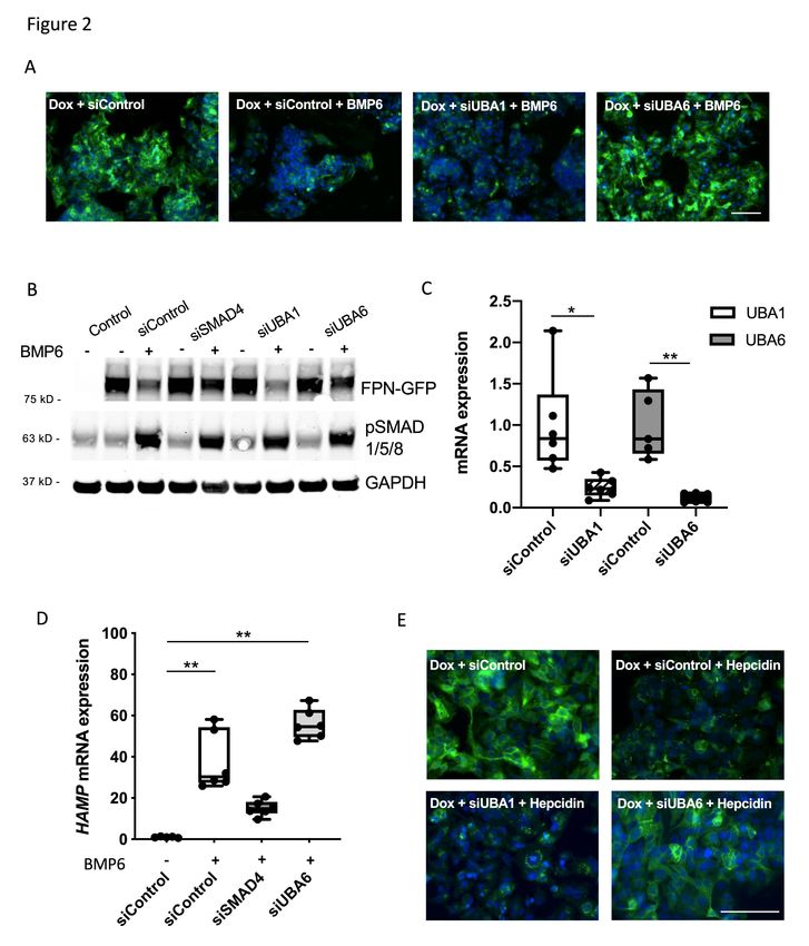
7The E1 enzyme UBA6 is required for ubiquitination of FPN
The human ubiquitin system encodes two E1 enzymes: UBA1 (also known as
UBE1) and UBA6. To identify the E1 enzyme that is involved in ferroportin
degradation, HepG2-FPN-GFP cells were transfected with siRNA that targeted each
of the E1 enzymes or with a control siRNA (siControl). Twenty-four hours after
transfection with siRNA, cells were treated overnight with doxycycline to induce the
expression of FPN-GFP and were then incubated with BMP6 for 18h. Cells that were
treated with siControl and BMP6 had decreased cell surface expression of FPN-GFP
(Figure 2A). Depletion of UBA1 did not prevent the BMP6-induced localization of
FPN-GFP to lysosomes and subsequent degradation. However, depletion of UBA6
prevented BMP6-mediated degradation of ferroportin, as indicated by the persistence
of the FPN-GFP fusion protein at the cell surface (Figure 2A). Immunoblotting
confirmed that depletion of UBA6, but not UBA1, impaired degradation of FPN-GFP
(Figure 2B, Supplemental Figure 2A). The successful depletion of each of the E1
enzymes by the appropriate siRNA was confirmed by qPCR (Figure 2C). Successful
depletion of UBA6 by siUBA6 was not affected by the addition of BMP6
(Supplemental Figure 2B).
Depletion of UBA6 might block degradation of FPN-GFP or prevent BMP6-
induced expression of hepcidin. To consider this latter possibility, the ability of UBA6
depletion to inhibit the BMP signal transduction pathway was investigated. Depletion
of UBA6 had no effect on BMP6-mediated phosphorylation of SMAD1/5/8 (Figure
2B). In addition, siRNA directed against UBA6 did not prevent expression of
endogenous hepcidin in HepG2-FPN-GFP cells (Figure 2D). In contrast, siRNA
directed against SMAD4 blunted BMP6 induced expression of hepcidin.
To further demonstrate that depletion of UBA6 blocks degradation of
ferroportin, independent of an effect on BMP-induced expression of hepcidin, the
effect of exogenous hepcidin on the cellular localization of FPN-GFP in HepG2 cells
was investigated. To determine the amount of hepcidin produced by HepG2 cells
after treatment with BMP6, HepG2 cells were incubated with BMP6 (10ng/ml) for 18h
and the amount of hepcidin in the tissue culture medium was measured by ELISA.
Under these conditions, BMP6 induced 3.9 ng/ml (± 0.4ng/ml) of hepcidin, and this
8concentration of hepcidin (rather than the much higher dose of 30-50ng/ml used in
other studies10,24) was used to treat cells in subsequent experiments. Cells were
transfected with siControl, siUBA1, or siUBA6 and treated overnight with hepcidin (4
ng/ml). In the presence of this low concentration of hepcidin, FPN-GFP expression at
the cell surface persisted in UBA6-depleted cells but not in siControl-treated- or
siUBA1-treated cells (Figure 2E). The inability of hepcidin to degrade the FPN-GFP
fusion protein in siUBA6 treated cells was confirmed by immunoblot (Supplemental
Figure 2C). Taken together, these results show that UBA6 is required for hepcidin
induced internalization and degradation of FPN-GFP.
The adaptor protein NDFIP1 regulates ferroportin degradation
To identify additional components of the ubiquitin pathway that might be
involved in ferroportin degradation, siRNAs directed against different E2 and E3
enzymes, as well as other known components of the ubiquitin pathway, were tested
for the ability to inhibit BMP6-mediated degradation of FPN-GFP (Supplemental
Table 1, N=77). A commercially available library (Dharmacon, Lafayette, CO, USA),
which contains siRNAs that were previously verified to silence the corresponding
targets and to minimize off-target effects, was used in these studies. HepG2 cells
transfected with siRNA directed against SMAD4 were used as positive controls for
inhibition of BMP6-mediated degradation of FPN-GFP; siControl was used as a
negative control. Eighteen hours after treatment with BMP6, the localization of FPN-
GFP was determined by immunofluorescence. In the first screen, we identified 23
siRNAs directed against different E2 and E3 enzymes that appeared to block FPN-
GFP re-localization to the lysosome based on FPN-GFP persistence at the cell
surface after BMP6 treatment. In second and tertiary screens, all positive candidates
were re-evaluated to exclude false positives.
Depletion of each of three E2 enzymes, UBE2R2, UBE2E2 and UBE2J2
partially blocked the internalization of FPN-GFP (Supplemental Figure 3A, B), while
depletion of other, individual E2 enzymes did not impair BMP6 mediated FPN-GFP
degradation (data not shown). Treatment with pairwise combinations of UBE2R2,
UBE2E2 and UBE2J2 or all three of the E2 enzymes did not further prevent the
degradation of ferroportin (data not shown), suggesting that additional E2 enzymes
participate in FPN ubiquitination.
9In an initial screen, depletion the NEDD family interacting protein NDFIP1 and
the E3 enzyme ARIH1 impaired BMP6 induced FPN-GFP localization to lysosomes
and subsequent degradation of the fusion protein (Figure 3A). siNDFIP1 successfully
depleted NDFIP1 mRNA in both the absence (Figure 3B) and the presence
(Supplemental Figure 3C) of exogenous BMP6. siRNA directed against NDFIP1 had
no effect on the ability of BMP6 to induce hepcidin expression, demonstrating that
the BMP signaling pathway was intact (Figure 3C). To confirm that depletion of
NDFIP1 blocks degradation of FPN-GFP, NDFIP1 depleted cells were treated with
exogenous hepcidin (4ng/ml). Compared to cells that were transfected with siControl,
depletion of NDFIP1 inhibited hepcidin-mediated degradation of the FPN-GFP fusion
protein (Figure 3D, Supplemental Figure 2C).
To investigate the possibility that NDFIP1 interacts with ferroportin, HepG2
cells were incubated in the presence or absence of exogenous hepcidin for 20min,
and protein lysates were immunoprecipitated with an antibody directed against
NDFIP1. In the absence of hepcidin, a small amount of FPN-GFP was detected in
the immunoprecipitated protein lysate. Treatment with hepcidin caused an increase
in the amount of FPN-GFP that co-immunoprecipitated with NDFIP1 (Figure 3E).
Taken together, the results suggest that NDFIP1 interacts with ferroportin and is
involved in hepcidin-induced ferroportin internalization and degradation.
A second adaptor protein (NDFIP2), which like NDFIP1 facilitates
ubiquitination by HECT E3 enzymes, shares 79% similarity with NDFIP127. To
investigate the potential role of NDFIP2 in the regulation of ferroportin, the effect of
NDFIP2 depletion on BMP6-induced FPN-GFP degradation was assessed. While
siNDFIP1 treatment prevented degradation of FPN-GFP, depletion of NDFIP2 had no
effect on BMP6-mediated degradation of the FPN-GFP (Figure 3F, Supplemental
Figure 3D).
NDFIP1 recruits members of the NEDD4 family of E3 ligases to target
proteins28. To investigate whether NEDD4 family members (NEDD4, NEDD4L, ITCH,
WWP1, WWP2, SMURF1, SMURF2, HECW1, HECW229) regulate ferroportin levels,
the localization of FPN-GFP in cells treated with siRNA directed against each of
these enzymes was examined. None of the siRNAs directed against members of the
NEDD4 family, either alone or in pair-wise combinations, prevented BMP6 mediated
FPN-GFP degradation (Supplemental Figure 4A, B). These results indicate that
either more than two of these enzymes are involved in BMP6-induced FPN
10degradation or additional, as yet unidentified, enzymes are able to interact with
NDFIP1 and mediate ferroportin degradation.
ARIH1 indirectly regulates ferroportin by inhibiting BMP6 mediated induction of
hepcidin
ARIH1 is a member of the Ariadne family of RBR E3 ligases. Treatment of
HepG2-FPN-GFP cells with siRNA directed against ARIH1 inhibited BMP6-mediated
degradation of FPN-GFP (Figure 3A). ARIH1 was successfully depleted by
transfection of siARIH1 in both the absence (Figure 4A) and the presence of BMP6
(Supplemental Figure 4C), as determined by qPCR. The addition of low dose
exogenous hepcidin to HepG2-FPN-GFP cells, however, reduced the level of FPN-
GFP on the surface of ARIH1-depleted cells (Figure 4B). The ability of exogenous
hepcidin to degrade FPN-GFP in siARIH1 treated cells was confirmed by immunoblot
(Supplemental Figure 2C). We considered the possibility that depletion of ARIH1
inhibits ferroportin degradation by interfering with the ability of BMP signaling to
induce hepcidin gene expression. In the absence of BMP6, the depletion of ARIH1
reduced basal hepcidin mRNA levels (Figure 4C). Depletion of siARIH1 impaired
BMP6-stimulated induction of hepcidin mRNA by 80% (Figure 4D). ARIH1 depletion
also inhibited BMP6-mediated induction of ID1, another target of the BMP signaling
pathway (Figure 4E). Interestingly, BMP6 induced phosphorylation of SMAD1/5/8
proteins was not affected by ARIH1 depletion (Figure 4F). These results suggest that
ARIH1 has an indirect effect on the stability of ferroportin by altering BMP6 mediated
hepcidin induction through a non-canonical pathway.
The Ariadne RBR E3 ligase ARIH2 (also known as TRIAD1) is the closest
relative to ARIH1 with 54% similarity30. To consider the possibility that this second
member of the Ariadne family is involved in the indirect regulation of ferroportin, the
effect of ARIH2 depletion on BMP6-induced FPN-GFP degradation was assessed. In
contrast to ARIH1, depletion of ARIH2 had no effect on BMP6-mediated degradation
of the FPN-GFP protein expression (Figure 4F, Supplemental Figure 4D).
Silencing of Ndfip1 stabilizes hepatic FPN in vivo
The adaptor protein NDFIP1 was identified as a protein that is involved in
ferroportin degradation in vitro. To address whether NDFIP1 is important for
11ferroportin degradation in vivo, mice were injected with an adeno-associated virus
(AAV2/8) encoding a short-hairpin directed against Ndfip1, under the control of a U6
promoter. The AAV serotype 8 was used in these studies because it has a high
efficiency of transduction in hepatocytes31. In both AAV2/8-shNdfip1 and AAV2/8-
shControl injected animals, GFP expression was detected in the liver, indicating
successful systemic administration of the virus (Figure 5A). In animals injected with
AAV2/8-shNdfip1, hepatic Ndfip1 mRNA levels were significantly reduced compared
to control animals (Figure 5B). Mice injected with AAV2/8-shNdfip1 had a 3-fold
increase in FPN protein level in the liver compared to control mice (Figure 5C-D).
Hamp mRNA and serum hepcidin levels were similar in both groups, suggesting that
higher ferroportin levels were not caused by induction of hepcidin (Figure 5E, F).
Increased hepatic ferroportin was associated with a 28% increase in serum iron
levels in AAV2/8-shNdfip1, compared to AAV2/8-shControl, mice (Figure 5G) and
there was a correlation between serum iron and ferroportin levels (Supplemental
Figure 5A). Hepatic FTL levels were increased and TfR1 mRNA was decreased in
AAV2/8-shNdfip1-treated mice (Supplemental Figure 5B-D). As expected because of
the targeting of AAV8 to the liver32, splenic Ndfip1 mRNA levels were not decreased
in AAV2/8-shNdfip1 mice (Supplemental Figure 5E). The results show that the
AAV2/8-mediated depletion of Ndfip1 increases the level of hepatic ferroportin and
that Ndfip1 is required for FPN degradation in the liver.
12Discussion
This study identified components of the ubiquitin system that are important for
ferroportin degradation. A HepG2 cell line that inducibly expresses functional FPN-
GFP fusion protein was established. BMP6 induced expression of hepcidin, which
caused the internalization and degradation of the fusion protein and permitted
analysis of ferroportin degradation under conditions in which the level of hepcidin
increases gradually. In vitro, the alternative E1 enzyme UBA6, as well as the adaptor
protein NDFIP1, were critical for hepcidin induced ferroportin degradation. Depletion
of either UBA6 or NDFIP1 inhibited hepcidin induced internalization and degradation
of FPN-GFP. The E3 ligase ARIH1 indirectly regulated ferroportin stability by altering
BMP6 mediated hepcidin induction through a non-canonical pathway. In vivo, the
depletion of Ndfip1 in the murine liver increased the level of hepatic ferroportin and
increased circulating iron.
In 2007, UBA6 was identified as a second ubiquitin activating E1 enzyme.
UBA1 and UBA6 have non-redundant functions and each enzyme is essential for
biological processes33,34. UBA6 is widely expressed in different tissues but
contributes to only approximately 1% of overall cellular ubiquitination33,35. In addition
to activating ubiquitin for subsequent transfer to the E2 enzyme, UBA6 also activates
the ubiquitin-like protein FAT10, which plays a role in the immune response, obesity
and aging. However, Fat10 deficient mice do not develop iron overload36,37,
suggesting that FAT10 does not play a direct role in iron homeostasis. In the present
study UBA6 was found to be the E1 enzyme involved in ferroportin regulation in vitro;
depletion of UBA6, but not UBA1, prevented hepcidin-induced ferroportin
degradation in HepG2 cells. In contrast to UBA1, which is known to charge multiple
E2 enzymes with ubiquitin, UBA6 transfers ubiquitin to a small number of E2
enzymes35. Although some E2 enzymes interact with both UBA1 and UBA6, one E2
enzyme (USE1, also known as UBE2Z) is exclusively charged by UBA635,38. In this
study, we found that inhibition of USE1 did not interfere with hepcidin induced
ferroportin degradation (data not shown), indicating that an E2 enzyme other than (or
in addition to) USE1 is involved in ferroportin regulation. In UBA6 depleted cells,
UBA1 failed to induce hepcidin mediated ferroportin degradation, indicating non-
redundant functions of UBA1 and UBA6 in ferroportin regulation. The results suggest
13that an as yet unidentified E2 enzyme, exclusively charged by UBA6, plays a role in
ferroportin degradation.
Members of the NEDD4 family of HECT-type E3 ligases contain a “WW”
domain that interacts with a proline rich PPXY (PY) motif in the target protein.
However, some target proteins lack a PY domain and ubiquitination of these proteins
requires the presence of adaptor proteins NDFIP1 or NDFIP2 to act as a scaffold
between the two proteins. NDFIP proteins contain three transmembrane domains as
well as two PY motifs, which interact with the WW domain of several members of the
NEDD4 family of E3 ligases28. In this study, NDFIP1 was shown to interact with
ferroportin in HepG2 cells in vitro and regulates the level of ferroportin in the liver in
vivo. None of the WW domain-containing NEDD4 family members that were tested
individually or in pair-wise combination prevented BMP6-induced FPN degradation.
The results suggest that several NEDD4 family members may have a redundant role
in ferroportin degradation. Another possibility is that an as yet unknown E3 ligase
interacts with the adaptor protein NDFIP1 to ubiquitinate ferroportin.
ARIH1 is a member of the Ariadne family of E3 RBR ligase. ARIH1 is highly
expressed in the nucleus, where it interacts with Cajal and PML nuclear bodies39.
ARIH1 associates with neddylated Cullin-RING E3 ligases (CRL) and
monoubiquitinates CRL targets40. In this study, ARIH1 was shown to indirectly
regulate ferroportin stability by altering BMP6-mediated hepcidin induction through a
non-canonical pathway. Depletion of ARIH1 blunted basal, as well as BMP6-
mediated, hepcidin and ID1 mRNA expression without altering the phosphorylation of
SMAD 1/5/8 proteins in response to BMP6. Further studies are needed to elucidate
the mechanism as to how ARIH1 regulates hepcidin expression in response to
BMP6.
NDFIP1 was previously shown to have a role in iron homeostasis41–43. NDFIP1
binds to divalent metal transporter 1 (DMT1), the major iron transporter for non-heme
iron import44. NDFIP1 recruits the NEDD4 family member WWP2 to ubiquitinate
DMT143. In vivo, Ndfip1 is involved in the regulation of DMT1 in enterocytes41. The
expression of Dmt1 in enterocytes of Ndfip1 deficient mice is increased under normal
iron conditions as well as during iron deficiency. The increased level of Dmt1 leads to
increased iron absorption, and under normal dietary iron conditions Ndfip1 deficient
14mice develop a phenotype resembling classic hereditary hemochromatosis, with
increased hepatic, duodenal and serum iron levels43,45. In this study we show that
depletion of Ndfip1 in the liver increased the level of ferroportin. Ndfip1 appears to
regulate two steps in iron metabolism: iron import by DMT1 in enterocytes and iron
export by ferroportin in the liver. Ndfip1-deficient mice were not used in this study,
because Ndfip1 deficiency results in a severe inflammatory phenotype caused by
hyperactivation of T-cells45,46.
In summary, this study demonstrated that the E1 enzyme UBA6 and the
adaptor protein NDFIP1 are important for iron homeostasis, regulating the
degradation of hepatic ferroportin. In the future, it may be possible to target specific
components of the ubiquitin pathway with small molecules47; the results of this study
may offer novel approaches to treating disorders of iron metabolism.
15References
1. Salahudeen AA, Bruick RK. Maintaining Mammalian iron and oxygen
homeostasis: sensors, regulation, and cross-talk. Ann N Y Acad Sci. 2009;1177:30-
38.
2. Pantopoulos K, Porwal SK, Tartakoff A, Devireddy L. Mechanisms of
mammalian iron homeostasis. Biochemistry. 2012;51(29):5705-5724.
3. Pigeon C, Ilyin G, Courselaud B, et al. A new mouse liver-specific gene,
encoding a protein homologous to human antimicrobial peptide hepcidin, is
overexpressed during iron overload. J Biol Chem. 2001;276(11):7811-7819.
4. Nicolas G, Viatte L, Lou D-Q, et al. Constitutive hepcidin expression prevents
iron overload in a mouse model of hemochromatosis. Nat Genet. 2003;34(1):97-101.
5. Park CH, Valore EV, Waring AJ, Ganz T. Hepcidin, a urinary antimicrobial
peptide synthesized in the liver. J Biol Chem. 2001;276(11):7806-7810.
6. Dev S, Babitt JL. Overview of iron metabolism in health and disease.
Hemodial Int. 2017;21 Suppl 1(Suppl 1):S6-S20.
7. Sebastiani G, Wilkinson N, Pantopoulos K. Pharmacological Targeting of the
Hepcidin/Ferroportin Axis. Front Pharmacol. 2016;7:160.
8. Wang C-Y, Xu Y, Traeger L, et al. Erythroferrone lowers hepcidin by
sequestering BMP2/6 heterodimer from binding to the BMP type I receptor ALK3.
Blood. 2020;135(6):453-456.
9. Wunderer F, Traeger L, Sigurslid HH, Meybohm P, Bloch DB, Malhotra R. The
role of hepcidin and iron homeostasis in atherosclerosis. Pharmacol Res.
2020;153:104664.
10. Nemeth E, Tuttle MS, Powelson J, et al. Hepcidin regulates cellular iron efflux
by binding to ferroportin and inducing its internalization. Science.
2004;306(5704):2090-2093.
11. Drakesmith H, Nemeth E, Ganz T. Ironing out Ferroportin. Cell Metab.
2015;22(5):777-787.
12. Lin L, Yee SW, Kim RB, Giacomini KM. SLC transporters as therapeutic
targets: emerging opportunities. Nat Rev Drug Discov. 2015;14(8):543-560.
13. Zhang D-L, Ghosh MC, Ollivierre H, Li Y, Rouault TA. Ferroportin deficiency in
erythroid cells causes serum iron deficiency and promotes hemolysis due to oxidative
stress. Blood. 2018;132(19):2078-2087.
14. Qiao B, Sugianto P, Fung E, et al. Hepcidin-induced endocytosis of ferroportin
is dependent on ferroportin ubiquitination. Cell Metab. 2012;15(6):918-924.
15. Ross SL, Tran L, Winters A, et al. Molecular mechanism of hepcidin-mediated
ferroportin internalization requires ferroportin lysines, not tyrosines or JAK-STAT. Cell
Metab. 2012;15(6):905-917.
16. Camaschella C, Nai A, Silvestri L. Iron metabolism and iron disorders revisited
in the hepcidin era. Haematologica. 2020;105(2):260-272.
17. Aschemeyer S, Qiao B, Stefanova D, et al. Structure-function analysis of
ferroportin defines the binding site and an alternative mechanism of action of
hepcidin. Blood. 2018;131(8):899-910.
18. Pickart CM, Eddins MJ. Ubiquitin: structures, functions, mechanisms. Biochim
Biophys Acta. 2004;1695(1-3):55-72.
19. Morreale FE, Walden H. Types of Ubiquitin Ligases. Cell. 2016;165(1):248-
248.
20. Shen M, Schmitt S, Buac D, Dou QP. Targeting the ubiquitin-proteasome
system for cancer therapy. Expert Opin Ther Targets. 2013;17(9):1091-1108.
1621. Stewart MD, Ritterhoff T, Klevit RE, Brzovic PS. E2 enzymes: more than just
middle men. Cell Res. 2016;26(4):423-440.
22. Ardley HC, Robinson PA. E3 ubiquitin ligases. Essays Biochem. 2005;41:15-
30.
23. Weber J, Polo S, Maspero E. HECT E3 Ligases: A Tale With Multiple Facets.
Front Physiol. 2019;10:370.
24. Fung E, Sugianto P, Hsu J, Damoiseaux R, Ganz T, Nemeth E. High-
throughput screening of small molecules identifies hepcidin antagonists. Mol
Pharmacol. 2013;83(3):681-690.
25. Meynard D, Kautz L, Darnaud V, Canonne-Hergaux F, Coppin H, Roth M-P.
Lack of the bone morphogenetic protein BMP6 induces massive iron overload. Nat
Genet. 2009;41(4):478-481.
26. Xiao X, Alfaro-Magallanes VM, Babitt JL. Bone morphogenic proteins in iron
homeostasis. Bone. 2020;138:115495.
27. Shearwin-Whyatt LM, Brown DL, Wylie FG, Stow JL, Kumar S. N4WBP5A
(Ndfip2), a Nedd4-interacting protein, localizes to multivesicular bodies and the Golgi,
and has a potential role in protein trafficking. J Cell Sci. 2004;117(Pt 16):3679-3689.
28. Harvey KF, Shearwin-Whyatt LM, Fotia A, Parton RG, Kumar S. N4WBP5, a
potential target for ubiquitination by the Nedd4 family of proteins, is a novel Golgi-
associated protein. J Biol Chem. 2002;277(11):9307-9317.
29. Ingham RJ, Gish G, Pawson T. The Nedd4 family of E3 ubiquitin ligases:
functional diversity within a common modular architecture. Oncogene.
2004;23(11):1972-1984.
30. Kelsall IR, Duda DM, Olszewski JL, et al. TRIAD1 and HHARI bind to and are
activated by distinct neddylated Cullin-RING ligase complexes. EMBO J.
2013;32(21):2848-2860.
31. Nam H-J, Lane MD, Padron E, et al. Structure of adeno-associated virus
serotype 8, a gene therapy vector. J Virol. 2007;81(22):12260-12271.
32. Snyder RO, Miao CH, Patijn GA, et al. Persistent and therapeutic
concentrations of human factor IX in mice after hepatic gene transfer of recombinant
AAV vectors. Nat Genet 1997;16(3):270-276.
33. Barghout SH, Schimmer AD. E1 Enzymes as Therapeutic Targets in Cancer.
Pharmacol Rev. 2021;73(1):1-56.
34. Groettrup M, Pelzer C, Schmidtke G, Hofmann K. Activating the ubiquitin
family: UBA6 challenges the field. Trends Biochem Sci. 2008;33(5):230-237.
35. Jin J, Li X, Gygi SP, Harper JW. Dual E1 activation systems for ubiquitin
differentially regulate E2 enzyme charging. Nature. 2007;447(7148):1135-1138.
36. Canaan A, Yu X, Booth CJ, et al. FAT10/diubiquitin-like protein-deficient mice
exhibit minimal phenotypic differences. Mol Cell Biol. 2006;26(13):5180-5189.
37. Canaan A, DeFuria J, Perelman E, et al. Extended lifespan and reduced
adiposity in mice lacking the FAT10 gene. Proc Natl Acad Sci U S A.
2014;111(14):5313-5318.
38. Wang F, Zhao B. UBA6 and Its Bispecific Pathways for Ubiquitin and FAT10.
Int J Mol Sci. 2019;20(9):2250.
39. Elmehdawi F, Wheway G, Szymanska K, et al. Human Homolog of Drosophila
Ariadne (HHARI) is a marker of cellular proliferation associated with nuclear bodies.
Exp Cell Res. 2013;319(3):161-172.
40. Scott DC, Rhee DY, Duda DM, et al. Two Distinct Types of E3 Ligases Work
in Unison to Regulate Substrate Ubiquitylation. Cell. 2016;166(5):1198-1214.
41. Foot NJ, Leong YA, Dorstyn LE, et al. Ndfip1-deficient mice have impaired
DMT1 regulation and iron homeostasis. Blood. 2011;117(2):638-646.
1742. Foot NJ, Gembus KM, Mackenzie K, Kumar S. Ndfip2 is a potential regulator
of the iron transporter DMT1 in the liver. Sci Rep. 2016;6:24045.
43. Foot NJ, Dalton HE, Shearwin-Whyatt LM, et al. Regulation of the divalent
metal ion transporter DMT1 and iron homeostasis by a ubiquitin-dependent
mechanism involving Ndfips and WWP2. Blood. 2008;112(10):4268-4275.
44. Yanatori I, Kishi F. DMT1 and iron transport. Free Radic Biol Med.
2019;133:55-63.
45. Oliver PM, Cao X, Worthen GS, et al. Ndfip1 protein promotes the function of
itch ubiquitin ligase to prevent T cell activation and T helper 2 cell-mediated
inflammation. Immunity. 2006;25(6):929-940.
46. Nemeth E, Valore EV, Territo M, Schiller G, Lichtenstein A, Ganz T. Hepcidin,
a putative mediator of anemia of inflammation, is a type II acute-phase protein.
Blood. 2003;101(7):2461-2463.
47. Deng L, Meng T, Chen L, Wei W, Wang P. The role of ubiquitination in
tumorigenesis and targeted drug discovery. Signal Transduct Target Ther.
2020;5(1):11.
18Figure Legends. Figure 1. Characterization of the HepG2-FPN-GFP cell line. (A) Images of untreated HepG2 cells, and cells treated with doxycycline (Dox; 2 µg/ml) alone, with Dox followed by hepcidin (50 ng/ml) for 90 min, with Dox followed by BMP6 (10 ng/ml) for 18 h are shown. (B) Treatment with Dox induced the expression of the FPN-GFP fusion protein. Dox-treated cells had reduced levels of intracellular ferritin light-chain (FTL), consistent with increased iron export in cells expressing FPN-GFP. GAPDH was used as a loading control. (C) In the absence of Dox, the fusion FPN- GFP protein was not detected by immunoblot (lanes 1-3). In the presence of Dox, FPN-GFP was expressed (lane 4). Treatment with hepcidin (50 ng/ml; lane 5), or BMP6 (10 ng/ml; lane 6), for 18 h caused degradation of the FPN-GFP fusion protein. (D) BMP6 stimulation (10 ng/ml) for 18 h induced hepcidin mRNA expression in HepG2-FPN-GFP cells, as determined by qPCR (mRNA expression relative to control; **=P
relative to control; *=P
18h). In the absence of Dox, control cells did not express the FPN-GFP fusion
protein. siRNA directed against NDFIP1, but not NDFIP2, prevented BMP6-induced
degradation of the FPN-GFP fusion protein, as determined by immunoblot. GAPDH
was used as a loading control. The immunoblot is representative of four separate
experiments.

Figure 4. ARIH1 regulates BMP6-mediated induction of hepcidin. (A)
Transfection with siARIH1 successfully depleted ARIH1 in HepG2-FPN-GFP cells, as
determined by qPCR (mRNA expression relative to control; ***=P< 0.001; Student’s
t-test). (B) HepG2-FPN-GFP cells were transfected with siControl or siARIH1 and
were treated with Dox or Dox followed by hepcidin (4 ng/ml for 18 h) as indicated. In
the presence of Dox, the expression of the FPN-GFP fusion protein was induced.
Treatment with hepcidin caused FPN-GFP localization to lysosomes and its
subsequent degradation in siControl-treated cells as well as in siARIH1-treated cells.
White bar indicates 100 µm. (C) Treatment of HepG2 cells with siARIH1 reduced the
basal expression of hepcidin mRNA, as determined by qPCR (mRNA expression
relative to control; ***=P≤0.001; Student’s t-test). (D) Pretreatment of HepG2 cells
with siRNA directed against ARIH1 reduced BMP6 mediated hepcidin mRNA
expression (relative to control), as determined by qPCR (***=P≤0.001; One-way
ANOVA). (E) BMP6 (10 ng/ml for 18h) induced the expression of ID1 in siControl
transfected cells. Pretreatment of HepG2 cells with siRNA directed against ARIH1
blunted BMP6 induced expression of ID1, as determined by qPCR (mRNA
expression relative to control; **=P
t-test). (C) The level of FPN in the liver of AAV2/8-shNdfip1-treated mice was
increased   compared    to   AAV2/8-shControl-treated   mice,   as   determined   by
immunoblot. GAPDH was used as a loading control. (D) Densitometric analysis of
immunoblot in (C) (***=P
1

Supplemental Material and Methods

Methods
siRNA screen

      The day before siRNA transfection, cells were seeded in 8-well tissue culture slides
in full growth medium without antibiotics to obtain a confluence of 30-50% on the day of
transfection. The siRNA-library Human Ubiquitin Conjugation Subset 1 (siGENOME®
SMART pool® siRNA library G005615 Lot11132, which contains a mixture of four
different siRNAs targeting each gene), as well as additional individual siRNAs directed
against specific components of the ubiquitin pathway, were obtained from Dharmacon
(Lafayette, CO, USA) (Supplemental Table 1). siRNA was transfected into cells using
Dharmafect transfection reagent 4 (Dharmacon) according to the manufacturer’s
instructions. Cells were transfected using a final concentration of 20nM siRNA. Twenty -
four hours after transfection, cells were washed, incubated with EMEM with 0.1% FBS
and expression of FPN-GFP was induced by the addition of doxycycline for 16-18h. The
following day, BMP6 (10ng/ml) or hepcidin (4ng/ml) or vehicle alone was added, and the
cells were incubated for an additional 18h. To prepare protein extracts for immunoblot,
cells were seeded and transfected in 6-well plates.

Immunofluorescence

      Cells were fixed with 2% PFA for 10 min and permeabilized with methanol
containing 4’,6-Diamidin-2-phenylindol (DAPI) (0.5µM) for 7 min. Cells were stained with
an antibody directed against GFP (Roche 11814460001, 1:250) for one hour at RT,
followed   by    Alexa-488     conjugated    donkey-anti-mouse      antibody    (Jackson
ImmunoResearch, Cat. No 715545150; 1:250) for one hour. Cells were visualized using
a Nikon Eclipse 80i (Nikon Instruments, Melville, NY, USA) microscope and the program
Retiga 2000R, QIMAGING (Surrey, BC, Canada).
2
Immunoblot analysis
Cells were lysed in RIPA buffer supplemented with a protease and phosphatase
inhibitor cocktail (Thermo Fisher Scientific). Plasma enriched membrane proteins were
prepared from liver using the Mem-PERTM Cell lysate kit (Thermo Fisher Scientific).
Samples were prepared in Laemmli buffer without reducing agent and incubated for 30
min at RT 1. Proteins were separated by SDS-Page, transferred to PDVF membranes,
and incubated with primary antibodies overnight. Immunoblots were incubated with
fluorescent dye-labeled secondary antibodies and imaged using the LI-COR Odyssey
detection system (LI-COR, Lincoln, NE, USA). All of the antibodies used in this study are
listed in supplemental table 2.
Quantitative Real-Time PCR
Total RNA was extracted using Trizol (Qiagen, Germantown, MD, USA) according
to the manufacturer’s instructions. Complementary DNA was synthesized using
MultiScribe Reverse Transcriptase (Applied Biosystems, Foster City, CA, USA).
Quantitative Real-Time PCR was performed on the Mastercycler Reaplex (Eppendorf,
Hamburg, Germany) using TaqMan Fast Advanced Master Mix (Applied Biosystems).
Target gene expression was normalized to levels of 18S ribosomal RNA and calculated
using the relative CT method2. TaqMan probes that were used for qPCR are listed in
supplemental table 3.
Hepcidin and Iron analysis
Hepcidin levels were determined using the Hepcidin-25 HS Elisa Kit (DRG
International, Inc.; Springfield, NJ, USA) according to the manufacturer’s instructions.
Serum iron levels were measured using the Iron-SL kit (Seksui Diagnostics, Lexington,
MA, USA) according to the manufacturer’s instructions.3
Material
Supplemental Table 1. List of siRNAs directed against components of the
ubiquitin pathway.
Initial E2/E3 Screen HECT E3 Screen
Gene
Gene Symbol Gene Symbol Gene Accession
Accession
UBE2C NM_181803 SMURF1 NM_181349
UBR5 NM_015902 ITCH NM_031483
UBE2K NM_001111113 NEDD4 NM_006154
HECTD1 NM_015382 WWP1 NM_007013
UBE2T NM_014176 SMURF2 NM_022739
CDC34 NM_004359 NEDD4L NM_015277
C12orf 51 NM_001109662 HECW2 NM_020760
DCUN1D1 NM_020640 HECW1 NM_015052
CUL2 NM_003591 WWP2 NM_001270453
HERC3 NM_014606
UBE2W NM_018299
UBE2V2 NM_003350
DCUN1D5 NM_032299
HERC2 NM_004667
UBE2N NM_003348
UBE2Z NM_023079
UBE2L3 NM_003347
HERC5 NM_016323
UBE2NL NM_001012989
DCUN1D3 NM_173475
BIRC6 NM_016252
UBE2J2 NM_194457
UBA7 NM_003335
HERC1 NM_003922
HACE1 NM_020771
CUL7 NM_014780
UBE2S NM_014501
CUL3 NM_003590
HUWE1 NM_031407
CAND2 NM_012298
UBE2D3 NM_1818934 UBA5 NM_198329 UBE2M NM_003969 UEVLD NM_018314 UBE2F NM_080678 DCUN1D2 NM_001014283 CUL1 NM_003592 UBE3B NM_130466 UBE2A NM_003336 UBE2E2 NM_152653 HECTD3 NM_024602 UBE2I NM_003345 UBE2Q2 NM_173469 HERC6 NM_017912 UBE2O NM_022066 UBE2V1 NM_001032288 UBE2R2 NM_017811 AKTIP NM_022476 UBE2E1 NM_003341 UBE3C NM_014671 TSG101 NM_006292 UBE2G2 NM_003343 UBA3 NM_003968 UBE2E3 NM_006357 UBE2D1 NM_003338 UBE3A NM_000462 TRIP12 NM_004238 HECTD2 NM_173497 CUL4A NM_003589 HERC4 NM_015601 UBE2B NM_003337 UBE2U NM_152489 TMEM189- NM_003349 UBE2V1 CAND1 NM_018448 KIAA0317 NM_001039479 UBE2D4 NM_015983 DCUN1D4 NM_015115 CUL4B NM_001079872 UBE2L6 NM_004223
5

 UBE2J1       NM_016021
 UBE2QL1      XM_940609
 UBE2Q1       NM_017582
 UBE2G1       NM_003342
 UBE2H        NM_182697
 CUL5         NM_003478
 ARIH1        NM_005744
 UBE2D2       NM_003339
 CACUL1       NM_153810
 NDFIP1       NM_030571

Supplemental Table 2. List of Antibodies
            Name                     Company          Catalog No.     Dilution
 Mouse-anti-GFP               Roche                  1181446000     1:1000
 Rabbit-anti-FPN              Novus                  NBP1-21502     1:1000
 Rabbit-anti-Ferritin H-chain Santa-Cruz             sc25617        1:1000
 Rabbit-anti pSMAD1/5/8       Maine Medical Center   Vl131          1:1000
                              Research Institute
 Rabbit-anti GAPDH            Cell Signaling         D16H11         1:2500
 Rabbit-anti-NDFIP1           Abcam                  ab236892       1:1000

Supplemental Table 3. List of qPCR TaqMan Probes
 Human S18 TaqMan gene expression assay                 Hs99999901_s1
 Human UBA1 TaqMan gene expression assay                Hs01031318_m1
 Human UBA6 TaqMan gene expression assay                Hs00414964_m1
 Human NDFIP1 TaqMan gene expression assay              Hs00228968_m1
 Human ARIH1 TaqMan gene expression assay               Hs00194934_m1
 Human HAMP TaqMan gene expression assay                Hs00221783_m1
 Human ID1 TaqMan gene expression assay                 Hs03676575_s1
 Mouse S18 TaqMan gene expression assay                 Mm03928990_g1
 Mouse Ndfip1 TaqMan gene expression assay              Mm01258331_m1
 Mouse Hamp TaqMan gene expression assay                Mm04231240_s1
 Mouse Slc40a1 (Fpn) TaqMan gene expression assay       Mm00489837_m1
6 Supplemental Figure Supplemental Figure 1. (A) Treatment of HepG2 FPN-GFP cells with Dox reduced the level of intracellular ferritin heavy-chain (FTH1), consistent with increased iron export in cells expressing FPN-GFP. GAPDH was used as a loading control. (B) Images of untreated HepG2 cells, and cells treated with doxycycline (Dox; 2 µg/ml) alone, with Dox followed by BMP6 (10 ng/ml) for 18 h and with Dox followed by chloroquine (100 µM) and BMP6 (10 ng/ml) for 18h are shown. Supplemental Figure 2. (A) The ratio of FPN levels in cells treated with Dox in the presence and absence of BMP6 was determined. The FPN protein ratio (Dox treatment/Dox+BMP6 treatment) was lower in siUBA6-treated cells compared to siControl- or siUBA1-treated cells (**=P
7

presence and absence of BMP6 (FPN protein ratio of Dox treatment/Dox+BMP6
treatment) was determined. The ratio is lower in siNDFIP1- treated cell compared to
siControl- and siNDFIP2-treated cells (*=P
8

References

1. Canonne-Hergaux F, Donovan A, Delaby C, Wang H, Gros P. Comparative studies
   of duodenal and macrophage ferroportin proteins. Am J Physiol Gastrointest Liver
   Physiol 2006;290(1):G156-163.

2. Pfaffl MW. A new mathematical model for relative quantification in real-time RT-
   PCR. Nucleic Acids Res 2001;29(9):e45.
9
10
11
12
13
You can also read