Adoptive transfer of bone marrow derived dendritic cells (BMDcs) alleviates oVA induced allergic airway inflammation in asthmatic mice - Nature
←
→
Page content transcription
If your browser does not render page correctly, please read the page content below
www.nature.com/scientificreports
OPEN Adoptive transfer of bone
marrow‑derived dendritic cells
(BMDCs) alleviates OVA‑induced
allergic airway inflammation
in asthmatic mice
Kan Xu1,8, Nan Wu1,8, Zhihui Min3,8, Zheng Li3, Tao Zhu4,6, Chunfang Liu7, Yuzhen Zeng2,
Juan Song2, Ruolin Mao2, Hong Ji 5,6*, Zhilong Jiang2* & Zhihong Chen2*
Airway dendritic cells (DCs) are recognized as important factors in the mechanisms of allergic
inflammatory diseases. Suppressor of cytokine signaling 3 (SOCS3) is involved in regulating the
functions of T cells and macrophages, but the roles of SOCS3-expressing DCs in the pathogeneses
of allergic inflammatory diseases are still controversial. We compared the effects of adoptively
transferred SOCS3−/− and SOCS3+/+ bone marrow-derived DCs (BMDCs) on airway inflammation in
ovalbumin (OVA)-sensitized asthmatic mice. Adoptive transfer of mature DCs (lipopolysaccharide
[LPS]-induced DCs, DClps) with or without SOCS3 gene expression significantly ameliorated allergic
airway inflammation. SOCS3−/− DCs slightly attenuated BMDC-induced immunogenic tolerance.
DClps migrated to OVA-sensitized lungs with higher efficiency than immature DCs (DCim). DClps
with or without SOCS3 greatly improved lung pathology scores and alleviated airway inflammatory
cell infiltration after adoptive transfer into mice; they also increased interleukin-10 (IL-10) and
transforming growth factor-β (TGF-β) production and inhibited signal transducer and activator of
transcription (STAT) 4 and STAT6 signaling in the lungs after OVA sensitization. In conclusion, the
BMDC adoptive transfer-induced immunogenic tolerance in OVA-sensitized mice might not be due to
SOCS3 gene depletion. BMDC adoptive transfer may be developed into a new approach that alleviates
asthma by modulating the balance between immune tolerance and inflammation.
Airway dendritic cells (DCs) play crucial roles in initiating effective adaptive immune responses against invad-
ing pathogens and inducing immune tolerance toward innocuous inhaled antigens. Exploiting the tolerogenic
function of DCs might be a novel way to treat allergic airway diseases. However, deletion of DCs in the lungs
is infeasible, as indicated by studies in which DC−/− mice have been found to exhibit severe viral respiratory
infections and systematic i llness1. Fine-tuning the balance between tolerogenic and immunogenic lung DCs is a
major goal in anti-inflammation research. Emerging literature has demonstrated that different DC subsets and
discrete functional states of DCs might be responsible for promoting tolerance to inhaled antigenic substances.
For example, Nakagome et al. reported that interleukin (IL)-10-treated DCs decrease airway allergic inflamma-
ice2. In addition, it has been shown that plasmacytoid DCs (pDCs) play an important role in inhalation
tion in m
1
Geriatric Department of Zhongshan Hospital, Shanghai Institute of Respiratory Disease, Fudan University,
Shanghai, China. 2Respiratory Division of Zhongshan Hospital, Shanghai Institute of Respiratory Disease, Fudan
University, No. 180 Fenglin Road, Shanghai, China. 3Research Center of Zhongshan Hospital, Fudan University,
Shanghai, China. 4Department of Respiratory Medicine, Second Affiliated Hospital of Chongqing Medical
University, Chongqing, China. 5Department of Anatomy, Physiology and Cell Biology, School of Veterinary
Medicine, University of California, Davis, CA, USA. 6California National Primate Research Center, Davis, CA,
USA. 7Department of Laboratory Medicine, Huashan Hospital, Shanghai Medical College, Fudan University,
Shanghai, China. 8These authors contributed equally: Kan Xu, Nan Wu, and Zhihui Min. *email: hgji@ucdavis.edu;
Jiang.zhilong@zs‑hospital.sh.cn; czh60@hotmail.com
Scientific Reports | (2020) 10:13915 | https://doi.org/10.1038/s41598-020-70467-3 1
Vol.:(0123456789)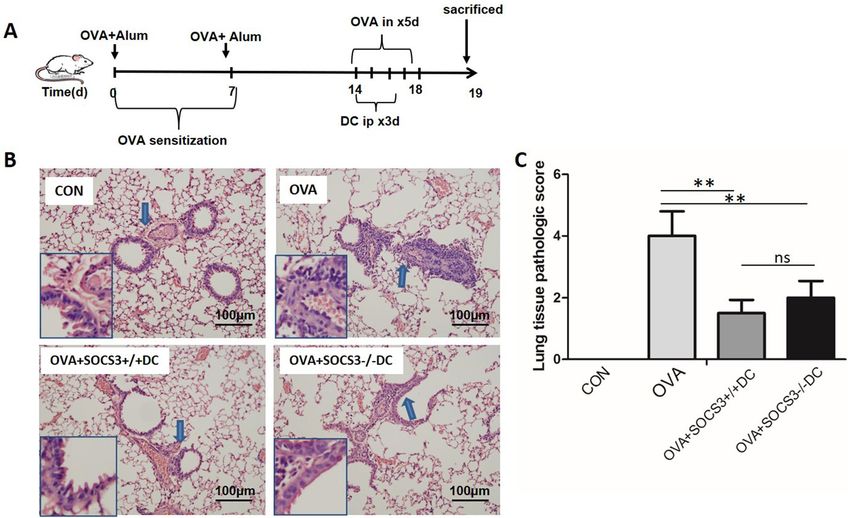
www.nature.com/scientificreports/
tolerance. Mice in which pDCs are specifically depleted develop the features of severe asthma after exposure to
nebulized harmless a ntigens3. Steroids can modulate the functions of DCs in the lungs of patients with allergic
asthma by activating indoleamine 2,3-dioxygenase (IDO) enzymes in D Cs4,5. Furthermore, vitamin D3-incubated
bone marrow-derived DCs (BMDCs) express relatively low levels of major histocompatibility complex class II
(MHCII) and costimulatory molecules, which ultimately attenuates DC-T cell interactions and T cell activation6.
Suppressor of cytokine signaling 3 (SOCS3) is central in negatively regulating signal transducer and activa-
tor of transcription (STAT) 3, STAT4, STAT1 and STAT5 signaling after stimulation with IL-6, IL-11, IL-27, etc.
Kubo et al. found that SOCS3 mRNA expression is increased in eosinophils and CD4+ T cells in asthma and
nonasthmatic eosinophilic bronchitis. T cell-specific deletion of SOCS3 impairs the T helper (Th) 2 response and
increases Th1 responses7. However, deletion of SOCS3 in hematopoietic cells results in severe inflammatory dis-
ease during adult life that is not rescued by IL-6 d eletion8. In addition, SOCS3 gene knockdown in macrophages
results in activation of STAT1 and induction of type I interferon (IFN) responses upon IL-6 s timulation9. Thus,
the roles of the SOCS3 gene in DC functional states and the cognate interaction of SOCS3 with T cells have
been controversial.
Herein, we critically assessed the effects of the SOCS3 gene in BMDCs on cell proliferation and activation by
coculturing SOCS3−/− BMDCs with CD4 T cells. Then, DCs with SOCS3 gene deletion in different functional
states were adoptively transferred into ovalbumin (OVA)-sensitized mice, and lung pathological injury and
airway inflammatory cell infiltration were evaluated. The underlying cellular and molecular mechanisms were
also studied.
Results
SOCS3 deficiency increased the DC‑induced proliferation and cytokine production of T lym‑
phocytes. To investigate the role of SOCS3 in airway inflammation, we created conditional SOCS3-knockout
(KO) mice according to the protocol in a previous study10. Briefly, SOCS3fl/fl mice were bred with mice trans-
genically expressing Cre under the control of the lysozyme 2 (Lyz2) promoter. The offspring SOCS3(Lyz2cre)
mice lacked exon 2 of the SOCS3 locus in myeloid cells; this exon was deleted under the control of the Lyz2
promoter (Fig. 1A). To identify BMDCs with SOCS3 deficiency, we screened bone marrow cells expressing
CD11c, CD80, and MHCII from each group and differentiated them into BMDCs in culture. Fluorescence-
activated cell sorting (FACS) analysis showed that SOCS3 protein expression was significantly lower (62% lower)
in SOCS3(Lyz2cre) mouse-derived BMDCs than in wild-type (WT) mouse-derived BMDCs (Fig. 1C). Western
blot analysis confirmed that the expression of SOCS3 was decreased by 56% in S OCS3−/− BMDCs (Supplemen-
tary Data 1).
We performed an allogeneic mixed lymphocyte reaction (MLR) to evaluate whether a lack of SOCS3 in
BMDCs affects interactions with T lymphocytes by assessing the function and proliferative ability of T lympho-
cytes. We distinguished T cells by staining cells in MLR culture with an anti-CD3 antibody. Carboxyfluorescein
diacetate succinimidyl ester (CFSE) analysis and differential cell counting both demonstrated that S OCS3−/− DCs
induced more robust T cell proliferation than SOCS3+/+DCs (Fig. 2A,B). Surprisingly, SOCS3 deficiency increased
the expression of IFN-γ by T cells (Fig. 2C1), but it did not influence the expression of IL-4 (Fig. 2C2).
Lack of SOCS3 did not alter the therapeutic effect of DCs on allergic airway inflammation. To
further evaluate the effects of SOCS3 deletion, we generated an OVA-induced asthma mouse model and adop-
tively transferred DCs into the asthmatic mice (Fig. 3A). Transfer of DCs ameliorated lung tissue damage and
decreased allergic airway inflammation; however, SOCS3 deficiency did not alter the therapeutic effect of DCs.
In other words, DC therapy attenuated the inflammatory response in the lungs regardless of whether the mature
DCs (lipopolysaccharide [LPS]-induced DCs, DClps) used for treatment expressed the SOCS3 gene (Fig. 3B,C).
Notably, SOCS3−/− DC therapy seemed to have a smaller beneficial effect on lung tissue pathological scores than
SOCS3+/+DC therapy. However, the difference was not statistically significant (Fig. 3C).
In addition, transfer of DCs reduced the total number of pulmonary inflammatory cells in the bronchoalveo-
lar lavage (BAL) fluid (BALF) (Fig. 4A). An obvious reduction in neutrophil count and increase in lymphocyte
count were observed. There was no change in eosinophil count after DC transfer (Fig. 4B). Correspondingly, the
expression of IL-13 and immunoglobulin (Ig) E, which are related to the asthmatic Th2 response, was diminished
after treatment with DCs (Fig. 4C). Unexpectedly, the inhibitory effect of BMDC adoptive transfer was not related
to SOCS3 gene status (Fig. 4C).
Proinflammatory and chemotactic effects of DClps and immature DCs (DCim). In the above
experiment, we demonstrated that transfer of DCs greatly ameliorated lung tissue damage and decreased allergic
airway inflammation. Next, we cultured DCs in vitro to further elucidate whether the therapeutic effect of DCs
was dependent on DC maturation.
DCim were induced to differentiate into mature DCs in the presence of LPS (producing DClps) and were
pulsed with OVA for 2 h. We then measured the levels of select related cytokines in DCim and DClps culture
supernatants. ELISA revealed that the levels of IL-10 and transforming growth factor-β (TGF-β) were higher in
DClps culture medium than in DCim culture medium and that the expression of MCP-3 and IFN-γ was unaf-
fected by DC maturation (Fig. 5).
Among the DClps groups, SOCS3 deficiency had no effect on the expression of MCP-3 or IFN-γ. Surpris-
ingly, the expression of TGF-β was mildly elevated in SOCS3−/− DClps compared to S OCS3+/+DClps (Fig. 5).
The maturity of DCs affects the efficiency of DC pulmonary migration following intraperito‑
neal injection. We attempted to track DCs that had been labeled with the fluorescent dye PKH26 to test
Scientific Reports | (2020) 10:13915 | https://doi.org/10.1038/s41598-020-70467-3 2
Vol:.(1234567890)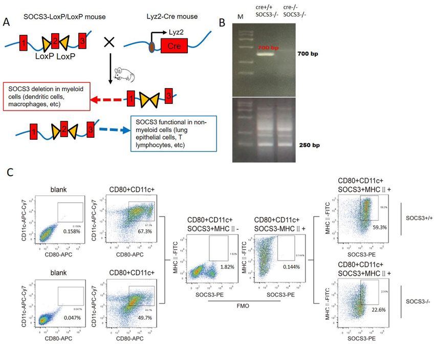
www.nature.com/scientificreports/
Figure 1. Generation of SOCS3(Lyz2cre) mice and identification of SOCS3−/− BMDCs. (A) Schematic
diagram of the generation of SOCS3(Lyz2cre) mice. Floxp-flanked SOCS3 mice were back-crossed with
Lyz2-Cre transgenic mice to create SOCS3 knockout mice with SOCS3 conditional knockout in myeloid
cells, such as DCs or macrophages. (B) The genotypes of SOCS3(Lyz2cre) mice identified by analyzing
mouse tails by PCR. The Cre + loci were identified as 700 bp. The FloxP-flanked exon 2 null SOCS3 loci
were identified as 250 bp. (C) Expression of SOCS3 in BMDCs evaluated by flow cytometry. BMDCs were
gated on CD11c + CD80 + MHCII + cells. A PE-conjugated anti-SOCS3 antibody was used to detect SOCS3
protein expression. In S OCS3−/− mice, SOCS3 protein expression was reduced by approximately 62.4%.
One representative dot plot is shown (SOCS3 and MHCII expression was assessed by comparing with the
corresponding fluorescence-minus-one (FMO) control).
whether DCs could migrate to the lungs successfully and to assess whether the efficiency of pulmonary migra-
tion was related to the maturation state of DCs.
We injected mice with OVA-induced asthma intraperitoneally with PKH26-labeled DCim or DClps or with
saline-diluted PKH26 as a control. We screened DCs by FACS with gating on CD11c + CD80 + MHCII + cells
in the mononuclear cell populations of lung digests. PKH26-labeled BMDCs were also detected by FACS. On
the first day after transplantation, 2.26% of DClps in the lungs were PKH26 positive. The proportion of PKH26-
positive BMDCs in the DClps group peaked at 5.42% on day 3; this proportion was significantly higher than that
in the DCim (1.52%) and control groups (0.86%). The number of PKH26-positive BMDCs in the DClps group
was reduced on day 7 but was still higher than the numbers in the DCim and control groups (Fig. 6). These data
showed that DCs delivered intraperitoneally accumulated in the lungs of OVA-sensitized asthmatic mice during
the week after passive transfer. The efficiency of DC pulmonary migration was related to the maturation state of
DCs, indicating that DClps could reach the lungs in more meaningful numbers than DCim.
Administration of DClps in an OVA‑induced mouse model significantly alleviated allergic air‑
way inflammation. After we confirmed that the efficiency of pulmonary migration was determined by the
maturation state of DCs, we attempted to further clarify whether the degree of DC maturation affected histo-
pathological changes in the lungs.
Scientific Reports | (2020) 10:13915 | https://doi.org/10.1038/s41598-020-70467-3 3
Vol.:(0123456789)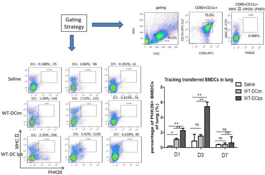
www.nature.com/scientificreports/
Figure 2. Mixed lymphocyte reaction (MLR) with SOCS3−/− BMDCs. (A) Mitomycin-activated BMDCs
cultured with CD4 + T cells (cell ratio of 1:4). T cell proliferation was assessed with CFSE staining. One
representative plot is shown. (B) Number of CD4 + T cells after a 5-days MLR. (C) Cytokine release from
CD4 + T cells after a 5-day MLR. (C-1) IFN-γ production; (C-2) IL-4 production. Data are representative of
3 independent experiments with similar results. The columns and error bars represent the mean and SEM
(*P < 0.05, **P < 0.01, ns: no significant difference).
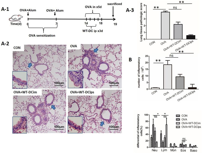
In vivo experiments revealed that compared with DCim injection or no DC injection, DClps injection sig-
nificantly reduced lung inflammatory cell infiltration and lung tissue pathological scores in mice with OVA-
induced asthma (Fig. 7A). In addition, DClps injection significantly reduced the total number of pulmonary
inflammatory cells in BALF, while there was no statistically significant difference between the DCim + OVA and
OVA groups (Fig. 7B). Moreover, neutrophil infiltration was reduced and lymphocyte counts were increased in
both the DClps + OVA and DCim + OVA groups (Fig. 7B).
Administration of DClps reversed impairments in the STAT4 and STAT6 pathways. To define
the roles of DCs in the Th1/Th2 immune signaling pathways, we evaluated the activation and expression of
STAT-1, STAT-4, and STAT-6 in different groups of mice (the control [CON], OVA + WT DC [WT-DC],
OVA + KO DC [KO-DC], and OVA groups). Western blot analysis revealed that administration of DCs signifi-
cantly reduced the phosphorylation of STAT-4 and STAT-6 in the lungs of OVA-sensitized mice, and this effect
was not related to the lack of SOCS3 (Fig. 8). There was also a trend toward reduced STAT1 phosphorylation
after administration of DCs in mice with OVA-induced asthma. However, the reduction was not significant
(Fig. 8). These findings indicate that DCs have the capacity to inhibit STAT6 and STAT4 signaling pathways to
regulate allergen-induced Th2 immune responses.
Scientific Reports | (2020) 10:13915 | https://doi.org/10.1038/s41598-020-70467-3 4
Vol:.(1234567890)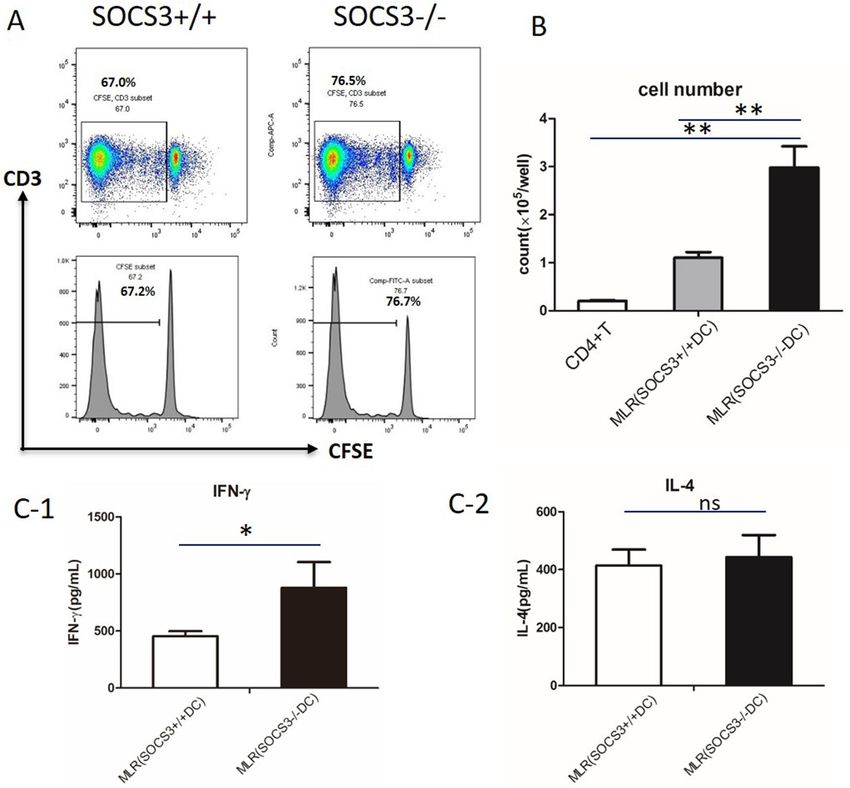
www.nature.com/scientificreports/
Figure 3. Effect of adoptively transferred BMDCs with or without the SOCS3 gene on lung pathology in
OVA-induced asthmatic mice. (A) Protocol for OVA-mediated induction of allergic asthma and SOCS3+/+ DC
or SOCS3−/− DC adoptive transfer into OVA-sensitized mice via intraperitoneal injection (DCs pulsed with
LPS before transfer). (B) Representative photomicrographs of lung sections stained with H&E and examined
at × 100 magnification. The same experiment was repeated 3 times with similar results (n = 6 in each group). C.
The scores for lung tissue pathology. The columns and error bars represent the mean and SEM (**P < 0.01, ns: no
significant difference).
Discussion
The role of the SOCS3 gene in inflammatory diseases, such as allergic asthma, has not been well defined. The
SOCS3 protein is the most widely studied member of the SOCS family, which includes negative regulators of
cytokine signaling. SOCS3 is central in negatively regulating Janus kinase (JAK) and STATs, such as STAT3,
STAT4, STAT1 and STAT5. Previously published evidence has shown that SOCS3 knockdown leads to improve-
ments in inflammation and airway hyperreactivity (AHR) in asthmatic mice and that T cell or CD4+ T cell
activation is restricted after specific SOCS3 d epletion11. However, Duan et al. found that deletion of SOCS3 in
macrophages enhanced the expression of STAT1-stimulated genes in response to IL-6. In addition, systemic
administration of a SOCS3-specific antagonistic peptide (pJAK2) resulted in the induction of IFN r esponses12.
Our previously published data revealed that SOCS3-deficient bone marrow-derived macrophages (BMDMs)
expressed relatively high levels of TNF-α and that adoptive transfer of SOCS3-deficient BMDMs into WT mice
enhanced the severity of acute lung injury (ALI)10. Such inconsistencies might be related to the different cell
subsets in which the SOCS3 gene was specifically depleted in our experiments (myeloid cells) and other experi-
ments (T cells). Based on previous data, BMDCs with or without SOCS3 gene expression were pulsed with
LPS in the current study to promote maturation. We found that either S OCS3−/− DC transfer or S OCS3+/+ DC
transfer markedly alleviated OVA-induced lung injury and dramatically decreased the total number of inflam-
matory cells in BALF. When we examined the proportions of cells in BALF, we found that a lower proportion
of neutrophils and a higher proportion of lymphocytes were present after BMDC adoptive transfer. Th2-type
cytokines and IgE levels were also markedly decreased after BMDC transfer. Interestingly, compared with WT
DCs, SOCS3−/− DCs slightly attenuated BMDC-induced immunogenic tolerance. In this regard, the effect of
SOCS3 depletion on BMDC function was evaluated in an MLR. After mixing cultured BMDCs with CD4+ T
cells, we found that the T cell proliferative capacity was enhanced in the SOCS3−/− BMDC group compared with
the SOCS3+/+BMDC group and the control group. SOCS3−/− BMDCs induced CD4 + T cells to produce more
IFN-γ but not IL-4. This finding partially explains why BMDC-induced immunogenic tolerance was slightly
attenuated by SOCS3−/− BMDCs compared with SOCS3+/+BMDCs.
Different DC subsets and their discrete functional states might be responsible for promoting immunologic
tolerance rather than inflammation. pDCs constitute a unique DC subset with the ability to induce regulatory
T (Treg) cell responses and inhibit Th2 cell production, which has the potential to induce antigen-specific toler-
ance in asthma13,14. Kool et al.15 previously reported that selective removal of pDCs during allergen stimulation
enhances airway inflammation, while adoptive transfer of pDCs before allergen stimulation inhibits airway
inflammation. In contrast, adoptive transfer of conventional DCs (cDCs) or monocyte-derived DCs (moDCs)
into OVA-sensitized mice augments airway inflammation. The various functional states of DCs affect allergic
Scientific Reports | (2020) 10:13915 | https://doi.org/10.1038/s41598-020-70467-3 5
Vol.:(0123456789)www.nature.com/scientificreports/
Figure 4. Adoptively transferred BMDCs with or without the SOCS3 gene alleviated allergic airway
inflammation. (A) The total cell number in the bronchoalveolar lavage fluid (BALF). (B) The differential cell
counts in the BALF. (C) The concentrations of Th2 cytokines in the BALF and serum IgE measured by ELISA.
Data are representative of 3 independent experiments with similar results. The columns and error bars represent
the mean and SEM (**P < 0.01, ns: no significant difference).
airway inflammation and AHR differently. Previous findings have shown that airway delivery of OVA-pulsed
splenic CD8α + DCs reverses AHR and Th2 responses but not allergen-specific IgE and IgG1 responses16. In
addition, adoptive transfer of TGF-β and IL-10-treated DCs can significantly attenuated asthma presentation in
sensitized mice17. On the other hand, DCs differentiated with GM-CSF enhance AHR and eosinophil numbers
and augment Th2 responses in recipient mice. Similar results have been observed in mice transferred with TNF-
treated DCs14. In this study, a subset of BMDCs were pulsed with LPS for 24 h to induce DClps development,
while another subset of BMDCs that were not pulsed with LPS were called DCim. We found that adoptive transfer
of DClps, but not DCim, via intraperitoneal injection greatly improved lung pathology scores and alleviated
airway inflammatory cell infiltration. We subsequently evaluated the biological activities of DClps and DCim.
DClps secreted more IL-10 and TGF-β than DCim, regardless of whether the SOCS3 gene was expressed. This
finding is consistent with previous data showing that IL-10 plays a very important role in IL-10-differentiated
DC (DC10)-mediated AHR improvement in allergic mice. The levels of MCP-3 and IFN-γ production were not
significantly changed by BMDCs.
Gordon et al.3 assessed the effectiveness of DC10 delivery to asthmatic animals via various routes, specifically
the transtracheal (t.t.), intraperitoneal (i.p.), intravenous (i.v.) and subcutaneous (s.c.) routes, and found that t.t.
DC10 delivery and i.p. DC10 delivery were equally effective in reversing AHR and rapidly inhibiting eosinophil
infiltration and the Th2 response. S.c. DC10 transfer inhibited airway Th2 responses to allergen attack, and i.v.
DC10 transfer was completely ineffective in inducing tolerance in asthmatic m ice6. In the present study, PKH26-
labeled BMDCs were transferred into OVA-sensitized mice via i.p. injection. We found that DClps could migrate
to OVA-sensitized lungs more effectively than DCim. DClps began to migrate to the mouse lungs one day after
delivery, and their numbers peaked on day 3 (5.42% vs 1.52% for DClps vs DCim; 5.42% vs 0.86% for DClps
vs saline control). The labeled BMDCs in both groups disappeared on day 7. We found that migrated BMDCs
prevented OVA-sensitized mice from reestablishing Th2 inflammation when BMDCs were administered intra-
peritoneally on days 14–16 during the OVA challenge period. This result indicates the rapid effects of BMDCs
on immunological tolerance after adoptive cell transfer. However, the published data regarding the time frames
of DC tolerance are inconsistent. For example, Nayyar et al.18 reported that the attenuation of AHR was first
Scientific Reports | (2020) 10:13915 | https://doi.org/10.1038/s41598-020-70467-3 6
Vol:.(1234567890)www.nature.com/scientificreports/
Figure 5. Activity of mature and immature DCs with or without the SOCS3 gene. BMDCs were generated from
SOCS3+/+ or SOCS3−/− mice and incubated with or without LPS (the cells incubated with LPS were considered
mature DCs). The cells were then pulsed with OVA for 2 h, and the supernatant was collected for ELISA
analysis. Data are representative of 3 independent experiments with similar results. The columns and error bars
represent the mean and SEM (*P < 0.05, **P < 0.01, ns: no significant difference).
apparent 2 weeks after DC10 delivery and that the effect lasted for 3–10 weeks. We note that there are differences
in experimental design, however; Nayyar et al. used mice with chronic OVA-induced asthma and performed
DC10 transfer 2 weeks after OVA challenge, which was quite different from our approach.
The tolerogenicity of DCs is controlled by a complex network of environmental signals and intrinsic cellular
mechanisms. DCs interact with T cells and determine the differentiation of distinct T cell subsets, such as the
Th1, Th2, and Treg cell subsets. Th1 cells are primed mainly through the IFN-γ/STAT1 pathway and the IL-12/
STAT4 pathway, while Th2 cells are primed mainly through the IL-4/STAT6 pathway. Medoff et al.19 demonstrated
that STAT6 in BMDCs was sufficient for the production of C–C motif chemokine ligand (CCL) 17, CCL22, and
CCL11, which are critical for Th2 lymphocyte recruitment to allergic airways, and found that STAT6 deficiency
abrogated Th2 cell-selective chemokine production in an asthmatic mouse model. In the present study, we found
that the phosphorylation of STAT4 and STAT6 in lung mononuclear cells was significantly decreased after BMDC
adoptive transfer. There was also a decreasing trend in the phosphorylation of STAT1 after BMDC adoptive
transfer. However, the difference was not significant. These results indicated that i.p. BMDC adoptive transfer
reversed OVA-sensitized airway inflammation by inhibiting proinflammatory signal transcription and ultimately
depressing both Th1- and Th2-mediated inflammation, especially Th2-mediated inflammation.
Overall, the findings suggested that BMDC adoptive transfer-induced immunogenic tolerance in OVA-
sensitized mice might not be due to SOCS3 gene depletion. SOCS3−/− DCs slightly attenuated BMDC-induced
immunogenic tolerance. In addition, DClps produced more IL-10 and TGF-β than DCim, leading to dramatic
decreases in the phosphorylation of both STAT4 and STAT6, which are critical in initiating Th1- and Th2-
mediated immunoinflammatory processes in asthma, respectively. Our study indicates that BMDC adoptive
transfer may be developed into a new approach for the treatment of asthma, as it enables fine modulation of the
balance between immune tolerance and inflammation. Further exploration is needed to elucidate the promising
Scientific Reports | (2020) 10:13915 | https://doi.org/10.1038/s41598-020-70467-3 7
Vol.:(0123456789)www.nature.com/scientificreports/
Figure 6. Tracking adoptively transferred wild-type BMDCs in OVA-induced asthmatic mice. Wild-type
BMDCs pulsed with or without LPS were labeled with PKH26 (WT-DClps and WT-DCim) or saline-diluted
PKH26 and transperitoneally transferred into OVA-sensitized mice. On day 1, 3, or 7, mice were sacrificed.
BMDCs were screened by gating on CD11c + CD80 + MHCII + cells in the mononuclear cell population of the
lungs. WT-DClps labeled with PKH26 were tracked and reached 5.42% of the BMDC population in the lungs
(the absolute number of WT-DClps migrating to the lungs was 1,220), and this proportion was higher than
that of WT-DCim and saline control on day 3. The gating strategy and the fluorescence-minus-one (FMO)
control (PKH26 negative) are presented. Histogram represents the percentage of PHK26 + BMDCs adoptively
transferred into the lung within a week. Data are representative of 3 independent experiments, and three mice
were used in each group. The columns and error bars represent the mean and SEM (**P < 0.01;*P < 0.05; ns: no
significant difference).
roles of BMDCs in alleviating allergic diseases. For example, the effects of distinct BMDC subsets on biologi-
cal functional states, BMDC-T cell interactions, BMDC-epithelium interactions, and relevant molecular and
biochemical mechanisms after BMDC transfer warrant additional investigation.
Methods
Establishment and identification of myeloid cell‑restricted SOCS3‑KO mice. The animal pro-
tocol for this study was approved by the Ethics Committee of Zhongshan Hospital of Fudan University. All
animals used in this study were maintained under specific pathogen-free conditions and treated in accordance
with the National Institutes of Health Guide for the Care and Use of Laboratory Animals (NIH Publication No.
8023, revised 1978). The study was approved by the Ethics Boards of Zhongshan Hospital of Fudan University
(Approval No. B2014-108).
The generation of SOCS3-KO mice has been described previously1. Briefly, conditional SOCS3(Lyz2cre) mice
were established by serial breeding of SOCS3fl/fl mice with Lyz2-Cre transgenic mice with Cre under the control
of the myeloid cell-restricted Lyz2 promoter (Fig. 1A). Exon 2 was deleted by the Cre protein in SOCS3 Floxp+/+/
Lyz2Cre+/− or SOCS3 Floxp+/+/Lyz2Cre+/+mice. Successful deletion of SOCS3 in SOCS3(Lyz2cre) mice was
confirmed by PCR methods using 4 pairs of primers according to our previously published m ethods1 (Fig. 1B).
The Cre + loci were identified by 700-bp bands, and exon 2-deleted SOCS3-null loci were identified by 250-bp
bands (Fig. 1B). The mice identified as having the Cre+/+SOCS3−/− genotype were considered SOCS3(Lyz2cre)
mice with myeloid cell-specific deletion of the SOCS3 gene.
To identify SOCS3 expression in BMDCs, bone marrow cells were collected from the femurs and tibiae of
WT and SOCS3(Lyz2cre) mice. The bone marrow cells were seeded in RPMI-1640 medium supplemented with
1% antibiotics/antimycotics and 10% heat-inactivated fetal calf serum (FCS) containing 20 ng/ml GM-CSF. On
Scientific Reports | (2020) 10:13915 | https://doi.org/10.1038/s41598-020-70467-3 8
Vol:.(1234567890)www.nature.com/scientificreports/
Figure 7. Effect of adoptively transferring LPS-pulsed mature BMDCs on lung pathology in OVA-induced
asthmatic mice. (A-1) Protocol for OVA-mediated induction of allergic asthma and transperitoneal adoptive
transfer of LPS-pulsed matured BMDCs (wild type) into OVA-sensitized mice. (A-2) Representative
photomicrographs of lung sections stained with H&E and examined at × 100 magnification. The same
experiment was repeated 3 times with similar results (n = 6 in each group). (A-3) The scores for lung
tissue pathology. (B) The total cell number in the bronchoalveolar lavage fluid (BALF) (top panel). The
differential cell counts in the BALF (bottom panel). The columns and error bars represent the mean and SEM
(**P < 0.01;*P < 0.05; ns: no significant difference).
days 3, 6, and 8, the nonadherent (dendritic) cell suspension was collected, and half of the supernatant was left.
Complete medium supplemented with 20 ng/ml GM-CSF was added for further culture. On day 9, cells were
collected for further study. The expression of cluster of differentiation (CD) 11c, CD80 and MHC-II was used
to phenotypically identify BMDCs. A total of 1 06 cells were washed and subsequently stained with APC-Cy7-
conjugated anti-CD11c (clone HL3, BD Pharmingen, USA), PE-Cy7-conjugated anti-CD80 (clone 16-10A1, BD
Pharmingen, USA), and FITC-conjugated anti-MHC-II (clone M5/114, BD Pharmingen, USA) antibodies. The
cells were stained with an anti-mouse SOCS3 antibody (ab16030, Abcam, UK) after being treated with a fixa-
tion/permeabilization agent (554722, BD Biosciences) to assess the SOCS3 expression deficiency in SOCS3-KO
mice. The cells were analyzed on a FACSCanto II instrument (BD Biosciences, San Jose, CA, USA), and the data
were analyzed with FlowJo software.
Generation of an OVA‑induced mouse asthma model and adoptive transfer of DCs into asth‑
matic mice. To establish an asthmatic mouse model, mice were sensitized with two i.p. injections of 100 µg
of OVA/alum (Sigma-Aldrich, Grade V; St. Louis, MO, USA) on day 0 and day 7 and were then exposed for
20 min to 100 μg of intranasal (i.n.) OVA on 5 consecutive days under light isoflurane anesthesia. The animals
were treated with 1 × 10⁶ DCs via i.p. injection on days 14–16 (Fig. 3A).
The mice were sacrificed within 24 h after the last OVA challenge, and BAL was immediately performed using
3 × 1 mL of 0.05 mM PBS-EDTA (Calbiochem, Darmstadt, Germany) as previously d escribed20–22. The cells in
the BALF were collected with a Cytospin centrifuge (1,200 rpm for 10 min at 4 °C) and stained with Wright’s
solution for differential cell counting. The supernatants were collected and frozen at -80 °C for IL-5 and IL-13
assessment by ELISA. Serum and tissue samples were obtained for further analyses.
Scientific Reports | (2020) 10:13915 | https://doi.org/10.1038/s41598-020-70467-3 9
Vol.:(0123456789)www.nature.com/scientificreports/
Figure 8. STAT signaling pathways were inhibited by adoptive transfer of BMDCs with or without the
SOCS3 gene. Lung tissue samples were homogenized after BMDC adoptive transfer into OVA-sensitized
mice. Mononuclear cells were isolated from homogenates. STATs and phosphorylated STATs were detected by
Western blotting (WB). The right columns represent the densitometry analysis of the WB results. (A) STAT1
and py-STAT1 detected by WB. (B) STAT4 and py-STAT4 detected by WB. C. STAT6 and py-STAT6 detected
by WB. Data are representative of 3 independent experiments with similar results (*P < 0.05, ns: no significant
difference).
Cell preparation and culture of BMDCs. BMDCs were generated from bone marrow cells collected
from WT C57/B6 mice and SOCS3-KO mice. The bone marrow cells were collected from the femurs and tibiae
of WT and SOCS3(Lyz2cre) mice. The bone marrow cells were then seeded in RPMI-1640 medium supple-
mented with 1% antibiotics/antimycotics and 10% heat-inactivated FCS containing 20 ng/ml GM-CSF. On days
3, 6, and 8, the nonadherent (dendritic) cell suspension was collected, and half of the supernatant was left. Com-
plete medium supplemented with 20 ng/ml GM-CSF was added for further culture. On day 9, a subset of cells
were collected for further study. Another subset of cells were incubated with LPS (100 ng/ml) for 24 h to generate
DClps. The cells were then pulsed with OVA (1 µM) for 2 h at 37 °C.
T cell purification and DC stimulation of T cells. To establish an MLR, CD4 + T cells were first obtained
from the spleen and lymph nodes of a native C57/B6 mouse. BMDCs were isolated and cultured as described
above. SOCS3+/+ and S OCS3−/− BMDCs were pretreated for 20 min at 37 °C in culture medium containing
200 μg/ml mitomycin C (Kyowa Hakko Kogyo, Tokyo, Japan). Murine CD4 + T cells were separated by negative
Scientific Reports | (2020) 10:13915 | https://doi.org/10.1038/s41598-020-70467-3 10
Vol:.(1234567890)www.nature.com/scientificreports/
isolation using a MagniSort™ Mouse CD4 T Cell Enrichment Kit (Thermo Fisher). Prior to culture, the purified
CD4 + T cells were labeled with CFSE (Invitrogen, Ltd., UK) for subsequent assessment of T cell proliferation.
Mitomycin C-treated SOCS3+/+ or SOCS3−/− DCs were cocultured with CD4 + T cells at a ratio of 1:4 in a 37 °C,
5% CO2 atmosphere for 5 days. T cell proliferation was assessed by evaluating CFSE dilution on the last day
of culture. After stimulation with phorbol 12-myristate 13-acetate (PMA; 100 ng/ml, Sigma) and ionomycin
(5 μmol/L, Sigma) overnight, the supernatants of the cocultured cells were collected, and IL-4 and IFN-γ pro-
duction was determined by ELISA.
Tracking of SOCS3−/− BMDCs in the lungs. SOCS3+/+ and S OCS3−/− BMDCs were labeled with the fluo-
rescent dye PKH26 (mini126, Sigma‐Aldrich) according to the manufacturer’s protocol. Briefly, 2 × 10⁶ BMDCs
were suspended in 1 ml of diluted buffer from the manufacturer’s labeling kit. The cell suspension was mixed
with an equal volume of a labeling solution containing 4 × 10⁻⁶ mol/L PKH26 dye in dilution buffer, and the mix-
ture was incubated for 4 min at room temperature. The reaction was terminated by adding 2 ml of fetal bovine
serum (FBS). After washing with a control medium, 5 × 10⁶ DCs labeled with PKH26 were mixed with 1 mol/L
PLA‐CMC solution and intraperitoneally injected into mice with OVA-induced asthma.
PKH26-labeled SOCS3+/+ and SOCS3−/− DCs were injected intraperitoneally into asthmatic mice (1 × 10⁶
cells/mouse) as described above. After 1, 3 or 7 days, we collected the lung tissue from each animal and generated
single-cell suspensions by enzymatic digestion of the lungs (which included cutting the lungs into small pieces,
incubating them with collagenase IV and DNase for one hour and filtering the resultant suspension through a
300-µm mesh filter). PKH26-labeled BMDCs in lung tissue samples were evaluated by assessing the expression
of CD11c, CD80, MHC-II and PKH26 with a FACSCanto II instrument (BD Biosciences, San Jose, CA, USA).
Histochemistry and assessment of pathological lung injury. After the BALF was collected, the lungs
were infused with 4% paraformaldehyde. Then, the trachea was clamped, and the lungs were excised. The left
lung was embedded in paraffin. Five-micrometer paraffin sections were obtained and stained with hematoxylin
and eosin (H&E). The right lung was collected and frozen at − 80 °C for protein assessment by Western blotting.
To quantify the degree of lung damage, H&E-stained slides were graded in a blinded fashion using a scoring
system described previously23. A score of 0 indicated that no detectable inflammation was present, a score of 1
indicated that occasional inflammatory cells were present, a score of 2 indicated that most bronchi or vessels
were surrounded by a thin layer (one to five cells thick) of inflammatory cells, and a score of 3 indicated that most
bronchi or vessels were surrounded by a thick layer (more than five cells thick) of inflammatory cells. Total lung
inflammation was defined as the average of the peribronchial and perivascular inflammation scores. All slides
were examined by two independent pathologists.
ELISA. Cell culture supernatants and BALF samples were evaluated by using commercially available ELISA
kits according to the manufacturers’ instructions. IL-4, IL-5, IL-10, IL-13, MCP-3, TGF-β, IFN-γ and IgE in
supernatants were detected with ELISA kits (R&D Systems).
Western blot analysis. The expression levels of STAT1, STAT4, STAT6 and the corresponding phosphoryl-
ated proteins in lung digests were analyzed by Western blot analysis. Cell lysates (40 μg) were separated by 10%
sodium dodecyl sulfate-PAGE and transferred to polyvinylidene fluoride (PVDF) membranes (Roche, USA).
After incubation in a blocking buffer containing 5% skim milk in TBST (12.5 mM Tris–HCl pH 7.5, 68.5 mM
NaCl, and 0.1% Tween 20) for 1 h, the blots were incubated overnight with primary antibodies including rabbit
anti-STAT1 (D1K9Y, Cell Signaling Technology, USA), rabbit anti-phosphorylated STAT1 (Tyr701, Cell Signal-
ing Technology, USA), rabbit anti-STAT4 (C46B10, Cell Signaling Technology, USA), rabbit anti-phosphoryl-
ated STAT4 (ab28815, Abcam, UK), rabbit anti-STAT6 (ab32520, Abcam, UK), and rabbit anti-phosphorylated
STAT6 (ab28829, Abcam, UK). An anti-mouse GAPDH antibody was used as a loading control. The blots were
washed with TBST, incubated with horseradish peroxidase (HRP)-conjugated anti-rabbit Ig (Jackson Immu-
noResearch) and then developed with an enhanced chemiluminescence (ECL) substrate solution (Millipore).
Statistical analysis. All data analysis and graph preparation were performed with GraphPad Prism 5.01
(GraphPad Software, San Diego, CA, USA). The data are presented as the mean ± standard error for each experi-
mental group. Multigroup comparisons were performed by either one-way ANOVA with Tukey’s post hoc test
or Wilcoxon signed rank tests (FACS analyses). P < 0.05 was regarded to indicate significance.
Received: 12 September 2019; Accepted: 30 June 2020
References
1. GeurtsvanKessel, C. H. et al. Clearance of influenza virus from the lung depends on migratory langerin+CD11b: but not plasma-
cytoid dendritic cells. J. Exp. Med. 205, 1621–1634 (2008).
2. Nakagome, K. et al. In vivo IL-10 gene delivery suppresses airway eosinophilia and hyperreactivity by down-regulating APC
functions and migration without impairing the antigen-specific systemic immune response in a mouse model of allergic airway
inflammation. J. Immunol. 174, 6955–6966 (2005).
3. Smit, J. J., Rudd, B. D. & Lukacs, N. W. Plasmacytoid dendritic cells inhibit pulmonary immunopathology and promote clearance
of respiratory syncytial virus. J. Exp. Med. 203, 1153–1159 (2006).
Scientific Reports | (2020) 10:13915 | https://doi.org/10.1038/s41598-020-70467-3 11
Vol.:(0123456789)www.nature.com/scientificreports/
4. Hammad, H. et al. Activation of the D Prostanoid 1 receptor suppresses asthma by modulation of lung dendritic cell function and
induction of regulatory T cells. J. Exp. Med. 204, 357–367 (2007).
5. Grohmann, U. et al. Reverse signaling through GITR Ligand enables dexamethasone to activate IDO in allergy. Nat. Med. 13,
579–586 (2007).
6. Gordon, J. R., Ma, Y., Churchman, L., Gordon, S. A. & Dawicki, W. Regulatory dendritic cells for immunotherapy in immunologic
diseases. Front. Immunol. 5, 7 (2014).
7. Kubo, M. & Inoue, H. Suppressor of cytokine signaling 3 (SOCS3) in Th2 cells evokes Th2 Cytokines, IgE, and Eosinophilia. Curr.
Allergy Asthma Rep. 6, 32–39 (2006).
8. Liu, X. et al. Key role of suppressor of cytokine signaling 3 in regulating Gp130 cytokine-induced signaling and limiting chondro-
cyte responses during murine inflammatory arthritis. Arthritis Rheumatol. 66, 2391–2402 (2014).
9. Lang, R. et al. SOCS3 Regulates the plasticity of Gp130 signaling. Nat. Immunol. 4, 546–550 (2003).
10. Jiang, Z., Chen, Z., Li, L., Zhou, W. & Zhu, L. Lack of SOCS3 increases LPS-induced murine acute lung injury through modulation
of Ly6C(+) macrophages. Respir. Res. 18, 217 (2017).
11. Moriwaki, A. et al. T cell treatment with small interfering RNA for suppressor of cytokine signaling 3 modulates allergic airway
responses in a murine model of asthma. Am. J. Respir. Cell. Mol. Biol. 44, 448–455 (2011).
12. Duan, W. N. et al. Protective effects of SOCS3 overexpression in high glucoseinduced lung epithelial cell injury through the JAK2/
STAT3 pathway. Mol. Med. Rep. 16, 2668–2674 (2017).
13. Chen, W., Liang, X., Peterson, A. J., Munn, D. H. & Blazar, B. R. The indoleamine 2,3-dioxygenase pathway is essential for human
plasmacytoid dendritic cell-induced adaptive t regulatory cell generation. J. Immunol. 181, 5396–5404 (2008).
14. Huang, H. et al. Regulatory dendritic cell expression of MHCII and IL-10 are jointly requisite for induction of tolerance in a murine
model of OVA-asthma. Allergy 68, 1126–1135 (2013).
15. Kool, M. et al. An anti-inflammatory role for plasmacytoid dendritic cells in allergic airway inflammation. J. Immunol. 183,
1074–1082 (2009).
16. Huang, H., Dawicki, W., Zhang, X., Town, J. & Gordon, J. R. Tolerogenic dendritic cells induce CD4+CD25hiFoxp3+ regulatory
T Cell differentiation from CD4+CD25-/loFoxp3- effector T Cells. J. Immunol. 185, 5003–5010 (2010).
17. Lu, M. et al. Therapeutic induction of tolerance by IL-10-differentiated dendritic cells in a mouse model of house dust mite-asthma.
Allergy 66, 612–620 (2011).
18. Nayyar, A. et al. Induction of prolonged asthma tolerance by IL-10-differentiated dendritic cells: differential impact on airway
hyperresponsiveness and the Th2 immunoinflammatory response. J. Immunol. 189, 72–79 (2012).
19. Medoff, B. D. et al. CD11b+ myeloid cells are the key mediators of Th2 cell homing into the airway in allergic inflammation. J.
Immunol. 182, 623–635 (2009).
20. Cataldo, D. D. et al. Matrix metalloproteinase-9 deficiency impairs cellular infiltration and bronchial hyperresponsiveness during
allergen-induced airway inflammation. Am. J. Pathol. 161, 491–498 (2002).
21. Gueders, M. M. et al. Matrix metalloproteinase-8 deficiency promotes granulocytic allergen-induced airway inflammation. J.
Immunol. 175, 2589–2597 (2005).
22. Schneider, A. M., Li, F., Zhang, X. & Gordon, J. R. Induction of pulmonary allergen-specific IgA responses or airway hyperrespon-
siveness in the absence of allergic lung disease following sensitization with limiting doses of ovalbumin-alum. Cell. Immunol. 212,
101–109 (2001).
23. Kwak, Y. G. et al. Involvement of PTEN in airway hyperresponsiveness and inflammation in bronchial asthma. J. Clin. Invest. 111,
1083–1092 (2003).
Acknowledgements
This work was supported by the National Natural Science Foundation of China (81470211 and 81970023 to ZHC),
the Shanghai Health Committee (201840288), the Shanghai Respiratory Research Institute and Yang Scientists
Training Program of Zhongshan Hospital, the Shanghai Top-Priority Clinical Key Disciplines Construction
Project (2017ZZ02013), and the Shanghai Municipal Key Clinical Specialty (shslczdzk02201).
Author contributions
Conceived and designed the study: C.Z; performed the biological experiments: K.X., N.W., Z.J., Z.M, and Z.L.;
performed the statistical analysis: T.Z., C.L., Y.Z., J.S., and R.M.; and wrote and modified the paper: N.W., Z.J.,
H.J. and C.Z. All authors read and approved the final manuscript.
Competing interests
The authors declare no competing interests.
Additional information
Supplementary information is available for this paper at https://doi.org/10.1038/s41598-020-70467-3.
Correspondence and requests for materials should be addressed to H.J., Z.J. or Z.C.
Reprints and permissions information is available at www.nature.com/reprints.
Publisher’s note Springer Nature remains neutral with regard to jurisdictional claims in published maps and
institutional affiliations.
Open Access This article is licensed under a Creative Commons Attribution 4.0 International
License, which permits use, sharing, adaptation, distribution and reproduction in any medium or
format, as long as you give appropriate credit to the original author(s) and the source, provide a link to the
Creative Commons license, and indicate if changes were made. The images or other third party material in this
article are included in the article’s Creative Commons license, unless indicated otherwise in a credit line to the
material. If material is not included in the article’s Creative Commons license and your intended use is not
permitted by statutory regulation or exceeds the permitted use, you will need to obtain permission directly from
the copyright holder. To view a copy of this license, visit http://creativecommons.org/licenses/by/4.0/.
© The Author(s) 2020
Scientific Reports | (2020) 10:13915 | https://doi.org/10.1038/s41598-020-70467-3 12
Vol:.(1234567890)You can also read


