Effects of an ethyl acetate extract of Daphne altaica stem bark on the cell cycle, apoptosis and expression of PPARγ in Eca 109 human esophageal ...
←
→
Page content transcription
If your browser does not render page correctly, please read the page content below
1400                                     Molecular Medicine REPORTS 22: 1400-1408, 2020

                Effects of an ethyl acetate extract of Daphne altaica
              stem bark on the cell cycle, apoptosis and expression of
               PPARγ in Eca‑109 human esophageal carcinoma cells
               MURAT KIZAIBEK1,2, AYIXIAMUGULI WUBULI3*, ZHENGBING GU4, DIDAR BAHETJAN1,2,
                  LAZZAT TURSINBAI1,2, KAMISHBEK NURHAMIT1,2, BIN CHEN5, JING WANG6,7,
                                     OMIRSHAT TAHAN8 and PENG CAO6,7*

       1
     Traditional Chinese Medicine Hospital of Ili Kazakh Autonomous Prefecture; 2Traditional Kazakh Medicine Research
     Institute of Ili Kazakh Autonomous Prefecture, Yining, Xinjiang 835000; 3Xinjiang Technical Institute of Physics and
     Chemistry, Chinese Academy of Science, Urumqi, Xinjiang 830011; 4Jiangsu Yongjian Medical Technology Ltd. Co.,
Taizhou, Jiangsu 225300; 5Nanjing Research Institute for Comprehensive Utilization of Wild Plants, Nanjing, Jiangsu 210042;
    6
      Affiliated Hospital of Integrated Traditional Chinese and Western Medicine, Nanjing University of Chinese Medicine;
               7
                 Jiangsu Province Academy of Traditional Chinese Medicine, Nanjing, Jiangsu 210028; 8College of
         Grassland and Environment Sciences, Xinjiang Agricultural University, Urumqi, Xinjiang 830052, P.R. China

                                        Received April 22, 2019; Accepted March 17, 2020

                                                    DOI: 10.3892/mmr.2020.11187

Abstract. Daphne altaica Pall. (D. altaica; Thymelaeaceae)            Cell morphology was observed under a phase contrast micro-
has long been used in traditional Kazakh medicine for                 scope. Cell apoptosis and cell cycle progression were assessed
the treatment of cancer and respiratory diseases. Previous            by flow cytometry following Annexin V/propidium iodide
studies have demonstrated the in vitro anticancer effects             (PI) double staining and PI single staining, respectively. The
of D. altaica extract and its constituents in certain cancer          mRNA and protein expression levels of PPARγ were deter-
cell lines; however, the underlying molecular mechanisms              mined by reverse transcription‑quantitative PCR and western
are not completely understooD. The present study aimed                blotting, respectively. Compared with the control group, the
to investigate the molecular mechanisms underlying the                percentage of apoptotic cells, cell cycle arrest at S phase and
activity of an ethyl acetate extract of D. altaica (Da‑Ea) by         apoptotic morphological cell characteristics were increased
assessing its effects on cell morphology, cell apoptosis, cell        in Da‑Ea‑treated Eca‑109 cells. Furthermore, Da‑Ea treat-
cycle progression and the expression levels of peroxisome             ment upregulated the mRNA and protein expression levels of
proliferator‑activated receptor γ (PPARγ) in Eca‑109 cells.           PPARγ compared with the control cells. High‑performance
                                                                      liquid chromatography with diode‑array detection indicated
                                                                      that daphnetin‑7‑O‑β‑D‑glucoside, daphnetin, demethyldaph
                                                                      noretin‑7‑O‑β‑D‑glucopyranoside and genkwanol A were the
                                                                      main constituents of Da‑Ea. Collectively, the results suggested
Correspondence to: Miss Ayixiamuguli Wubuli, Xinjiang                 that Da‑Ea displayed antiproliferative activities in Eca‑109
Technical Institute of Physics and Chemistry, Chinese Academy         cells by inducing apoptosis and S phase cell cycle arrest, as
of Science, 40‑1 Beijing South Road, Urumqi, Xinjiang 830011,         well as upregulating PPARγ expression levels.
P.R. China
E‑mail: ayixia1212@163.com
                                                                      Introduction
Dr Peng Cao, Jiangsu Province Academy of Traditional Chinese
Medicine, 100 Shizi Road, Nanjing, Jiangsu 210028, P.R. China         Throughout history, natural products have served an important
E‑mail: njpcao@126.com                                                role in the treatment of human diseases. At present, natural
                                                                      products are the major source of pharmaceutical agents,
*
    Contributed equally
                                                                      particularly for cancer therapy. Natural products and their
Abbreviations: Da‑Ea, ethyl acetate extract of Daphne altaica;        derivatives account for ~80% of all drugs approved for cancer
PPARγ, peroxisome proliferator‑activated receptor‑γ; HPLC‑DAD,        therapy by the USA Food and Drug Administration during the
high‑performance liquid chromatography‑diode‑array detector           last three decades (1,2).
                                                                          Daphne altaica Pall. (D. altaica) is a medicinal herb used
Key words: Daphne altaica Pall., cell apoptosis, cell cycle, PPARγ,   in traditional Kazakh medicine for the treatment of numerous
traditional Kazakh medicine                                           diseases, including cancer of the digestive tract, tracheitis,
                                                                      common cold, sore throat, rheumatism and snakebite (3). The
                                                                      plant is endemically distributed in the North of the Jungar Basin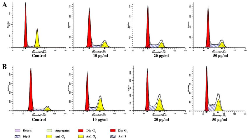
KIZAIBEK et al: EFFECTS OF Daphne altaica ON CELL CYCLE, APOPTOSIS AND EXPRESSION OF PPARγ                    1401

of Xinjiang (Tacheng and Habahe areas), the Altai, Manrak            solvents of increased polarity, including pure petroleum ether,
and Tarbagatai Mountains of Kazakhstan, the Altai region             pure chloroform and pure ethyl acetate. The only extract used
of Russia and Northwest Mongolia (3). The medicinal use of           in subsequent experiments was Da‑Ea.
D. altaica was first recorded in the Kazakh medical classic              High performance liquid chromatography (HPLC) with
work Shipagerlik Bayan (3). However, the anticancer effects of       diode‑array detection (DAD) was used to analyse the main
D. altaica were reported for the first time by Kizaibek et al (3),   ingredients in the extracts. HPLC was conducted using a
who demonstrated that different D. altaica extracts, except          1260 HPLC system (Agilent Technologies, Inc.) equipped
for the aqueous extract, displayed moderate to significant           with a 1260 Infinity Diode Array Detector (cat. no. G4212A;
in vitro cytotoxicity against several cancer cell lines, including   Agilent Technologies, Inc.) in gradient elution mode. An
Eca‑109, AGS, SMMC‑7721 and HeLa. Kizaibek et al (4) also            XBridge™ C18 column (4.6x250 mm; diameter, 5 µm; Waters
identified antiproliferative activities of D. altaica in the human   Chromatography Europe BV) set at 25˚C with a 0.5 ml/min
CCRF‑CEM leukaemia and MDA‑MB‑231 breast cancer                      flow rate was useD. For each experiment, a 5 µl injection volume
cell lines, and identified the constituents of the D. altaica        was useD. The mobile phase consisted of 0.1% (v/v) acetic acid
CH2Cl2 extract using liquid chromatography (LC)‑diode‑array          aqueous solution (solvent A) and acetonitrile (solvent B). The
detection (DAD)‑mass spectrometry and LC‑DAD‑high reso-              gradient conditions used were as follows: 10% solvent B for
lution electrospray ionization mass spectrometry in positive         0 min; 62% solvent B for 37 min; 62% solvent B for 39 min; and
mode. Nugroho et al (5) reported that three new daphnane             10% solvent B for 40 min. The chromatogram was analysed
diterpenoids (Altadaphnans A‑C) from the aerial parts of             using OpenLAB CDS Chemstation software (version A.01.05;
D. altaica significantly inhibited the proliferation of A549         Agilent Technologies, Inc.). To obtain stock solution, 22.7 mg
cancer cells. However, the mechanisms underlying the anti-           Da‑Ea was sonicated with 1,135 µl 80% MeOH‑DMSO (1:1)
proliferative activities of D. altaica have not been previously      at 40 MHz frequency and room temperature for 10 min.
reporteD. Therefore, the present study aimed to identify the         Subsequently, the stock solution was further diluted with
mechanism underlying the antiproliferative activity of an            methanol to generate 1.25 mg/ml sample solution for HPLC
ethyl acetate extract of D. altaica (Da‑Ea) by assessing cell        analysis. Standard compounds [daphnetin‑7‑O‑β‑D‑glucoside,
apoptosis, cell cycle progression and the expression of peroxi-      daphnetin, demethyldaphnoretin‑7‑O‑ β ‑D‑glucopyrano
some proliferator‑activated receptor γ (PPARγ) in the human          side and genkwanol A; provided by Professor Zhengbing
Eca‑109 oesophageal squamous cell carcinoma cell line.               Gu (Jiangsu Yongjian Medical Technology Ltd., Co.)] were
                                                                     dissolved in methanol. Compounds were identified based on
Materials and methods                                                their retention time and UV spectra compared with reference
                                                                     standards.
Cell culture. The human Eca‑109 oesophageal cancer cell
line was purchased from The Cell Bank of Type Culture                Evaluation of cell morphology. Eca‑109 cells were seeded
Collection of the Chinese Academy of Sciences and main-              (2x10⁵ cells/well) into 6‑well culture plates and incubated with
tained at 37˚C with 5% CO2 in RPMI‑1640 (Gibco; Thermo               Da‑Ea (10, 20 or 50 µg/ml) at 37˚C for 24, 48 or 72 h. Cells
Fisher Scientific, Inc.) supplemented with 10% fetal bovine          treated with DMSO (10, 20 or 50 µg/ml) were used as the
serum (Gibco; Thermo Fisher Scientific, Inc.) and 1%                 control. For each test and control medium, the final concentra-
penicillin‑streptomycin solution (HyClone; GE Healthcare             tion of DMSO was adjusted to 1%. Under x100 magnification,
Life Sciences). At 90% confluency, cells were harvested using        cell morphology was assessed using an IX71‑12FL/PH phase
0.25% trypsin‑EDTA (Gibco; Thermo Fisher Scientific, Inc.).          contrast microscope (Olympus Corporation) with Olympus
Cells in the exponential phase of growth were used for subse-        cellSens Standard imaging software, version 1.0 (Olympus
quent experiments.                                                   Corporation).

Plant materials. The D. altaica plant was collected from Habahe      Detection of cell apoptosis. Early cell apoptosis was exam-
County of Xinjiang Uyghur Autonomous Region, P.R. China              ined by Annexin V‑FITC Apoptosis Detection kit (BD
in July 2017. The plant was identified by Dr Omirshat Tahan          Bioscience) according to the manufacturer's protocol. Cells
(College of Grassland and Environment Sciences, Xinjiang             (2x10⁵ cells/well) were seeded into 6‑well culture plates and
Agricultural University, Urumqi, China). A voucher specimen          allowed to grow overnight. Subsequently, cells were treated
(no. HB‑2017001) was deposited at the Traditional Kazakh             with Da‑Ea (10, 20 or 50 µg/ml) for 24, 48 and 72 h. Following
Medicine Research Institute of Traditional Chinese Medicine          treatment, cells were collected and washed twice with PBS.
Hospital of Ili Kazakh Autonomous Prefecture.                        Cells were incubated with 5 µl Annexin V and 5 µl PI (BD
                                                                     Biosciences; Becton, Dickinson and Company) for 15 min
Extraction of Da‑Ea. Da‑Ea was extracted as previously               at room temperature in the dark. Following incubation, each
described (3). Briefly, dried bark of the plant (150 g) was cut      sample was filtrated on a nylon membrane (pore size, 48 ìm).
into small pieces and macerated with 95% EtOH for 2 weeks            Flow cytometry was performed using a FACSAriaII flow
at room temperature in the dark. The extraction process              cytometer (Beckman Coulter, Inc.) and FlowJo software,
was repeated twice. The extracted mixtures were combined,            version 7.6 (Tree Star Inc.).
concentrated by distillation under vacuum and freeze‑dried to
yield the EtOH extract. Petroleum ether, chloroform and ethyl        Detection of cell cycle. For cell cycle analysis, cells were
acetate (Da‑Ea; 0.8243 g) extracts were obtained from the            seeded (2x10⁵ cells/well) into 6‑well culture plates and main-
EtOH extract using a sequential liquid‑liquid extraction with        tained overnight. Subsequently, cells were treated with Da‑Ea
1402 Molecular Medicine REPORTS 22: 1400-1408, 2020
(10, 20 or 50 µg/ml) for 24 or 48 h. Cells were washed twice and reverse, 5'‑CTAAGTCATAGTCCGCCTAGAAGCA‑3'.
with PBS. Cells were fixed in 75% ethanol at 4˚C for 2 h and The following thermocycling conditions were used for qPCR:
subsequently washed three times with PBS. Cells were stained Initial denaturation for 3 min at 95˚C; followed by 40 cycles of
with 500 µl PI/RNase staining buffer (BD Biosciences; denaturation at 95˚C for 30 sec, annealing at 60˚C for 30 sec
Becton, Dickinson and Company) at room temperature for and elongation at 72˚C for 10 min; and melt curve analysis from
15 min in dark. Following staining, cells were filtrated on a 65‑95˚C at a heating rate of 20˚C/sec. PPARγ mRNA levels
nylon membrane (pore size, 48 ìm). Cells were analysed using were quantified using the 2‑ΔΔCq method and normalized to the
a FACSAriaII flow cytometer (Beckman Coulter, Inc.) and internal reference gene β‑actin (6).
ModFit LT software, version 3.2 (Verity Software House, Inc.).
Assessment of PPARγ protein expression by western blotting.
Assessment of PPARγ mRNA expression by reverse tran‑ Cells were treated with Da‑Ea (10, 20 or 50 µg/ml) for 24, 48
scription‑quantitative PCR (RT‑qPCR). Eca‑109 cells in the or 72 h. Total protein was extracted from cells using RIPA
logarithmic phase of growth were seeded (3x10⁵ cells/ml) into buffer (cat. no. P0013B; Beyotime Institute of Biotechnology)
6‑well culture plates. Cells were incubated with Da‑Ea (10, containing PMSF. Subsequently, samples were centrifuged
20 or 50 µg/ml) for 24, 48 and 72 h at 37˚C with 5% CO2. at 12,000 x g for 10 min at 4˚C. Total protein was quantified
Subsequently, cells were washed with PBS and total RNA was using a bicinchoninic acid assay. Protein (20 µg per lane)
extracted using TRIzol® reagent (Thermo Fisher Scientific, was separated via 10% SDS‑PAGE and transferred to PVDF
Inc.) according to the manufacturer's protocol. Subsequently, membranes. Ponceau S staining was performed to confirm
the cell lysate was incubated at room temperature for 5 min and successful transfer of the proteins to the membrane, for 5 min
transferred to a fresh 1.5 ml Eppendorf tube containing 200 µl at room temperature. Subsequently, the membranes were
chloroform. The tubes were vortexed for 15 sec, incubated at blocked with 5% non‑fat milk in TBST (Tris‑buffered saline
room temperature for 2‑3 min and centrifuged at 15,400 x g containing 0.1% Tween‑20) for 1 h at room temperature. The
for 15 min at 4˚C until the liquid separated into three phases. membranes were incubated overnight at 4˚C with primary
The upper aqueous phase (400‑500 µl) was transferred to a antibodies targeted against: PPARγ (1:5,00; cat. no. sc‑7273;
new RNase‑free Eppendorf tube, mixed with an equal volume Santa Cruz Biotechnology, Inc.) and β ‑actin (1:5,000;
of isopropanol and incubated for 10 min at room tempera- cat. no. 60008‑1‑Ig; ProteinTech Group, Inc.) After washing
ture. Subsequently, the sample was centrifuged for 10 min at with TBST, the membranes were incubated with a horse-
15,400 x g and 4˚C to precipitate the RNA. The supernatant radish peroxidase‑conjugated secondary antibody (1:5,000;
was discarded and 75% ethanol was added to the RNA prior cat. no. SA00001‑1; ProteinTech Group, Inc.) for 1 h at 37˚C.
to centrifugation at 3,800 x g for 15 min at 4˚C. Subsequently, Subsequently, the membranes were washed three times with
the supernatant was discarded and the RNA was air‑dried TBST for 5 min. Protein bands were visualized using an
at room temperature for 5‑10 min. Total RNA was stored at ECL detection kit (7Sea Biotech; http://7seapharmtech.com/)
‑20˚C until further analysis. RNA purity was assessed by and imaged using the ChemiScope 5300 Pro (Clinx Science
measuring the A260/A280 absorbance ratio using a Nanodrop Instruments Co., Ltd.) imaging system. β‑actin was used as
spectrophotometer (Thermo Fisher Scientific, Inc.). RNA with the loading control. The bands were then quantified using
an A260/A280 ratio of 1.7‑2.0 was considered high quality. image analysis software (ImageJ2x; version 2.1.4.5; National
RNA quality and integrity were verified by visualising 28S Institutes of Health).
and 18S RNA bands on an ethidium bromide‑stained 1.5%
agarose gel using the Gel DOC XR imaging system (Bio‑Rad Statistical analysis. Statistical analyses were performed using
Laboratories, Inc.). RNA samples with a clear and sharp 28S GraphPad Prism software (version 6.0; GraphPad Software,
band that were twice as intense as the 18S band were used Inc.). Comparisons among groups were analysed using
for reverse transcription. Total RNA was reverse transcribed one‑way ANOVA followed by Dunnett's post hoc test. Data are
to cDNA using RevertAid First strand cDNA Synthesis kit expressed as the mean ± standard deviation of three replicates.
(Thermo Fisher Scientific, Inc.). Reverse transcription was PKIZAIBEK et al: EFFECTS OF Daphne altaica ON CELL CYCLE, APOPTOSIS AND EXPRESSION OF PPARγ 1403 Figure 1. High performance liquid chromatography chromatograms. Compounds were identified by comparing the retention times and UV spectra of the (A) sample solution and (B) standard solution. The average UV spectrum of (C) daphnetin‑7‑O‑β‑D‑glucoside, (D) daphnetin, (E) demethyldaphnoretin‑7‑O‑β‑ D‑glucopyranoside and (F) genkwanol A. The red line represents the sample solution and the blue line represents the standard solution. concentrations of Da‑Ea for 24‑72 h underwent notable 7.350±0.043 and 7.516±0.015% in the 10, 20 and 50 µg/ml morphological alterations (Fig. 3). For example, round or Da‑Ea groups, respectively, compared with the control group polygonal‑shaped cells became elongated, cells appeared to (1.763±0.045%; P
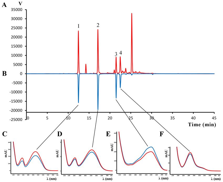
1404                                          Molecular Medicine REPORTS 22: 1400-1408, 2020

Figure 2. Chemical structures of the main components of an ethyl acetate extract of Daphne altaica. The chemical structure of (1) daphnetin‑7‑O‑β‑D‑glucos
ide, (2) daphnetin, (3) demethyldaphnoretin‑7‑O‑β‑D‑glucopyranoside and (4) genkwanol A.

Figure 3. Effect of Da‑Ea on Eca‑109 cell morphology. Eca‑109 cell morphology following treatment with 0, 10, 20, 50 µg/ml Da‑Ea for (A) 24, (B) 48 and
(C) 72 h (magnification, x100). The white arrows indicate cell shrinkage and elongation. Da‑Ea, ethyl acetate extract of Daphne altaica.

alterations to the cell cycle distribution occurred in a time‑                 PPARγ mRNA expression level. Agarose gel electrophoresis
and dose‑dependent manner, which suggested that Da‑Ea                          demonstrated that the bands corresponding to 28S RNA and
inhibited cell proliferation by inducing S phase cell cycle                    18S RNA were sharp and clear. Furthermore, the intensity of
arrest in Eca‑109 cells.                                                       the 28S RNA band was approximately twice as intense as the
KIZAIBEK et al: EFFECTS OF Daphne altaica ON CELL CYCLE, APOPTOSIS AND EXPRESSION OF PPARγ                                                                                         1405

Table I. Rate of Eca‑109 cell apoptosis following treatment with ethyl acetate extract of Daphne altaica for 24, 48 or 72 h.

	Rate of apoptosis (%)
                      ---------------------------------------------------------------------------------------------------------------------------------------------------------------
Concentration (µg/ml)         24 h                                                               48 h                                                                72 h

Control                                                             1.463±0.055                                               1.630±0.155                                                 1.763±0.045
10                                                                  1.756±0.040a                                              4.430±0.060a                                                7.173±0.251a
20                                                                  2.016±0.015a                                              5.800±0.010a                                                7.350±0.043a
50                                                                  2.700±0.200a                                              6.876±0.025a                                                7.516±0.015a

P
1406 Molecular Medicine REPORTS 22: 1400-1408, 2020 Figure 5. Da‑Ea induces S phase cell cycle arrest in Eca‑109 cells. The cell cycle distribution of Eca‑109 cells treated with Da‑Ea for (A) 24 and (B) 48 h was determined by flow cytometry. Da‑Ea, ethyl acetate extract of Daphne altaica. Figure 6. Effect of the ethyl acetate extract of Daphne altaica on the mRNA expression level of PPARγ in Eca‑109 cells. *P
KIZAIBEK et al: EFFECTS OF Daphne altaica ON CELL CYCLE, APOPTOSIS AND EXPRESSION OF PPARγ                         1407

Discussion                                                                 Additionally, the effects of D. altaica on the expression
                                                                       level of PPARγ in Eca‑109 cells were investigateD. PPARs
In the present study, the effects of Da‑Ea on Eca‑109 cell             are ligand‑activated transcription factors that regulate the
apoptosis and cell cycle distribution were analysed, and the           expression of genes involved in lipid metabolism, glucose
underlying mechanisms were investigated by detecting the               homeostasis, cell proliferation, differentiation and survival.
expression levels of PPARγ.                                            PPARs are divided into three subfamilies: PPARα, PPARβ/δ
    A previous study reported that petroleum ether, chloro-            and PPARγ, with the PPARγ subfamily being the most
form, DA‑Ea and n‑butanol extracts of D. altaica display               intensively investigated (12,13). A number of studies have
moderate to significant in vitro cytotoxicity against several          demonstrated that natural bioactive compounds can exert
cancer cell lines (Eca‑109, AGS, SMMC‑7721 and HeLa).                  chemopreventive effects by modulating PPARγ (14‑16).
Moreover, Da‑Ea inhibited Eca‑109 cell proliferation to the            According to the literature, triterpenoids, f lavonoids,
greatest extent out of the four extracts; therefore, DA‑Ea was         carotenoids and linoleic acid are cancer chemoprotective
used in the present study (3).                                         compounds that effectively activate PPARγ (13). Among
    The present study demonstrated that the main constituents          these compounds, triterpenoids (17), flavonoids (18) and
of Da‑Ea were daphnetin‑7‑O‑ β ‑D‑glucoside, daphnetin,                linoleic acid (19), which display antitumor activities, have
genkwanol A and demethyldaphnoretin‑7‑O‑β ‑D‑glucopyra                 been identified in Daphne species (20); therefore, investi-
noside. Demethyldaphnoretin‑7‑O‑β‑D‑glucopyranoside has                gating whether the bioactive extract of D. altaica can activate
been reported to display potent cytotoxicity against HepG2             PPARγ expression is important.
and Hep3B cells (7), which indicates that this compound could              In the present study, Da‑Ea treatment for 48 h increased the
be at least partially responsible for the antiproliferative activity   protein expression level of PPARγ in Eca‑109 cells compared with
of Da‑Ea. To the best of our knowledge, the effects of the four        the control cells. Similarly, the mRNA expression levels of PPARγ
identified compounds on PPARγ expression have not been                 in Eca‑109 cells were increased following treatment with Da‑Ea
previously reporteD. However, a previous study demonstrated            compared with the control cells, which indicated that D. altaica
that D. gnidium, which contains daphnetin‑7‑O‑β‑D‑glucoside            extract may inhibit cell proliferation and induce cell apoptosis by
and daphnetin, activates PPARγ (8,9). Therefore, the effect            upregulating PPARγ gene expression. However, Da‑Ea‑induced
of Daphne‑derived chemical compounds, including the four               PPARγ protein expression was not time‑ or concentration‑depen-
compounds identified in the present study, on PPARγ expres-            dent, which may be associated with the complexity of components
sion requires further investigation.                                   present in the D. altaica extract. Daphne species contain various
    In the present study, Da‑Ea‑treated cells displayed                coumarins (21), diterpenes (22), triterpenes (23), flavonoids (24),
characteristic morphological features of apoptotic cells,              biflavionoids (25), lignans (22,26), norlignans (27), simple
including cell shrinkage, membrane blebbing, pyknotic cells            phenylpropanoids (28) and steroids (26). Interactions, including
with broken nuclei and floating cell formation. In addition,           synergism or antagonism, among the Daphne‑derived compo-
the rate of apoptosis increased in a time‑ and dose‑dependent          nents have been suggested (29); therefore, further investigation
manner in Da‑Ea‑treated Eca‑109 cells. Cancer is a disease             into the effects of D. altaica extract‑derived purified compounds
that is associated with uncontrolled cell proliferation, which         on PPARγ is required.
is mediated by antiapoptotic mechanisms. When cancer cells                 In summary, phase contrast microscopy was used to observe
undergo apoptosis, no additional damage to surrounding                 Da‑Ea induced morphological alterations in Eca‑109 cells.
normal cells and tissues is induced; therefore, enhancing              Flow cytometry was performed to investigate cell apoptosis
apoptosis may serve as an effective therapeutic strategy for           and cell cycle arrest in the Eca‑109 cells. RT‑qPCR and western
cancer (10). The results of the present study suggested that the       blotting were performed to detect the mRNA and protein
anticancer effects of Da‑Ea on Eca‑109 cells were partly due           expression levels of PPARγ, respectively. The results suggested
to apoptosis induction. However, the rate of Da‑Ea‑induced             that Da‑Ea induced apoptosis and S phase cell cycle arrest, and
apoptosis was not as high as expected, which may have been             also upregulated the mRNA and protein expression levels of
caused by low purity of the extract. Therefore, future studies         PPARγ in Eca‑109 cells. In conclusion, the results suggested
investigating the active principles isolated from D. altaica           that Da‑Ea inhibited Eca‑109 cell proliferation by inducing cell
are required.                                                          cycle arrest and apoptosis via PPARγ‑mediated pathways.
    Cell cycle progression dysregulation is also a common
characteristic of cancer. The cell cycle is separated into four        Acknowledgements
sequential phases, G1, S, G2 and M, which are regulated by
a series of proteins, including cyclin‑dependent kinases and           The authors would like to thank Mrs Aerziguli Tuerxun and
cyclins, at a number of checkpoints. Cells can be arrested at          Mrs Xue Zhang (Clinical Medical Research Institute, The
a cell cycle checkpoint for a number of reasons, including             First Affiliated Hospital of Xinjiang Medical University,
DNA damage, which can ultimately result in apoptosis induc-            Urumqi, P.R. China) for their assistance in performing the cell
tion (10). Uncontrolled cell cycle progression is one of the most      biology assays.
common causes of the transformation of normal cells to cancer
cells (7); therefore, components of the cell cycle machinery           Funding
may serve as molecular therapeutic targets for cancer (11). In
the present study, Eca‑109 cell S phase arrest was increased           The present study was supported by the National Natural
following treatment with Da‑Ea for 24 and 48 h compared                Science Foundation of China (grant no. 81360499) and
with the control cells, as determined by flow cytometry.               the Training Project for Scientific and Technological
1408 Molecular Medicine REPORTS 22: 1400-1408, 2020
Talents of Xinjiang Uighur Autonomous Region (grant 9. Deiana M, Rosa A, Casu V, Cottiglia F, Bonsignore L and
Dessì MA: Chemical composition and antioxidant activity of
no. QN2016YX0759). extracts from Daphne gnidium L. J Am Oil Chemists' Society 80:
65‑70, 2003.
Availability of data and materials 10. Chen Y, Zhu L, Yang X, Wei C, Chen C, He Y and Ji Z: Ailanthone
induces G2/M cell cycle arrest and apoptosis of SGC‑7901
human gastric cancer cells. Mol Med Report 16: 6821‑6827, 2017.
The datasets used and/or analyzed during the current study are 11. Shapiro GI and Harper JW: Anticancer drug targets: Cell cycle
available from the corresponding author on reasonable request. and checkpoint control. J Clin Invest 104: 1645‑1653, 1999.
12. Ota K, Ito K, Suzuki T, Saito S, Tamura M, Hayashi S, Okamura K,
Sasano H and Yaegashi N: Peroxisome proliferator‑activated
Authors' contributions receptor gamma and growth inhibition by its ligands in uterine
endometrial carcinoma. Clin Cancer Res 12: 4200‑4208, 2006.
13. Sainis I, Vareli K, Karavasilis V and Briasoulis E: PPARgamma:
MK analysed the chemical constituents, performed the data The portrait of a target ally to cancer chemopreventive agents.
analysis and drafted and revised the manuscript. AW performed PPAR Res 2008: 436489, 2008.
the cell‑based assays and data analysis. ZG provided the stan- 14. Mora FD, Jones DK, Desai PV, Patny A, Avery MA, Feller DR,
Smillie T, Zhou YD and Nagle DG: Bioassay for the identification of
dard compounds and interpreted HPLC‑DAD data. DB, LT natural product‑based activators of peroxisome proliferator‑activated
and KN prepared the extract. DB also cooperated with AW receptor‑gamma (PPARgamma): The marine sponge metabolite
on performing the cell‑based assays. BC and JW provided the psammaplin A activates PPARgamma and induces apoptosis in
human breast tumor cells. J Nat Prod 69: 547‑552, 2006.
related materials and performed the HPLC‑DAD assay. OT 15. Edwards IJ and O'Falherty JT: Omega‑3 fatty acids and
identified the plant taxonomically and collaborated with MK PPARgamma in cancer. PPAR Res 2008: 358052, 2008.
and AW on statistical analyses of the data. PC designed the 16. Josep BR, Kathryn R, Susan MC, Yongzhi C, Lothar H, Frank G,
Jurg R, Alejandro UB and Raquel H: Activation of PPAR gamma
study. All authors read and approved the final manuscript. and delta by conjugated linoleic acid mediates protection from
experimental inflammatory bowel disease. Gastroenterology 127:
777‑791, 2004.
Ethics approval and consent to participate 17. Ullah N, Ahmed Z, Ahmed S, Muhammad P and Malik A: A
pentacyclic triterpene from Daphne oleoides. Phytochemistry 50:
Not applicable. 839‑841, 1999.
18. Xu WC, Shen JG and Jiang JQ: Phytochemical and biological studies of
the plants from the genus Daphne. Chem Biodivers 42: 1215‑1233, 2011.
Patient consent for publication 19. Pang NN, Yong YU, Bi KS, Yan BQ and Chen XH: Simultaneous
determination of the contents of palmitic acid and linoleic acid in
Genkwa Flos by GC. J Shenyang Pharmaceutical University 28:
Not applicable. 47‑50, 2011 (In Chinese).
20. Riaz M, Saleem A, Siddique S, Khan BA, Nurealam M,
Competing interests Shahzadulhussan S, Miana GA and Khan MQ: Phytochemistry
of Daphne oleoides. Nat Prod Res 30: 880‑897, 2016.
21. Cabrera E, Garcia‑Granados A and Maqueda M: Antibacterial
The authors declare that they have no competing interests. activity of coumarins isolated from Daphne gnidium L.
Microbios Letters 37: 153‑159, 1988.
22. Pan L, Zhang XF, Deng Y, Zhou Y, Wang H and Ding LS:
References Chemical constituents investigation of Daphne tangutica.
Fitoterapia 81: 38‑41, 2010.
1. Cragg GM and Pezzuto JM: Natural products as a vital source for 23. Ulubelen A, Tan N and Tuzlaci E: Constituents of Daphne
the discovery of cancer chemotherapeutic and chemopreventive mucronata. Fitoterapia 61: 281, 1990.
agents. Med Princ Pract 25 (Suppl 2): 41‑59, 2016. 24. Park BY, Min BS, Oh SR, Kim JH, Bae KH and Lee HK: Isolation
2. Bishayee A and Sethi G: Bioactive natural products in cancer of flavonoids, a biscoumarin and an amide from the flower buds
prevention and therapy: Progress and promise. Semin Cancer of Daphne genkwa and the evaluation of their anti‑complement
Biol 40: 1‑3, 2016. activity. Phytother Res 20: 610‑613, 2006.
3. Kizaibek M, Daniar M, Li L and Upur H: Antiproliferative 25. Baba K, Takeuchi K, Doi M, Inoue M and Kozawa M: Chemical
activity of different extracts from Daphne altaica Pall. On studies on the constituents of the thymelaeaceous plants. II.
selected cancer cells. J Med Plants Res 5: 3448‑3452, 2011. Stereochemistry of daphnodorin A and daphnodorin B. Chem
4. Kizaibek M, Pferschy‑Wenzig EM, Kretschmer N, Hamburger M Pharm Bull 34: 1540‑1545, 1986.
and Bauer R: LC‑MS‑based phytochemical characterization of 26. Yuan XH, Xu CX, Zhou M, Zhang XY and Li BG: Chemical
an antiproliferative Daphne altaica stem bark extract. Planta constituents of Daphne tangutica. Tianran Chanwu Yanjiu Yu
Med 81: PM‑128, 2015. Kaifa 19: 55‑58, 2007.
5. Nugroho AE, Chin‑Piow W, Hirasawa Y, Janar J, Kaneda T, 27. Zhang W, Zhang WD, Liu RH, Shen YH, Zhang C, Cheng HS,
Shirota O and Morita H: Daphnane diterpenoids from Daphne Fu P and Shan L: Two new chemical constituents from Daphne
altaica. Nat Prod Commun 11: 1073‑75, 2016. odora Thunb. var. marginata. Nat Prod Res 20: 1290‑1294, 2006.
6. Kong TC, Xiang L, Wang X, Jun EL, Xi LF and Schweinfurth JM: 28. Ullah N, Ahmad S and Malik A: Phenylpropanoid glycosides
High level expression of human epithelial β‑defensins (hBD‑1, 2 from Daphne oleoides. Chem Pharm Bull 47: 114‑115, 1999.
and 3) in papillomavirus induced lesions. Virol J 3: 75, 2006. 29. Boik JC, Kirakosyan A, Kaufman PB, Seymour EM and
7. Yang X, Huang M, Zheng S, Ma X, Wan D and Feng Y: Liquid Spelman K: Interactions of bioactive plant metabolites:
chromatography with mass spectrometry and NMR spectroscopy Synergism, antagonism, and additivity. In: Recent advances in
based discovery of cytotoxic principles from Daphne tangutica plant biotechnology. Springer US, Boston, MA, pp213‑230, 2009.
Maxim. J Sep Sci 39: 2179, 2016.
8. Yang MH, Avula B, Smillie T, Khan IA and Khan SI: Screening This work is licensed under a Creative Commons
of medicinal plants for PPARα and PPARγ activation and evalu- Attribution-NonCommercial-NoDerivatives 4.0
ation of their effects on glucose uptake and 3T3‑L1 adipogenesis. International (CC BY-NC-ND 4.0) License.
Planta Medica 79: 1084‑1095, 2013.You can also read