Symbiosis and evolution: at the origin of the eukaryotic cell - Auteurs : 01-04-2019
←
→
Page content transcription
If your browser does not render page correctly, please read the page content below
Symbiosis and evolution: at the origin of the eukaryotic cell
     Auteurs :

                                                              01-04-2019

Encyclopédie de l'environnement   1/14                    Généré le 24/10/2020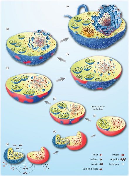
The cell of eukaryotic organisms (animals, plants, fungi) differs from that of prokaryotic organisms (Archaea and
      Bacteria) by the presence of several specialized organelles, such as: the nucleus (containing the genetic information
      of the cell), the mitochondria (site of cellular respiration), or the chloroplast (site of photosynthesis in plants). The
      existence and organization of mitochondrial and chloroplast DNA, as well as their biochemistry and some structural
      traits, have led to their being considered as ancient bacteria integrated into a host cell by an endosymbiosis process.
      One possible hypothesis would be that current eukaryotes would descend from an archaeal ancestor who acquired a
      proteobacteria, the present mitochondria. Once this step was established, some cells would have incorporated
      cyanobacteria that are the origin of the chloroplast. At the same time, they have acquired the ability to carry out
      photosynthesis, and thus an autotrophic metabolism, a particularity of plants. Throughout the process, gene transfer
      phenomena between symbionts, the taking over of the coding of some organelle proteins by the nucleus and the
      relocation of gene products into the organelles have closely integrated these prokaryotes within the host cell. The
      phenomenon of endosymbiosis is therefore very largely responsible for the biodiversity of eukaryotes that appeared
      during evolution. Thus, photosynthesis has developed in a wide variety of organisms: red and green algae, green
      plants through primary endosymbiosis, brown algae and many other organisms through secondary or tertiary
      endosymbiosis.

Encyclopédie de l'environnement                               2/14                                            Généré le 24/10/2020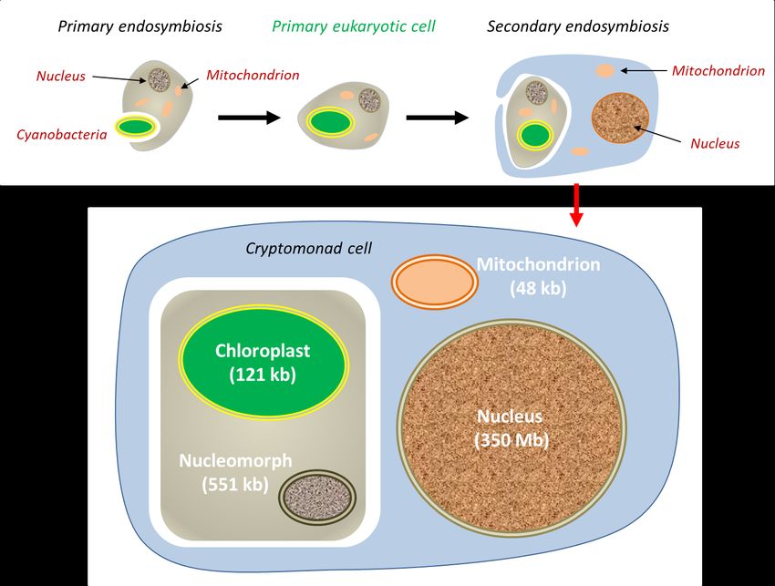
1. The eukaryotic cell is a chimera
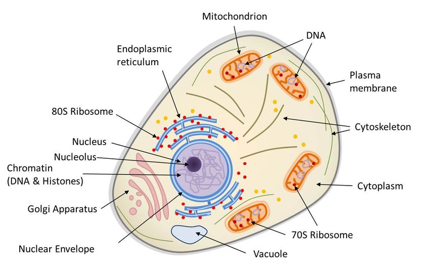
Figure 1. Diagram of the structure of a eukaryotic animal cell. The animal cell is compartmentalized, it contains an endomembrane
system (nuclear envelope, Golgi apparatus, endoplasmic reticulum, vacuoles…), mitochondria (limited by a double membrane), a
cytoskeleton bathed in the cytoplasm. The nucleus and mitochondria contain DNA. Ribosomes (protein synthesis machinery) are present in
two forms: 70S in mitochondria and 80S, generally in association with the reticulum.
Eukaryotes{ind-text} Single-cell or multicellular organisms whose cells have a nucleus and organelles (endoplasmic reticulum,
Golgi apparatus, various plastids, mitochondria, etc.) delimited by membranes. Eukaryotes are, along with bacteria and archaea,
one of the three groups of living organisms.{end-tooltip} correspond to multicellular organisms (animals, plants, fungi) and some
unicellular organisms (protozoa, for example). The main characteristic of the eukaryotic cell (Figure 1) is the existence of a
nucleus (in prokaryotes, the genome is only very rarely surrounded by a membrane) surrounded by a cytoplasm containing many
organelles, such as mitochondria{ind-text} Organelles of the cytoplasm of eukaryotic cells (plants, algae, animals). As the site of
cellular respiration, mitochondria convert the energy of organic molecules from digestion (glucose) into energy that can be
directly used by the cell (ATP) during the "Krebs cycle". This reaction requires the presence of oxygen and releases CO2, so it
plays an essential role in the carbon cycle. Mitochondria originate from a prokaryotic organism (α-proteobacteria) integrated into
eukaryotic protocells 2 billion years ago.{end-tooltip} (where respiration, present in all eukaryotic cells, takes place) and
chloroplasts{ind-text} Organelles of the cytoplasm of photosynthetic eukaryotic cells (plants, algae). As a site of photosynthesis,
chloroplasts produce O2 oxygen and play an essential role in the carbon cycle: they use light energy to fix CO2 and synthesize
organic matter. They are thus responsible for the autotrophy of plants. Chloroplasts are the result of the endosymbiosis of a
photosynthetic prokaryote (cyanobacterium type) in a eukaryotic cell about 1.5 billion years ago.{end-tooltip} (site for
photosynthesis{ind-text} Bioenergetic process that allows plants, algae and some bacteria to synthesize organic matter from
atmospheric CO2 by using sunlight. Solar energy is used to oxidize water and reduce carbon dioxide in order to synthesize
organic substances (carbohydrates). The oxidation of water leads to the formation of O2 oxygen found in the atmosphere.
Photosynthesis is the basis of autotrophy, it is the result of the integrated functioning of the chloroplast within the
cell.{end-tooltip}, in plants in the broad sense, terrestrial plants and algae). These organelles are frequently displaced or
reorganized by the cytoskeleton that triggers intracellular mobility (Figure 1).
The eukaryotic nucleus is delimited by a double membrane called the nuclear envelope (Figure 1). It contains the nuclear
genome characteristic of the eukaryotic cell, i.e. the genetic material of an individual encoded in its DNA (deoxyribonucleic
acid). It is usually this genome that is referred to when the genome of a eukaryote is mentioned. However, the eukaryotic cell
also contains non-nuclear genomes within the organelles:
- the mitochondrial genome, within the mitochondrial matrix (Figure 1);
- the chloroplastic genome, within the chloroplast stroma (e.g. plants or algae).
The DNA constituting these three genomes is not organized in the same way. In the nucleus, the genome is distributed over
several linear DNA molecules, and organized into well-differentiated chromosomes. DNA contains all coding sequences
(transcribed into messenger RNA, mRNA, and translated into proteins) and non-coding sequences (not transcribed, or
transcribed into RNA, but not translated). The three-dimensional configuration of the nuclear genome has a functional
importance: the winding (or “condensation”) of DNA on itself and around proteins, the histones{ind-text} Basic proteins
Encyclopédie de l'environnement 3/14 Généré le 24/10/2020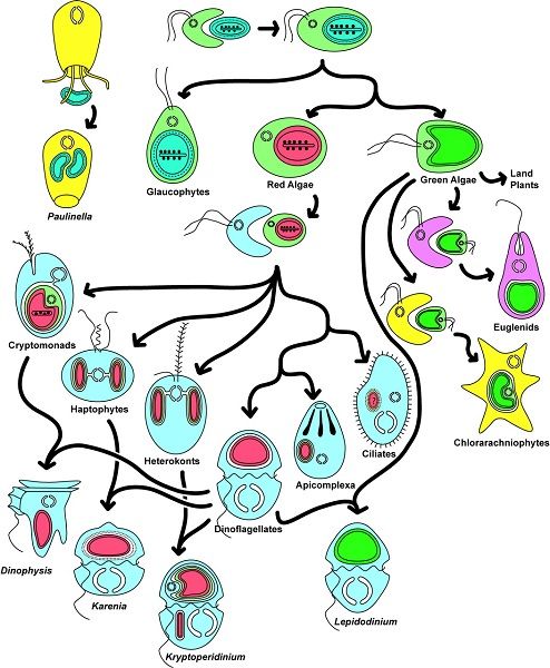
associating with DNA to form the basic structure of chromatin. Histones play an important role in DNA packaging and
     folding{end-tooltip}, allowing a large amount of genetic information to be packaged in the tiny nucleus of a cell. Mitochondrial
     or chloroplastic DNA do not have the same organization: it is generally circular, rarely linear (plant mitochondria), generally
     without intron, and is not associated with histone proteins.

     Prokaryotic{ind-text}Microorganisms (usually unicellular) with a simple cellular structure, no nucleus, and almost never internal
     compartmentalization (the only exception being thylakoids in cyanobacteria). Two of the three groups that make up living
     organisms are prokaryotes: Archaea and Bacteria.{end-tooltip}-type cells (Bacteria and Archaea{end-text} Single-celled
     prokaryotic microorganisms living in particular in extreme environments (anaerobic, high salinity, very hot...). Phylogenetic
     research by Carl Woese and George E. Fox (1977) differentiated between archaea and other prokaryotic organisms (bacteria).
     Currently, living organisms are considered to consist of three groups: Archaea, Bacteria and Eukaryota.{end-tooltip}), do not
     have a nucleus and their DNA is circular (or -in some rare cases- linear) and organized like that of chloroplasts or mitochondria.
     In this way, DNA replication, transcription and translation directly take place into the cytoplasm. It should be noted, however,
     that Archaea are only superficially similar to Bacteria in their cellular aspect: their metabolism differs greatly, and the
     mechanisms and proteins involved in the replication, transcription and translation processes have similar characteristics to those
     of eukaryotes. Finally, prokaryotes -in general- do not have internal compartments and, if present, they are less complex
     (cyanobacteria are an example of an exception). Above all, compartments, when they exist, are not mobile in the cell: the
     cytoskeleton, which is beginning to be discovered, does not move the cellular components within it.

     Table 1. Comparison of eukaryotic and prokaryotic cells

Encyclopédie de l'environnement                                   4/14                                                Généré le 24/10/2020
Table 1 compares the properties of prokaryotic and eukaryotic cells (with their mitochondria and possibly their chloroplasts). It
shows that mitochondria and chloroplasts have many characteristics in common with those of prokaryotic cells. Beyond the
structure of DNA, eukaryotic cell organelles are formed from pre-existing organelles, dividing by fission to multiply, like a
bacteria. Similarly, they have the same protein synthesis machinery (free 70S ribosomes{ind-text}A huge complex composed of
RNAs and ribosomal proteins that allows the translation of mRNAs into proteins. Common to all cells (prokaryotes and
eukaryotes), the ribosome varies according to the organisms: 80S ribosome in eukaryotes and 70S ribosome in prokaryotes and
cellular organelles (mitochondria, chloroplast).{end-tooltip} in the matrix or stroma) while in the cytoplasm of the eukaryotic
cell, this machinery consists of 80S ribosomes, sometimes fixed on the membranes of the endoplasmic reticulum{ind-text}
Membrane network of the eukaryotic cell cytoplasm, essential for cellular metabolism (lipid and protein synthesis, calcium
storage). Associated with ribosomes, it is the place of synthesis of proteins secreted outside the cell and, on the other hand,
proteins and lipids constituting the membranes of cellular organelles (Golgi apparatus, lysosomes, mitochondria, nucleus,
ribosomes, vesicles...).{end-tooltip}. Finally, bacteria also have the metabolism of mitochondria (i.e. respiration) and, in some
peculiar cases, of chloroplasts (i.e. photosynthesis). On the other hand, the eukaryotic cell is distinguished by the existence of an
active protein network, the cytoskeleton, a self-organized system capable of mobility, which positions and displaces the
Encyclopédie de l'environnement 5/14 Généré le 24/10/2020organelles in the cell. Such a protein network is static, or even absent, in prokaryotes, and poorly developed in mitochondria and
chloroplasts.
Figure 2. Unrooted phylogenetic tree of the three domains of living organisms, produced using a gene from the small ribosomal subunit
(bar: 0.1 substitution per site). The positions of the three genomes (nuclear, mitochondrial and chloroplastic) contained in maize (Zea
mays) are indicated - Synechococcus is a cyanobacterium [Source: adapted from Ref. [1]]
The analysis of genome sequence by DNA sequencing techniques has provided information on the evolutionary history of living
beings, including their relationship, also known as their phylogeny{ind-text} Study of the links between related species. Allows
to trace the main stages of the evolution of organisms from a common ancestor and to establish relationships of kinship between
living beings.{end-tooltip} (see What is biodiversity? and Inheritance or convergence?...). Molecular phylogenetic analysis
carried out on the nuclear genome of maize, as well as on its mitochondrial or chloroplastic genomes, makes it possible to
determine the phylogenetic position of this plant within the tree of life (Figure 2). The analysis shows that three lines (two of
which belong to Bacteria) are associated within what is considered, as they are structurally and functionally intertwined, as a
single organism - showing the triple origin of the species.
All these properties show that the eukaryotic cell is a chimera containing both characteristic components of the eukaryotic cell
(the nucleus) and organelles with typically prokaryotic properties (chloroplasts, mitochondria).
The distinction between Procaryotes and Eukaryotes was proposed in 1925 by Edouard Chatton [2] (who named these two cell
types), although it was not recognized until the 1950s and 1960s. The chimeric nature of eukaryotic cells had been observed
since the turn of the 19th to 20th centuries. If the botanist Andreas Schimper (born in France) had the idea in 1883 that
photosynthetic organisms were the result of the combination of distinct organisms, it was the Russian biologist Constantin
Mereschkowsky, who first provided solid arguments that some cells come from an intracellular union of two different types of
cells (endosymbiosis). In his 1905 article [3], Mereschkowsky proposed three essential ideas: (a) chloroplasts resemble
cyanobacteria that very early in the evolution established a symbiosis with a heterotrophic host, (b) the host that acquired the
plastids was itself the product of an earlier symbiosis between a larger, heterotrophic, amoeboid-type host cell and a smaller,
micrococcus-type endosymbionte that formed the nucleus, and (c) the autotrophy of plants is entirely inherited from
cyanobacteria. Mereschkowsky had not considered the origin of the mitochondria. It is to the credit of the French microbiologist
Paul Portier who wrote in a text in 1918 [4] that "all living beings, all animals (...), all plants (...) are constituted by the
association, the interlocking of two different beings. Each living cell contains (...) formations that cytologists refer to as
"mitochondria". For me, these organelles would be nothing more than symbiotic bacteria, what I call symbionts. "These observations
received no more attention from scientists than that, and the theory fell into disgrace, especially because plastids and
mitochondria were not successfully cultured, which in the 19th century was considered as evidence of a bacterial nature [5]. It
took new methods of studying the cell using electron microscopy, biochemistry and molecular biology for the theory of the
endosymbiotic origin of organelles in the eukaryotic cell to be brought back to life around 1970 by the American microbiologist
Lynn Margulis.
2. How did the eukaryotic cell evolved?
Encyclopédie de l'environnement 6/14 Généré le 24/10/2020Figure 3. Hypotheses for the origin of eukaryotes. (A) In this so-called "Three Domains" scheme, the two Eukaryotic and Archaea lines
have the same origin, each line being as old as the other. (B) The "two-domain" hypothesis comes from recent phylogenetic analyses.
Several hypotheses have been put forward to explain the appearance of the eukaryotic cell about 1.5 billion years ago, almost a
billion years after the first prokaryotic organisms appeared on Earth. This question can be addressed in very different ways,
depending on whether one considers palaeontological evidence, energy aspects, the origin of the characteristics of the eukaryotic
cell or the relationships of the different prokaryotic and eukaryotic lines with respect to each other [6]. Figure 3 shows
hypothesis for the origin of eukaryotes and other lines (Archaea and Bacteria).
Some models assume that eukaryotes emerged from a single ancestral lineage via successive mutations during the evolutionary
process. Other models postulate that eukaryotes have emerged from a symbiotic association of prokaryotic cells whose fusion
would have resulted in the transition from prokaryotic to eukaryotic. These various hypotheses can be partly tested by
experience, in particular through the analysis of the genomes of current organisms (prokaryotes or eukaryotes) [7,8].
Figure 4. Schematic representation of the integration between the Archaea host cell and an α-proteobacteria to give a eukaryotic cell
containing a mitochondria [see ref. 5]. (a-h) Illustrations of various steps describing the transition from an H2-dependent Archaea host
cell (red) to an optional anaerobic protein bacteria (blue) to a eukaryotic cell. At first, the two organisms live nearby, the Archaea needs
the hydrogen produced by the Bacteria, which does not support well the accumulation of hydrogen from its metabolism. The integration of
the bacteria will follow and this transition is accompanied on one hand by gene transfer between the two organisms (c) and on the other
hand by the establishment of the nucleus (e-h). [Source: reproduced with the permission of the authors (see ref. [5]) © 2015 (CC BY 4.0)]
The nature of the original host cell is a very controversial issue. The idea that it is an Archaea constitutes one of the possible
scenarios for the appearance of the eukaryotic cell: according to this scenario, an Archaea host cell and a
α-proteobacteria{ind-text}A large group of bacteria called Gram-negative bacteria, because they have a cell wall rich in
lipopolysaccharides and low in peptidoglycans. The mitochondria of current eukaryotic cells are thought to derive from one of
these types of bacteria: alpha-proteobacteria. {end-tooltip} have established a stable symbiotic relationship (Figure 4, [5]).
Encyclopédie de l'environnement 7/14 Généré le 24/10/2020Among the many possibilities for establishing such a relationship [6], the existence of trophic relationships between two
anaerobic prokaryotes living together, and drawing their food from this association (syntrophy), suggests an origin for
mitochondrial symbiosis. In this hypothesis [5], the host would be an Archaea needing hydrogen in its environment to live
(so-called H2-dependent Archaea which produces methane as metabolic waste) and the symbiote an optional anaerobic organism
(α-proteobacteria) which could either breathe in the presence of O2 or perform H2-producing fermentations under anaerobic
conditions. The latter metabolism produces energy only in low concentrations of H2 and benefits from the presence of H2
-dependent archaea. Figure 4 shows how such a situation could have evolved into a eukaryotic cell [5, 9]. The strength of this
hypothesis is that the partners need each other mutually, and that this scenario involves archaea and bacteria present in the
current biosphere.
In this context, Bacteria and Archaea tend to interact closely in the same way as many current symbiotic associations. This can
lead, in principle, to the situation described in Figure 4, in which the bacterial symbiote will be retained by the archeal host and
eventually reside inside. In this case, the host does not feed on the symbiote, so integration is not due to a phenomenon of
phagocytosis{ind-text} The process by which a cell encompasses and then digests a foreign substance or organism (e g. bacteria)
{end-tooltip} - although there is no doubt that phagocytosis has increased the frequency of integration of endosymbiotes in the
evolution of eukaryotic cells [5].
The question of the origin of the eukaryotic cell is also linked to that of the nucleus, the emblematic structure of this cell. The
establishment of a new membrane system, the nuclear membrane, in the host after mitochondria acquisition could be due to the
aggregation of membrane vesicles composed of bacterial lipids. This separation between nucleus and cytoplasm could have
responded to the need to separate, following gene transfer between host and symbiote, the splicing of RNA from DNA
translation. It is then the selective pressure that would have led to the fixation of the compartmentalization between the newly
formed nucleus and the cytoplasm [5] (Figure 4).
Thus, all eukaryotes currently known would descend from an archaean ancestor who acquired a proteobacterium during the
Precambrian period, which became the mitochondria. This step is crucial: the integration of the mitochondria is therefore
inseparable from the appearance of the eukaryotic cell as we know it today. The strong energy constraints exerted on the
organization of prokaryotic cells were a major factor of innovation at the origin of the evolution of this cell. Only cells that had
mitochondria had sufficient energy resources to reach the complexity of the eukaryotic cell, which is why there are no real
intermediaries in the transition from prokaryotes to eukaryotes. It is often considered that it is only after this stage has been
established that some of these cells have acquired the characteristics of the eukaryotic cell (nucleus, compartmentalization) and,
eventually, integrated cyanobacteria. At the same time, they have acquired the ability to carry out photosynthesis, which is the
origin of chloroplast, thus giving them an autotrophic metabolism.
Recently, some work has nuanced the age of mitochondrial symbiosis [10]. They are based on the age of acquisition of bacterial
genes present in the eukaryotic nucleus (i.e. the date of their divergence from homologous genes currently found in free
bacteria). They revealed that many genes, some of which contribute to the complexity of the eukaryotic cell, were most likely
acquired before mitochondria. This does not imply that the cell into which the mitochondria entered was as complex as it is now,
but it may have already been capable of phagocytosis, for example. This feature (specific to eukaryotes because it depends on
the mobility of the cytoskeleton) could have helped in the placement of the mitochondria. The development of eukaryotic
complexity therefore remains speculative, but may have begun before mitochondria, even if it undoubtedly benefited from it
afterwards.
3. The endosymbiotic origin of the chloroplast
During phagocytosis in white blood cells or many protozoa (Figure 5), ingested cells are often directly digested (as in the case of
prey), but sometimes they are permanently lodged in the cells (endosymbiotes). In the endosymbiosis process, organelle
therefore results from internalization by phagocytosis without digestion of a prokaryote within a eukaryote (Figure 5). This is the
case for chloroplasts in terrestrial plants, but also for red and green algae that are close to them [11,12].
Encyclopédie de l'environnement 8/14 Généré le 24/10/2020Figure 5. Phagocytosis and primary endosymbiosis. During phagocytosis, ingested prey is often directly digested, but sometimes
permanently lodged in the cells during primary endosymbiosis, the plasma membrane of the cell invades around the prokaryote and
isolates it within an endocytosis vesicle. Then, when the prokaryote is integrated into the eukaryotic cell, the membrane of this vesicle
disappears as well as the layer of peptidoglycans located between the two membranes of the cyanobacterium [see ref. 9 & 10].
During phagocytosis processes, the plasma membrane of the cell invades around the prey and isolates them into endocytosis
vesicles where they are then digested as these vesicles fuse with others, the lysosomes, which contain enzymes. By analogy, it
was generally considered that the outer membrane of the organelles came from this endocytosis membrane. Things are likely to
be more complex (Figure 5). Indeed, the prokaryotes at the origin of chloroplasts or mitochondria are Gram- bacteria,
characterized by the existence of a double membrane on the periphery of the bacteria. The outer membrane of chloroplasts, and
in particular its outer surface immersed in the cell's cytosol, contains characteristic glycolipids found in cyanobacteria [9, 10, 13]
. It is therefore possible that the endocytosis membrane may have disappeared during the integration of the prokaryote into the
eukaryotic cell. This is currently observed in Elysia chlorotica (see Focus), a marine mollusc that grazes algae, digests part of
their cells but not the chloroplasts which is integrated into the cytoplasm of some of its cells. These chloroplasts remain
functional throughout the life of the mollusc, which benefits from photosynthesis.
Primary and secondary endosymbioses
During the evolution, several endosymbiosis events repeated themselves and led to the formation of particular organisms. In
primary endosymbiosis, the eukaryotic cell integrates a living prokaryotic. Thus, the chloroplasts of green line plants (red and
green algae, to which terrestrial plants are attached) are derived from primary endosymbioses involving cyanobacteria. In some
eukaryotes, mitochondria have evolved as a result of adaptation to anaerobic environments, but have never disappeared: they
have produced particular mitochondria (hydrogenosomes{ind-text} Organelles producing hydrogen, derived from a
mitochondria. It is found in some anaerobic ciliates, Trichomonas, and fungi.{end-tooltip}) carrying out H2-producing
fermentation (for example in some Ciliates) [14], but also small organelles, only involved in biosynthesis for the host cell, the
mitosomes{ind-text} Organelles present in some single-celled eukaryotic organisms, probably lacking DNA but with
biosynthesis functions.{end-tooltip} [15].
Encyclopédie de l'environnement 9/14 Généré le 24/10/2020Figure 6. Secondary chloroplastic endosymbiosis model in the cryptophyte Guillardia theta [see ref. 18]. Here, the nucleus of the
internalised red algae (primary host) persists within the secondary host in the form of a vestigial nucleus (or nucleomorph), but with a very
small genome (551 kb instead of the 350 Mb of the nucleus).
Secondary endosymbioses are a reiteration of the process, when a eukaryote already containing an endosymbiont realizes a
secondary endosymbiosis within another eukaryote (Figure 6). This is the origin of plastids with more than two membranes
present in some groups: internalisation of a green algae in Euglenes; independent internalisation of a red algae in brown algae,
etc. Tertiary endosymbiosis, less frequent, have also been described. These various symbioses constitute as many founding
endosymbioses of evolutionary lines [16,17].
4. Integration of the prokaryote into the eukaryotic cell
All these lines have a common characteristic: a strong genetic regression of endosymbiotes. Compared to free proteobacteria
such as Escherichia coli, mitochondria have lost 99% of their genes. In the extreme, hydrogenosomes and mitosomes simply no
longer have a genome! Green line plastids show a 95% genetic regression compared to single-celled free cyanobacteria: the
number of genes has increased from several thousand in cyanobacteria to about 100 to 200 in chloroplasts... or even none in the
regressed plastids of the parasitic plant Rafflesia.
The cause of this regression is obviously the loss of genes necessary for free living, or even for certain metabolic functions. For
example, as with all Gram-{ind-text} bacteria detected by a staining technique called Gram staining: they then appear pink under
the microscope. The staining technique is based on the membrane and wall characteristics of the bacteria. However, this is not a
phylogenetic classification factor: indeed, the'Gram +' and'Gram -' groups are both non-monophyletic. {end-tooltip}, a layer of
peptidoglycan{end-text} composed of the wall of Gram positive and Gram negative bacteria. Consists of a carbohydrate part (=
polysaccharide) and a peptide part. It maintains the shape of the cells and provides mechanical protection against osmotic
pressure. {end-tooltip} is located between the two membranes of cyanobacteria, essential for maintaining the structure of
bacteria in the natural environment, of low osmolarity{ind-text} Number of moles of "osmotically active" particles in solution in
1 litre of solution. Concept related to the osmotic pressure exerted by the particles in solution, and responsible for osmosis.
Sucrose is a small osmotically active molecule while starch is a huge osmotically inactive glucose polymer. The accumulation of
sucrose in a compartment leads to an increase in osmotic pressure in that compartment, which is not the case with
starch.{end-tooltip}. Once integrated into the host cell, the prokaryote will find itself in a medium, the cytoplasm, whose
osmolarity is very close to that of its inner medium. The peptidoglycan layer then becomes useless, and the genes responsible for
the placement of the peptidoglycan layer are then lost in chloroplasts (except in glaucophytes{ind-text} Unicellular plankton
eukaryotes living in lakes, ponds or wetlands in temperate regions. They have flagella (2 of unequal length). They have
chloroplasts (called cyanelles) that are blue-green in colour, due to the presence of phycocyanins and allophycocyanins in
phycobilisomes. This is a group of reduced diversity. {end-tooltip}).
Although the organelle genome is regressing, the organelle protein repertoire (the proteome), when known, remains similar to
that of the free bacteria proteome: proteins operating with new functions have therefore compensated for the losses. Their
coding has in fact been supported by the host's nuclear genome: genes located in the nucleus are translated into proteins in the
cytosol which are addressed to the organelle through a transit peptide{ind-text}Peptide sequence located at the NH2-terminal end
of the newly synthesized proteins in the cytoplasm and which allows them to be addressed to the specific organelle
(mitochondria, etc.) where they function. We also speak of addressing peptide. {end-tooltip}. This phenomenon of relocation of
the gene product into the organelle is an absolutely essential phenomenon for the integration of the prokaryote into the host cell.
Encyclopédie de l'environnement 10/14 Généré le 24/10/2020The addressing machinery responsible for these transfers is a converging innovation in plastids and mitochondria. It is also an
example of the new functions linked to intracellular life. These machines, which allow the import of proteins synthesized in the
cytosol through the two limiting membranes of mitochondria and chloroplasts, contain a large number of proteins whose
evolutionary origin is complex: proteins of both prokaryotic and eukaryotic origin, encoded in the organelle and nucleus, are
found there. Together, they allow the recognition of the protein being addressed, its unfolding followed by import (the protein
must be maintained in an unfolded state to cross the membranes), then the cleavage of the addressing peptide before its precise
location in its functional compartment [19].
What is the origin of the genes that encode in the nucleus for functions in organelles? There are actually two (Figure 7) [16].
Sometimes, original nuclear genes have replaced organelle genes: the corresponding gene product has acquired the ability to be
addressed in the organelle. This activation may have in the past led to a redundancy situation whenever a gene already encoded
the same function in the organelle. From this redundancy, the organelle gene could be lost without damage (Figure 7a) [16].
Figure 7. Evolutionary mechanisms leading to the replacement of organelle genes by genes located in the nucleus. Substitution (A) involves
genes of "true" nuclear origin while transfer (B) involves a nuclear relocation of genes from organelles. [Source: According to Selosse et al
(2001) Reference [16]]
Other cases involve gene transfers from organelle to the nucleus, which takes place in two major steps (Figure 7b). First, a DNA
fragment encoding the organelle protein is relocated and then integrated into the nuclear genome. The transferred sequence will
only code if, through mutations, it adapts to the nuclear genetic code, and if it acquires regulatory sequences for transcription. It
must also acquire the pre-sequence corresponding to the transit peptide, which will ensure proper addressing of the mature
protein to the organelle and therefore its correct location. As above, this leads to genetic redundancy: one or the other of the
copies can be lost without damage. The loss of functionality and/or the disappearance of the organelle copy then seals the
transfer (Figure 7) [16].
The passage of DNA fragments from organelles to the nucleus is not uncommon: large blocks of organelle DNA are inserted into
the genome of some plants. These can be activated: nearly 10% of Arabidopsis thaliana's nuclear genes are thus derived from
transfers from plastids, often followed by duplications [20]. It is not known how the DNA of the endosymbiote could have been
integrated into the host genome, but it is assumed that this occurs during degradations of damaged or aged organelles accidentally
releasing pieces of DNA into the host cytoplasm which are then randomly integrated into the host's nuclear DNA.
The cytoplasmic genomes of organelles are at the crossroads of various selective forces, some of which favour their regression
(such as the need for co-expression of certain genes), others favour the persistence of certain genes in the organelle genome. This
could be the case for selection for a small genomic size that accelerates the multiplication of organelles and allows better
transmission to daughter cells: it selects in particular the transfer of genes to the nucleus. The latter thus accumulates genetic
potentialities from different lines coexisting with him in the cell [16]. Thus, while endosymbiosis reduces the genomes of
endosymbiotes, it nourishes the genome of the host nucleus, contributing to its genetic diversification, and pushing for closer
espouses the endosymbiotic association. Endosymbiosis therefore mixes the evolutionary lines present, by nesting but also by
genetic chimerization in the nucleus of the host cell.
Finally, vertical transmission of the endosymbiote through generations is essential for the endosymbiosis to persist. Plastids must
divide before the division of the host cell and must be half distributed in the two daughter cells. If their division is too rapid, they
could take advantage of the host cell. On the contrary, a low division rate could lead to their disappearance. In this context, the
establishment of coordination of cell division and symbiote division has been an essential element in the success of
endosymbiosis. While most of the proteins involved in chloroplast division come from the cell division machinery present in
cyanobacteria, some proteins are apparently of eukaryotic origin, and all are encoded in the nucleus: this is a way for the host to
Encyclopédie de l'environnement 11/14 Généré le 24/10/2020exercise control over chloroplast division.
5. Is symbiosis driving evolution?
Figure 8. Endosymbiosis (primary, secondary and tertiary) in the history of plastid evolution. They are responsible for organisms as
diverse as red and green algae, terrestrial plants such as apicomplexes (parasites responsible for malaria and toxoplasmosis) or
dinoflagellates (components of marine plankton that are particularly important in primary ocean production). [Source: reproduced from
Keeling et al [12]. Copyright 2016 by American Journal of Botany, Inc]
In conclusion, extremely diverse symbioses that led to the formation of the eukaryotic cell [1,9], are at the origin of the
development of eukaryotic biodiversity during evolution. Endosymbioses found new evolutionary lines. An example of the
extreme diversity of organisms derived from endosymbiosis that causes chloroplasts is shown in Figure 8. However, things are
not fixed: evolution continues to repeat itself! Today, some unicellular algae, cryptophytes{ind-text} Single-celled organisms,
mostly photosynthetic. Their chloroplasts are limited by four membranes, indicating an endosymbiosis of a photosynthetic
eukaryote. Cryptophytes occur in many environments, particularly aquatic ones (oceanic environments, fresh waters, wetland
pore waters). Some species have become intestinal parasites of metazoans. Some are Dinophyte endosymbiotes {end-tooltip} and
heterocontes (Figure 8), whose four-membrane plastid derives from a secondary endosymbiosis, live in symbiosis in the
cytoplasm of dinoflagellates that have lost their own plastids: there are three successive endosymbioses there!
More than a biological curiosity, symbiosis is certainly one of the most powerful motors of the evolution of the living world. It
very quickly creates chimeric organisms that can generate new lines. It brings partners closer together and promotes massive
gene transfers that also create chimeric genomes: the nuclear genome thus contains eukaryotic genes, but also genes of bacterial
origin, derived from mitochondria, or even plastids, with which it borders. Such events may explain the major evolutionary leaps
whose evolution seems to be punctuated, which have given rise to the great lines of life and shaped current biological diversity.
Thus, renewing the Darwinian vision of evolution by descent with modification, where one species is likely to give two, the
endosymbiosis mechanisms remind us that sometimes two species, previously free and recognizable, merge into one. Man
himself can be considered as an extremely integrated symbiotic community, formed by the eukaryotic cytoplasm and
mitochondria, but also by the archaea and bacteria that populate, for instance, his gut microbiota...
References and notes
[1] Lang T. et al (2000) Autophagy and the cvt pathway both depend on AUT9. J Bacteriol 182, 2125-2133.
[2] Chatton E. (1938) Titres et travaux scientifiques (1906-1937). Sette, Sottano, Italy. L’histoire des conditions dans lesquelles
Encyclopédie de l'environnement 12/14 Généré le 24/10/2020Chatton a établi le concept de procaryote et eucaryote est décrite par Sapp J. (2005) The Prokaryote-Eukaryote Dichotomy:
Registering
Meanings and Mythology, Microbiol Mol Biol Rev. 69, 292–305.
[3] Mereschkowsky C. 1905 Uber Natur und Ursprung der Chromatophoren im Pflanzenreiche. Centralbl. 25, 593-604; translated
by Martin W, Kowallik K. (1999) Annotated English translation of Mereschkowsky's 1905 paper 'Uber Natur und Ursprung der
Chromatophoren im Pflanzenreiche'. Eur. J. Phycol. 34, 287–295.
[4] Portier P. (1918) Les Symbiotes. Masson (ed.), Paris. (in french)
[5] Martin W.F., Garg S. & Zimorski V. (2015) Endosymbiotic theories for eukaryote origin. Phil. Trans. R. Soc. B370,
20140330.
[6] Selosse M.A. (2012). Gloire et disgrâce de la théorie endosymbiotique. La Recherche 468: 92-94. (in french)
[7] Archibald J.M. (2014) One plus one equals one: symbiosis and the evolution of complex life. Oxford, UK: Oxford University
Press.
[8] McFadden G.I. (2014) Origin and Evolution of Plastids and Photosynthesis in Eukaryotes, Cold Spring Harb.Perspect. Biol. 6,
a016105
[9] Martin W. & Müller M. (1998) The hydrogen hypothesis for the first eukaryote. Nature 392, 37-41.
[10] Ettema T.J.G. (2016) Mitochondria in the second act. Nature 531, 39-40doi:10.1038/nature16876
[11] Archibald J.M. & Keeling P.J. (2002) Recycled plastids: a "green movement" in eukaryotic evolution. Trends Genetics 18,
577-584.
[12] Keeling P.J. (2004) Diversity and evolutionary history of plastids and their hosts. Am. J. Bot. 91, 1481-1493.
[13] Douce R., Block M.A., Dorne A.J., Joyard J. (1984) The plastid envelope membranes: their structure, composition, and role in
chloroplast biogenesis. Subcell. Biochem. 10, 1-84, Springer US (Ed.)
[14] Selosse M.A. & Loiseaux-de Goër S. (1997) La Saga de l’endosymbiose, La Recherche 296, 36 (in french)
[15] Embley T.M. & Martin W. (2006) Eukaryotic evolution, changes and challenges. Nature 440, 623-630
[16] Lefèvre T., Renaud F., Selosse M.-A. & Thomas F. (2010). Évolution des interactions entre espèces, in F. Thomas, T.
Lefèvre & M. Raymond (ed.), Biologie évolutive, p. 530-613. De Boeck, Paris. (in french)
[17] Keeling P.J. (2010) The endosymbiotic origin, diversification and fate of plastids. Phil. Trans. R. Soc. B 365, 729-748
[18] Douglas S. et al (2001) The highly reduced genome of an enslaved algal nucleus. Nature 410, 1091-1096.
[19] Selosse M.A., Albert B. & Godelle B. (2001) Small is successful: selection for reducing organelle's genome size favours gene
transfer to the nucleus. Trends Ecol Evol 16, 135-141.
[20] Jarvis P. (2004) Organellar Proteomics: Chloroplasts in the Spotlight. Current Biology 14, R317-9.
http://www.cell.com/current-biology/references/S0960-9822%2804%2900231-3
Encyclopédie de l'environnement 13/14 Généré le 24/10/2020L’Encyclopédie de l’environnement est publiée par l’Université Grenoble Alpes - www.univ-grenoble-alpes.fr
Pour citer cet article: Auteurs : (2019), Symbiosis and evolution: at the origin of the eukaryotic cell, Encyclopédie de
l’Environnement, [en ligne ISSN 2555-0950] url : http://www.encyclopedie-environnement.org/?p=6954
Les articles de l’Encyclopédie de l’environnement sont mis à disposition selon les termes de la licence Creative Commons
Attribution - Pas d'Utilisation Commerciale - Pas de Modification 4.0 International.
Encyclopédie de l'environnement
Powered by TCPDF (www.tcpdf.org)
14/14 Généré le 24/10/2020You can also read