Norcantharidin Suppresses YD-15 Cell Invasion Through Inhibition of FAK/Paxillin and F-Actin Reorganization - MDPI
←
→
Page content transcription
If your browser does not render page correctly, please read the page content below
molecules
Article
Norcantharidin Suppresses YD-15 Cell Invasion
Through Inhibition of FAK/Paxillin and
F-Actin Reorganization
Kyoung-Ok Hong, Chi-Hyun Ahn, In-Hyoung Yang, Jung-Min Han, Ji-Ae Shin,
Sung-Dae Cho * and Seong Doo Hong *
 Department of Oral Pathology, School of Dentistry and Dental Research Institute, Seoul National University,
 Seoul 03080, Korea; hongko95@snu.sc.kr (K.-O.H.); chihyun610@snu.ac.kr (C.-H.A.);
 inhyoung3@naver.com (I.-H.Y.); 2017-20655@snu.sc.kr (J.-M.H.); sky21sm@snu.ac.kr (J.-A.S.)
 * Correspondence: efiwdsc@snu.ac.kr (S.-D.C.); hongsd@snu.ac.kr (S.D.H.); Tel.: +82-2-740-8647 (S.-D.C.);
   Tel.: +82-2-740-8682 (S.D.H.)

 Received: 19 April 2019; Accepted: 17 May 2019; Published: 19 May 2019

 Abstract: Norcantharidin (NCTD), a demethylated derivative of cantharidin, has been reported to
 exhibit activity against various types of cancers. However, the anti-invasive effects of NCTD and its
 molecular mechanism in human mucoepidermoid carcinoma (MEC) remain incompletely elucidated.
 Clonogenic, wound healing, invasion, zymography, western blotting and immunocytochemistry
 assays were performed in YD-15 cells to investigate the anti-invasive effect of NCTD and its molecular
 mechanism of action. The inhibitory effects of NCTD on invasiveness were compared with those
 of a novel focal adhesion kinase (FAK) kinase inhibitor, PF-562271. NCTD markedly suppressed
 the colony formation, migration, and invasion of YD-15 cells as well as the activities of MMP-2 and
 MMP-9. It disrupted F-actin reorganization through suppressing the FAK/Paxillin axis. Moreover,
 NCTD exhibited a powerful anti-invasive effect compared with that of PF-562271 in YD-15 cells.
 Collectively, these results suggest that NCTD has a potential anti-invasive activity against YD-15 cells.
 This study may clarify the impact of NCTD on migration and invasion of human MEC cells.

 Keywords: mucoepidermoid carcinoma; norcantharidin; invasion; F-actin reorganization; focal
 adhesion kinase; paxillin

1. Introduction
     Mucoepidermoid carcinoma (MEC) is the most common malignant tumor of the major
salivary glands and histologically comprises variable mixtures of mucin-secreting, intermediated,
and epidermoid cells [1,2]. MEC is characterized by ready penetration of surrounding tissues and is
likely to recur easily [3]. As with other solid tumors, traditional methods for treating MEC, such as
chemotherapy, can cause strong side effects and drug resistance [4]. Therefore, it is necessary to
continue searching for novel, efficient, and less toxic cancer therapeutic drug candidates.
     Invasion and metastasis belong to the six cancer biological capability hallmarks which were
described by Hanahan and Weinberg, and represent distinct traits between benign and malignant
tumors [5,6]. To metastasize, cancer cells require a mechanical event that involves adhesion, shape
change, movement, and force generation; only then can they finally undergo infiltration through
the extracellular matrix (ECM) in order to form new tumors at another site in the human body [7].
With remodeling of the ECM, reorganization of the actin cytoskeleton is required for invasion and
metastasis of cancer and is regulated by a variety of proteins, including actin regulatory proteins,
adhesion molecules, signaling proteins, and matrix degradation enzymes [8–10]. Focal adhesion
kinase (FAK) is a non-receptor tyrosine kinase that acts as an important mediator of integrin-mediated

Molecules 2019, 24, 1928; doi:10.3390/molecules24101928                        www.mdpi.com/journal/molecules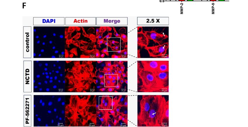
Molecules 2019, 24, x                                                                                       2 of 11
    Molecules 2019, 24, 1928                                                                                     2 of 11
kinase (FAK) is a non-receptor tyrosine kinase that acts as an important mediator of integrin-
mediated signaling between cells and the ECM. It serves as a scaffolding protein for the binding sites
    signaling between cells and the ECM. It serves as a scaffolding protein for the binding sites of multiple
of multiple oncogenic tyrosine kinases and regulates paxillin function through phosphorylation to
    oncogenic tyrosine kinases and regulates paxillin function through phosphorylation to induce diverse
induce diverse cellular processes, including adhesion, migration, invasion, and metastasis [11,12].
    cellular processes, including adhesion, migration, invasion, and metastasis [11,12]. Indeed, FAK is
Indeed, FAK is found at an elevated level in most human cancers, particularly when the cancer is
    found at an elevated level in most human cancers, particularly when the cancer is transformed into
transformed into highly invasive metastases [13,14], suggesting that FAK is a key signaling molecule
    highly invasive metastases [13,14], suggesting that FAK is a key signaling molecule for an effective
for an effective cancer therapeutic strategy. However, the functional significance of FAK activity in
    cancer therapeutic strategy. However, the functional significance of FAK activity in MEC has not yet
MEC has not yet been fully understood.
    been fully understood.
      Natural products such as plant and animal-derived drugs play an increasingly important role in
          Natural products such as plant and animal-derived drugs play an increasingly important role in
cancer treatment due to their low side-effects and high efficacy [15,16]. Recently, NCTD, a synthetic
    cancer treatment due to their low side-effects and high efficacy [15,16]. Recently, NCTD, a synthetic
demethylated analog of cantharidin isolated from Mylabris phalerata Pallas (blister beetle), has been
    demethylated analog of cantharidin isolated from Mylabris phalerata Pallas (blister beetle), has been
reported to have potent anticancer activity, including the to inhibit capacity tumor invasiveness and
    reported to have potent anticancer activity, including the to inhibit capacity tumor invasiveness
metastasis in colorectal and breast cancer cells [17,18]. NCTD is known as an inhibitor of protein
    and metastasis in colorectal and breast cancer cells [17,18]. NCTD is known as an inhibitor of
Ser/Thr phosphatases (PPP), which regulate various important physiological and pathological
    protein Ser/Thr phosphatases (PPP), which regulate various important physiological and pathological
processes, such as cell cycle and proliferation [19]. Despite several studies on the biological activities
    processes, such as cell cycle and proliferation [19]. Despite several studies on the biological activities of
of NCTD, its anti-proliferative and anti-invasive potential are not yet defined. In the present study,
    NCTD, its anti-proliferative and anti-invasive potential are not yet defined. In the present study, we
we investigated the anti-invasive potential of NCTD in the YD-15 human MEC cell line and compared
    investigated the anti-invasive potential of NCTD in the YD-15 human MEC cell line and compared its
its anti-cancer function with that of PF562271, a potent FAK inhibitor.
    anti-cancer function with that of PF562271, a potent FAK inhibitor.
2. Results
    2. Results

2.1.2.1.
    LowLow
         Concentrations of NCTD
            Concentrations       Affect
                            of NCTD     Survival
                                     Affect      without
                                            Survival     Apoptosis
                                                     without       in the
                                                             Apoptosis in YD-15 CellCell
                                                                          the YD-15  LineLine
     To examine
         To examinethe the
                        inhibitory   effect
                            inhibitory       of NCTD
                                         effect of NCTD on cell growth,
                                                           on cell      a Trypan
                                                                   growth,  a TrypanBlue
                                                                                       Blueexclusion   assay
                                                                                               exclusion     waswas
                                                                                                          assay
performed   on  YD-15   cells after exposure    to various concentrations  of NCTD    or  0.1%   DMSO
   performed on YD-15 cells after exposure to various concentrations of NCTD or 0.1% DMSO (vehicle       (vehicle
control) for for
    control) 24 h.
                 24Low   concentrations
                    h. Low    concentrations(2.5–10 μM)µM)
                                                (2.5–10   of NCTD   did did
                                                              of NCTD   not not
                                                                            inhibit  cell cell
                                                                                inhibit   viability (Figure
                                                                                               viability     1B),1B),
                                                                                                         (Figure
but but
     high concentrations     (20–40  μM)    of  NCTD   significantly reduced   cell viability   (Supplementary
        high concentrations (20–40 µM) of NCTD significantly reduced cell viability (Supplementary
Figure  S1A).
    Figure S1A).

          Figure 1. NCTD inhibits colony formation of YD-15 cells. (A) Chemical structure of NCTD. (B) Trypan
      Figure 1. NCTD inhibits colony formation of YD-15 cells. (A) Chemical structure of NCTD. (B) Trypan
          blue exclusion assay for cell viability after the treatment of NCTD (2.5, 5, and 10 µM) for 24 h. (C,D)
      blue exclusion assay for cell viability after the treatment of NCTD (2.5, 5, and 10 μM) for 24 h. (C,D)
          Clonogenic assay for colony formation of YD-15 cells. Graphs show the mean ± SD of triplicate
      Clonogenic assay for colony formation of YD-15 cells. Graphs show the mean ± SD of triplicate
          experiments and significance compared with the vehicle control (* p < 0.05).
      experiments and significance compared with the vehicle control (* p < 0.05).

        To further investigate the effects of NCTD on the survival and proliferation of YD-15 cells, we
     To further investigate the effects of NCTD on the survival and proliferation of YD-15 cells, we
   performed a 10-day clonogenic assay. As shown in Figure 1C,D, 10 µM NCTD exhibited significant
performed a 10-day clonogenic assay. As shown in Figure 1C,D, 10 μM NCTD exhibited significant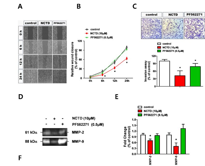
Molecules 2019, 24, 1928                                                                                                   3 of 11
Molecules 2019, 24, x                                                                                                      3 of 11

inhibition of colony formation. Western blotting and flow cytometric analyses with Annexin V/PI
 inhibition of colony formation. Western blotting and flow cytometric analyses with Annexin V/PI
double staining were performed to confirm whether the inhibition of colony formation by NCTD
 double staining were performed to confirm whether the inhibition of colony formation by NCTD
induces apoptosis. There was no change in YD-15 cells treated with low concentrations of NCTD
 induces apoptosis. There was no change in YD-15 cells treated with low concentrations of NCTD
(data not shown), but high concentrations of NCTD significantly increased cleavages of caspase 3 and
 (data not shown), but high concentrations of NCTD significantly increased cleavages of caspase 3
PARP (Supplementary Figure S1B). In addition, the number of annexin V-positive cells in 40 μM
 and PARP (Supplementary Figure S1B). In addition, the number of annexin V-positive cells in 40 µM
NCTD-treated YD-15 cells increased (Supplementary Figure S1C,D). These results suggest that low
 NCTD-treated YD-15 cells increased (Supplementary Figure S1C,D). These results suggest that low
concentrations of NCTD affect survival without causing apoptotic cell death in YD-15 cells. Thus, we
 concentrations of NCTD affect survival without causing apoptotic cell death in YD-15 cells. Thus,
used NCTD of 10 μM or less for all subsequent experiments.
 we used NCTD of 10 µM or less for all subsequent experiments.

2.2.
 2.2.NCTD
      NCTDRepresses
             RepressesYD-15
                       YD-15Cell
                             CellMigration
                                 Migrationand
                                           andInvasion
                                               Invasionthrough
                                                        throughDown-Regulation
                                                               Down-RegulationofofMMP-2
                                                                                  MMP-2and
                                                                                        andMMP-
9MMP-9
  ActivityActivity
    AAwound
        woundhealing   assay
                 healing      was
                          assay   performed
                                 was          to measure
                                      performed           the effects
                                                  to measure          of NCTD
                                                               the effects       on cell
                                                                            of NCTD    onmigration.
                                                                                          cell migration.
YD-15
 YD-15cells
        cellsshowed
               showedsignificantly  reduced
                        significantly        wound
                                       reduced  woundclosure  levels
                                                         closure      afterafter
                                                                  levels    NCTD    treatment
                                                                                 NCTD          in a in a
                                                                                         treatment
concentration-dependent
 concentration-dependentmanner
                           mannercompared
                                    comparedwith
                                               withvehicle
                                                    vehiclecontrol
                                                            control(Figure
                                                                    (Figure2A,B).
                                                                             2A,B).

                  NCTDinhibits
       Figure2.2.NCTD
      Figure               inhibitsthe
                                     themigration
                                          migrationand
                                                     andinvasion
                                                          invasionofofYD-15
                                                                       YD-15cells.
                                                                                cells.(A,B)
                                                                                       (A,B)Wound-healing
                                                                                              Wound-healingassay
                                                                                                               assayofofcell
                                                                                                                         cell
       migration   after treatment     of NCTD    (5 and  10 µM)   in YD-15    cells for 24 h. The  dotted
      migration after treatment of NCTD (5 and 10 μM) in YD-15 cells for 24 h. The dotted lines delineate   lines delineate
       clearzones
      clear  zones at
                    at various
                       various time-points.
                                 time-points. (C,D)
                                                (C,D)Transwell
                                                       Transwellassay
                                                                    assayforfor
                                                                              cell invasive
                                                                                cell        potential
                                                                                     invasive         after
                                                                                                potential   treatment
                                                                                                          after         with
                                                                                                                treatment
       NCTD    in YD-15   cells.  (E,F) Effects of NCTD    on  MMP    activity  using   gelatin zymography
      with NCTD in YD-15 cells. (E,F) Effects of NCTD on MMP activity using gelatin zymography and             and western
       blotting.blotting.
      western     GraphsGraphs
                            show the    mean
                                     show   the±mean
                                                 SD of ±triplicate  experiments
                                                          SD of triplicate          and significance
                                                                             experiments              compared
                                                                                             and significance      with the
                                                                                                                compared
       vehicle  control (*  p < 0.05).
      with the vehicle control (* p < 0.05).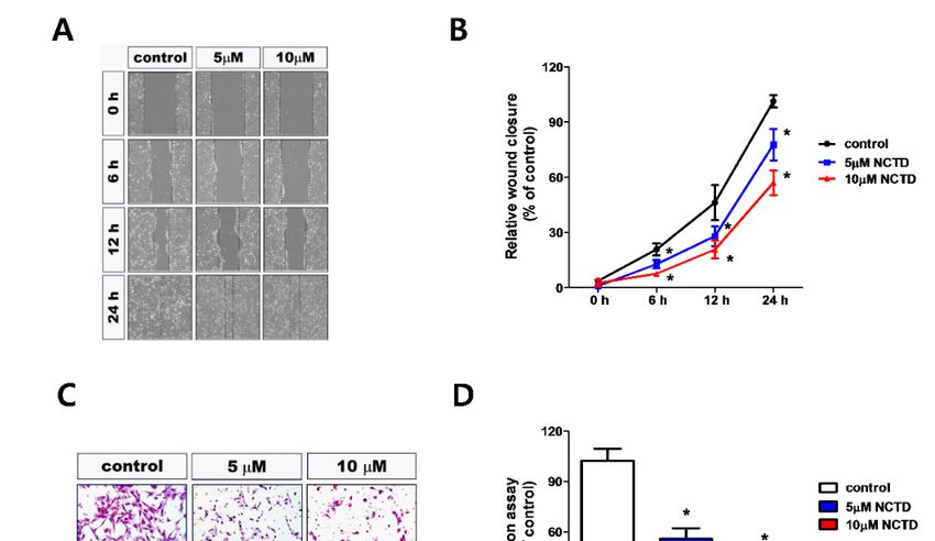
Molecules 2019, 24, 1928 4 of 11
Molecules 2019, 24, x 4 of 11
The
Theeffect
effectofofNCTD
NCTDon oncell
cellinvasion
invasionwaswasalso
also investigated
investigated using
using aa transwell chamber pre-coated
transwell chamber pre-
coated with matrigel.
with matrigel. Cells treated
Cells treated with NCTD with showed
NCTD showed a significant
a significant decreasedecrease in cell similar
in cell invasion invasion to those
similar to those obtained from the wound healing assay (Figure 2C,D). In order to
obtained from the wound healing assay (Figure 2C,D). In order to evaluate the mechanism underlying evaluate the
mechanism underlying
their inhibitory their
action on inhibitory
migration andaction on migration
invasion, we measured andtheinvasion,
activitiesweof measured
MMP-2 and the
MMP-9
activities
involvedof in MMP-2
tumor cell and MMP-9 and
migration involved in by
invasion tumor cellzymography
gelatin migration and invasion
[7]. The resultsby gelatin
showed that the
zymography [7]. The results showed that the activities of both MMP-2 and
activities of both MMP-2 and MMP-9 were significantly decreased by NCTD treatment (Figure 2E,F). MMP-9 were
significantly
These results decreased by NCTD
suggest that treatmentMMP-2
NCTD regulates (Figureand2E,F). Thesetoresults
MMP-9 inhibit suggest
migration that
and NCTD
invasion of
regulates MMP-2 and
the human YD-15 cell line.MMP-9 to inhibit migration and invasion of the human YD-15 cell line.
2.3.
2.3.NCTD
NCTDInhibits
InhibitsF-Actin
F-ActinReorganization
ReorganizationofofYD-15
YD-15Cells
Cellsthrough
throughInactivation
InactivationofofFAK
FAKand
andPaxillin
Paxillin
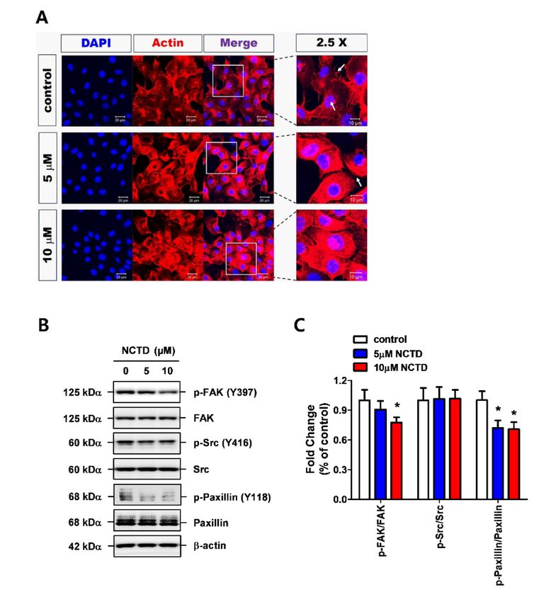
InInorder
ordertoto
determine
determinewhether
whethercytoskeletal
cytoskeletalreorganization
reorganizationsignaling
signalingpathways
pathwaysare areimplicated
implicatedinin
the
the anti-invasive effects of NCTD, immunofluorescence analysis was performed on fixedcells.
anti-invasive effects of NCTD, immunofluorescence analysis was performed on fixed cells.Actin
Actin
stress
stressfiber
fiberand
and filopodia were largely
filopodia were largelyabolished
abolishedandanddisorganized
disorganized byby NCTD
NCTD treatment
treatment compared
compared with
with the vehicle
the vehicle controlcontrol (Figure
(Figure 3A). FAK,
3A). Next, Next,Src,
FAK,
andSrc, and the
paxillin, paxillin, the mostsignaling
most common commoncomponents
signaling
components of F-actin reorganization,
of F-actin reorganization, were evaluated
were evaluated usingblotting.
using western western blotting. Activation
Activation of FAK (Tyrof FAK
397)(Tyr
and
397) and (Tyr
paxillin paxillin
118) (Tyr 118) wasattenuated
was strongly strongly attenuated by NCTDbut
by NCTD treatment, treatment, but NCTD
NCTD exhibited exhibitedeffect
no apparent no
apparent effect on Src
on Src activation (Tryactivation (Try3B,C).
416) (Figure 416) (Figure 3B,C).
These data These data
suggested thatsuggested
NCTD may thatact
NCTD
as anmay act asof
inhibitor
anthe
inhibitor of the signaling
FAK/paxillin FAK/paxillin
axissignaling
to promote axis to promote
F-actin F-actin reorganization
reorganization in YD-15 cells.in YD-15 cells.
Figure 3. NCTD induces F-actin reorganization and activates FAK/Src/paxillin signaling. (A) Confocal
Figure
images 3. of
NCTD
YD-15 induces
cells inF-actin reorganization
the absence andofactivates
and presence NCTD (5 FAK/Src/paxillin
and 10 µM). Actin signaling.
staining(A) Confocal
by fluorescent
images of YD-15 cells in the absence and presence of NCTD (5 and 10 μM). Actin
phalloidin (red) and nuclear staining by DAPI (blue). The white arrow indicates actin with elongated staining by
fluorescent phalloidin (red) and nuclear staining by DAPI (blue). The white arrow indicates
filopodia and stress fibers. The images are representative of three independent experiments. (B,C) actin with
elongated
Western blot filopodia
analysisandof stress fibers. The images
the phosphorylation states ofareFAK/Src/paxillin
representative signaling.
of three independent
Total protein
experiments. (B,C) Western blot analysis of the phosphorylation states of
expressions of FAK, Src, and paxillin were used for normalization. Graphs show the meanFAK/Src/paxillin signaling.
± SD of
Total protein expressions of FAK, Src, and paxillin were used for normalization.
triplicate experiments and significance compared with the vehicle control (* p < 0.05). Graphs show the
mean ± SD of triplicate experiments and significance compared with the vehicle control (* p < 0.05).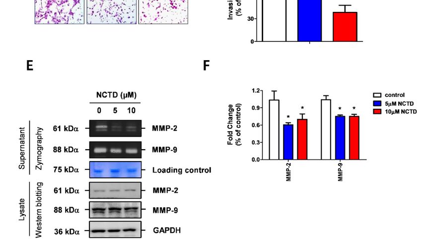
Molecules 2019, 24, x 5 of 11
2.4. NCTD Inhibits the Invasiveness of YD-15 Cells More Potently than PF562271
Molecules 2019, 24, 1928 5 of 11
PF562271 is a 24,
Molecules 2019, potent
x small molecule inhibitor of FAK and has been reported to inhibit the
5 of 11
proliferation of cancer cells [20,21]. Thus, we first evaluated the effects of PF562271 on the expression
levels of2.4. NCTDSrc,
p-FAK, Inhibits
andthepaxillin
Invasiveness of YD-15
in YD-15 Cells
cells Moreevaluating
before thanthe
Potently than PF562271
PF562271
anti-invasive effect of NCTD.
The results PF562271
showed that
PF562271 PF562271
is a potent smallsignificantly inactivated
molecule inhibitor
inhibitor of FAKFAKand(Tyr 397) reported
has been and paxillin (Tyr the
to inhibit 118)
similar proliferation
to NCTD (Figure
proliferation 4).cells
of cancer
of cancer Next,
cells the anti-invasive
[20,21].
[20,21]. Thus, we firsteffect of NCTD
evaluated wasofcompared
the effects PF562271 on with
thePF562271
expressionin
YD-15 cells.
levelsNCTD
of p-FAK,dramatically
Src, and decreased
paxillin
Src, and paxillin in wound
YD-15 cells closure
before speed
evaluating(Figure
the 5A,B) and
anti-invasive led to
anti-invasive effect of
effect inhibition
NCTD.
The results
of invasion
The resultsshowed
(Figure showed
5C). that that PF562271
PF562271 significantly
significantly inactivated
inactivated FAKFAK (Tyr and
(Tyr 397) 397)paxillin
and paxillin (Tyr
(Tyr 118) 118)
similar
similar
to NCTDto(Figure
NCTD4).(Figure
Next, 4).
theNext, the anti-invasive
anti-invasive effect was
effect of NCTD of NCTD was with
compared compared withinPF562271
PF562271 in
YD-15 cells.
YD-15 cells. NCTD dramatically decreased wound closure speed (Figure 5A,B) and led to
NCTD dramatically decreased wound closure speed (Figure 5A,B) and led to inhibition of invasion inhibition
of invasion
(Figure 5C). (Figure 5C).
Figure 4. Comparison of the activation of FAK/Src/paxillin signaling between NCTD and PF562271
in YD-15 Figure
cells.4. Comparison
(A,B) Westernof the activation of FAK/Src/paxillin signaling between NCTD and PF562271after
in
Figure 4. Comparison ofblot
the analysis
activationofofthe phosphorylation
FAK/Src/paxillin of FAK/Src/paxillin
signaling between NCTD signaling
and PF562271
YD-15 cells.
the treatment with (A,B)
NCTD Western
(10 μM)blot analysis of the (0.5
or analysis
PF562271 phosphorylation
μM) for 24 of h.FAK/Src/paxillin
Graphs signaling
show the meanafter the
± SD
in YD-15 cells. (A,B) Western blot of the phosphorylation of FAK/Src/paxillin signaling afterof
treatment with NCTD (10 µM) or PF562271 (0.5 µM) for 24 h. Graphs show the mean ± SD of triplicate
triplicatethe
experiments
treatment with andNCTD
significance
(10 μM)compared
or PF562271 with
(0.5the vehicle
μM) for 24control (* pshow
h. Graphs < 0.05).
the mean ± SD of
experiments and significance compared with the vehicle control (* p < 0.05).
triplicate experiments and significance compared with the vehicle control (* p < 0.05).
Figure 5. Cont.
Molecules 2019, 24, x                                                                                                     6 of 11
Molecules 2019, 24, 1928                                                                                            6 of 11

      Figure 5. Comparison of the anti-invasive efficacy between NCTD and PF562271 in YD-15 cells. (A,B)
               Figure 5. Comparison of the anti-invasive efficacy between NCTD and PF562271 in YD-15 cells. (A,B)
      Wound-healing assay of cell migration after treatment with NCTD or PF562271. (C) Transwell assay
               Wound-healing assay of cell migration after treatment with NCTD or PF562271. (C) Transwell assay
      for the cell invasive potential after treatment with NCTD or PF562271 in YD-15 cells. (D,E) Effects of
               for the cell invasive potential after treatment with NCTD or PF562271 in YD-15 cells. (D,E) Effects of
      NCTD or PF562271 on MMP activity using gelatin zymography. (F) Confocal images of YD-15 cells
               NCTD or PF562271 on MMP activity using gelatin zymography. (F) Confocal images of YD-15 cells
      after treatment with NCTD or PF562271. Graphs show the mean ± SD of triplicate experiments and
               after treatmentwith
      significance compared     withthe
                                      NCTD
                                        vehicleorcontrol
                                                  PF562271.
                                                         (* p
Molecules 2019, 24, 1928 7 of 11
NCTD dephosphorylated FAK (Y397). These results are inconsistent with the findings of Hsia et al.
who showed that the treatment of platelets with NCTD inhibited phosphorylation of both Src and
FAK [33]. Collectively, NCTD has the potential to inhibit F-actin reorganization in human MEC cells,
and the underlying mechanism may be related to the FAK/Paxillin signaling axis.
To further clarify whether the FAK/paxillin signaling axis may be involved in the anti-invasive
activity of NCTD, we compared its activity with a potent, selective FAK inhibitor, PF562271 [34].
PF562271 was previously shown to display inhibitory effects on FAK phosphorylation and migration
in human umbilical vascular endothelial cells, Ewing sarcoma, and human keratinocytes [35–37].
PF562271 attenuated the phosphorylation expression levels of FAK and paxillin. However, unexpectedly,
PF562271 weakly suppressed only the invasiveness of YD-15 cells in the transwell invasion assay and
had no effect on migration, F-action reorganization, or MMPS activity of YD-15 cells. Contrary to
PF562271, NCTD completely inhibited the invasion of YD-15 cells. Vinculin is localized in focal adhesion
as well as cell-adherence junctions (AJ) and is phosphorylated on residues Y100 and Y1065 by members
of the Src family before binding to F-actin [38]. Recently, Bays et al., reported that phosphorylation
of vinculin at Y822 can increase when forces are applied to cell–cell junctions (E-cadherin), which
indicates the regulatory function of Y822 occurs by distribution of the phosphorylation of some focal
adhesion proteins including paxillin and FAK [39]. One possible explanation for the different responses
of NCTD and PF562271 is that p-vinculin can be affected differently by the two chemicals. Although
future studies on the current issue are recommended, these finding clearly demonstrate more potent
efficacy of NCTD on F-actin disorganization compared with PF562271.
In conclusion, the present in vitro study indicates that NCTD may have an anti-invasive potential
for human MEC through a novel mechanism involving inhibition of F-actin binding-related proteins.
This provides tremendous support for the role of NCTD as an attractive anticancer drug candidate
against human MEC.
4. Materials and Methods
4.1. Cell Culture and Chemical Treatment
The YD-15 cell line was obtained from Yonsei University (Seoul, Korea) and was cultured in
RPMI-1640 medium supplemented with 10% fetal bovine serum (FBS) and antibiotics at 37 ◦ C in a 5%
CO2 incubator. NCTD (Figure 1A) was purchased from Sigma-Aldrich Chemical Co. (St. Louis, MO,
USA) and used for treatment at various concentrations ranging from 0 to 40 µM for 24 h. PF562271
(a focal adhesion protein-tyrosine kinase inhibitor) was purchased from Selleckchem (Houston, TX,
USA). Each chemical was dissolved in dimethyl sulfoxide (DMSO), aliquoted, and stored at −20 ◦ C.
All treatments were performed after cells reached 50–60% confluence.
4.2. Trypan Blue Exclusion Assay
YD-15 cells were treated with different concentrations of NCTD for 24 h, and cell viability was
measured using trypan blue staining (Gibco, Paisley, UK). Cells were stained with 0.4% trypan blue
solution, and viable cells were counted using a hemocytometer.
4.3. Clonogenic Formation Assay
Assays were performed as previously described [40]. Briefly, YD-15 cells were seeded into a
12-well culture plate with 1000 cells per well. After 18 h, NCTD was added to the wells, and the plate
was re-incubated. The medium was replaced with fresh medium containing NCTD every three days
for 1 week. The colonies were then stained with 1% crystal violet and counted.
4.4. Scratch Wound Healing Assay
When cells reached 90–100% confluence in 6-well culture plates, YD-15 cells were slowly scratched
using a sterile plastic pipette tips across the center of the well. The cells were gently washed twice inMolecules 2019, 24, 1928 8 of 11
warm medium to remove the detached cells, immediately imaged (0 h), and treated with DMSO, NCTD
(5 and 10 µM), or PF562271 (0.5 µM) for 24 h. Images of wound gaps were then time-dependently
acquired using a CKX53 microscope (Olympus, Tokyo, Japan). The wound closure areas were quantified
as a percentage of cell migration into the wound with respect to the clear area at 24 h. The migration
area was measured using the ImageJ software (Version 1.51k; NIH, Bethesda, MD, USA).
4.5. Matrigel Invasion Assay
YD-15 cells (5 × 10⁴ cells/well) were treated with NCTD or PF562271 for 24 h on pre-coated 24-well
inserts (8 µm pore size) with Matrigel (BD Bioscience, Bedford, MA, USA). The lower chamber was
filled with medium containing 10% FBS as a chemoattractant. The transwell chamber was incubated at
37 ◦ C with 5% CO2 for 24 h. The cells on the upper surface of the filter were then wiped off with a
cotton swab, and the filter was removed from the chamber and stained with hematoxylin and eosin.
The number of cells on lower chamber were counted under a light microscope (Leica DM5000B; Leica
Microsystems, Wetzlar, Germany). For each assay, 10 different microscopic fields (×100 magnification)
were randomly chosen.
4.6. Gelatin Zymography
YD-15 cells were treated in serum-free medium with NCTD or PF562271 for 72 h. The conditioned
medium was collected and concentrated using Amicon Ultra Centrifugal Filter Units (Millipore,
Billerica, MA, USA). To analyze MMP-2 and MMP-9 activities, equal amounts of protein (30 µg) were
loaded onto a gelatin-containing gel (8% acrylamide gel containing 1.5 mg/mL gelatin) and separated
by electrophoresis. The gel was renatured in 2.5% Tween-20 solution with gentle agitation for 30 min at
room temperature (RT), developed overnight at 37 ◦ C in zymogram incubation buffer (50 mM Tris–HCl
(pH 7.6) and 5 mM CaCl2 ), and stained with Coomassie Brilliant Blue R250 (Bio-Rad Laboratories,
Hercules, CA, USA). The gel was then de-stained with a solution of 50% methanol and 10% acetic acid
until the part of the membrane degraded by MMP-2 and MMP-9 became clear. The density of the clear
bands was determined using ImageJ software.
4.7. F-Actin Staining Using TRITC-Conjugated Phalloidin
YD-15 cells were seeded on coverslips and treated with DMSO, NCTD, or PF562271 for 24 h.
The cells were fixed and permeabilized with Cytofix/Cytoperm (BD Bioscience) for 40 min at 4 ◦ C and
incubated with TRITC-labelled phalloidin for 40 min, washed with PBS containing 0.1% BSA, and then
reacted with 5 mg/mL 4,6-diamidino-2-phenylindole (DAPI; Sigma) for nuclei staining. After washing,
the samples were rinsed with PBS and mounted with a mounting media. Immunofluorescence images
were obtained using LSM700 confocal laser scanning microscope (Carl Zeiss, Oberkochen, Germany).
4.8. Western Blot Analysis
Proteins were extracted from cell pellets by homogenization with RIPA buffer (EMD Millipore,
Billerica, CA, USA), and the protein concentration of each sample was measured using a DC
Protein Assay Kit (Bio-Rad Laboratories). After normalization, equal amounts of protein were
separated by sodium dodecyl sulfate-polyacrylamide gel electrophoresis and transferred to immunoblot
polyvinylidene difluoride membranes (Pall Corporation, Port Washington, NY, USA). The membranes
were blocked with 5% skim milk at RT for 2 h, incubated with the specific primary antibodies, and
probed with corresponding horseradish peroxidase-conjugated secondary antibodies (GTX213110
for anti-Rabbit and GTX213111 for anti-mouse). Rabbit anti-human polyclonal antibodies against
cleaved poly (ADP-ribose) polymerase (PARP; 1:3000; #9541), cleaved caspase-3 (1:1000; #9664),
p-FAK(Y397; 1:1000; #3283), FAK (1:1000; #32853), p-Paxillin (Y118; 1:1000; #2541), Paxillin (1:1000;
#2542), p-Src (Y416; 1:1000; #6943), and Src (1:1000; #2109) were purchased from Cell Signaling
Technology (Charlottesville, VA, USA). Mouse anti-human monoclonal antibodies against β-actin
(1:3000; SC-47778) and GAPDH (1:3000; ab9484) were obtained from Santa Cruz BiotechnologyMolecules 2019, 24, 1928 9 of 11
(Santa Cruz, CA, USA) and Thermo Fisher Scientific (Rockford, IL, USA), respectively. Immunoreactive
proteins were detected by SuperSignal West Pico Chemiluminescent Substrate (sc-2048; Santa Cruz
Biotechnology). The immunoreactive bands were visualized using ImageQuant LAS 500 (GE Healthcare
Life Sciences, Pittsburgh, PA, USA). The densitometric analysis of western blotting was quantified
using ImageJ software (NIH).
4.9. Statistical Analysis
Data are expressed as mean ± S.D. One-way ANOVAs analysis was applied to determine the
significance of differences between the control and treatment groups using SPSS v22 (SPSS, Chicago,
IL, USA); values of p < 0.05 were considered statistically significant (*).
Supplementary Materials: The following is available online at http://www.mdpi.com/1420-3049/24/10/1928/s1,
Figure S1: Effects of NCTD on apoptosis in YD-15 cells.
Author Contributions: K.-O.H. designed the study, analyzed and interpreted the results, and drafted the
manuscript; C.-H.A. contributed to the manuscript preparation, the study design, statistical analysis; I.-H.Y.
managed the samples and the data acquisition; J.-M.H. contributed to quality control of data, statistical analysis
and wrote the methods section; J.-A.S. performed statistical analysis, data interpretation and wrote parts of the
results; S.-D.C. and S.D.H. supervised the project and contributed to manuscript review and editing, and revised
critically the manuscript. All authors were involved in the final version of the manuscript.
Funding: This research received no external funding.
Acknowledgments: This work was supported by the Basic Science Research Program through the National
Research Foundation of Korea (NRF) funded by the Ministry of Science, ICT, and Future Planning
[2017R1D1A1B03029317] and [2018R1D1A1B07043080].
Conflicts of Interest: The authors declare no conflict of interest.
References
1. Barnes, L.; Eveson, J.; Reichart, P.; Sidransky, D. World Health Organization Classification of Tumours: Pathology
and Genetics of Tumours of the Head and Neck; IARC: Lyon, France, 2005.
2. Bell, D.; El-Naggar, A.K. Molecular heterogeneity in mucoepidermoid carcinoma: Conceptual and practical
implications. Head Neck Pathol. 2013, 7, 23–27. [CrossRef]
3. Spiro, R.H.; Huvos, A.G.; Berk, R.; Strong, E.W. Mucoepidermoid carcinoma of salivary gland origin.
A clinicopathologic study of 367 cases. Am. J. Surg. 1978, 136, 461–468. [CrossRef]
4. Terhaard, C.H.; Lubsen, H.; Rasch, C.R.; Levendag, P.C.; Kaanders, H.H.; Tjho-Heslinga, R.E.;
van Den Ende, P.L.; Burlage, F.; Dutch, H.; Neck Oncology Cooperative Group. The role of radiotherapy
in the treatment of malignant salivary gland tumors. Int. J. Radiat. Oncol. Biol. Phys. 2005, 61, 103–111.
[CrossRef]
5. Hanahan, D.; Weinberg, R.A. Hallmarks of cancer: The next generation. Cell 2011, 144, 646–674. [CrossRef]
6. Lazebnik, Y. What are the hallmarks of cancer? Nat. Rev. Cancer 2010, 10, 232–233. [CrossRef] [PubMed]
7. Poltavets, V.; Kochetkova, M.; Pitson, S.M.; Samuel, M.S. The Role of the Extracellular Matrix and Its
Molecular and Cellular Regulators in Cancer Cell Plasticity. Front. Oncol. 2018, 8, 431. [CrossRef] [PubMed]
8. Condeelis, J.; Singer, R.H.; Segall, J.E. The great escape: When cancer cells hijack the genes for chemotaxis
and motility. Annu. Rev. Cell Dev. Biol. 2005, 21, 695–718. [CrossRef] [PubMed]
9. Sahai, E. Mechanisms of cancer cell invasion. Curr. Opin. Genet. Dev. 2005, 15, 87–96. [CrossRef]
10. Yamaguchi, H.; Wyckoff, J.; Condeelis, J. Cell migration in tumors. Curr. Opin. Cell Biol. 2005, 17, 559–564.
[CrossRef] [PubMed]
11. Parsons, J.T. Focal adhesion kinase: The first ten years. J. Cell Sci. 2003, 116, 1409–1416. [CrossRef]
12. Kanteti, R.; Batra, S.K.; Lennon, F.E.; Salgia, R. FAK and paxillin, two potential targets in pancreatic cancer.
Oncotarget 2016, 7, 31586–31601. [CrossRef] [PubMed]
13. Gabarra-Niecko, V.; Schaller, M.D.; Dunty, J.M. FAK regulates biological processes important for the
pathogenesis of cancer. Cancer Metast. Rev. 2003, 22, 359–374. [CrossRef]Molecules 2019, 24, 1928 10 of 11
14. Itoh, S.; Maeda, T.; Shimada, M.; Aishima, S.; Shirabe, K.; Tanaka, S.; Maehara, Y. Role of expression of focal
adhesion kinase in progression of hepatocellular carcinoma. Clin. Cancer Res. 2004, 10, 2812–2817. [CrossRef]
[PubMed]
15. Rajesh, E.; Sankari, L.S.; Malathi, L.; Krupaa, J.R. Naturally occurring products in cancer therapy. J. Pharm.
Bioallied Sci. 2015, 7, S181–S183. [CrossRef]
16. Nobili, S.; Lippi, D.; Witort, E.; Donnini, M.; Bausi, L.; Mini, E.; Capaccioli, S. Natural compounds for cancer
treatment and prevention. Pharmacol. Res. 2009, 59, 365–378. [CrossRef] [PubMed]
17. Chen, Y.J.; Chang, W.M.; Liu, Y.W.; Lee, C.Y.; Jang, Y.H.; Kuo, C.D.; Liao, H.F. A small-molecule
metastasis inhibitor, norcantharidin, downregulates matrix metalloproteinase-9 expression by inhibiting Sp1
transcriptional activity in colorectal cancer cells. Chem. Biol. Interact 2009, 181, 440–446. [CrossRef] [PubMed]
18. Huang, Y.; Liu, Q.; Liu, K.; Yagasaki, K.; Zhang, G. Suppression of growth of highly-metastatic human breast
cancer cells by norcantharidin and its mechanisms of action. Cytotechnology 2009, 59, 201–208. [CrossRef]
[PubMed]
19. Li, Y.; Ge, Y.; Liu, F.Y.; Peng, Y.M.; Sun, L.; Li, J.; Chen, Q.; Sun, Y.; Ye, K. Norcantharidin, a protective
therapeutic agent in renal tubulointerstitial fibrosis. Mol. Cell Biochem. 2012, 361, 79–83. [CrossRef]
20. Roberts, W.G.; Ung, E.; Whalen, P.; Cooper, B.; Hulford, C.; Autry, C.; Richter, D.; Emerson, E.; Lin, J.;
Kath, J.; et al. Antitumor activity and pharmacology of a selective focal adhesion kinase inhibitor, PF-562,271.
Cancer Res. 2008, 68, 1935–1944. [CrossRef]
21. Slack-Davis, J.K.; Hershey, E.D.; Theodorescu, D.; Frierson, H.F.; Parsons, J.T. Differential requirement for
focal adhesion kinase signaling in cancer progression in the transgenic adenocarcinoma of mouse prostate
model. Mol. Cancer Ther. 2009, 8, 2470–2477. [CrossRef]
22. Irani, S. Distant metastasis from oral cancer: A review and molecular biologic aspects. J. Int. Soc. Prev.
Community Dent. 2016, 6, 265–271. [CrossRef] [PubMed]
23. Johnson, K.A.; Brown, P.H. Drug development for cancer chemoprevention: Focus on molecular targets.
Semin. Oncol. 2010, 37, 345–358. [CrossRef] [PubMed]
24. Steele, V.E.; Lubet, R.A. The use of animal models for cancer chemoprevention drug development.
Semin. Oncol. 2010, 37, 327–338. [CrossRef] [PubMed]
25. Jiang, Y.L.; Liu, Z.P. Natural products as anti-invasive and anti-metastatic agents. Curr. Med. Chem. 2011,
18, 808–829. [CrossRef] [PubMed]
26. Hsieh, C.H.; Chao, K.S.; Liao, H.F.; Chen, Y.J. Norcantharidin, derivative of cantharidin, for cancer stem cells.
Evid. Based Complement. Alternat. Med. 2013, 2013, 838651. [CrossRef]
27. Zhu, W.; Sun, W.; Zhang, J.T.; Liu, Z.Y.; Li, X.P.; Fan, Y.Z. Norcantharidin enhances TIMP2 antivasculogenic
mimicry activity for human gallbladder cancers through downregulating MMP2 and MT1MMP. Int. J. Oncol.
2015, 46, 627–640. [CrossRef] [PubMed]
28. Yeh, C.B.; Hsieh, M.J.; Hsieh, Y.H.; Chien, M.H.; Chiou, H.L.; Yang, S.F. Antimetastatic effects of norcantharidin
on hepatocellular carcinoma by transcriptional inhibition of MMP-9 through modulation of NF-kB activity.
PLoS ONE 2012, 7, e31055. [CrossRef] [PubMed]
29. Mon, N.N.; Ito, S.; Senga, T.; Hamaguchi, M. FAK signaling in neoplastic disorders: A linkage between
inflammation and cancer. Ann. N. Y. Acad. Sci. 2006, 1086, 199–212. [CrossRef] [PubMed]
30. Peng, C.; Li, Z.; Niu, Z.; Niu, W.; Xu, Z.; Gao, H.; Niu, W.; Wang, J.; He, Z.; Gao, C.; et al. Norcantharidin
Suppresses Colon Cancer Cell Epithelial-Mesenchymal Transition by Inhibiting the alphavbeta6-ERK-Ets1
Signaling Pathway. Sci. Rep. 2016, 6, 20500. [CrossRef]
31. Geiger, B.; Bershadsky, A.; Pankov, R.; Yamada, K.M. Transmembrane crosstalk between the extracellular
matrix–cytoskeleton crosstalk. Nat. Rev. Mol. Cell Biol. 2001, 2, 793–805. [CrossRef]
32. Roskoski, R., Jr. Src kinase regulation by phosphorylation and dephosphorylation. Biochem. Biophys.
Res. Commun. 2005, 331, 1–14. [CrossRef]
33. Hsia, C.H.; Lu, W.J.; Lin, K.H.; Chou, D.S.; Geraldine, P.; Jayakuma, T.; Chang, N.C.; Sheu, J.R. Norcantharidin,
a clinical used chemotherapeutic agent, acts as a powerful inhibitor by interfering with fibrinogen-integrin
alphaIIb beta3 binding in human platelets. J. Cell Mol. Med. 2018, 22, 2142–2152. [CrossRef]
34. Hu, C.; Chen, X.; Wen, J.; Gong, L.; Liu, Z.; Wang, J.; Liang, J.; Hu, F.; Zhou, Q.; Wei, L.; et al. Antitumor effect
of focal adhesion kinase inhibitor PF562271 against human osteosarcoma in vitro and in vivo. Cancer Sci.
2017, 108, 1347–1356. [CrossRef] [PubMed]Molecules 2019, 24, 1928 11 of 11
35. Bi, Y.L.; Mi, P.Y.; Zhao, S.J.; Pan, H.M.; Li, H.J.; Liu, F.; Shao, L.R.; Zhang, H.F.; Zhang, P.; Jiang, S.L.
Salinomycin exhibits anti-angiogenic activity against human glioma in vitro and in vivo by suppressing the
VEGF-VEGFR2-AKT/FAK signaling axis. Int. J. Mol. Med. 2017, 39, 1255–1261. [CrossRef]
36. Crompton, B.D.; Carlton, A.L.; Thorner, A.R.; Christie, A.L.; Du, J.; Calicchio, M.L.; Rivera, M.N.;
Fleming, M.D.; Kohl, N.E.; Kung, A.L.; et al. High-throughput tyrosine kinase activity profiling identifies
FAK as a candidate therapeutic target in Ewing sarcoma. Cancer Res. 2013, 73, 2873–2883. [CrossRef]
[PubMed]
37. Wang, Y.; Zheng, J.; Han, Y.; Zhang, Y.; Su, L.; Hu, D.; Fu, X. JAM-A knockdown accelerates the
proliferation and migration of human keratinocytes and improves wound healing in rats via FAK/Erk
signaling. Cell Death Dis. 2018, 9, 848. [CrossRef] [PubMed]
38. Goldmann, W.H.; Auernheimer, V.; Thievessen, I.; Fabry, B. Vinculin, cell mechanics and tumour cell invasion.
Cell Biol. Int. 2013, 37, 397–405. [CrossRef] [PubMed]
39. Goldmann, W.H. Role of vinculin in cellular mechanotransduction. Cell Biol. Int. 2016, 40, 241–256. [CrossRef]
40. Aggarwal, S.; Das, S.N. Garcinol inhibits tumour cell proliferation, angiogenesis, cell cycle progression and
induces apoptosis via NF-kappaB inhibition in oral cancer. Tumour Biol. 2016, 37, 7175–7184. [CrossRef]
Sample Availability: Samples of the compounds are not available from the authors.
© 2019 by the authors. Licensee MDPI, Basel, Switzerland. This article is an open access
article distributed under the terms and conditions of the Creative Commons Attribution
(CC BY) license (http://creativecommons.org/licenses/by/4.0/).You can also read