A Retrospective Study of the Diagnostic Accuracy of In Vivo Reflectance Confocal Microscopy for Basal Cell Carcinoma Diagnosis and Subtyping - MDPI
←
→
Page content transcription
If your browser does not render page correctly, please read the page content below
Journal of
           Clinical Medicine

Article
A Retrospective Study of the Diagnostic Accuracy of
In Vivo Reflectance Confocal Microscopy for Basal
Cell Carcinoma Diagnosis and Subtyping
Mihai Lupu 1 , Iris Maria Popa 2 , Vlad Mihai Voiculescu 1,3 , Daniel Boda 4 ,
Constantin Caruntu 4,5, *, Sabina Zurac 5, * and Calin Giurcaneanu 1,3
 1   Department of Dermatology, “Carol Davila” University of Medicine and Pharmacy, 050474 Bucharest,
     Romania; lupu.g.mihai@gmail.com (M.L.); voiculescuvlad@yahoo.com (V.M.V.);
     calin.giurcaneanu@gmail.com (C.G.)
 2   Department of Plastic and Reconstructive Surgery, “Bagdasar-Arseni” Clinical Emergency Hospital,
     041915 Bucharest, Romania; irismpopa@gmail.com
 3   Department of Dermatology, “Elias” University Emergency Hospital, 011461 Bucharest, Romania
 4   Department of Dermatology, “Prof. N. Paulescu” National Institute of Diabetes, Nutrition and Metabolic
     Diseases, 011233 Bucharest, Romania; daniel.boda@yahoo.com
 5   Department of Physiology, “Carol Davila” University of Medicine and Pharmacy, 050474 Bucharest,
     Romania
 *   Correspondence: costin.caruntu@gmail.com (C.C.); sabina_zurac@yahoo.com (S.Z);
     Tel.: +40-7450-869-78 (C.C)

 Received: 11 March 2019; Accepted: 2 April 2019; Published: 3 April 2019

 Abstract: Current national and European guidelines recommend distinct management approaches
 for basal cell carcinoma (BCC) based on tumor location, size, and histopathological subtype. In vivo
 reflectance confocal microscopy (RCM) is a non-invasive skin imaging technique which may change
 the diagnostic pathway for BCC patients. This study aimed to determine the sensitivity and specificity
 of RCM for BCC diagnosis, assess the predictive values of several confocal criteria in correctly
 classifying BCC subtypes, and evaluate the intraobserver reliability of RCM diagnosis for BCC.
 We conducted a retrospective study in two tertiary care centers in Bucharest, Romania. We included
 adults with clinically and dermoscopic suspect BCCs who underwent RCM and histopathological
 examination of excision specimens. For RCM examinations, we used the VivaScope 1500 and
 histopathology of the surgical excision specimen was the reference standard. Of the 123 cases
 included in the analysis, BCC was confirmed in 104 and excluded in 19 cases. RCM showed both high
 sensitivity (97.1%, 95% CI (91.80, 99.40)) and specificity (78.95%, 95% CI (54.43, 93.95)) for detecting
 BCC. Several RCM criteria were highly predictive for BCC subtypes: cords connected to the epidermis
 for superficial BCC, big tumor islands, peritumoral collagen bundles and increased vascularization for
 nodular BCC, and hyporefractile silhouettes for aggressive BCC. Excellent intraobserver agreement
 (κ = 0.909, p < 0.001) was observed. This data suggests that RCM could be used for preoperative
 diagnosis and BCC subtype classification in patients with suspected BCCs seen in tertiary care centers.

 Keywords: Carcinoma; basal cell; dermoscopy; microscopy; confocal; retrospective studies;
 skin neoplasms

1. Introduction
     Basal cell carcinoma (BCC) is the most prevalent skin cancer worldwide. In Europe, BCC incidence
has been constantly rising by approximately 5% annually over recent decades [1], causing a major
burden on healthcare systems [2,3]. Adding to this, an abrupt increase in BCC incidence in the young

J. Clin. Med. 2019, 8, 449; doi:10.3390/jcm8040449                                 www.mdpi.com/journal/jcm
J. Clin. Med. 2019, 8, 449                                                                           2 of 14

population is reported [4–6]. Although BCC mortality is low (0.0028% to 0.55%) [7], these tumors are
locally invasive and can induce significant morbidity owing to their frequent development on the head
and neck.
     Current national and European guidelines [8,9] advise distinct therapeutic approaches of BCC
based on tumor location, size, and histopathological subtype. With the increasing number of efficient
non-surgical treatment options for superficial BCC (sBCC) [10], the histopathological subtype becomes
of special interest in choosing the most appropriate management course [11–14]. Guidelines also
recommend BCC diagnosis confirmation and histological subtyping through a punch biopsy [15,16].
However, a punch biopsy fails to diagnose an aggressive BCC subtype in one out of six tumors [17].
Therefore, histopathological examination of the entire tumor specimen remains the most accurate
approach of establishing BCC histopathological subtype [18–20].
     The preoperative assessment of BCC histological subtypes through non-invasive techniques may
reduce the number of painful invasive diagnostic procedures, lower the delay between diagnosis and
treatment, and lower the burden on healthcare systems through reducing administrative workloads
and financial costs [21,22]. Several recent studies have tried to correlate dermoscopic criteria to certain
BCC subtypes [23,24], without consistent results.
     High resolution non-invasive skin imaging may change the diagnostic pathway in the case of
BCC patients [25,26]. In vivo reflectance confocal microscopy (RCM) is a novel, non-invasive imaging
technique capable of producing horizontal optical sections of the skin [27]. RCM enables examination
of the entire lesion, while confocal resolution and morphologic features are similar to histology [28].
This non-invasive imaging technique has been proven useful not only in the evaluation and follow-up
of melanocytic [29,30] and non-melanocytic lesions [31–37], but also in the diagnosis of inflammatory
skin diseases [38–48]. However, one fundamental risk of techniques such as RCM is that they rely on
morphology-based analysis, thus being subject to interpretation bias. Previous studies addressing this
subject exist [49,50], however, there is still a need for more research performed in accordance with the
Standards of Reporting of Diagnostic Accuracy (STARD) [51].
     The primary objective of this study was to determine the agreement between RCM and
histopathology in correctly detecting BCC presence. The secondary objectives were to assess the
accuracy of predefined confocal criteria in correctly classifying BCC histopathological subtypes and
evaluate the intraobserver reliability of preoperative, non-invasive BCC diagnosis through RCM.

2. Materials and Methods
     A retrospective multicenter study was performed at the following 2 sites: the Dermatology
Research Laboratory, “Carol Davila” University of Medicine and Pharmacy, Bucharest, Romania and
the Department of Dermatology at Medas Medical Center, Bucharest, Romania. Patient data was
collected retrospectively by searching the electronic archives of the participating centers for patients
registered between 1 May 2017 and 31 October 2018.
     We included consecutively identified patients older than 18 years with a clinical and dermoscopic
suspicion of previously untreated BCC, whose medical records included medical history, clinical,
dermoscopic, and RCM images as well as a histopathologic report of the excisional biopsy of the
lesion. We excluded patients with missing or incomplete data, patients with lesions that were
reported to be recurrences, previously treated lesions, or lesions extending to mucosal surfaces.
Immunocompromised patients were not excluded from the study. The study was conducted in
accordance with the Declaration of Helsinki, and the protocol was approved by the Ethics Committee
of the “Carol Davila” University of Medicine and Pharmacy Bucharest (Project Number 185, approved
on 26.12.2018). All participants gave written informed consent as part of their investigation and
treatment procedures, at the time of their registration.
     In both centers, RCM examination was conducted using the same commercially available confocal
microscope (VivaScope 1500® ; Caliber ID, Henrietta, NY, USA.; MAVIG GmbH, München, Germany).
J. Clin. Med. 2019, 8, 449                                                                        3 of 14

RCM imaging at the Medas Medical Center was performed by ML and by CC at the “Prof. N. Paulescu”
National Institute of Diabetes, Nutrition, and Metabolic Diseases.
     The VivaScope 1500 uses an 830 nm laser diode, reaching a maximum output power of 20 mW at
the skin level, allowing for skin imaging without causing injury to investigated tissues. A dermoscopic
image captured using the VivaCam serves as a surface map to guide confocal imaging. Five level cubes
(30 µm increments, vertically), including the corneal, granular/spinous, dermal-epidermal junction,
and papillary dermis, are acquired in the center of the lesion. Each level is a mosaic with a minimum
surface of 4 × 4 mm and a maximum of 8 × 8 mm. Individual stacks (4.5 µm increments, vertically)
are also acquired in one or more areas of interest, up to a depth of 200 to 250 µm. Individual images of
cellular and tissular architecture are also obtained. Only patients with lesions investigated following
this RCM imaging protocol were included in the study. Verification that the RCM image set respects
protocol was done through an inspection of the log file generated for each confocal examination.
     Prior to the study, ML was trained in RCM use and interpretation during a one week confocal laser
scanning microscopy course organized by MAVIG GmbH (distributor of the VivaScope® device) at the
University of Modena and Reggio Emilia in Italy. ML had more than three years of RCM experience
prior to the start of the study. CC had more than nine years RCM experience.
     Based on previous findings [49,50], a set of 14 confocal imaging criteria was formulated:
keratinocyte atypia, epidermal streaming, ulceration, cords connected to the epidermis, small tumor
islands (diameter 300 µm), hyporefractile silhouettes,
peripheral palisading, clefting, increased vascularization, “onion-like” structures (corresponding to
milia-like cysts), peritumoral collagen bundles, inflammation represented by bright dots and plump
bright cells (corresponding to lymphocytes and melanophages), and dendritic cells inside tumor
islands (corresponding to melanocytes).
     Imaging, at the time of patient evaluation, was not conducted in a blinded fashion as patient
history and clinical examination had to be conducted as part of the standard clinical care. However,
the database of static RCM images was analyzed in a blinded fashion by ML immediately after
completion and locking, and four weeks after, to document the presence or absence of BCC and of the
aforementioned criteria.
     All lesions included in the study were surgically treated with margins between 3 to 5 mm.
Histopathologic confirmation of BCC presence and subtype, and excision margins inspection using
hematoxylin and eosin stained bread-loafed sections was defined as the reference standard. The
reporting of histopathological findings was performed by experienced pathologists. During assessment
of the reference standard, the pathologist was masked to the findings of the RCM examination, but not
to the clinical description of the lesions and patients’ clinical history.
     We recorded the following characteristics of participants and tumors and summarized them with
descriptive statistics: age, gender, tumor topographic location, and tumor histopathological subtype.
A distinction was made between superficial, nodular, and aggressive (micronodular, infiltrative, and
basosquamous) BCC growth patterns. For the purposes of this study sclerodermiform/morpheaform
BCC was considered equivalent to infiltrative BCC. In the case of mixed-type histopathological
diagnosis, defined as two or more growth patterns, the most aggressive component was taken into
account for analysis.
     The primary objective was the agreement between the index test (RCM) and reference standard
(histopathology of the excision specimen) in correctly determining BCC presence. The secondary
outcomes were estimating the accuracy of predefined confocal criteria in correctly classifying BCC
subtypes and determining the intraobserver agreement of preoperative BCC diagnosis through RCM.
     One rater (ML) reviewed all RCM images of de-identified cases twice, at a four-week interval.
The rater was blinded to clinical and dermoscopic images, histopathological report, and to his
previous interpretation. Between evaluations, RCM case numbers were shuffled and recoded by
an online software-based algorithm (available at https://www.graphpad.com/quickcalcs/) to prevent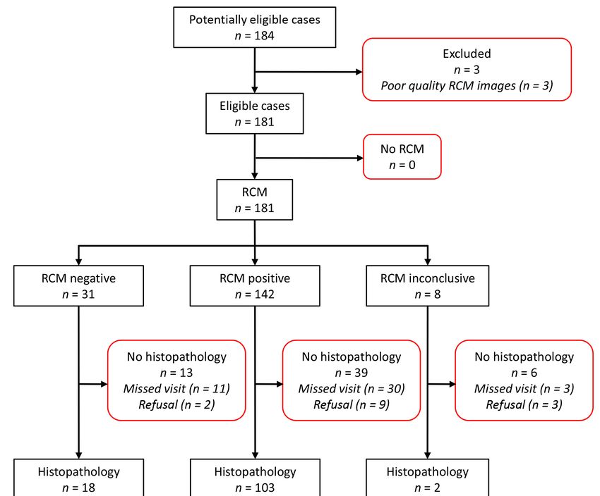
J. Clin. Med. 2019, 8, 449                                                                                                       4 of 14

identification. Evaluation data were recorded in a standardized manner to BCC presence (yes or no)
and presence of the 14 selected criteria (yes or no).
     Lesions in which subsequent surgical excision was not performed (reasons were recorded) were
      J. Clin. Med. 2019, 8, x FOR PEER REVIEW                                                                              4 of 14
excluded. According to Shinkins et al. [52], the ideal approach to including and analyzing inconclusive
valid test Lesions
              resultsin  (nwhich
                             = 8) is    to treat them
                                    subsequent     surgicalas excision
                                                              if in a clinical
                                                                       was not scenario.
                                                                                  performed We        have,were
                                                                                                (reasons     therefore,
                                                                                                                 recorded) considered
                                                                                                                             were
theseexcluded.
       inconclusive         valid results
                     According                 as RCM
                                     to Shinkins           positive
                                                      et al.          cases,
                                                               [52], the   idealandapproach
                                                                                     includedtothe      cases which
                                                                                                     including          had received
                                                                                                                 and analyzing
the reference
      inconclusive standard      (n =results
                        valid test       2) in the
                                               (n = analysis.    The
                                                    8) is to treat    numbers
                                                                    them   as if inof  true and
                                                                                     a clinical     false positives
                                                                                                scenario.   We have, and     negatives
                                                                                                                       therefore,
      considered      these   inconclusive     valid results   as RCM   positive   cases,  and
were recorded. We established the sensitivity, specificity, positive, and negative likelihood   included   the  cases which   had ratios,
      received     the  reference    standard    (n =  2) in the  analysis.   The  numbers
and positive and negative predictive values for BCC diagnosis by RCM using 2 × 2 contingency  of  true  and  false positives  and
tablesnegatives
        analysis.were      recorded. the
                      To calculate        We overall
                                              established   the sensitivity,
                                                        diagnostic    accuracy,specificity, positive,formula
                                                                                    the following      and negative
                                                                                                                  was likelihood
                                                                                                                       used: Overall
      ratios, and positive and negative predictive values for BCC diagnosis by RCM using 2 × 2
diagnostic accuracy = sensitivity × prevalence + specificity × (1 − prevalence) [53,54]. We used
      contingency tables analysis. To calculate the overall diagnostic accuracy, the following formula was
binomial logistic regression to determine the odds ratio (OR) of the predefined confocal criteria for
      used: Overall diagnostic accuracy = sensitivity × prevalence + specificity × (1 − prevalence) [53,54].
each individual
      We used binomial BCC histological         subtype.
                                 logistic regression        Confidence
                                                         to determine     theintervals
                                                                               odds ratiowere
                                                                                            (OR) 95%        a p value of
                                                                                                       andpredefined
                                                                                                   of the
J. Clin. Med. 2019, 8, 449 5 of 14
Eighty-seven patients (36 males and 51 females) with a mean age of 68.1 ± 12.17 years and median
disease duration of 2 years were included in the study. Most lesions were of the nodular subtype,
with 11 aggressive BCCs (aBCCs) represented (7 infiltrative BCCs, 3 basosquamous BCCs, and one
micronodular BCC), which is consistent with the natural incidence of BCC subtypes. The distribution
of the 104 BCCs in terms of subtype and the histopathological diagnoses for the remaining 19 lesions
are summarized in Table 1.
Table 1. Characteristics of patients and imaged lesions.
BCC 1 Subtype N (%)
Superficial BCC 24 (23.1)
Nodular BCC 69 (66.3)
Aggressive BCC 11 (10.6)
Total = 104
Non-BCC Lesions N (%)
Bowen’s disease 3 (2.4)
Seborrheic keratosis 3 (2.4)
Actinic keratosis 4 (3.3)
Keratoacanthoma 2 (1.6)
Lichen planus-like keratosis 2 (1.6)
Tubular apocrine adenoma 1 (0.8)
Moderately differentiated SCC 2 1 (0.8)
Poorly differentiated SCC 1 (0.8)
Poroid hidradenoma 1 (0.8)
Chronic radiation dermatitis 1 (0.8)
Total = 19
BCC 1 , basal cell carcinoma; SCC 2 , squamous cell carcinoma.
Most lesions were located in the head and neck area (n = 72), followed by the trunk (n = 30),
lower extremities (n = 10), upper extremities (n = 8), and abdomen (n = 3). The number of BCCs in
our study (n = 104) is sufficient to confidently calculate sensitivity and specificity with a maximum
error of estimation of 6% and 14.1%, respectively, with a confidence interval of 1-alpha = 0.95 (95%).
The average time between RCM examination and surgical treatment was 50.99 days.
3.2. Test Results
3.2.1. Basal Cell Carcinoma Diagnosis by Preoperative Reflectance Confocal Microscopy
In our sample of 123 lesions, RCM detected BCC presence with a sensitivity of 97.1%
(95% CI 91.80, 99.40) and a specificity of 78.95% (95% CI 54.43, 93.95) at a disease prevalence of 84.55%.
The positive likelihood ratio was 4.61 (95% CI 1.93, 11.03) while the negative likelihood ratio was 0.04
(95% CI 0.01, 0.11). Positive and negative predictive values were 96.19% (95% CI 91.35, 98.37) and
83.33% (95% CI 61.55, 93.98), respectively. The overall accuracy of preoperative RCM for detection of
BCC was 94.31% (95% CI 88.63, 97.68).
If only conclusive RCM analysis results were included in the analysis (n = 121), RCM sensitivity
was unchanged at 97.1% (95% CI 91.80, 99.40), but specificity was higher at 88.2% (95% CI 63.56, 98.54),
as was disease prevalence (85.95%). The positive likelihood ratio was 8.25 (95% CI 2.24, 30) while the
negative likelihood ratio was 0.03 (95% CI 0.01, 0.1). Positive and negative predictive values were 98.1%
(95% CI 93.21, 99.46) and 83.3% (95% CI 61.79, 93.92), respectively. The overall accuracy of preoperative
RCM for detection of BCC in this case was only slightly higher, at 95.87% (95% CI 90.62, 98.64).J.J. Clin.
Clin. Med.
Med. 2019,
2019, 8,
8, xx FOR
FOR PEER
PEER REVIEW
REVIEW 66 of
of 14
14
accuracy
J. Clin. of preoperative
Med. 2019,
accuracy of preoperative
8, 449 RCM for
RCM for detection
detection of
of BCC
BCC in
in this
this case
case was
was only
only slightly
slightly higher,
higher, at
at 95.87%
95.87%
6 of 14
(95% CI
(95% CI 90.62,
90.62, 98.64).
98.64).
3.2.2. Evaluation
3.2.2. ofofRCM
Evaluation RCMCriteria
CriteriaAccording
According to
to BCC Subtype
BCC Subtype
Subtype
3.2.2. Evaluation of RCM Criteria According to BCC
In In
superficial
In BCCs
superficial
superficial BCCs(sBCCs),
BCCs (sBCCs),RCM
(sBCCs), RCMexamination
RCM
revealedthe
examination revealed
examination revealed thepresence
the presenceof
presence ofofcords
cords
cords connected
connected
connected to to
to thethe
the
epidermis (13/24)
epidermis (13/24)
epidermis with
(13/24) with peripheral
with peripheral palisading
peripheral palisading (19/24)
palisading (19/24) (Figure
(19/24) (Figure 2).
(Figure 2).
2).
Figure 2.
Figure 2. Superficial
Superficial basal
basal cell carcinoma.
carcinoma. (A) Dermoscopy
Dermoscopy showing
showing multiple,
multiple, brown
brown globules
globules and
and
Figure 2. Superficial basal cellcell
carcinoma. (A)(A) Dermoscopy showing multiple, brown globules and dots,
dots,
dots, and leaf-like
and leaf-like peripheral
peripheral structures.
structures. (B) reflectance
(B) reflectance confocal microscopy (RCM) revealed the
and leaf-like peripheral structures. (B) reflectance confocalconfocal microscopy
microscopy (RCM) (RCM)
revealedrevealed the
the presence
presence of of sharply
sharply demarcated
demarcated cords
cords connected to to the
the epidermis
epidermis (white
(white arrows), dark
dark peritumoral
peritumoral
of presence
sharply demarcated cords connectedconnected
to the epidermis (white arrows),arrows),
dark peritumoral clefting
clefting (red arrowheads), and peripheral palisading (white arrowhead),
clefting (red arrowheads), and peripheral palisading (white arrowhead), corresponding to corresponding to the
the
(red arrowheads), and peripheral palisading (white arrowhead), corresponding to the basaloid cords
basaloid cords and aggregates seen on histopathology (hematoxylin and
basaloid cords and aggregates seen on histopathology (hematoxylin and eosin (H&E) stain, eosin (H&E) stain,
and aggregates seen on histopathology (hematoxylin and eosin (H&E) stain, magnification 4×) (C).
magnification 4×)
magnification 4×) (C).
(C).
Moreover, dendritic structures inside tumor islands and cords (14/24) were frequently seen.
Moreover, dendritic
Moreover, dendritic structures
structures inside
inside tumor
tumor islands
islands and
and cords
cords (14/24)
(14/24) were
were frequently
frequently seen.
seen. For
For
Fornodular
nodularBCCs
BCCs(nBCCs),
(nBCCs),bigbigtumor
tumorislands
islands(52/69)
(52/69)associated
associatedwith
withperipheral
peripheralpalisading
palisading(42/69),
(42/69),
nodular BCCs (nBCCs), big tumor islands (52/69) associated with peripheral palisading (42/69),
clefting (34/69),
clefting (34/69),and
andhypervascularization (52/69)
hypervascularization (52/69) werecharacteristic
(52/69) were
were characteristic findings
findings (Figure
(Figure 3). 3).
clefting (34/69), and hypervascularization characteristic findings (Figure 3).
Figure
Figure
Figure 3. Nodularbasal
Nodular
3. 3. Nodular basalcell
basal cellcarcinoma.
cell carcinoma. (A)
carcinoma. (A) Dermoscopy
Dermoscopy image
Dermoscopy image
imageof ofofaaapigmented
pigmented nodular
pigmentednodular
nodular basal
basal
basal cellcell
cell
carcinoma
carcinoma
carcinoma (nBCC)with
(nBCC)
(nBCC) withblue-gray
with blue-grayovoid
blue-gray ovoid nests,
ovoid nests, brown
nests, brown globules,
brown globules, and
globules, and arborizing
and arborizing vessels.
arborizingvessels. (B)(B)
vessels.(B) RCM
RCMRCM
revealed
revealed
revealed large,well
large,
large, welldefined
well definedtumor
defined tumor islands
tumor islands (TI),
islands (TI), peritumoral
peritumoral clefting
peritumoral clefting (red
clefting(red arrows),
(redarrows), and
arrows),and clumped
andclumped
clumped
melanophages
melanophages
melanophages (green
(green
(green rectangle).
rectangle).
rectangle). (C)
(C)(C) Histopathology
Histopathology
Histopathology showed
showed
showed large
large
large basaloid
basaloid
basaloid islands
islands
islands with
with
with palisading
palisading
palisading and
and
stromal stromal
and stromal
retraction retraction
retraction in the
in the
in the dermis (H&E dermis
dermis (H&E
stain,(H&E stain, magnification
stain, magnification
magnification 4×). (D) Dermoscopy 4×). (D)
4×). (D) of Dermoscopy
Dermoscopy
hypopigmented of
of
hypopigmented nBCC nBCC with
hypopigmented
nBCC with ulceration andwith ulceration and
ulceration
arborizing and arborizing
vessels. arborizing vessels.
vessels. (E)
(E) RCM showed (E) RCM
largeRCM showed
tumor showed large
islandslarge tumor
(TI) tumor islands
islands
with peripheral
(TI) with
(TI) withand peripheral
peripheral palisading and clefting (white arrows). (F) Histopathologic correlates of the
the
palisading cleftingpalisading and clefting
(white arrows). (white arrows).
(F) Histopathologic (F) Histopathologic
correlates correlates
of the structures seen ofthrough
structures
structures seen through
seen through RCM
RCM (H&E(H&E stain, magnification 40×).
RCM (H&E stain, magnification 40×).stain, magnification 40×).J. Clin. Med. 2019, 8, 449 7 of 14
J. Clin. Med. 2019, 8, x FOR PEER REVIEW 7 of 14
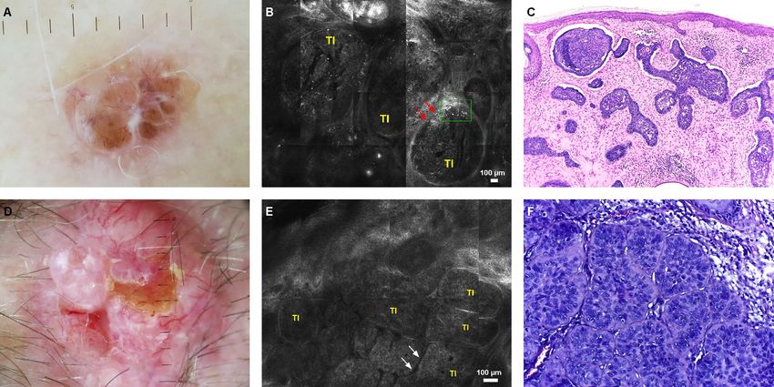
Aggressive BCCs were typified by the presence of hyporefractile silhouettes (7/11), peripheral
Aggressive BCCs were typified by the presence of hyporefractile silhouettes (7/11), peripheral
palisading (7/11),
palisading (7/11),and
andclefting
clefting(7/11) (Figure4).
(7/11) (Figure 4).
Figure
Figure 4. 4. Infiltrativebasal
Infiltrative basalcell
cellcarcinoma.
carcinoma. (A)
(A) Dermoscopy
Dermoscopy showing
showingaahypopigmented
hypopigmented lesion with
lesion with
structureless red shiny areas, chrysalis pattern, short fine telangiectasia, and erosion. (B) RCM
structureless red shiny areas, chrysalis pattern, short fine telangiectasia, and erosion. (B) RCM showed
showed multiple hyporefractile silhouettes, which appear as imprints (white arrows) outlined by
multiple hyporefractile silhouettes, which appear as imprints (white arrows) outlined by bundles of
bundles of bright collagen (red asterisks). (C) Histopathology shows tumor islands and strands that
bright collagen (red asterisks). (C) Histopathology shows tumor islands and strands that resemble the
resemble the hyporefractile silhouettes observed through RCM (BerEP4 stain, magnification 4×).
hyporefractile silhouettes observed through RCM (BerEP4 stain, magnification 4×).
Keratinocyte atypia, epidermal streaming, ulceration, and inflammation were observed with
Keratinocyte atypia, epidermal streaming, ulceration, and inflammation were observed with
comparable frequencies in all tumor subtypes. The analytic descriptive results of the confocal image
comparable frequencies in all tumor subtypes. The analytic descriptive results of the confocal image
analysis are summarized in Table 2.
analysis are summarized in Table 2.
Table 2. Frequencies of confocal criteria in different histologic basal cell carcinoma subtypes.
Table 2. Frequencies of confocal criteria in different histologic basal cell carcinoma subtypes.
BCC 1 Histologic Subtype
BCC 1 Histologic Subtype
ConfocalConfocal Criterion,
Criterion, N (%) N (%) Nodular Superficial Aggressive
Nodular Superficial Aggressive
(N = 69) (N = 24) (N = 11)
(N = 69) (N = 24) (N = 11)
Keratinocyte
Keratinocyte atypia atypia 49
49 (71) (71) 17 (70.8)
17 (70.8) 10 (90.9)
10 (90.9)
Epidermal streaming
Epidermal streaming 21 (30.4)
21 (30.4) 99(37.5)
(37.5) 5 (45.5)
5 (45.5)
Ulceration 24 (34.8) 5 (20.8) 4 (36.4)
Cords connected to the Ulceration
epidermis 24 (34.8)
3 (4.3) 513(20.8)
(54.2) 4 (36.4)
2 (18.2)
Small tumor islands
Cords connected to the epidermis 25 (36.2)
3 (4.3) 3 (12.5)
13 (54.2) 2 (18.2) (54.5)
6
Big tumor islands 52 (75.4) 8 (33.3) 4 (36.4)
HyporefractileSmall tumor islands
silhouettes 25 (36.2)
21 (30.4) 3 1(12.5)
(4.2) 6 (54.5)
7 (63.6)
Peripheral Big
palisading
tumor islands 42 (60.9)
52 (75.4) 819(33.3)
(79.2) 7 (63.6)
4 (36.4)
Clefting 34 (49.3) 11 (45.8) 7 (63.6)
IncreasedHyporefractile
vascularization silhouettes 21 (30.4)
52 (75.4) 18 (4.2)
(33.3) 7 (63.6)
4 (36.4)
Onion-like structures
Peripheral palisading 22 (31.9)
42 (60.9) 3 (12.5)
19 (79.2) 7 (63.6) (45.5)
5
Peritumoral collagen bundles 32 (46.4) 0 (0) 4 (36.4)
Inflammation Clefting 34 (49.3)
58 (84.1) 1119(45.8)
(79.2) 7 (63.6)
9 (81.8)
Dendritic structures inside tumor islands 35 (50.7) 14 (58.3) 4 (36.4)
Increased vascularization 52 (75.4) 8 (33.3) 4 (36.4)
BCC 1 , basal cell carcinoma.
Onion-like structures 22 (31.9) 3 (12.5) 5 (45.5)
Peritumoral collagen bundles 32 (46.4) 0 (0) 4 (36.4)
3.2.3. Logistic Regression Analysis for RCM Criteria in BCC Subtyping
Inflammation 58 (84.1) 19 (79.2) 9 (81.8)
We used both univariate and multivariate logistic regression to model the influence of RCM
Dendritic structures inside tumor islands 35 (50.7) 14 (58.3) 4 (36.4)
criteria on BCC subtype classification (odds ratios in Table 3 correspond to each BCC subtype).
BCC 1, basal cell carcinoma.
3.2.3. Logistic Regression Analysis for RCM Criteria in BCC Subtyping
We used both univariate and multivariate logistic regression to model the influence of RCM
criteria on BCC subtype classification (odds ratios in Table 3 correspond to each BCC subtype).J. Clin. Med. 2019, 8, 449 8 of 14
Table 3. Multivariate reflectance confocal microscopy criteria predictors for nodular, superficial, and
aggressive subtypes of basal cell carcinoma.
p Value OR 1 95% CI 2 for OR
Nodular
Collagen surrounding tumor islands 0.014 11.454 1.636–80.188
Increased vascularization 0.04 4.359 1.071–17.730
Cords connected to the epidermis 0.008 0.096 0.017–0.543
Superficial
Cords connected to the epidermis 0.017 6.794 1.399–32.991
Aggressive
Hyporefractile silhouettes 0.01 16.92 1.915–149.499
Big tumor islands 0.048 0.227 0.052–0.988
OR 1 , Odds Ratio; CI 2 , Confidence Interval.
In univariate analysis, nBCC was almost six times more likely (OR = 5.863, 95% CI (2.415, 14.236),
p < 0.01) if big tumor islands had been observed, and more than six times more likely if peritumoral
collagen bundles were present (OR = 6.703, 95% CI (2.136, 21.036), p = 0.01). Superficial BCC was
almost 14 times more likely if cords connected to the epidermis had been observed (OR = 13.636,
95% CI (4.247, 43.784), p < 0.001). The presence of hyporefractile silhouettes was associated with
five-fold higher odds for aBCC (OR = 5.648, 95% CI (1.511, 21.105), p = 0.01).
We entered all 14 RCM criteria in a multivariate logistic regression analysis with backward
elimination according to likelihood ratios and a classification cutoff of 0.5. Three separate models were
created, one for each BCC subtype. The nBCC model correctly classified 66.3% of cases before including
regression criteria and 81.7% after adding predictors, gaining a substantial increase in percentage
accuracy in classification (PAC). Nodular BCC was more likely in the presence of peritumoral collagen
bundles (OR = 11.454, 95% CI (1.636, 80.188), p = 0.014), increased vascularization (OR = 4.359, 95% CI
(1.071, 17.730), p = 0.04), and if cords connected to the epidermis were absent (10.41 times lower
odds; p = 0.008). For sBCC, the constant model correctly classified 77.9% of cases and 87.5% with
the predictors added. Superficial BCC was the most common diagnosis if cords connected to the
epidermis were observed (6.794-fold higher odds; p = 0.017). For aBCC, the change in PAC after
adding the RCM criteria was smaller (1%). Aggressive BCC was most common in the presence of
hyporefractile silhouettes (OR = 16.92, 95% CI (1.915, 149.499), p = 0.01) and the absence of big tumor
islands (4.4-fold lower odds; p = 0.048).
Even though big tumor islands and peritumoral collagen bundles were strongly associated with
nBCC in the univariate analysis, this effect was diminished by the influence of other variables in the
multivariate statistical model. In aBCC, hyporefractile silhouettes remained a potent predictor in the
univariate, but even more so in the multivariate model.
3.2.4. Intraobserver Agreement
The intraobserver agreement for BCC presence calculated from the cross-tabulation was 97.56%.
Cohen’s kappa was run to determine the intraobserver agreement between the two evaluations.
The analysis showed that there was excellent agreement between the two evaluations, κ = 0.909
(95% CI 0.807, 1), p < 0.001.
3.2.5. Adverse Events for Index Test and Reference Standard
There were no adverse events after performing RCM. Adverse reactions after surgical excision
included five patients with post-operative wound infections. All cases were successfully treated with
oral antibiotics, without the need of hospitalization. There were no serious adverse reactions.J. Clin. Med. 2019, 8, 449 9 of 14
4. Discussion
Previous studies assessing RCM for BCC diagnosis report varying sensitivity and specificity
values ranging from 85%–97% and 89%–99%, respectively [56]. Our results confirm the high sensitivity
(97.1%) and specificity (78.95%) of RCM for diagnosing BCC.
There is considerable dermoscopic pattern variability among different BCC histologic subtypes
and, recently, dermoscopy has been shown to accurately discriminate between superficial BCC and all
other histopathologic subtypes in approximately 80% of cases, based on a series of criteria [24], meaning
that the remaining 20% of tumors that do not fit these criteria require histopathologic examination for
subtyping. However, this study included only histopathologically proven BCCs, thus the validity of
the criteria for differentiating BCC from other diseases was not assessed. Furthermore, differences in
the incidence and frequency of various BCC subtypes among different populations should be taken
into account [57].
Reflectance confocal microscopic criteria associated with BCC have been previously described by
several authors, with tumor islands and cords being considered as this tumor’s trademark [50,58–66].
Our study reveals significant differences in the confocal patterns among BCC subtypes, confirming
that RCM provides additional morphologic information and suggesting that RCM enhances the
preoperative diagnosis of BCC as well as its subtype classification. This aspect is particularly
important in clinical practice since the therapeutic approach of BCC is largely determined by its
histopathological subtype.
Our findings confirm previous reported data on RCM findings in BCC and associates specific
criteria with different BCC subtypes. First and foremost, tumor cords connected to the epidermis
strongly and significantly predicted sBCC, thus supporting previous data [49]. Epidermal streaming is
described as one of the most important RCM criterion for the diagnosis of BCC [50,67], however, in our
study, epidermal streaming was found in only 37.7% of sBCCs and was not statistically significant
in predicting histotype. This result is in accordance with a previous study [49], which reports a
50% frequency of epidermal streaming in sBCC and 50% increased odds of sBCC, although without
statistical significance. This may be connected to, as the authors have observed, the increased degree
of subjectivity that comes into play when assessing this parameter. Nodular BCC was typified
by the presence of big tumor islands and peritumoral collagen bundles, confirming the findings
of Longo et al. [49]. Although increased vascularization was detected in all tumoral subtypes in
our dataset, in multivariate analysis, this parameter was a predictor only for nBCC. In Nori et al.’s
study [50], increased vascularization had a sensitivity of 83.9% and 95.7% and a specificity of 53.6%
and 53.6% for nodular and superficial BCC, respectively. A cord connected to the epidermis was,
in our study, a negative predictor for nBCC, their absence resulting in 10-times lower odds for this
subtype. Previous results also show 93% lower odds for nBCC in the presence of cords connected to
the epidermis [49]. Aggressive BCC was characterized by the presence of hyporefractile silhouettes
(63.6%), while others have found these structures in 77.3% of infiltrative BCCs [49]. Furthermore,
aBCC was the most common diagnosis in the absence of big tumor islands, a finding corroborated by
others [49]. However, due to the particular appearance of hyporefractile silhouettes, their recognition
might require substantial experience with confocal microscopy. Histopathologically, these structures
correspond to non-pigmented tumor islands. While previous studies [49] report a high frequency
(95.5%) of collagen fibers surrounding tumor islands in infiltrative BCCs, although without statistical
significance, in our study, this criterion was present in only 36.4% of aBCCs and was not a statistically
significant predictor.
Keratinocyte atypia and inflammation were present in the majority of tumors of all subtypes,
while ulceration, onion-like structures, and dendritic structures inside tumor islands were less
frequently seen, results also confirmed by previous findings [49,66]. Nori et al. [50] found the
sensitivity of pleomorphic epidermis (keratinocyte atypia) for sBCC and nBCC to be 56.5% and
65.5%, while specificity was 63.8% for both subtypes.J. Clin. Med. 2019, 8, 449 10 of 14
In our study, the intraobserver agreement was assessed based on static de-identified RCM images.
We report an intraobserver agreement for BCC presence of 97.56%, thus confirming previous findings
of the reliability of RCM in correctly and consistently diagnosing BCC [56,68]. However, we believe
this simple method of assessing agreement is flawed because it does not take into account chance
agreement [69]. Therefore, Cohen’s kappa was run, showing excellent agreement (κ = 0.909, 95% CI
(0.807, 1), p < 0.001). We consider this to be one of the strengths of our study, along with the adherence
to the STARD guidelines [51]. Using predefined RCM criteria and the same generation VivaScope 1500
device at both participating centers helped prevent heterogeneity of the results.
Limitations of our study include the retrospective design, which is subject to recall and observer
bias. These have been addressed by the use of de-identified RCM images and the shuffling of images
between evaluations. The use of static RCM images brings external validity issues into discussion,
as there are significant differences between diagnosing and subtyping BCCs using blinded static
images and real-time RCM combined with clinical information and dermoscopy [70]. We believe that
in a real clinical scenario, where real-time RCM is typically used as an adjunct imaging tool to patient
history, clinical examination, and dermoscopy, diagnostic accuracy measures of this complementary
approach could be even higher. However, this assumption needs to be corroborated through further,
preferably prospective, studies. Secondly, our study included a limited number of patients and our
findings need to be confirmed prospectively to more precisely determine the sensitivity and specificity
of these diagnostic criteria. Thirdly, our sample did not include any melanomas, one of the biggest
differential diagnostic concerns of clinicians evaluating skin tumors, making this another limitation.
Two of the biggest challenges RCM users face in accurately discriminating between BCC subtypes
are the relative lack of studies reporting the reliability of subtype-specific confocal criteria and the
limited depth of imaging of the RCM device (approximately 200–250 µm). The latter is already being
addressed by several ongoing studies [71,72].
The use of RCM to avoid skin biopsy in selected cases could lead to a significant cost reduction
if we consider that RCM requires one user and one confocal imaging device, while a skin biopsy
necessitates a minimum of four persons: dermatologist, nurse, histopathology laboratory technician,
and pathologist. However, prospective cost-effectiveness studies of RCM versus skin biopsy should be
conducted in order to determine if there is a financial advantage to be gained. Previous diagnostic
RCM studies have focused on sensitivity and specificity for diagnosing BCC and its histopathological
subtypes, however, other aspects, such as time between diagnosis and treatment, should also be
considered. The average time period between RCM and surgery in our study was 50.99 days, although
this was due mostly to patient related factors. In our experience, RCM imaging only takes about 10 to
15 min per lesion, therefore optimizing patient flow from presentation to the operating room. Thus,
one of the main advantages using RCM is on the spot diagnosis and treatment of BCCs compared to
painful procedures, such as skin biopsies, with all the delays this implies. In the future, RCM could
potentially replace the skin biopsy before Mohs micrographic surgery procedures, saving time, funds,
and an avoidable and painful procedure. Moreover, by using the more flexible hand-held VivaScope
3000 (VivaScope 3000; Caliber ID, Henrietta, NY, USA.), clinically suspicious lesions can be evaluated
even faster. Moreover, selected cases of sBCC patients could potentially benefit from completely
non-invasive management [73].
5. Conclusions
In conclusion, our study shows that RCM is reliable in correctly diagnosing BCC and identifies
specific confocal criteria associated with BCC subtypes. If accurate subtyping is achieved, RCM could
play a key role in BCC management, therefore additional prospective studies are required to investigate
whether the combination of dermoscopy and RCM would help increase the accuracy of preoperative
BCC subtype classification.J. Clin. Med. 2019, 8, 449 11 of 14
Author Contributions: M.L., C.C., S.Z., and C.G. contributed to the conception of this study and performed
the preliminary documentation. All authors participated in the design of the study and implemented the
research. M.L., C.C., D.B., and S.Z. were responsible for the data acquisition, selection and analysis, and clinical
interpretation of the data. M.L., I.M.P., V.M.V., D.B., C.C., S.Z., and C.G. participated in the statistical analysis
and contributed to the interpretation of the results as well as the manuscript drafting and writing of the study.
M.L., C.C., S.Z., and C.G. have revised critically the manuscript for important intellectual content. All authors
reviewed and approved the final manuscript.
Funding: This research and APC were funded by a grant of Romanian Ministry of Research and Innovation,
CCCDI-UEFISCDI, (project number 61PCCDI⁄2018 PN-III-P1-1.2-PCCDI-2017-0341), within PNCDI-III.
Conflicts of Interest: The authors declare no conflict of interest. The funders had no role in the design of the
study; in the collection, analyses, or interpretation of data; in the writing of the manuscript, or in the decision to
publish the results.
References
1. Lomas, A.; Leonardi-Bee, J.; Bath-Hextall, F. A systematic review of worldwide incidence of nonmelanoma
skin cancer. Br. J. Dermatol. 2012, 166, 1069–1080. [CrossRef] [PubMed]
2. Verkouteren, J.A.C.; Ramdas, K.H.R.; Wakkee, M.; Nijsten, T. Epidemiology of basal cell carcinoma: Scholarly
review. Br. J. Dermatol. 2017, 177, 359–372. [CrossRef] [PubMed]
3. Papagheorghe, L.M.L.; Lupu, M.; Pehoiu, A.G.; Voiculescu, V.M.; Giurcaneanu, C. Basal cell
carcinoma—Increasing incidence leads to global health burden. Rom. J. Clin. Exp. Dermatol. 2015, 2, 106–111.
4. De Vries, E.; Louwman, M.; Bastiaens, M.; de Gruijl, F.; Coebergh, J.W. Rapid and continuous increases in
incidence rates of basal cell carcinoma in the southeast netherlands since 1973. J. Investig. Dermatol. 2004,
123, 634–638. [CrossRef] [PubMed]
5. Christenson, L.J. Incidence of basal cell and squamous cell carcinomas in a population younger than 40 years.
JAMA 2005, 294, 681–690. [CrossRef]
6. Bivens, M.-M.; Bhosle, M.; Balkrishnan, R.; Camacho, F.T.; Feldman, S.R.; Fleischer, A.B. Nonmelanoma skin
cancer: Is the incidence really increasing among patients younger than 40? A reexamination using 25 years
of US Outpatient data. Dermatol. Surg. 2006, 32, 1473–1479. [CrossRef] [PubMed]
7. Lo, J.S.; Snow, S.N.; Reizner, G.T.; Mohs, F.E.; Larson, P.O.; Hruza, G.J. Metastatic basal cell carcinoma: Report
of twelve cases with a review of the literature. J. Am. Acad. Dermatol. 1991, 24, 715–719. [CrossRef]
8. Ministerul Sanatatii Romania. Ghid de Diagnostic Si Tratament Pentru Carcinomul Bazocelular. Available
online: http://old.ms.ro/documente/1218%20Anexa%205_8724_6606.doc (accessed on 7 March 2019).
9. Trakatelli, M.; Morton, C.A.; Nagore, E.; Ulrich, C.; del Marmol, V.; Peris, K.; Basset-Seguin, N. Guideline
on the Treatment of Basal Cell Carcinoma. Available online: https://www.euroderm.org/dam/jcr:
d69bdaac-4b86-4cc5-bd41-ba0a5036f96b/Guidelines-on-Basal-Cell-Carcinoma_Update2012_prolonged-
until2017.pdf (accessed on 7 March 2019).
10. Voiculescu, V.M.; Lisievici, C.V.; Lupu, M.; Vajaitu, C.; Draghici, C.C.; Popa, A.V.; Solomon, I.;
Sebe, T.I.; Constantin, M.M.; Caruntu, C. Mediators of inflammation in topical therapy of skin cancers.
Mediators Inflamm. 2019, 2019, 15. [CrossRef]
11. Kelleners-Smeets, N.W.J.; Mosterd, K.; Nelemans, P.J. Treatment of low-risk basal cell carcinoma.
J. Investig. Dermatol. 2017, 137, 539–540. [CrossRef]
12. Tyrrell, J.; Paterson, C.; Curnow, A. Regression analysis of protoporphyrin IX measurements obtained during
dermatological photodynamic therapy. Cancers 2019, 11, 72. [CrossRef]
13. Piccolo, D.; Kostaki, D. Photodynamic therapy activated by intense pulsed light in the treatment of
nonmelanoma skin cancer. Biomedicines 2018, 6, 18. [CrossRef]
14. Spallone, G.; Botti, E.; Costanzo, A. Targeted therapy in nonmelanoma skin cancers. Cancers 2011, 3,
2255–2273. [CrossRef] [PubMed]
15. Trakatelli, M.; Morton, C.; Nagore, E.; Ulrich, C.; Del Marmol, V.; Peris, K.; Basset-Seguin, N. Update of the
european guidelines for basal cell carcinoma management. Eur. J. Dermatol. 2014, 24, 312–329. [PubMed]
16. Telfer, N.R.; Colver, G.B.; Morton, C.A. Guidelines for the management of basal cell carcinoma. Br. J. Dermatol.
2008, 159, 35–48. [CrossRef]J. Clin. Med. 2019, 8, 449 12 of 14
17. Kadouch, D.J.; van Haersma de With, A.; Limpens, J.; van der Wal, A.C.; Wolkerstorfer, A.; Bekkenk, M.W.;
de Rie, M.A. Is a punch biopsy reliable in subtyping basal cell carcinoma? A systematic review. Br. J. Dermatol.
2016, 175, 401–403. [CrossRef] [PubMed]
18. Haws, A.L.; Rojano, R.; Tahan, S.R.; Phung, T.L. Accuracy of biopsy sampling for subtyping basal cell
carcinoma. J. Am. Acad. Dermatol. 2012, 66, 106–111. [CrossRef] [PubMed]
19. Paolino, G.; Donati, M.; Didona, D.; Mercuri, S.R.; Cantisani, C. Histology of non-melanoma skin cancers:
An update. Biomedicines 2017, 5, 71. [CrossRef] [PubMed]
20. Fahradyan, A.; Howell, A.C.; Wolfswinkel, E.M.; Tsuha, M.; Sheth, P.; Wong, A.K. Updates on the
management of non-melanoma skin cancer (NMSC). Healthcare 2017, 5, 82. [CrossRef]
21. Hoorens, I.; Vossaert, K.; Ongenae, K.; Brochez, L. Is early detection of basal cell carcinoma worthwhile?
Systematic review based on the who criteria for screening. Br. J. Dermatol. 2016, 174, 1258–1265. [CrossRef]
22. Edwards, S.J.; Mavranezouli, I.; Osei-Assibey, G.; Marceniuk, G.; Wakefield, V.; Karner, C. Vivascope® 1500
and 3000 systems for detecting and monitoring skin lesions: A systematic review and economic evaluation.
Health Technol. Assess. 2016, 20, 1–260. [CrossRef] [PubMed]
23. Zalaudek, I.; Kreusch, J.; Giacomel, J.; Ferrara, G.; Catricala, C.; Argenziano, G. How to diagnose
nonpigmented skin tumors: A review of vascular structures seen with dermoscopy: Part II. Nonmelanocytic
skin tumors. J. Am. Acad. Dermatol. 2010, 63, 377–386. [CrossRef] [PubMed]
24. Lallas, A.; Tzellos, T.; Kyrgidis, A.; Apalla, Z.; Zalaudek, I.; Karatolias, A.; Ferrara, G.; Piana, S.; Longo, C.;
Moscarella, E.; et al. Accuracy of dermoscopic criteria for discriminating superficial from other subtypes of
basal cell carcinoma. J. Am. Acad. Dermatol. 2014, 70, 303–311. [CrossRef] [PubMed]
25. Rossi, A.M.; Sierra, H.; Rajadhyaksha, M.; Nehal, K. Novel approaches to imaging basal cell carcinoma.
Future Oncol. 2015, 11, 3039–3046. [CrossRef] [PubMed]
26. Giavedoni, P.; Puig, S.; Carrera, C. Noninvasive imaging for nonmelanoma skin cancer. Semin. Cutan.
Med. Surg. 2016, 35, 31–41. [CrossRef]
27. Longo, C.; Zalaudek, I.; Argenziano, G.; Pellacani, G. New directions in dermatopathology: In vivo confocal
microscopy in clinical practice. Dermatol. Clin. 2012, 30, 799–814. [CrossRef]
28. Rajadhyaksha, M.; Grossman, M.; Esterowitz, D.; Webb, R.H.; Rox Anderson, R. In vivo confocal scanning
laser microscopy of human skin: Melanin provides strong contrast. J. Investig. Dermatol. 1995, 104, 946–952.
[CrossRef]
29. Diaconeasa, A.; Boda, D.; Neagu, M.; Constantin, C.; Căruntu, C.; Vlădău, L.; Guţu, D. The role of confocal
microscopy in the dermato–oncology practice. J. Med. Life 2011, 4, 63–74.
30. Agozzino, M.; Russo, T.; Ardigo, M.; Piccolo, V.; Mascolo, M.; Staibano, S.; Alfano, R.; Argenziano, G.
Challenging facial pigmented lesions: Values and limits of confocal microscopy. Dermatol. Pract. Concept.
2018, 8, 188–190. [CrossRef]
31. Lupu, M.; Caruntu, C.; Solomon, I.; Popa, A.; Lisievici, C.; Draghici, C.; Papagheorghe, L.; Voiculescu, V.M.;
Giurcaneanu, C. The use of in vivo reflectance confocal microscopy and dermoscopy in the preoperative
determination of basal cell carcinoma histopathological subtypes. DermatoVenerol. (Buc.) 2017, 62, 7–13.
32. Lupu, M.; Căruntu, A.; Moraru, L.; Voiculescu, V.M.; Boda, D.; Tănase, C.; Căruntu, C. Non-invasive
imaging techniques for early diagnosis of radiation-induced squamous cell carcinoma of the lip. Rom. J.
Morphol. Embryol. 2018, 59, 949–953.
33. Ardigo, M.; Donadio, C.; Vega, H.; Cota, C.; Moscarella, E.; Agozzino, M. Concordance between in vivo
reflectance confocal microscopy and optical histology of lymphomatoid papulosis. Skin Res. Technol. 2013,
19, 308–313. [CrossRef]
34. Lupu, M.; Caruntu, A.; Caruntu, C.; Boda, D.; Moraru, L.; Voiculescu, V.; Bastian, A. Non-invasive imaging
of actinic cheilitis and squamous cell carcinoma of the lip. Mol. Clin. Oncol. 2018, 8, 640–646. [CrossRef]
[PubMed]
35. Ghita, M.A.; Caruntu, C.; Rosca, A.E.; Kaleshi, H.; Caruntu, A.; Moraru, L.; Docea, A.O.; Zurac, S.; Boda, D.;
Neagu, M. Reflectance confocal microscopy and dermoscopy for in vivo, non-invasive skin imaging of
superficial basal cell carcinoma. Oncol. Lett. 2016, 11, 3019–3024. [CrossRef] [PubMed]
36. Caruntu, C.; Boda, D.; Gutu, D.E.; Caruntu, A. In vivo reflectance confocal microscopy of basal cell carcinoma
with cystic degeneration. Rom. J. Morphol. Embryol. 2014, 55, 1437–1441.J. Clin. Med. 2019, 8, 449 13 of 14
37. Ilie, M.A.; Caruntu, C.; Lupu, M.; Lixandru, D.; Georgescu, S.-R.; Bastian, A.; Constantin, C.; Neagu, M.;
Zurac, S.A.; Boda, D. Current and future applications of confocal laser scanning microscopy imaging in skin
oncology. Oncol. Lett. 2019. [CrossRef]
38. Ardigo, M.; Agozzino, M.; Franceschini, C.; Lacarrubba, F. Reflectance confocal microscopy algorithms for
inflammatory and hair diseases. Dermatol. Clin. 2016, 34, 487–496. [CrossRef]
39. Batani, A.; Brănis, teanu, D.E.; Ilie, M.A.; Boda, D.; Ianosi, S.; Ianosi, G.; Caruntu, C. Assessment of dermal
papillary and microvascular parameters in psoriasis vulgaris using in vivo reflectance confocal microscopy.
Exp. Ther. Med. 2018, 15, 1241–1246. [CrossRef]
40. Fuchs, C.S.K.; Andersen, A.J.B.; Ardigo, M.; Philipsen, P.A.; Haedersdal, M.; Mogensen, M. Acne vulgaris
severity graded by in vivo reflectance confocal microscopy and optical coherence tomography. Lasers Surg.
Med. 2019, 51, 104–113. [CrossRef]
41. Caruntu, C.; Boda, D. Evaluation through in vivo reflectance confocal microscopy of the cutaneous neurogenic
inflammatory reaction induced by capsaicin in human subjects. J. Biomed. Opt. 2012, 17, 085003. [CrossRef]
42. Agozzino, M.; Noal, C.; Lacarrubba, F.; Ardigo, M. Monitoring treatment response in psoriasis: Current
perspectives on the clinical utility of reflectance confocal microscopy. Psoriasis (Auckland N.Z.) 2017, 7, 27–34.
[CrossRef] [PubMed]
43. Căruntu, C.; Boda, D.; Căruntu, A.; Rotaru, M.; Baderca, F.; Zurac, S. In vivo imaging techniques for psoriatic
lesions. Rom. J. Morphol. Embryol. 2014, 55, 1191–1196. [PubMed]
44. Lacarrubba, F.; Verzi, A.E.; Errichetti, E.; Stinco, G.; Micali, G. Darier disease: Dermoscopy, confocal
microscopy, and histologic correlations. J. Am. Acad. Dermatol. 2015, 73, e97–e99. [CrossRef] [PubMed]
45. Nazzaro, G.; Farnetani, F.; Moltrasio, C.; Passoni, E.; Pellacani, G.; Berti, E. Image gallery: Demodex
folliculorum longitudinal appearance with reflectance confocal microscopy. Br. J. Dermatol. 2018, 179, e230.
[CrossRef] [PubMed]
46. Ianos, i, S.L.; Forsea, A.M.; Lupu, M.; Ilie, M.A.; Zurac, S.; Boda, D.; Ianosi, G.; Neagoe, D.; Tutunaru, C.;
Popa, C.M. Role of modern imaging techniques for the in vivo diagnosis of lichen planus. Exp. Ther. Med.
2019, 17, 1052–1060. [CrossRef]
47. Ilie, M.A.; Caruntu, C.; Lixandru, D.; Tampa, M.; Georgescu, S.R.; Constantin, M.M.; Constantin, C.;
Neagu, M.; Zurac, S.A.; Boda, D. In vivo confocal laser scanning microscopy imaging of skin inflammation:
Clinical applications and research directions. Exp. Ther. Med. 2019, 17, 1004–1011. [CrossRef]
48. Ghita, M.A.; Caruntu, C.; Rosca, A.E.; Caruntu, A.; Moraru, L.; Constantin, C.; Neagu, M.; Boda, D. Real-time
investigation of skin blood flow changes induced by topical capsaicin. Acta Dermatovenerol. Croat. 2017, 25,
223–227.
49. Longo, C.; Lallas, A.; Kyrgidis, A.; Rabinovitz, H.; Moscarella, E.; Ciardo, S.; Zalaudek, I.; Oliviero, M.;
Losi, A.; Gonzalez, S.; et al. Classifying distinct basal cell carcinoma subtype by means of dermatoscopy and
reflectance confocal microscopy. J. Am. Acad. Dermatol. 2014, 71, 716–724. [CrossRef]
50. Nori, S.; Rius-Díaz, F.; Cuevas, J.; Goldgeier, M.; Jaen, P.; Torres, A.; González, S. Sensitivity and specificity of
reflectance-mode confocal microscopy for in vivo diagnosis of basal cell carcinoma: A multicenter study.
J. Am. Acad. Dermatol. 2004, 51, 923–930. [CrossRef]
51. Bossuyt, P.M.; Reitsma, J.B.; Bruns, D.E.; Gatsonis, C.A.; Glasziou, P.P.; Irwig, L.; Lijmer, J.G.; Moher, D.;
Rennie, D.; de Vet, H.C.W.; et al. STARD 2015: An updated list of essential items for reporting diagnostic
accuracy studies. Radiology 2015, 277, 826–832. [CrossRef] [PubMed]
52. Shinkins, B.; Thompson, M.; Mallett, S.; Perera, R. Diagnostic accuracy studies: How to report and analyse
inconclusive test results. BMJ 2013, 346, f2778. [CrossRef] [PubMed]
53. Grunau, G.; Linn, S. Detection and diagnostic overall accuracy measures of medical tests. Rambam Maimonides
Med. J. 2018, 9, e0027. [CrossRef]
54. Alberg, A.J.; Park, J.W.; Hager, B.W.; Brock, M.V.; Diener-West, M. The use of “overall accuracy” to evaluate
the validity of screening or diagnostic tests. J. Gen. Intern. Med. 2004, 19, 460–465. [CrossRef] [PubMed]
55. Mokkink, L.B.; Terwee, C.B.; Gibbons, E.; Stratford, P.W.; Alonso, J.; Patrick, D.L.; Knol, D.L.; Bouter, L.M.;
de Vet, H.C. Inter-rater agreement and reliability of the cosmin (consensus-based standards for the selection
of health status measurement instruments) checklist. BMC Med. Res. Methodol. 2010, 10, 82. [CrossRef]
[PubMed]
56. Que, S.K.T.; Grant-Kels, J.M.; Longo, C.; Pellacani, G. Basics of confocal microscopy and the complexity of
diagnosing skin tumors. Dermatol. Clin. 2016, 34, 367–375. [CrossRef] [PubMed]J. Clin. Med. 2019, 8, 449 14 of 14
57. Kyrgidis, A.; Tzellos, T.G.; Vahtsevanos, K.; Triaridis, S. New concepts for basal cell carcinoma. Demographic,
clinical, histological risk factors, and biomarkers. A systematic review of evidence regarding risk for tumor
development, susceptibility for second primary and recurrence. J. Surg. Res. 2010, 159, 545–556. [CrossRef]
[PubMed]
58. González, S.; Tannous, Z. Real-time, in vivo confocal reflectance microscopy of basal cell carcinoma. J. Am.
Acad. Dermatol. 2002, 47, 869–874. [CrossRef] [PubMed]
59. Agero, A.L.C.; Busam, K.J.; Rajadhyaksha, M.; Patel, Y.; Scope, A.; Benvenuto-Andrade, C.; Gill, M.;
Marghoob, A.A.; González, S.; Halpern, A.C. Reflectance confocal microscopy for imaging pigmented
basal cell cancers in vivo. In Biomedical Optics, Fort Lauderdale, FL, USA, 19–22 March 2006; OSA publishing:
Washington, DC, USA, 2006.
60. Segura, S.; Puig, S.; Carrera, C.; Palou, J.; Malvehy, J. Dendritic cells in pigmented basal cell carcinoma:
A relevant finding by reflectance-mode confocal microscopy. Arch. Dermatol. 2007, 143, 883–886. [CrossRef]
61. Scope, A.; Mecca, P.S.; Marghoob, A.A. Skinsight lessons in reflectance confocal microscopy: Rapid diagnosis
of pigmented basal cell carcinoma. Arch. Dermatol. 2009, 145, 106–107. [CrossRef]
62. Braga, J.C.T.; Scope, A.; Klaz, I.; Mecca, P.; González, S.; Rabinovitz, H.; Marghoob, A.A. The significance of
reflectance confocal microscopy in the assessment of solitary pink skin lesions. J. Am. Acad. Dermatol. 2009,
61, 230–241. [CrossRef]
63. Segura, S.; Puig, S.; Carrera, C.; Palou, J.; Malvehy, J. Development of a two-step method for the diagnosis of
melanoma by reflectance confocal microscopy. J. Am. Acad. Dermatol. 2009, 61, 216–229. [CrossRef]
64. Ulrich, M.; Roewert-Huber, J.; Gonzalez, S.; Rius-Diaz, F.; Stockfleth, E.; Kanitakis, J. Peritumoral clefting
in basal cell carcinoma: Correlation of in vivo reflectance confocal microscopy and routine histology.
J. Cutan. Pathol. 2011, 38, 190–195. [CrossRef]
65. Casari, A.; Pellacani, G.; Seidenari, S.; Cesinaro, A.M.; Beretti, F.; Pepe, P.; Longo, C. Pigmented nodular
basal cell carcinomas in differential diagnosis with nodular melanomas: Confocal microscopy as a reliable
tool for in vivo histologic diagnosis. J. Skin Cancer 2011, 2011, 406859. [CrossRef]
66. Peppelman, M.; Wolberink, E.A.W.; Blokx, W.A.M.; van de Kerkhof, P.C.M.; van Erp, P.E.J.; Gerritsen, M.-J.P.
In vivo diagnosis of basal cell carcinoma subtype by reflectance confocal microscopy. Dermatology 2013, 227,
255–262. [CrossRef]
67. Guitera, P.; Menzies, S.W.; Longo, C.; Cesinaro, A.M.; Scolyer, R.A.; Pellacani, G. In vivo confocal microscopy
for diagnosis of melanoma and basal cell carcinoma using a two-step method: Analysis of 710 consecutive
clinically equivocal cases. J. Investig. Dermatol. 2012, 132, 2386–2394. [CrossRef]
68. Kadouch, D.; van Haersma de With, A.; Elshot, Y.; Peppelman, M.; Bekkenk, M.; Wolkerstorfer, A.; Eekhout, I.;
Prinsen, C.; de Rie, M. Interrater and intrarater agreement of confocal microscopy imaging in diagnosing and
subtyping basal cell carcinoma. J. Eur. Acad. Dermatol. Venereol. 2018, 32, 1278–1283. [CrossRef] [PubMed]
69. Artstein, R.; Poesio, M. Inter-coder agreement for computational linguistics. Comput. Linguist. 2008, 34,
555–596. [CrossRef]
70. Borsari, S.; Pampena, R.; Lallas, A.; Kyrgidis, A.; Moscarella, E.; Benati, E.; Raucci, M.; Pellacani, G.;
Zalaudek, I.; Argenziano, G.; et al. Clinical indications for use of reflectance confocal microscopy for skin
cancer diagnosis. JAMA Dermatol. 2016, 152, 1093–1098. [CrossRef] [PubMed]
71. Sahu, A.; Yelamos, O.; Iftimia, N.; Cordova, M.; Alessi-Fox, C.; Gill, M.; Maguluri, G.; Dusza, S.W.;
Navarrete-Dechent, C.; Gonzalez, S.; et al. Evaluation of a combined reflectance confocal microscopy-optical
coherence tomography device for detection and depth assessment of basal cell carcinoma. JAMA Dermatol.
2018, 154, 1175–1183. [CrossRef]
72. Iftimia, N.; Peterson, G.; Chang, E.W.; Maguluri, G.; Fox, W.; Rajadhyaksha, M. Combined reflectance
confocal microscopy-optical coherence tomography for delineation of basal cell carcinoma margins: An ex
vivo study. J Biomed. Opt. 2016, 21, 16006. [CrossRef]
73. Manubens, E.; Barreiro, A.; Bennassar, A.; Podlipnik, S.; Moreno, N.; Iglesias, P.; Malvehy, J.; Puig, S. Fast
evaluation and monitoring of ingenol mebutate treatment of multiple basal cell carcinomas by in vivo
hand-held reflectance confocal microscopy. J. Eur. Acad. Dermatol. Venereol. 2016, 31, e284–e286. [CrossRef]
© 2019 by the authors. Licensee MDPI, Basel, Switzerland. This article is an open access
article distributed under the terms and conditions of the Creative Commons Attribution
(CC BY) license (http://creativecommons.org/licenses/by/4.0/).You can also read