Downregulation of the CCL2/ CCR2 and CXCL10/CXCR3 axes contributes to antitumor effects in a mouse model of malignant glioma - Nature
←
→
Page content transcription
If your browser does not render page correctly, please read the page content below
www.nature.com/scientificreports
OPEN Downregulation of the CCL2/
CCR2 and CXCL10/CXCR3 axes
contributes to antitumor effects
in a mouse model of malignant
glioma
Kenji Shono1,3, Izumi Yamaguchi1,3, Yoshifumi Mizobuchi1*, Hiroshi Kagusa1, Akiko Sumi1,
Toshitaka Fujihara1, Kohei Nakajima1, Keiko T. Kitazato1, Kazuhito Matsuzaki1,
Hideyuki Saya 2 & Yasushi Takagi1
Glioblastoma multiforme involves glioma stem cells (GSCs) that are resistant to various therapeutic
approaches. Here, we studied the importance of paracrine signaling in the glioma microenvironment
by focusing on the celecoxib-mediated role of chemokines C–C motif ligand 2 (CCL2), C-X-C ligand 10
(CXCL10), and their receptors, CCR2 and CXCR3, in GSCs and a GSC-bearing malignant glioma model.
C57BL/6 mice were injected with orthotopic GSCs intracranially and divided into groups administered
either 10 or 30 mg/kg celecoxib, or saline to examine the antitumor effects associated with chemokine
expression. In GSCs, we analyzed cell viability and expression of chemokines and their receptors in the
presence/absence of celecoxib. In the malignant glioma model, celecoxib exhibited antitumor effects
in a dose dependent manner and decreased protein and mRNA levels of Ccl2 and CxcL10 and Cxcr3
but not of Ccr2. CCL2 and CXCL10 co-localized with Nestin+ stem cells, CD16+ or CD163+ macrophages
and Iba-1+ microglia. In GSCs, celecoxib inhibited Ccl2 and Cxcr3 expression in a nuclear factor-kappa
B-dependent manner but not Ccr2 and CxcL10. Moreover, Ccl2 silencing resulted in decreased GSC
viability. These results suggest that celecoxib-mediated regulation of the CCL2/CCR2 and CXCL10/
CXCR3 axes may partially contribute to glioma-specific antitumor effects.
Glioblastoma multiforme (GBM) is the most malignant type of astrocytic glioma and has the highest mortality
rate among intracranial tumors. Despite the availability of many therapeutic approaches including surgery, radio-
therapy, and chemotherapy, patient prognosis remains poor1. GBM involves glioma stem cells (GSCs) that exhibit
self-renewal capacity and resistance to chemotherapy and radiotherapy. Although they affect infiltration and
proliferation of cancer cells, there are few studies on the pathophysiology of and therapeutic approaches for GSCs.
Recently, increasing attention has been focused on the importance of paracrine-signaling networks within
the microenvironment on GSC growth and maintenance2,3. Chemokines recruit monocytes to tumor sites, where
monocytes are differentiated into tumor-associated macrophages and promote tumor growth, with interactions
between chemokines and chemokine receptors proposed as important to cancer initiation and p rogression4–8.
C–C motif chemokine ligand 2 (CCL2) and C-X-C motif chemokine ligand 10 (CXCL10) are associated with
macrophage infiltration and cell proliferation. CCL2 bound to C–C motif chemokine receptor 2 (CCR2) is
involved in promoting cancer angiogenesis, proliferation, and metastasis4,5, whereas chemokine CXCL10 binds
to C-X-C chemokine receptor 3 (CXCR3) and exhibits dual functions on tumorigenesis depending on the spliced
variant: CXCR3-A promotes cell proliferation and CXCR-B exerts growth inhibition6,7. A previous study sug-
gested that the expression of chemokine receptor/ligand pairs such as CXCR3/CXCL10 plays an important role
1
Department of Neurosurgery, Graduate School of Biomedical Sciences, Tokushima University, 3‑18‑15,
Kuramoto‑cho, Tokushima 770‑8503, Japan. 2Division of Gene Regulation, Institute for Advanced Medical
Research, School of Medicine, Keio University, Shinjuku‑ku, Tokyo 160‑8582, Japan. 3These authors contributed
equally: Kenji Shono and Izumi Yamaguchi. *email: y.mizobuchi1203@gmail.com
Scientific Reports | (2020) 10:15286 | https://doi.org/10.1038/s41598-020-71857-3 1
Vol.:(0123456789)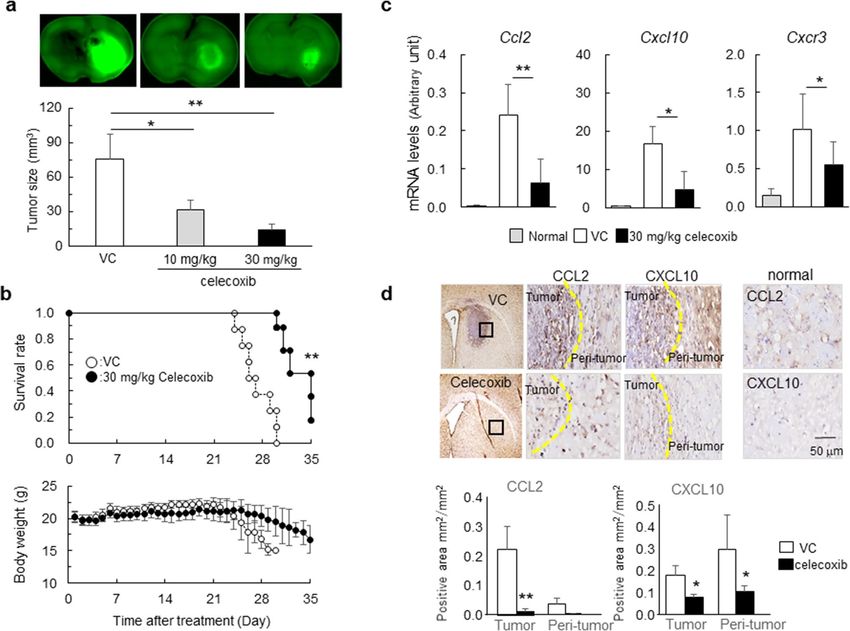
www.nature.com/scientificreports/
in the proliferation of glioma c ells8. Therefore, we focused on the paracrine-signaling networks involved in the
growth microenvironment and maintenance of GSCs related to antitumor effects.
Cyclooxygenase (COX)-2 levels increase in response to various stimuli, and COX-2 increases the malig-
nant potential of human glioma cells9,10. A new generation of nonsteroidal anti-inflammatory drugs (celecoxib)
selectively inhibit COX-2 activity, which exerts COX-2-dependent and -independent antitumor e ffects11. We
recently demonstrated that celecoxib treatment of low-grade glioma cells induces apoptosis and inhibits their
proliferation via the Akt/survivin and Akt/inhibitor of differentiation (Id)-3 pathways12. Additionally, Kurtova
et al.13 reported that blocking prostaglandin E2-induced tumor re-population abrogates bladder cancer chemore-
sistance. Furthermore, Fujita et al.14 demonstrated that COX-2 blockade suppresses gliomagenesis by inhibiting
myeloid-derived suppressor cells accompanied by chemokine regulation. Moreover, the nuclear factor-kappaB
(NF-κB)-dependent cytokine pathway is reportedly associated with mesenchymal g lioblastoma15.
Based on these findings, we hypothesized that in GSCs and a GSC-bearing malignant glioma model, modula-
tion of the CCL2/CCR2 and CXCL10/CXCR3 axes by celecoxib might contribute to antitumor effects. Therefore,
we examined the relationships between celecoxib-specific antitumor effects and regulation of chemokines in a
mouse GSC-bearing glioma model that closely resembled human G BM16. Furthermore, we studied the mecha-
nisms underlying regulation of the expression of chemokines and their receptors in GSCs. Here, we show that
suppression of the CCL2/CCR2 and CXCL10/CXCR3 axes by celecoxib in the tumor and the tumor microenvi-
ronment might contribute to antitumor effects.
Results
Celecoxib‑mediated induction of apoptosis and inhibition of cell proliferation in mouse
GSCs. We first examined the effects of celecoxib on apoptosis-related molecules and cell proliferation. In
GSCs exposed to celecoxib for 24 h, cell viability was inhibited in a dose-dependent manner, and the 50% inhibi-
tion concentration of celecoxib was 60 μM (Fig. 1a). Additionally, in GSCs treated with 60 μM celecoxib for 24 h,
the number and area of colony formation decreased (Fig. 1b). Furthermore, celecoxib increased levels of cleaved
caspase-8, -9, and -3 and poly (ADP-ribose) polymerase (PARP) compared with those in the vehicle control
(Fig. 1c). The increased PARP cleavage and the number of celecoxib-induced Annexin-V+ cells were attenuated
after the administration of 40 μM Boc-D-FMK, a pan-caspase inhibitor (Fig. 1d). These results indicated the
induction of both intrinsic and extrinsic caspase-dependent apoptosis by celecoxib.
The transcription factor β-catenin and c-Myc play an important role in stem cell renewal and organ
regeneration17. Additionally, a cell cycle regulator cyclin D1 is linked to cancer development and progression,
and acts as a transcription co-regulator18. Treatment with 60 μM celecoxib for 24 h reduced levels of β-catenin
and cyclin D1 but not of c-Myc (Fig. 1e), and lowered the number of 4′,6-amino-2-phenylindole dihydrochloride
(DAPI)+ nuclei and Ki-67+ cells as a cell-proliferation marker (Fig. 1f). Collectively, these findings suggest that
celecoxib might not only induce apoptosis but also inhibit GSC proliferation.
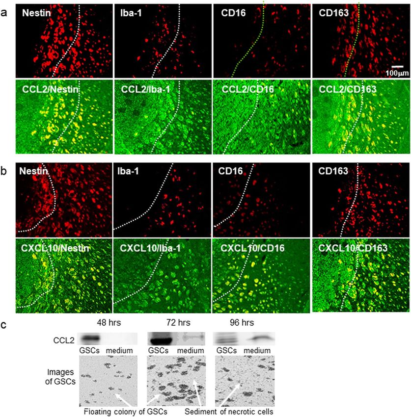
Chemokine regulation associated with antitumor effects by celecoxib in a mouse malignant
glioma model. To evaluate the antitumor effects of celecoxib, we administered celecoxib at 10 mg/kg/day
or 30 mg/kg/day for 14 days to GSC-bearing glioma models, and found that celecoxib suppressed the tumor
volume in a dose-dependent manner (Fig. 2a). Another set of mice treated with 30 mg/kg/day celecoxib or vehi-
cle followed by a 35-day observation period revealed that celecoxib significantly extended the survival period
without affecting body weight, whereas all mice in the vehicle control died at day ~ 28 accompanied by a decrease
in body weight (Fig. 2b).
To address the mechanisms underlying the antitumor effects of celecoxib, we focused on the tumor microen-
vironment and examined the levels of chemokines CCL2 and CXCL10 and their receptors, CCR2 and CXCR3,
respectively. Treatment with celecoxib notably decreased the mRNA levels of Ccl2 and Cxcl10 and Cxcr3 (Fig. 2c)
but not of Ccr2 (data not shown). Immunohistochemically, the expression of C CL2+ cells and C
XCL10+ cells in
tumor and peri-tumor were higher than in normal brain tissues, and celecoxib decreased the number of these
cells (Fig. 2d).
Localization of CCL2+ cells and CXCL10+ cells in a malignant glioma model. Annovazzi et al.19
reported that glioma-associated microglia macrophages (GAMs) assimilated to peripheral macrophages are
polarized and have two functional profiles: pro-inflammatory M1-type- and immunosuppressive, anti-inflam-
matory M2-type macrophages. The phenotype of GAMs overlaps only partially with M1 and M2 phenotypes.
To identify GAMs, 4 antibodies are currently used: Iba1, CD16, CD68, and C D16320. To analyze which cells
+ +
co-localized with CCL2 or CXL10 cells in the glioma model, we used the stem cell marker Nestin, a microglia
marker Iba-1, and the M1- and M2-type monocyte macrophage markers CD16 and CD163, respectively. As
shown in Fig. 3a,b, we found that in the tumor and the tumor microenvironment, CCL2 and CXCL10 localized
in Nestin+, Iba-1+, CD16+ and CD163+ cells.
Since CCL2 is secreted by necrotic and endothelial cells21, we analyzed CCL2 expression during culture for
48–96 h. (Fig. 3C). GSCs proliferated in a time-dependent manner. In the presence of dead cells, CCL2 expression
increased in the culture medium as well as in GSCs. The expression of CCL2 in the culture medium increased
in the presence of dead cells may suggest the secretion of CCL2 from necrotic GSCs.
Downregulation of CCL2 and CXCR3 via the NF‑κB pathway by celecoxib inhibits GSC viabil-
ity. We addressed the regulation of chemokines (CCL2 and CXCL10) and their receptors CCR2 and CXCR3)
in GSCs treated with 60 µM celecoxib for 24 h in GSCs. In GSCs, celecoxib revealed significant decrease in
mRNA levels of Ccl2 and Cxcr3 but not of Ccr2 and Cxcl10 (Fig. 4a), which was consistent with the high mRNA
Scientific Reports | (2020) 10:15286 | https://doi.org/10.1038/s41598-020-71857-3 2
Vol:.(1234567890)
www.nature.com/scientificreports/
Figure 1. Effects of celecoxib on GSC viability, colony formation, and apoptosis. (a) GSC viability evaluated at
24-h post-treatment with celecoxib using water-soluble tetrazolium (WST-8 assay) (n = 8). (b) Representative
images of GSCs 24-h post-treatment with 60 μM celecoxib (50% inhibition concentration; I C50) or vehicle
control (VC). The number of colony (≥ 2000 μm2) and the colony area (mm2) per mm2 were determined (n = 5).
(c) Representative western blot (WB) images of cleaved caspase (cCasp)-8, -9, and -3 and PARP levels in GSCs
treated with 60 μM celecoxib or VC for 6 h (n = 4). (d) Representative images of WB and Annexin-V (red)
staining in GSCs treated with 60 μM celecoxib for 12 h in the presence or absence of 40 μM of the pan-caspase
inhibitor Boc-D-FMK (n = 3). Scale bar, 50 µm. (e) Representative WB images of c-Myc, β-catenin, and cyclin
D1 levels in GSCs after treatment with 60 μM celecoxib or VC for 24 h (n = 4). (f) Ki-67 positive cells (Ki-67+,
red) detected by immunohistochemistry in GSCs. The number of Ki-67+ cells and DAPI+ (blue) nuclei was
calculated (n = 4). Data represent the mean ± SD. *p < 0.05, **p < 0.01 vs. VC by Student’s t test.
levels of Ccl2 in the glioma model and GSCs but none of Cxcl10 in GSCs (Fig. 4b). The protein expression of
pNFkB, Ccl2, and Cxcr3 was decreased (Fig. 4c) without affecting CCR2 and CXCL10 (data not shown). Then,
using caffeic acid phenethyl ester (CAPE), which is a potent NF-κB inhibitor that suppresses the translocation
of NF-κB p65 from the cytoplasm to the nucleus, we confirmed that the levels of pIκB, pNF-kB, CCL2, and
CXCR3 were significantly decreased in GSCs (Fig. 4d). The proteasomal degradation of IkBa is a key step in the
regulation of the NF-kB pathway and liberates the NF-kB proteins from IkBa, leading to their nuclear translo-
cation and increasing their interaction with NF-kB DNA-binding properties to result in the activation of gene
expression22. These results suggested NF-kB-dependent transcriptional regulation of CCL2 and CXCR3 levels
by celecoxib (Fig. 4d).
Finally, we assessed the functional relevance of CCL2 in GSCs. Ccl2 silencing by small-interfering (si)RNA
decreased CCL2 mRNA and protein levels (Fig. 5a,b) 24 h after transfection. Additionally, 48 h after transfection,
cyclin D1 levels decreased (Fig. 5c) accompanied by decreased GSC viability (Fig. 5d), suggesting that reduced
CCL2 level might be partly associated with GSC growth inhibition.
Discussion
This is the first study showing that the suppression of the CCL2/CCR2 and CXCL10/CXCR3 axes by celecoxib
may attribute to antitumor effects in a mouse malignant glioma model, in addition to intrinsic and extrinsic
apoptotic induction by celecoxib in GSCs (Fig. 5e). We newly found that in the mouse glioma model, celecoxib
decreased mRNA levels of Ccl2, CxcL10 and Cxcr3 but not of Ccr2 and protein expression of CCL2 and CXCL10
in the tumor and peri-tumor. Moreover, we demonstrated that in GSCs, celecoxib treatment decreased protein
and mRNA level of Ccl2 and Cxcr3 levels without affecting Ccr2 and CxcL10, which were associated with the
inhibition of NF-κB signaling, and that Ccl2 silencing by siRNA inhibited cell proliferation. These results showed
Scientific Reports | (2020) 10:15286 | https://doi.org/10.1038/s41598-020-71857-3 3
Vol.:(0123456789)www.nature.com/scientificreports/
Figure 2. Antitumor effects of celecoxib in the mouse glioma model. (a) GFP+ tumor areas (green) measured
in mice treated with 10 mg/kg/day or 30 mg/kg/day celecoxib or vehicle for 7 days (n = 6). *p < 0.05, **p < 0.01
vs. VC by Tukey–Kramer test. (b) Survival curves estimated with the Kaplan–Meier method and changes in
body weight for 35 days (n = 8). **p < 0.01 (log-rank test). (c) Ccl2, Cxcl10 and Cxcr3 mRNA levels determined
in mice treated with 30 mg/kg/day celecoxib or VC for 7 days and compared with the normal tissue obtained
from age-matched C57BL/6 J mice. *p < 0.05, **p < 0.01 vs. normal by Student’s t-test. (n = 6). (d) Representative
immunohistochemistry stained with DAB in mice treated with 30 mg/kg/day celecoxib or VC for 7 days.
Dashed line, tumor border. The positive areas of CCL2 and CXCL10 in tumor and peri-tumor were analyzed
by BZ-X710 microscope equipped analyzing system. Scale bar, 50 µm. Each column data indicates mean ± SD
(n = 6). *p < 0.05, **p < 0.01 by student’s t-test.
that celecoxib-induced the modulation of chemokine and chemokine-receptor levels in GSCs and the tumor
microenvironment may be at least partly attributable to its antitumor effects in a mouse malignant glioma model.
GBM comprises a heterogeneous cell population that includes tumor and immune cells, with interactions among
these different cell types and chemokines/cytokines promoting tumor development and p rogression20. CCL2 is
a major chemokine involved in attracting bone-marrow-derived mesenchymal cells to the tumor microenviron-
ment, and CCL2 loss decreases accumulation of these c ells21. In the present study, we identified CCL2 in GSCs,
the mouse glioma model, and in the culture medium in the presence of a large number of dead cells. Moreover,
CCL2 and CXCL10 was present in the tumor and the tumor microenvironment and co-localized with almost all
Nestin+, Iba-1+, CD163+ or CD16+ cells, suggesting the same distribution and localization. However, in GSCs,
the high protein and mRNA levels of Ccl2 and Cxcr3 were decreased by celecoxib, whereas the low protein and
mRNA level of Ccr2, Cxcl10 were not affected by celecoxib in our in vitro study. On the other hand, in the mouse
glioma model, the high protein and mRNA level of Ccl2, Cxcl10 and Cxcr3 were decreased by celecoxib. These
findings suggest that CCL2 and CXCL10 and their receptors may be generated from different cell types and
have distinct crucial role (Fig. 5e) in tumor growth. The downregulation of these chemokines and receptors by
celecoxib might contribute to antitumor effects in both tumor and peri-tumor areas.
Jung et al.23 reported that CCL2/MCP1 is secreted by necrotic glioma cells and endothelial cells in glioblas-
toma. We found high expression levels of CCL2 in GSCs and GSCs-derived glioma model. To demonstrate the
secretion of CCL2 by glioma cells, we extended the culture period as shown in Fig. 3c. In the presence of dead
cells, the GSCs proliferated in a time-dependent manner, and we observed the increase in CCL2 expression in
the culture medium as well as in GSCs. Takeshima et al.24 reported that the degree of macrophage infiltration
Scientific Reports | (2020) 10:15286 | https://doi.org/10.1038/s41598-020-71857-3 4
Vol:.(1234567890)www.nature.com/scientificreports/
Figure 3. Localization of CCL2+ and CXCL10+ cells in the mouse glioma model. (a) Representative
immunohistochemical images of CCL2+ cells (green) localized among Nestin+, Iba-1+. CD16+ or CD163+ cells
(red). (b) Representative immunohistochemical images of C XCL10+ cells (green) localized with Nestin+, Iba-1+.
+ +
CD16 or CD163 cells (red). (c) Representative image of western blot analysis of CCL2 in GSCs and the culture
medium. The experiment was repeated 3 times. The cell image was taken by BZ-X710 microscope. Each culture
medium was centrifuged using Microcon Centrifugal Filters for protein concentration.
found among the glioma cells was grossly correlated with the level of CCL2/MCP1 expression, suggesting that
CCL2/MCP1 produced by the glioma cells may mediate macrophage infiltration into glioma tissues. These
findings support our results and a potential therapeutic approach to reduce glioma growth through promoting
M1-polarized macrophages and inhibiting M2-phenotypic m acrophages25,26.
Continuous treatment with CCL2-neutralizing antibodies inhibits tumor progression in hepatocellular
carcinoma27. CCL2 binds the cognate receptor CCR2, and together this signaling pair has been shown to have
multiple pro-tumorigenic roles28. Because Ccl2 mRNA levels are not reduced by treatment with a CCL2 antibody,
the recovery of CCL2 level after cessation of CCL2 antibody treatment is suggested. In the present study, NF-κB
signaling mediated by celecoxib decreased mRNA and protein levels of CCL2, suggesting greater therapeutic
potential than therapy involving the CCL2 antibody. However, celecoxib treatment did not alter CCR2 in the
glioma model, we could not clarify the role of CCR2.
Although CXCL10 mediates the chemotaxis of tumor-promoting c ells29 and exerts dual effects depending
on the splicing variant of its receptors7, CXCL10 induces DNA synthesis and might promote proliferation of
anaplastic astrocytoma (grade III) and GBM8. Although CXCL10-specific effects against glioma associated with
celecoxib treatment might differ depending on the circumstances7, we observed that increased levels of CXCL10+
cells in the tumor and peri-tumor, and that CXCL10 levels decreased following treatment with celecoxib in
our glioma model. These findings in the glioma model are similar to a previous study showing that CXCL10 is
produced in bone-marrow-derived mesenchymal stem cells induced by h ypoxia30. Therefore, we were unable
to address its regulatory mechanisms in GSCs. In contrast, we observed reduced CXCR3 levels associated with
attenuated NFκB signaling in GSCs. Because increased CXCR3 levels are indicative of poor survival rates, and
targeted silencing of Cxcr3 inhibits the proliferation of hepatocellular carcinoma cells31, these findings support
Scientific Reports | (2020) 10:15286 | https://doi.org/10.1038/s41598-020-71857-3 5
Vol.:(0123456789)www.nature.com/scientificreports/
Figure 4. Effect of celecoxib treatment on Ccl2, Cxcl10, Ccr2, and Cxcr3 expression in mouse GSCs. (a) mRNA
levels of Ccl2, Cxcl10, Ccr2, and Cxcr3 determined in mouse GSCs treated with 60 µM celecoxib or vehicle
control (VC) for 24 h (n = 6). (b) Comparison of mRNA levels of Ccl2 and Cxcl10 in the mouse glioma model
and GSCs (n = 6). (c) Representative images of western blot analysis and the expression levels of phosphorylated
NF-κB (pNF-κB), CCL2 and CXCR3 in mouse GSCs treated with VC or celecoxib (n = 4). (d) Representative
images of western blot analysis and the expression levels of phosphorylated IκBα (pIκBα), pNF-κB, CCL2, and
CXCR3 in mouse GSCs treated with VC, and CAPE (n = 4). Data represent the mean ± SD. **p < 0.01 vs. VC by
Student’s t test.
the present results showing that celecoxib-specific inhibition of growth and/or proliferation might partially
contribute to suppression of the CXCL10/CXCR3 axis in GSCs.
The tumorigenic potential of increased COX-2 levels is linked to apoptosis resistance in some cell types,
and celecoxib-induced apoptosis involves the extrinsic/death receptor- and/or intrinsic/mitochondria-mediated
pathways32. In agreement with these findings, we found that celecoxib treatment increased caspase-8, -9, and -3
levels, as well as those of PARP levels in GSCs, indicating that apoptosis occurred via both pathways. Moreover,
β-catenin and cyclin D1 levels were decreased, and Ki-67 level were reduced by celecoxib treatment, which
agreed with a study of human glioma cell lines treated with c elecoxib33. Therefore, the antitumorigenic effects of
celecoxib might include not only induced apoptosis but also suppressed cell proliferation (Fig. 5e).
We initiated celecoxib treatment on day 1 following intracranial GSC injection, which is akin to celecoxib
administration after surgical resection of human gliomas. Our results suggest the possibility that glioma patients
having undergone tumor resection might benefit from celecoxib treatment to reduce the risk of GSC-driven
tumor growth.
There are some limitations to this study. First, our GSCs were established from mouse neural stem cells,
and this animal model exhibited pathological similarities to human GBM.16 with a more accurate reflection of
its features and malignant behavior. However, the glioma in the mouse model might differ from that of human
gliomas, which are heterogeneous, unlike the homogeneous GSCs derived from oncogene knock-in mice. Sec-
ond, we analyzed the effects of celecoxib treatment using only one cell line derived from mice and one animal
model. Further studies using GSCs derived from humans are required. Moreover, a number of distinct molecular
pathways might play a role in the antitumor effects of celecoxib in a COX-2-dependent and/or -independent
manner11. Because previous studies suggested alternative targets implicated in the tumor-inhibitory effects of
celecoxib, we cannot rule out other targets associated with its antitumor effects.
In conclusion, our findings suggest that downregulation of the CCL2/CCR2 and CXCL10/CXCR3 axes via
inhibition of the NF-κB pathway in the tumor and the tumor microenvironment might have contributed to the
antitumor effects of celecoxib observed in the mouse glioma model. These results warrant further investigation
to clarify the therapeutic benefits of celecoxib in humans.
Scientific Reports | (2020) 10:15286 | https://doi.org/10.1038/s41598-020-71857-3 6
Vol:.(1234567890)www.nature.com/scientificreports/
Figure 5. GSC viability following Ccl2 silencing. (a) Ccl2 mRNA levels in GSCs transfected with 10 nM Ccl2
siRNA (siCcl2) or negative control siRNA (siCon) for 24 h (n = 6). (b) Representative western blot results for
CCL2 (n = 4). (c) Representative western blot results for cyclin D1 from GSCs at 48-h post-transfection with
siCcl2 or siCon (n = 4). (d) Representative images of GSCs and cell viability evaluated by WST-8 assay at 48-h
post-transfection with siCcl2 or siCon (n = 4). Scale bar, 50 µm. (e) Schematic illustration of the antitumor
effects of celecoxib in the mouse glioma model. Data represent the mean ± SD. *p < 0.05, **p < 0.01 vs. siCon by
Student’s t test.
Materials and methods
All experiments and protocols were approved by the ethics committee of the Graduate School of Biomedical
Sciences, Tokushima University (Tokushima, Japan).
Cell line. GSCs were established by Saya et al.16 and comprised H-RasV12 overexpressed in Ink4a/Arf-null
neural stem cells and the progenies of GSCs were maintained as a sphere culture in Dulbecco’s modified Eagle
medium F-12 (Sigma-Aldrich, St. Louis, MO, USA) supplemented with 20 ng/mL recombinant human epider-
mal growth factor (PeproTech, Rocky Hill, NJ, USA), 20 ng/mL recombinant human basic fibroblast growth
factor (PeproTech), B-27 supplement without vitamin A (Life Technologies, Carlsbad, CA, USA), 200 ng/mL
heparin sulfate, 100 U/mL penicillin, and 100 μg/mL streptomycin (Nacalai Tesque, Kyoto, Japan) at 37 °C in an
atmosphere of 5% CO2/95% humidified air.
Cell viability and colony formation assays. GSCs were seeded in 96-well culture plates (1 × 10⁴ cells/
well). Celecoxib was purchased from Sigma-Aldrich (Tokyo, Japan) which was dissolved in dimethyl sulfoxide
(DMSO) and diluted to 0.1% with culture medium for use as the vehicle control. The number of viable cells was
determined using a Cell Counting Kit-8 (Dojindo Laboratories, Kumamoto, Japan) and measured on an Infinite
F200 Pro microplate reader (Tecan, Mannedorf, Switzerland) at 450 nm. Cell viability following celecoxib treat-
ment was calculated as the percentage of the vehicle control. For the colony-formation assay, GSCs were seeded
in 96-well plates (5 × 10³ cells/well) and treated with celecoxib or vehicle for 24 h. The number of colonies with
size > 2000 μm2 and the area of colony were analyzed using a BZ-X710 microscope (Keyence, Osaka, Japan)
equipped image analysis system.
Animal experiments. Experiments were performed according to the National Institutes of Health Guide
for the Care and Use of Laboratory Animals. The GSC-bearing glioma model, which resemble human malignant
glioma, was previously established by Saya et al.16 and used according to their protocol.
Male C57BL/6 J mice (6-weeks old) were obtained from CLEA Japan (Tokyo, Japan). Animals were anesthe-
tized and placed in a stereotaxic apparatus, and a small hole was bored into the skull of each mouse (2.0-mm
lateral to bregma) using a dental drill. Viable cells (1 × 10³) resuspended in 2 μl of Hank’s balanced salt solution
were injected into the right hemisphere 3-mm below the brain surface using a 10-μL Hamilton syringe with an
unbeveled 30-gauge needle. The injection was performed over 5 min, and the needle was left in the brain for an
Scientific Reports | (2020) 10:15286 | https://doi.org/10.1038/s41598-020-71857-3 7
Vol.:(0123456789)www.nature.com/scientificreports/
additional 5 min before it was gradually withdrawn and the hole sealed. Celecoxib was dissolved in DMSO and
diluted four-fold with sterile water immediately before injection. Celecoxib doses were established based on our
previous study12. Treatment with 10 mg/kg or 30 mg/kg celecoxib or vehicle (control) by intraperitoneal injec-
tion was initiated on the first day of GSC injection. To determine tumor size, fixed brain tissue was sliced on a
brain-slicer matrix at 1.0-mm intervals. Based on the tumor area, represented by the green fluorescent protein
(GFP)+ area in the slice, the tumor volume was calculated using a microscope equipped with an image analysis
system (BZ-X710; Keyence).
Western blot analysis. Protein samples were separated by sodium dodecyl sulfate–polyacrylamide gel
electrophoresis and transferred to a polyvinylidene difluoride membrane (Bio-Rad, Hercules, CA, USA) that
was incubated in blocking solution. Membranes were incubated with primary antibodies (Supplementary
Table 1), followed by incubation with horseradish peroxidase-conjugated secondary antibody. Immunoreactiv-
ity was detected using enhanced chemiluminescence prime western blot detection reagent and visualized with
an Image Quant LAS4000 mini (GE Healthcare, Little Chalfont, UK). Band intensities were quantified using
ImageJ software (v.1.49; National Institutes of Health, Bethesda, MD, USA) and normalized to β-actin. To detect
extracellular CCL2 in the culture medium, we collected the medium containing proliferated GSCs at 48, 72, and
96 h after cell culture. The medium was fractionated according to molecular weight using Microcon centrifugal
filters (Merk, Tokyo, Japan) for protein concentration. An inhibitor of NF-κB (Caffeic acid phenethyl ester;
CAPE; R&D System, Minneapolis, MN, USA) was dissolved in DMSO.
Quantitative real‑time polymerase chain reaction (qRT‑PCR). Total RNA was purified using a
MagNA Pure compact RNA isolation kit (Roche Diagnostics, Burgess Hill, UK) according to manufacturer
instructions. cDNA was synthesized using Transcriptor Universal cDNA master mix (Roche Diagnostics), and
qRT-PCR was performed using FastStart DNA M asterPLUS SYBR Green I (Roche Diagnostics) on a Light Cycler
2.0 instrument (Roche Diagnostics) under conditions recommended by the manufacturer. The forward and
reverse primer sequences were as follows: mouse Ccl2, 5′-AGCAAGATGATCCCAATGAGT-3′ and 5′-GAGCTT
GGTGACAAAAACTACAG-3′; mouse Cxcl10, 5′-CCCACGTGTTGAGATCATTG-3′ and 5′-CAGTTAAGG
AGCCCTTTTAGACC-3′; mouse Ccr2, 5′-CAAAATGGATGCCTTAGCACTG-3′ and 5′-CCAGGTTTTCAT
GTTTGGCTATTC-3′; mouse Cxcr3, 5′-CCAAGCCATGTACCTTGAGGTTAG-3′ and 5′-GCTCTCGTTTTC
CCCATAATCGTAG-3′; mouse glyceraldehyde-3-phosphate dehydrogenase (Gapdh), 5′-CAGAACATCATC
CCTGCATC-3′ and 5′-CTGCTTCACCACCTTCTTGA-3′. mRNA levels were normalized to that of Gapdh.
Small‑interfering RNA (siRNA) transfection. Transfections were conducted using Lipofectamine
RNAiMAX transfection reagent (Life Technologies) according to manufacturer instructions. Briefly, siRNA and
transfection reagent were diluted in OPTI-MEM medium (Life Technologies), and transfection reagent/siRNA
complexes were allowed to form for 20 min. The complexes were added to each well, and GSCs were seeded
into 6-well culture plates (2 × 10⁵ cells/well) or 96-well culture plates (4 × 10³ cells/well) and incubated for 24 h
or 48 h. The target sequence for mouse Ccl2 siRNA (QIAGEN, Valencia, CA, USA) was as follows: 5′-ACCCGT
AAATCTGAAGCTAAT-3′. Non-targeting siRNA was used as a negative control.
Annexin‑V assay. Apoptotic cells were labeled with the Annexin-V-PE assay kit (BioVision, Milpitas, CA,
USA) according to manufacturer instructions. Briefly, cells were treated with 0.2% DMSO or 60 μM celecoxib
and the pan-caspase inhibitor boc-aspartyl-(OMe)-fluoromethyl-ketone (Boc-D-FMK) (40 μM; Abcam, Cam-
bridge, UK) to confirm caspase-dependent apoptosis.
Immunocytochemistry and immunohistochemistry. GSCs were collected and smeared using Smear
Gel (GenoStaff, Tokyo, Japan) according to manufacturer instructions and then fixed with 4% paraformaldehyde
(PFA) in phosphate-buffered saline (PBS). After permeabilization with 0.2% Triton X-100 in PBS, cells were
labeled with antibody of Ki-67 as a cell proliferation marker (Abcam), the antigen was detected with Alexa Fluor
594-conjugated donkey antirabbit IgG (Life Technologies), and nuclei were counterstained with 4′,6-diamino-
2-phenylindole dihydrochloride (DAPI) solution (Dojindo Laboratories).
Tissue samples were fixed with 4% PFA in PBS, and 10-µm-thick frozen sections were mounted on glass slides.
For immunoperoxidase staining, sections were blocked and then incubated overnight with primary antibodies.
Antibodies were visualized using a VECTASTAIN Elite ABC kit (Vector Laboratories, Burlingame, CA, USA),
H2O2/diaminobenzidine (Merck Millipore, Billerica, MA, USA), and Mayer’s hematoxylin solution according
to manufacturer instructions. For double-immunofluorescence staining, sections were immunoreacted with an
antibody for nestin-, IBA-1-, CD163 and CD16 positive cells followed by incubation with anti-CCL2 and anti-
CXCL10 antibodies. To visualize each marker, sections were incubated with green fluorescence and chemokines
with red fluorescence, and sections were incubated with an Alexa 488- or Alexa 594-conjugated secondary anti-
body (Life Technologies), respectively. Nuclei were counterstained with DAPI solution (Dojindo Laboratories).
The detailed information for each antibody is provided in Supplementary Table 2. All images were obtained
using BZ-X710 microscope (Keyence).
Statistical analysis. Data are presented as the mean ± standard deviation (SD) and were analyzed with
a Student’s t test for two-group comparisons and analysis of variance, followed by the Tukey–Kramer test for
multiple comparisons. A survival curve was estimated by the Kaplan–Meier method, and statistical significance
was tested with the log-rank test. Differences between groups were considered statistically significant at p < 0.05.
Scientific Reports | (2020) 10:15286 | https://doi.org/10.1038/s41598-020-71857-3 8
Vol:.(1234567890)www.nature.com/scientificreports/
Received: 20 October 2019; Accepted: 17 August 2020
References
1. Stupp, R. et al. European Organisation for Research and Treatment of Cancer Brain Tumor and Radiotherapy Groups; National
Cancer Institute of Canada Clinical Trials Group. Radiotherapy plus concomitant and adjuvant temozolomide for glioblastoma.
N. Engl. J. Med. 352, 987–996 (2009).
2. Wei, Z., Kale, S., El Fatimy, R., Rabinovsky, R. & Krichevsky, A. M. Co-cultures of glioma stem cells and primary neurons, astrocytes,
microglia, and endothelial cells for investigation of intercellular communication in the brain. Front. Neurosci. 13, 361 (2019).
3. Thomas, T. M. & Yu, J. S. Metabolic regulation of glioma stem-like cells in the tumor micro-environment. Cancer Lett. 408, 174–181
(2017).
4. Chang, A. L. et al. CCL2 produced by the glioma microenvironment is essential for the recruitment of regulatory T cells and
myeloid-derived suppressor cells. Cancer Res. 76, 5671–5682 (2016).
5. Huang, B. et al. CCL2/CCR2 pathway mediates recruitment of myeloid suppressor cells to cancers. Cancer Lett. 252, 86–92 (2007).
6. Honeth, G., Staflin, K., Kalliomaki, S., Lindvall, M. & Kjellman, C. Chemokine-directed migration of tumor-inhibitory neural
progenitor cells towards an intracranially growing glioma. Exp. Cell. Res. 312, 1265–1276 (2006).
7. Urra, S. et al. Differential expression profile of CXCR3 splicing variants is associated with thyroid neoplasia. Potential role in
papillary thyroid carcinoma oncogenesis? Oncotarget. 9, 2445–2467 (2018).
8. Maru, S. V. et al. Chemokine production and chemokine receptor expression by human glioma cells: role of CXCL10 in tumour
cell proliferation. J. Neuroimmunol. 199, 35–45 (2008).
9. Shono, T., Tofilon, P. J., Bruner, J. M., Owolabi, O. & Lang, F. F. Cyclooxygenase-2 expression in human gliomas: prognostic sig-
nificance and molecular correlations. Cancer Res. 61, 4375–4381 (2001).
10. Xu, K., Wang, L. & Shu, H. K. COX-2 overexpression increases malignant potential of human glioma cells through Id1. Oncotarget.
5, 1241–1252 (2014).
11. Grosch, S., Maier, T. J., Schiffmann, S. & Geisslinger, G. Cyclooxygenase-2 (COX-2)-independent anticarcinogenic effects of selec-
tive COX-2 inhibitors. J. Natl. Cancer. Inst. 98, 736–747 (2006).
12. Sato, A. et al. Blocking COX-2 induces apoptosis and inhibits cell proliferation via the Akt/survivin- and Akt/ID3 pathway in
low-grade-glioma. J. Neurooncol. 132, 231–238 (2017).
13. Kurtova, A. V. et al. Chan KS Blocking PGE2-induced tumour repopulation abrogates bladder cancer chemoresistance. Nature
517, 209–213 (2015).
14. Fujita, M. et al. COX-2 blockade suppresses gliomagenesis by inhibiting myeloid-derived suppressor cells. Cancer Res. 71, 2664–
2674 (2011).
15. Zanotto-Filho, A. et al. Inflammatory landscape of human brain tumors reveals an NFkappaB dependent cytokine pathway associ-
ated with mesenchymal glioblastoma. Cancer Lett. 390, 176–187 (2017).
16. Sampetrean, O. et al. Invasion precedes tumor mass formation in a malignant brain tumor model of genetically modified neural
stem cells. Neoplasia. 13, 784–791 (2011).
17. Zhang, K. et al. WNT/β-Catenin directs self-renewal symmetric cell division of h TERThigh prostate cancer stem cells. Cancer Res.
77, 2534–3254 (2017).
18. Jagadeesh, S. & Banerjee, P. P. Telomerase reverse transcriptase regulates the expression of a key cell cycle regulator, cyclin D1.
Biochem. Biophys. Res. Commun. 347, 774–780 (2006).
19. Annovazzi, L. et al. Microglia immunophenotyping in gliomas. Oncol. Lett. 15, 998–1006 (2018).
20. Jordan, J. T. et al. Preferential migration of regulatory T cells mediated by glioma-secreted chemokines can be blocked with
chemotherapy. Cancer Immunol. Immunother. 57, 123–131 (2008).
21. Bonapace, L. et al. Cessation of CCL2 inhibition accelerates breast cancer metastasis by promoting angiogenesis. Nature 515,
130–133 (2014).
22. Ushio, A. & Eto, K. RBM3 expression is upregulated by NF-κB p65 activity, protecting cells from apoptosis, during mild hypother-
mia. J. Cell. Biochem. 119, 5734–5749 (2018).
23. Jung, Y. et al. MCP-1 and MIP-3α secreted from necrotic cell-treated glioblastoma cells promote migration/infiltration of microglia.
Cell Physiol. Biochem. 48, 1332–1346 (2018).
24. Takeshima, H., Kuratsu, J., Takeya, M., Yoshimura, T. & Ushio, Y. Expression and localization of messenger RNA and protein for
monocyte chemoattractant protein-1 in human malignant glioma. J. Neurosurg. 80, 1056–1062 (1994).
25. Xue, N. et al. Chlorogenic acid inhibits glioblastoma growth through repolarizating macrophage from M2 to M1 phenotype. Sci
Rep. 7, 39011. https://doi.org/10.1038/srep39011 (2017).
26. Szulzewsky, F. et al. Glioma-associated microglia/macrophages display an expression profile different from M1 and M2 polarization
and highly express Gpnmb and Spp1. PLoS ONE 10, e0116644 (2015).
27. Teng, K. Y. et al. Blocking the CCL2-CCR2 axis using CCL2-neutralizing antibody is an effective therapy for hepatocellular cancer
in a mouse model. Mol. Cancer Ther. 16, 312–322 (2017).
28. Lim, S. Y., Yuzhalin, A. E., Gordon-Weeks, A. N. & Muschel, R. J. Targeting the CCL2-CCR2 signaling axis in cancer metastasis.
Oncotarget. 7, 28697–28710 (2016).
29. Liu, M., Guo, S. & Stiles JK. The emerging role of CXCL10 in cancer (Review). Oncol. Lett. 2, 583–589 (2011).
30. Xia, J. B. et al. Hypoxia/ischemia promotes CXCL10 expression in cardiac microvascular endothelial cells by NFkB activation.
Cytokine 81, 63–70 (2016).
31. Ren, Y., Kan, Y. Z. & Kong, L. F. Study on the effects of target-silencing CXCR3 expression on malignant proliferation of hepatocel-
lular carcinoma. Zhonghua Gan Zang Bing Za Zhi. 26, 508–512 (2018).
32. Kern, M. A. et al. Cyclooxygenase-2 inhibition induces apoptosis signaling via death receptors and mitochondria in hepatocellular
carcinoma. Cancer Res. 66, 7059–7066 (2006).
33. Sareddy, G. R., Kesanakurti, D., Kirti, P. B. & Babu, P. P. Nonsteroidal anti-inflammatory drugs diclofenac and celecoxib attenuates
Wnt/beta-catenin/Tcf signaling pathway in human glioblastoma cells. Neurochem. Res. 38, 2313–2322 (2013).
Acknowledgements
We thank Emiko Nishikawa for excellent technical assistance. This work was supported by a Grant-in-Aid for
Scientific Research [Japan Society for the Promotion of Science (JSPS) KAKENHI, Grant number JP25462264]
and a Grant-in-Aid for Young Scientists (B) (JSPS KAKENHI Grant number JP26861153).
Scientific Reports | (2020) 10:15286 | https://doi.org/10.1038/s41598-020-71857-3 9
Vol.:(0123456789)www.nature.com/scientificreports/
Author contributions
K.S. and I.Y. performed the in vivo and in vitro experiments, and H.K. revised immunohistochemistry; A.S., T.F.,
K.N. and K.M. performed the in vitro experiments; H.S. provided transgenic cell; Y.M. and K.T.K. contributed
at the revision stage of the paper; Y.T. designed and organized the work; all of the authors contributed to the
final version of the paper.
Competing interests
The authors declare no competing interests.
Additional information
Supplementary information is available for this paper at https://doi.org/10.1038/s41598-020-71857-3.
Correspondence and requests for materials should be addressed to Y.M.
Reprints and permissions information is available at www.nature.com/reprints.
Publisher’s note Springer Nature remains neutral with regard to jurisdictional claims in published maps and
institutional affiliations.
Open Access This article is licensed under a Creative Commons Attribution 4.0 International
License, which permits use, sharing, adaptation, distribution and reproduction in any medium or
format, as long as you give appropriate credit to the original author(s) and the source, provide a link to the
Creative Commons licence, and indicate if changes were made. The images or other third party material in this
article are included in the article’s Creative Commons licence, unless indicated otherwise in a credit line to the
material. If material is not included in the article’s Creative Commons licence and your intended use is not
permitted by statutory regulation or exceeds the permitted use, you will need to obtain permission directly from
the copyright holder. To view a copy of this licence, visit http://creativecommons.org/licenses/by/4.0/.
© The Author(s) 2020
Scientific Reports | (2020) 10:15286 | https://doi.org/10.1038/s41598-020-71857-3 10
Vol:.(1234567890)You can also read