Clinical characteristics and related risk factors of disease severity in 101 COVID-19 patients hospitalized in Wuhan, China - Nature
←
→
Page content transcription
If your browser does not render page correctly, please read the page content below
www.nature.com/aps

                  ARTICLE
                  Clinical characteristics and related risk factors of
                  disease severity in 101 COVID-19 patients hospitalized
                  in Wuhan, China
                  Xue-qing Liu1,2, Shan Xue1,2, Jia-bo Xu1,2, Heng Ge2,3, Qing Mao2,4, Xin-hui Xu2,5 and Han-dong Jiang1,2

                      Coronavirus disease 2019 (COVID-19) broke out in December 2019. Due its high morbility and mortality, it is necessary to
                      summarize the clinical characteristics of COVID-19 patients to provide more theoretical basis for future treatment. In the current
                      study, we conducted a retrospective analysis of the clinical characteristics of COVID-19 patients and explored the risk factors for the
                      severity of illness. A total of 101 COVID-19 patients hospitalized in Leishenshan Hospital (Wuhan, China) was classified into three
                      sub-types: moderate (n = 47), severe (n = 36), and critical (n = 18); their clinical data were collected from the Electronic Medical
                      Record. We showed that among the 101 COVID-19 patients, the median age was 62 years (IQR 51–74); 50 (49.5%) patients were
                      accompanied by hypertension, while 25 (24.8%) and 22 (21.8%) patients suffered from diabetes and heart diseases, respectively,
                      with complications. All patients were from Wuhan who had a definite history of exposure to the epidemic area. Multivariate logistic
                      regression analysis revealed that older age, diabetes, chronic liver disease, percentage of neutrophils (N%) > 75%, CRP > 4 mg/L,
1234567890();,:

                      D-dimer > 0.55 mg/L, IL-2R > 710 U/mL, IL-8 > 62 pg/mL, and IL-10 > 9.1 pg/mL were independent variables associated with severe
                      COVID-19. In conclusion, we have identified the independent risk factors for the severity of COVID-19 pneumonia, including older
                      age, diabetes, chronic liver disease, higher levels of N%, CRP, D-dimer, IL-2R, IL-8, and IL-10, providing evidence for more accurate
                      risk prediction.

                      Keywords: coronavirus disease 2019 (COVID-19); severe acute respiratory syndrome coronavirus 2 (SARS-CoV-2); clinical features;
                      risk factors; disease severity; retrospective analysis

                      Acta Pharmacologica Sinica (2021) 0:1–12; https://doi.org/10.1038/s41401-021-00627-2

                  INTRODUCTION                                                                             COVID-19 patients to provide a theoretical basis for future
                  As a highly contagious disease, coronavirus disease 2019 (COVID-                         research and assistance in COVID-19 prevention and control. In
                  19)-induced pneumonia was first reported in Wuhan, Hubei                                  this study, we retrospectively collected and analyzed the detailed
                  Province, China, in December 2019 and then broke out in other                            clinical data of COVID-19 patients with varying disease severity
                  provinces of China and overseas [1]. The World Health Organiza-                          from a hospital in Wuhan. The clinical characteristics of patients
                  tion declared ongoing COVID-19 a public health emergency of                              with COVID-19 pneumonia were described, and risk factors for the
                  international concern on January 30, 2020 [2]. Under strict                              severity of COVID-19 were explored.
                  prevention and control strategies, new cases of COVID-19 in
                  China have been rapidly reduced, and the epidemic has been
                  effectively managed. However, the epidemic has constituted a                             METHODS
                  considerable challenge worldwide, and the global situation                               Study design, setting, and participants
                  remains grim. As of June 29, 2020, there were over 10 million                            In this retrospective cohort study, 101 case subjects (≥18 years
                  COVID-19 patients and more than 500,000 deaths worldwide. The                            old) with COVID-19 infection who were discharged from or died at
                  numbers are still growing, greatly exceeding the numbers of cases                        Leishenshan Hospital (Wuhan, China) between February 23, 2020,
                  and deaths caused by SARS or the Middle East respiratory                                 and April 4, 2020, were included. According to the seventh edition
                  syndrome (MERS) outbreaks in 2003 and 2013, respectively.                                of the COVID-19 diagnosis and treatment guide of the National
                  However, the source of COVID-19 has not been identified, and                              Health Commission of China, the above patients were divided into
                  there are currently no specific antiviral treatments. Therefore, it is                    three groups: moderate (mild and common, n = 47), severe (n =
                  necessary to summarize and analyze the clinical characteristics of                       36), and critical (n = 18). The study was approved by the Research

                  1
                   Department of Respiratory and Critical Care Medicine, Ren Ji Hospital, School of Medicine, Shanghai Jiao Tong University, Shanghai 200001, China; 2Department of Intensive
                  Infection, Leishenshan Hospital, Wuhan 430212, China; 3Department of Cardiology, Ren Ji Hospital, School of Medicine, Shanghai Jiao Tong University, Shanghai 200001, China;
                  4
                   Department of Neurosurgery, Ren Ji Hospital, School of Medicine, Shanghai Jiao Tong University, Shanghai 200001, China and 5Department of Emergency, Ren Ji Hospital,
                  School of Medicine, Shanghai Jiao Tong University, Shanghai 200001, China
                  Correspondence: Heng Ge (dr.geheng@foxmail.com) or Qing Mao (neurojack@163.com) or Xin-hui Xu (xinhui_72@hotmail.com) or Han-dong Jiang (jianghd@163.com)
                  These authors contributed equally: Xue-qing Liu, Shan Xue, Jia-bo Xu.

                  Received: 3 August 2020 Accepted: 10 February 2021

                  © The Author(s), under exclusive licence to CPS and SIMM 2021
Characteristics of 101 hospitalized COVID-19 patients in Wuhan
XQ Liu et al.
2
Ethics Commission of the hospital, and the requirement for history among the three groups. Nearly half of the patients (50,
informed consent was given by the Ethics Commission. 49.5%) had hypertension, which was also the most common
primary comorbidity, followed by diabetes (25, 24.8%) and heart
Data collection disease (22, 21.8%). The differences in the above three complica-
Epidemiological, clinical, laboratory, radiography, treatment, and tions were statistically significant among the three groups.
outcome data were collected from electronic medical records. All Additionally, clinical complications, such as chronic renal disease
data were extracted and examined by three clinicians and cerebrovascular diseases, were significantly different among
independently. the groups. The difference in pulse and differential pressure
among the three groups was statistically significant, and the
Statistical analysis differential pressure in critically ill patients was higher than that in
For continuous variables, the mean (SD) and median (IQR) were moderate patients (P < 0.05). In terms of clinical presentation,
used for normally and abnormally distributed data. Categorical fever (65, 64.4%) and cough (59, 58.4%) were the most common
variables were expressed as counts (%). For laboratory tests, it was clinical features in these patients, while chest distress, dyspnea,
determined whether the measurement results were outside the fatigue, diarrhea and myalgia were also found. Symptoms such as
normal range. runny nose and sore throat appeared less frequently and were
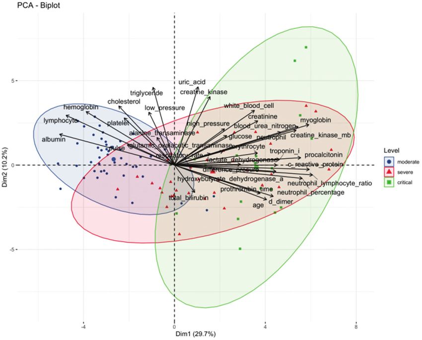
Principal component analysis (PCA) was carried out to extract therefore classified as other symptoms. Based on the SOFA score
the features, and dimension reduction was completed without and CURB-65 score at admission, critically ill patients were in a
losing important information on the variables. Based on the data more critical condition than patients with milder illness.
integrated by PCA, cluster analysis was performed to determine
the COVID-19 subgroups. Laboratory markers
In this article, the chi-square test or Fisher’s test was used to test Key laboratory indicators for 101 patients on admission are
an association between categorical variables, while ANOVA was summarized in detail (Table 2). In terms of routine blood tests,
used for continuous variables. We have presented our method in there were significant differences in the counts of white blood
the table and figure legends to ensure that the readers can cells (WBCs), lymphocytes (L), and neutrophils (N); the
understand what statistical approaches we adopted for the given neutrophil–lymphocyte ratio (NLR); the percentage of neutrophils
calculation. To identify the risk factors for the severity of COVID-19 (N%); the number of platelets (PLTs); and the hemoglobin (HB)
illness in 101 patients, we performed univariate ordinal logistic level among the three groups. Among them, the L count (0.74
regression by including 52 key characteristics as independent [0.36]), PLT count (125.00 [85.00, 194.00]) and HB level (97.50
variables in the calculation, followed by fitting a multivariate [87.50, 106.75]) were lowest in critical patients, while the values for
ordinal logistic regression model with the significant variables WBC (9.32 [6.21, 11.87]), N (8.18 [4.71]), NLR (12.33 [6.96]), and N%
selected from univariate ordinal logistic regression. Before fitting (82.70 [8.94]) were relatively high. Concerning the infection-
the ordinal logistic regression model, continuous variables of the related parameters, the levels of inflammatory markers such as C-
indicators were converted into categorical variables according to reactive protein (CRP, 68.73 [44.82]) and the erythrocyte sedi-
their reference values. The univariate and multivariate cumulative mentation rate (ESR, 48.22 [37.95]) were dramatically elevated in
logit models were fitted, with the levels of indicators in the severely and critically ill patients and were positively correlated
moderate group as the reference. First, univariate ordinal logistic with the severity of pneumonia. In addition, the critical patients
regression was used to investigate the potential predictors of had higher levels of inflammatory cytokines, including interleukin
severity. To exclude the insignificant variables in the univariate (IL)-6 (556.44 [1390.84]), IL-2R (1410.06 [1490.60]), IL-8 (43.39
cumulative logit model, we further carried out a backward, [73.60]), IL-10 (26.99 [33.9]) and tumor necrosis factor-alpha (TNF-
stepwise, multivariate, ordered, and logistic regression analysis. α, 17.70 [21.51]), than moderately severe patients. Additionally,
The odds ratio of the 95% confidence interval (CI) was estimated there were also significant differences in PT among the three
by the ordinal logistic regression model. A two-sided P value < groups (P < 0.001), while the D-dimer level showed no difference
0.05 was considered statistically significant. The R.3.6.3 and R among the groups. Moreover, the levels of indicators reflecting
packages (gtsummary, MASS, FactoMineR, tableone) were used for cardiac function, such as creatine kinase-MB (CK-MB, 4.19 [4.63]),
data analysis and statistics [3]. myoglobin (Mb, 402.45 [928.54]), troponin I (TNI, 0.04 [0.04]),
lactate dehydrogenase (LDH, 335.19 [170.24]), hydroxybutyrate
dehydrogenase-α (HBDH, 254.94 [119.18]), and brain natriuretic
RESULTS peptide (BNP, 230.47 [398.42]), were significantly increased in
Demographics and clinical characteristics patients with critical illness (P < 0.05). Moreover, the indicators of
A total of 101 adult patients with COVID-19 infection who were hepatorenal function, including glutamic oxaloacetic transaminase
hospitalized at a Wuhan hospital between February 23, 2020, and (GOT, 63.83 [97.64]), amylase (31.64 [36.75]), blood urea nitrogen
April 4, 2020, were included in this study. On the basis of the (BUN, 17.18 [15.23]) and creatinine (CR, 17.18 [15.23]) levels, were
seventh edition of the COVID-19 diagnosis and treatment guide of higher in critical patients (P < 0.05), while the levels of albumin
the National Health Commission in China, the above patients were (ALB, 30.37 [4.94]) were lower. In addition, patients with severe
divided into three groups: moderate (mild and common, n = 47), and critically ill conditions had higher fasting blood glucose levels
severe (n = 36), and critical (n = 18). Among these patients, 14 (6.42 [4.65]; 8.38 [6.54], respectively), which also corresponded to a
patients died during hospitalization, and 87 were discharged or higher rate of history of diabetes in the severe and critical patients.
transferred to a general hospital for other treatments. As shown in Otherwise, the levels of serum potassium (K), sodium (Na), and
Table 1, the median age of the 101 patients was 62 years (IQR chlorine (Cl) did not differ significantly among the three groups.
51–74). The severe or critical patients were older (69.5 years, (IQR To explore the similarities and differences among patients in
62–80.5), and 72.5 years, (IQR 62.25–79.75) than the moderate the moderate, severe and critical groups, PCA was performed
patients (52 years, (IQR 40.5–62)). There were more male (56, based on a set of clinical factors. PCA is an unsupervised learning
55.4%) than female (45, 44.6%) patients. All patients were from method and enables us to identify subgroups of patients sharing
Wuhan and had a definite history of exposure to the epidemic similar characteristics. As shown in the present study, the biplot
area, of whom 55 patients (54.4%) had been in close contact with from our PCA not only captured the patients with the same clinical
patients diagnosed with COVID-19, 7 patients (6.9%) had a cluster symptoms (i.e., severity), but also showed the variables that were
onset, and 39 patients (38.6%) had no clear history of direct positively or negatively associated with the different severity
contact. There was no difference in exposure history or smoking categories. The contribution of each variable to the given
Acta Pharmacologica Sinica (2021) 0:1 – 12Characteristics of 101 hospitalized COVID-19 patients in Wuhan
XQ Liu et al.
3
Table 1. Demographics and clinical characteristics of 101 patients.
Demographics and clinical characteristics Total (n = 101) Moderate (n = 47) Severe (n = 36) Critical (n = 18) P value
Age (median [IQR]) 62.00 [51.00, 74.00] 52.00 [40.50, 62.00] 69.50 [62.00, 80.25] 72.50 [62.25, 79.75]Characteristics of 101 hospitalized COVID-19 patients in Wuhan
XQ Liu et al.
4
Table 2. Laboratory data of 101 patients.
Laboratory data and clinical features Total (n = 101) Moderate (n = 47) Severe (n = 36) Critical (n = 18) P value
Blood routine
WBC (*109/L, median [IQR]) 6.34 [4.93, 7.79] 5.68 [4.61, 6.98] 6.82 [5.16, 9.11] 9.32 [6.21, 11.87] 0.002
L (*109/L, mean (SD)) 1.28 (0.65) 1.69 (0.56) 1.02 (0.53) 0.74 (0.36)Characteristics of 101 hospitalized COVID-19 patients in Wuhan
XQ Liu et al.
5
Fig. 1 PCA of the laboratory markers in COVID-19 patients with different disease severity. This section shows the correlation degree of
continuous variables and the influence degree of each continuous variable on PCs.
Table 3. Image characteristics of 101 patients.
Imaging features Total (n = 101) Moderate (n = 47) Severe (n = 36) Critical (n = 18) P value
Ground glass opacification (GGO) 92 (91.1%) 41 (87.2%) 34 (97.1%) 17 (94.4%) 0.24
Bilateral involvement 92 (91.1%) 44 (93.6%) 32 (91.4%) 16 (88.9%) 0.811
Consolidation 74 (73.3%) 31 (66.0%) 29 (82.9%) 14 (77.8%) 0.208
Subpleural distribution 53 (52.5%) 28 (59.6%) 17 (48.6%) 8 (44.4%) 0.445
Fibrosis 41 (40.6%) 13 (27.7%) 20 (57.1%) 8 (47.1%) 0.024
Fig. 2 Representative CT image of the COVID-19 patients. The CT image shows ground-glass opacification (GGO) in bilateral upper lungs
with a little consolidation (a); CT radiography shows consolidation mainly in both lungs (b).
Acta Pharmacologica Sinica (2021) 0:1 – 12Characteristics of 101 hospitalized COVID-19 patients in Wuhan
XQ Liu et al.
6
Complications and treatments time from illness onset to ICU admission was 21 days (IQR
Secondary infection (53 patients, 52.5%) was the most common 6.25–33.75) and 29 days (IQR 15–36) for severely ill patients.
complication, followed by hypoproteinemia (52 patients, 51.5%), Patients with critical illness had a longer length of stay in the
respiratory failure (44 patients, 43.6%), anemia (44 patients, hospital, with an average length of stay in the ICU of 12 days (IQR
43.6%), coagulation disorder (35 patients, 34.7%), and acute 5–20.75). The median time from admission to discharge was
respiratory distress syndrome (ARDS) (32 patients, 31.7%). The 20 days (IQR 14–27) for moderate patients, 10 days (IQR
results showed that the populations of severe or critical patients 7.75–16.25) for severe patients, and 13.5 days (IQR 5–18.5) for
experiencing the above complications were greater than those of critically ill patients. The above indicators were significantly
patients with moderate illness. Beyond that, sepsis, septic shock, different between each two groups (P < 0.001).
heart failure, and acute renal injury also occurred more frequently
in severe or critically ill patients than in mild patients (P < 0.05). Risk factors for the severity of disease in 101 patients with
All patients were held in isolation with empiric and supportive COVID-19
medication. A total of 95 patients (94.1%) received antiviral To identify the risk factors for the severity of COVID-19 illness in
treatments, including ribavirin, arbidol, interferon-α sprays, 101 patients, we performed univariate ordinal logistic regression
oseltamivir and ganciclovir, of which the first two kinds of by including 52 key characteristics as independent variables in
medicines were listed and most commonly used. As a representa- the calculation. Before fitting the ordered logistic regression
tive proprietary Chinese medicine, Lianhua Qingwen capsule was model, continuous variables were converted into categorical
used by 57 patients (56.4%) in total, including 35 patients (74.5%) variables according to their reference values, and moderate
with mild and moderate disease. Given that IL-6 may be involved illness was fitted as the reference level in the univariate
in the activation of multiple immune and inflammatory mediators cumulative logit model. As shown in Table 6, 36 variables were
contributing to respiratory failure in SARS-COV-2-infected patients, found to be associated with COVID-19 severity. Older age,
a human recombinant IL-6 receptor (IL-6R)—tocilizumab (not diabetes, heart diseases, chronic renal disease, cerebrovascular
listed in Table 4)—was administered to two of the critical patients diseases, chronic liver disease, WBC (>10*109/L), N% (>75%), N
with significantly elevated IL-6 levels. Unfortunately, both patients ((1.8–6.3)*109/L; >6.3*109/L), NLR, CRP (>4 mg/L), ESR (>20 mm/h),
ultimately died. Vitamin C is a well-known antioxidant that has PCT (>0.05 ng/mL), GOT (>35 IU/L), TBIL (>21 µmol/L), BUN (>7.6
anti-inflammatory, antipathogen, and immune-enhancing proper- mmol/L), CR (>110 µmol/L), CKI (>4.97 ng/mL), Mb (>65 ng/mL),
ties [4, 5]. Thirty-five patients (34.7%) were given vitamin TNI (>0.04 ng/mL), LDH (>243 IU/L), PT (>13 s), D-dimer
C treatments. In addition, 72 patients (71.3%) received antibiotic (>0.55 mg/L), IL-6 (>7 pg/mL), IL-1β (>7 pg/mL), IL-2R ((223–710)
therapy, which included cephalosporins, quinolones, carbapenem, U/mL; >710 U/mL), IL-8 (>62 pg/mL), TNF-α (>8.1 pg/mL), IL-10
an enzyme inhibitor, linezolid, etc. Antifungal drugs were used at (>9.1 pg/mL), and fibrosis in chest imaging examination were
the appropriate time. In addition, 28% of patients (27.7%) received associated with increased risks of severe COVID-19. However,
glucocorticoids, and 17 patients (16.8%) used gamma immuno- WBC ((4–10)*109/L), PLT ((125–350)*109/L; >350*109/L), HB
globulins. Additionally, anticoagulant therapy was administered in (≥130 g/L), L ((0.5–1.1)*109/L; >1.1*109/L), ALB (>40 g/L), GLU
34 patients (33.7%). Continuous renal replacement therapy was ((3.9–6.1) mmol/L; >6.1 mmol/L), and BUN ((2.8–7.6) mmol/L) were
administered to seven severe and critical patients (6.9%) in the relevant to decreased risks of COVID-19 severity.
ICU. Proper nutrition support, such as albumin, early effective fluid We further performed a multivariate ordinal logistic regression
infusion, and correction of electrolyte imbalances and acidosis, analysis to obtain the statistically significant independent
helped to save lives. determinants of COVID-19 illness severity in the final model
Oxygen therapy plays a critical role in the treatment process of (Table 7). Multivariable logistic regression analysis revealed that
patients. At the onset of illness, the majority of patients (69, 68.3%) older age (OR = 1.06, 95% CI: 1.02–1.11; P = 0.007), diabetes
received oxygen therapy, and only 32 patients (31.7%) were (OR = 4.82, 95% CI: 1.55–16.64; P = 0.01), chronic liver disease
treated without oxygen. Among the patients initially receiving (OR = 6.53, 95% CI: 1.38–35.81; P = 0.025), percentage of neutro-
nasal catheters, 16 patients (34.0%) had moderate disease, 30 phils greater than 75% (OR = 9.12, 95% CI: 2.49–41.41; P = 0.002),
patients (83.3%) had severe disease, and 7 patients (38.9%) had CRP more than 4 mg/L (OR = 7.52, 95% CI: 1.83–35.36; P = 0.008),
critical disease. Mask oxygen inhalation was used by only one D-dimer more than 0.55 mg/L (OR = 19.96, 95% CI: 2.37–272.47
severely ill patient and one critically ill patient (2.8% and 5.6%, P = 0.013), IL-2R greater than 710 U/mL (OR 2.55, 95% CI:
respectively), while high-flow oxygen inhalation was used by 1.06–6.58; P = 0.044), IL-8 more than 62 pg/mL (OR = 236.35,
4 severe and 2 critical patients (11.1% and 11.1%, respectively). 95% CI: 5.62–384.1; P = 0.018) and IL-10 more than 9.1 pg/mL (OR
Among critical patients, 3 (16.7%) received noninvasive ventila- = 14.64, 95% CI: 2.14–162.46; P = 0.016) were independent risk
tion, and 5 (27.8%) underwent tracheal intubation. In the course of factors associated with severe COVID-19 (Table 7).
the illness, an intermittent nasal catheter was used for oxygen
inhalation in the vast majority of patients with mild symptoms. On
discharge, 46 moderately ill patients (97.9%) were able to DISCUSSION
discontinue oxygen, and only 1 (2.1%) remained on oxygen. Currently, the COVID-19 outbreak is still spreading around the
Among the critically ill patients, 6 (33.3%) underwent noninvasive world, posing a serious threat to human health. The origin and
ventilation, 10 (55.6%) underwent endotracheal intubation, and 2 pathogenesis of COVID-19 are still unclear, and there are still no
(11.1%) underwent ECMO. Encouragingly, one of the patients specific drugs to treat the disease. Unfortunately, some patients
successfully weaned from ECMO, transitioning to ventilator- progress so rapidly that they develop respiratory failure and even
assisted ventilation and eventually to nasal catheterization for die within a short period of time. Therefore, the potential for early
oxygen (Table 4). identification of severe and critically ill patients has become a
The course of hospitalization of these patients is shown in priority in improving effective treatment, reducing mortality and
Table 5. In general, the total hospitalization time of these patients contributing to the allocation of medical resources. Therefore, we
was 40 days (IQR 28–49), of which the hospital stay of critically ill investigated the clinical characteristics and prognostic factors of
patients was the shortest, only 21.5 days (IQR 13.5–46). The COVID-19 patients in a hospital in Wuhan to identify the risk
median time from illness onset to first admission was 4 days (IQR factors for illness severity, thus providing evidence for strengthen-
2–8) in patients with moderate disease, 19 days (IQR 10.5–30) in ing the effective management of COVID-19 patients hospitalized
patients with severe disease, and 10.5 days (IQR 1.25–28.5) in with severe illness. The median age of the 101 COVID-19 patients
patients with critical disease. For critically ill patients, the median included in this study was 62 years (IQR 51–74); 50 (49.5%)
Acta Pharmacologica Sinica (2021) 0:1 – 12Characteristics of 101 hospitalized COVID-19 patients in Wuhan
XQ Liu et al.
7
Table 4. Complications and treatments of 101 patients.
Total (n = 101) Moderate (n = 47) Severe (n = 36) Critical (n = 18) P value
Complications
Sepsis 8 (7.9%) 0 (0.0%) 3 (8.3%) 5 (27.8%) 0.001
Septic shock 4 (4.0%) 0 (0.0%) 0 (0.0%) 4 (22.2%)Characteristics of 101 hospitalized COVID-19 patients in Wuhan
XQ Liu et al.
8
Table 5. The hospital course of 101 patients.
The hospital course (median [IQR]) Total (n = 101) Moderate (n = 47) Severe (n = 36) Critical (n = 18) P value
Hospital time 40.00 [28.00, 49.00] 40.00 [30.00, 47.50] 41.50 [27.75, 51.00] 21.50 [13.50, 46.00] 0.132
Time from illness onset to first admission 8.00 [3.00, 21.00] 4.00 [2.00, 8.00] 19.00 [10.50, 30.00] 10.50 [1.25, 28.50]Characteristics of 101 hospitalized COVID-19 patients in Wuhan
XQ Liu et al.
9
Table 6.Results of univariate ordinal logistic model for the severity of Table 6. continued
COVID-19.
Variable Univariate
Variable Univariate Clinical characteristics Level OR 95% CIa P value
Clinical characteristics Level OR 95% CIa
P value
ALT (IU/L) ≤45 – –
Sex Female >45 0.44 0.15–1.17 0.1
Male 1.84 0.87–3.97 0.11 GOT (IU/L) ≤35 – –
Age 1.08 1.05–1.12 35 2.79 1.03–7.68 0.043
Hypertension 0 ALB (g/L) ≤40 – –
1 2.01 0.96–4.29 0.065 >40 0.1 0.02–0.32 21 5.92 1.51–26.1 0.011
Heart diseases 0 GLU (mmol/L) 145 2.98 0.76–12.1 0.11
Sputum 0 LDH (IU/L) ≤243 – –
1 1.17 0.31–4.29 0.81 >243 12.8 5.21–34.6 13 9.03 3.48–25.2 0.55 56.7 14.9–376 7 42.1 14.5–144Characteristics of 101 hospitalized COVID-19 patients in Wuhan
XQ Liu et al.
10
assessed, and thromboprophylaxis should be undertaken for all
Table 7.Results of multivariate ordinal logistic model for the severity
these high-risk patients [27].
of COVID-19.
In terms of laboratory tests, the lymphocyte counts decreased in
Variable Multivariate most patients, and the absolute value of lymphocytes was
negatively correlated with the severity of COVID-19, indicating that
Clinical characteristics Level OR 95% CIa P value COVID-19 mainly attacked lymphocytes. T lymphocytes play an
important role in viral clearance, with CD8+ cytotoxic T cells (CTLs)
Age 1.06 1.02–1.11 0.007 and CD4+ helper T cells (Th) enhancing the host’s ability to remove
Diabetes 0 pathogens. However, continuous stimulation by the virus may lead
1 4.82 1.55–16.64 0.01 to T-cell exhaustion, resulting in reduced immune function and
Chronic liver disease 0 aggravation of the patient’s condition. Studies have shown a
1 6.53 1.38–35.81 0.025
decrease in all subsets of lymphocytes, including total T cells, CD4+
and CD8+ T cells, memory and regulatory T cells, and B cells, in
N% ≤75 – – COVID-19 infection. Moreover, lower lymphocyte counts are closely
>75 9.12 2.49–41.41 0.002 correlated with worse prognosis [28, 29]. Due to the limited
CRP (mg/L) ≤4 – – detection conditions, we failed to detect lymphocyte subsets in our
>4 7.52 1.83–35.36 0.008 clinical work. Nevertheless, urgent intervention may be necessary to
Fibrosis 0
prevent the development of disease in patients with lower T-cell
counts. Cytokine storms have been considered an important factor
1 2.37 0.81–7.36 0.125 in ARDS and have also been associated with respiratory viral
PCT (ng/mL) ≤0.05 – – infections, such as SARS in 2000 and H7N9 infection in 2013 [30, 31].
>0.05 3.56 0.78–17.86 0.112 In addition, studies have shown that the number of T cells is
D-dimer (mg/L) ≤0.55 – – negatively correlated with serum IL-6, IL-10, and TNF-α [32]. Here, we
>0.55 19.96 2.37–272.47 0.013
found that serum IL-2R, IL-6, IL-8, TNF-α, and IL-10 levels were
elevated in severe/critical patients compared with moderate
IL-6 (pg/mL) ≤7 – – patients. Furthermore, the multivariate logistic models indicated
>7 1.44 0.23–8.22 0.685 that higher levels of IL-8 (>62 pg/mL) were independent significant
IL-2R (U/mL) 710 2.55 1.06–6.58 0.044
regulator of lung neutrophil and monocyte chemotaxis. Studies
have shown that IL-8 may promote the inflammatory response by
IL-8 (pg/mL) ≤62 – – recruiting immune cells into the lung, which may be directly
>62 236.35 5.62–384.10 0.018 involved in the pathogenesis of ARDS [33, 34]. It has been shown
IL-10 (pg/mL) ≤9.1 – – that increased plasma IL-8 levels are related to the potential
>9.1 14.64 2.14–162.46 0.016 mortality of patients with acute lung injury [35]. Consistent with
previous investigations [36], our study found that plasma pro-
Statistically significant p < 0.05 values are in bold. inflammatory cytokines IL-8, along with IL-6 and TNF-α, were
Observations 99, R2 Nagelkerke 0.76.
significantly increased in both severe and critical cases and were
a
CI Confidence Interval.
positively correlated with COVID-19 severity, suggesting that a
vigorous inflammatory response plays a crucial role in the
pathogenesis of SARS-COV-2 infection. IL-10 is an inhibitory cytokine
shown that the median CRP value is approximately 40 mg/L for that can not only inhibit T-cell proliferation but also induce T-cell
survivors and 125 mg/L for nonsurvivors, which is closely related exhaustion. It has been reported that higher levels of IL-10 are
to disease severity and prognosis [24]. associated with better survival of ARDS patients and that lower IL-10
Coagulation disorders are common in COVID-19 patients, levels are correlated with more severe illness in SARS patients
especially in severe cases [25]. Multiple studies have confirmed [37, 38]. However, the IL-10 level was found to be significantly higher
that D-dimer dynamics can reflect the severity of COVID-19 and in severe MERS patients than in mild MERS patients and was
that elevated levels are associated with poor outcomes in positively correlated with mortality [39]. Similarly, we demonstrated
COVID-19 patients. In a multicentric retrospective study, D-dimer here that severe COVID-19 patients displayed significantly higher
levels (≥0.5 mg/L) were increased in 260 (46.4%) of 560 levels of IL-10 following SARS-CoV-2 infection, and an IL-10 level
confirmed COVID-19 patients and were more pronounced in greater than 9.1 pg/mL was an independent predictor of COVID-19
severe patients (59.6%) [26]. Another study showed that patients severity, with an OR of 14.64 (95% CI: 2.14–162.46; P = 0.016) in the
requiring ICU support had higher levels of D-dimer and multivariate analysis. These results may be due to the anti-
prothrombin time (PT) on admission. Furthermore, among 201 inflammatory regulation of IL-10, which may be a compensatory
patients with COVID-19 pneumonia, prolonged PT and increased response to the increased pro-inflammatory cytokines. In addition,
D-dimer were associated with an increased risk of ARDS (P < elevation of IL-10 is associated with increased expression of the T-
001). In a retrospective cohort study, multivariate analysis cell exhaustion markers PD-1 and Tim-3, and their ability to clear
showed that elevated D-dimer levels (>1 g/L) were closely viral infections is thus impaired, especially in severe COVID-19
related to in-hospital mortality [4]. In our study, D-dimer and patients. Therefore, the increase in IL-10 levels may be associated
PT levels were increased in severe or critical COVID-19 patients, with poor prognosis. In contrast to IL-10, serum IL-2R is considered a
and a multivariate model revealed that patients with D-dimer marker of T-cell activation [40]. In patients with acute lung injury, IL-
greater than 0.55 mg/L had a higher risk of disease deterioration 2R concentrations have been shown to be higher than those in
(OR = 19.96, 95% CI: 2.37–272.47; P = 0.013). The elevated D- patients without ALI, with enhanced T-cell activity [41, 42]. A study
dimer level reflects hypercoagulability in vivo and may promote carried out by Ni et al. found that IL-2R levels were significantly
the formation of deep venous thrombosis or even the possibility associated with disease severity, showing markedly higher
of lethal pulmonary thromboembolism (PE) caused by detach- serum levels in severe COVID-19 patients [43]. Our multivariate
ment of thrombi in COVID-19. Therefore, the risk of venous logistic models revealed that COVID-19 patients with IL-2R levels
thrombus embolism in all hospitalized patients must be greater than 710 U/mL had an increased risk of disease progression
Acta Pharmacologica Sinica (2021) 0:1 – 12Characteristics of 101 hospitalized COVID-19 patients in Wuhan
XQ Liu et al.
11
(OR = 2.55, 95% CI: 1.06–6.58; P = 0.044) compared to patients with 8. Wu C, Chen X, Cai Y, Ja Xia, Zhou X, Xu S, et al. Risk factors associated with acute
IL-2R levels less than 223 U/mL, indicating that an increase in IL-2R respiratory distress syndrome and death in patients with coronavirus disease
levels was associated with poor prognosis. Based on our results, IL-8, 2019 pneumonia in Wuhan, China. JAMA Intern Med. 2020;180:934–43.
IL-10, and IL-2R levels can serve as potential prognostic indicators for 9. Schiffrin EL, Flack JM, Ito S, Muntner P, Webb RC. Hypertension and COVID-19. Am
J Hypertens. 2020;33:373–4.
risk stratification in COVID-19 patients, which further suggests that
10. Weynand B, Jonckheere A, Frans A, Rahier J. Diabetes mellitus induces a thick-
both pro-inflammatory and anti-inflammatory responses may occur ening of the pulmonary basal lamina. Respiration. 1999;66:14–9.
in COVID-19 patients; however, the role of immunosuppression in 11. Liu WJ, Zhao M, Liu K, Xu K, Wong G, Tan W, et al. T cell immunity of SARSCoV:
disease progression remains to be determined by further research. implications for vaccine development against MERSCoV. Antivir Res. 2017;
The rapid spread of COVID-19 has prompted efforts to find 137:82–92.
effective treatments, mainly for its severe form. Therefore, factors 12. Palmieri L, Vanacore N, Donfrancesco C, Lo Noce C, Canevelli M, Punzo O, et al.
that predict progression of the disease to a more severe form are Clinical characteristics of hospitalized individuals dying with COVID-19 by age
most urgently needed to be identified. According to our findings, group in Italy. J Gerontol A Biol Sci Med Sci. 2020;75:1796–800.
older age, diabetes, chronic liver disease, percentage of neutrophils 13. Oyelade T, Alqahtani J, Canciani G. Prognosis of COVID-19 in patients with liver
and kidney diseases: an early systematic review and meta-analysis. Trop Med
above 75%, CRP more than 4 mg/L, IL-2R greater than 710 U/mL, IL-8
Infect Dis. 2020;5:80.
more than 62 pg/mL, and IL-10 above 9.1 pg/mL were independent 14. Chen N, Zhou M, Dong X, Qu J, Gong F, Han Y, et al. Epidemiological and clinical
risk factors for severe COVID-19 pneumonia. Furthermore, the characteristics of 99 cases of 2019 novel coronavirus pneumonia in Wuhan,
multivariate logistic analysis indicated that older age, diabetes, China: a descriptive study. Lancet. 2020;395:507–13.
chronic liver disease, percentage of neutrophils above 75%, CRP 15. Wang D, Hu B, Hu C, Zhu F, Liu X, Zhang J, et al. Clinical characteristics of 138
more than 4 mg/L, D-dimer more than 0.55 mg/L, IL-2R greater than hospitalized patients with 2019 novel coronavirus-infected pneumonia in Wuhan,
710 U/mL, IL-8 more than 62 pg/mL, and IL-10 above 9.1 pg/mL China. JAMA. 2020;323:1061–9.
were independent variables associated with severe COVID-19. 16. Chai X, Hu L, Zhang Y, Han W, Lu Z, Ke A, et al. Specific ACE2 expression in
Thus, in specific patients, treatment targeting pro-inflammatory cholangiocytes may cause liver damage after 2019-nCoV infection. BioRxiv. 2020.
https://doi.org/10.1101/2020.02.03.931766.
cytokines such as IL-8 and IL-2R suppresses excessive inflamma-
17. Zhang C, Shi L, Wang FS. Liver injury in COVID-19: management and challenges.
tion, and some cytokines theoretically trigger immune recovery. Lancet Gastroenterol Hepatol. 2020;5:428–30.
Given the severity of this global public health emergency, 18. Terpos E, Ntanasis-Stathopoulos I, Elalamy I, Kastritis E, Sergentanis TN, Politou M,
although our work was based on a small sample size, we believe et al. Hematological findings and complications of COVID-19. Am J Hematol.
that this report is important for understanding the clinical 2020;95:834–47.
characteristics of COVID-19 infection and identifying the risk 19. Zhang HM, Cao XC, Kong M, Mao XL, Huang LF, He PW, et al. Clinical and
factors associated with the severity of the disease. hematological characteristics of 88 patients with COVID-19. Int J Lab Hematol.
2020;42:780–7.
20. Han Q, Wen X, Wang L, Han X, Shen Y, Cao J, et al. Role of hematological
ACKNOWLEDGEMENTS parameters in the diagnosis of influenza virus infection in patients with
We really acknowledge all the medical workers fighting shoulder to shoulder in respiratory tract infection symptoms. J Clin Lab Anal. 2020:e23191.
Wuhan who were involved in the diagnosis and treatment of the patients. 21. Guzik TJ, Mohiddin SA, Dimarco A, Patel V, Savvatis K, et al. COVID-19 and the
cardiovascular system: implications for risk assessment, diagnosis, and treatment
options. Cardiovasc Res. 2020;116:1666–87.
AUTHOR CONTRIBUTIONS 22. Hahn WH, Song JH, Kim H, Park S. Is procalcitonin to C-reactive protein ratio
XQL and SX collected and organized the clinical data. JBX were responsible for useful for the detection of late onset neonatal sepsis? J Matern Fetal Neonatal
reviewing data and ensuring the accuracy of data. XQL wrote the main paper; HG, Med. 2018;31:822–6.
QM, and XHX analyzed the data and assisted the statistical analysis; HDJ designed 23. Liu Fang, Li Lin, Xu MD, Wu J, Luo D, et al. Prognostic value of interleukin-6, C-
this study and directed the overall project. All authors reviewed the paper. reactive protein, and procalcitonin in patients with COVID-19. J Clin Virol.
2020;127:104370.
24. Ruan Q, Yang K, Wang W, Jiang L, Song J. Clinical predictors of mortality due to
COVID-19 based on an analysis of data of 150 patients from Wuhan, China.
ADDITIONAL INFORMATION
Intensive Care Med. 2020;46:846–8.
Supplementary information The online version contains supplementary material 25. Deng Y, Liu W, Liu K, Fang YY, Shang J, Zhou L, et al. Clinical characteristics of
available at https://doi.org/10.1038/s41401-021-00627-2. fatal and recovered cases of coronavirus disease 2019 (COVID-19) in Wuhan,
China: a retrospective study. Chin Med J. 2020;133:1261–7.
Competing interests: The authors declare no competing interests. 26. Guan WJ, Ni ZY, Hu Y, Liang WH, Ou CQ, He JX, et al. Clinical characteristics of
coronavirus disease 2019 in China. N Engl J Med. 2020;382:1708–20.
27. Kahn SR, Lim W, Dunn AS, Cushman M, Dentali F, Akl EA, et al. Prevention of VTE
REFERENCES in nonsurgical patients: antithrombotic therapy and prevention of thrombosis,
1. Huang C, Wang Y, Li X, Ren L, Zhao J, Hu Y, et al. Clinical features of patients 9th ed: American College of Chest Physicians Evidence-Based Clinical Practice
infected with 2019 novel coronavirus in Wuhan, China. Lancet. 2020;395:497–506. Guidelines. Chest. 2012;141:e195S–226S.
2. Lai CC, Shih TP, Ko WC, Tang HJ, Hsueh PR. Severe acute respiratory syndrome 28. Qin C, Zhou L, Hu Z, Zhang S, Yang S, Tao Y, et al. Dysregulation of immune
coronavirus 2 (SARS-CoV-2) and coronavirus disease-2019 (COVID-19): the epi- response in patients with COVID-19 in Wuhan, China. Clin Infect Dis.
demic and the challenges. Int J Antimicrob Agents. 2020;55:105924. 2020;71:762–8.
3. R Core Team. R: A language and environment for statistical computing. Vienna, 29. Wang F, Nie J, Wang H, Zhao Q, Xiong Y, Deng L, et al. Characteristics of per-
Austria: R Foundation for Statistical Computing; 2017. ipheral lymphocyte subset alteration in COVID-19 pneumonia. J Infect Dis.
4. Zhou F, Yu T, Du R, Fan G, Liu Y, Liu Z, et al. Clinical course and risk factors for 2020;221:1762–9.
mortality of adult inpatients with COVID-19 in Wuhan, China: a retrospective 30. Channappanavar R, Perlman S. Pathogenic human coronavirus infections: causes
cohort study. Lancet. 2020;395:1054–62. and consequences of cytokine storm and immunopathology. Semin Immuno-
5. Lian J, Jin X, Hao S, Jia H, Cai H, Zhang X, et al. Epidemiological, clinical, and pathol. 2017;39:529–39.
virological characteristics of 465 hospitalized cases of coronavirus disease 2019 31. Huang KJ, Su IJ, Theron M, Wu YC, Lai SK, Liu CC, et al. An interferongamma-
(COVID-19) from Zhejiang Province in China. Influenza Other Respir Viruses. related cytokine storm in SARS patients. J Med Virol. 2005;75:185–94.
2020;14:564–74. 32. Diao B, Wang C, Tan Y, Chen X, Liu Y, Ning L, et al. Reduction and functional
6. Hong KH, Choi JP, Hong SH, Lee J, Kwon JS, Kim SM, et al. Predictors of mortality exhaustion of T cells in patients with coronavirus disease 2019 (COVID-19). Front
in Middle East respiratory syndrome (MERS). Thorax. 2018;73:286–9. Immunol. 2020;11:827.
7. Yang X, Yu Y, Xu J, Shu H, Xia J, Liu H, et al. Clinical course and outcomes of 33. García-Laorden MI, Lorente JA, Flores C, Slutsky AS, Villar J. Biomarkers for the
critically ill patients with SARS-CoV-2 pneumonia in Wuhan, China: a single- acute respiratory distress syndrome: how to make the diagnosis more precise.
centered, retrospective, observational study. Lancet Respir Med. 2020;8:475–81. Ann Transl Med. 2017;5:283.
Acta Pharmacologica Sinica (2021) 0:1 – 12Characteristics of 101 hospitalized COVID-19 patients in Wuhan
XQ Liu et al.
12
34. Goodman RB, Strieter RM, Martin DP, Steinberg KP, Milberg JA, Maunder RJ, et al. 39. Min CK, Cheon S, Ha NY, Sohn KM, Kim Y, Aigerim A, et al. Comparative and
Inflammatory cytokines in patients with persistence of the acute respiratory kinetic analysis of viral shedding and immunological responses in MERS patients
distress syndrome. Am J Respir Crit Care Med. 1996;154:602–11. representing a broad spectrum of disease severity. Sci Rep. 2016;6:25359.
35. Parsons PE, Eisner MD, Thompson BT, Matthay MA, Ancukiewicz M, Bernard 40. Lin M, Park S, Hayden A, Giustini D, Trinkaus M, Pudek M, et al. Clinical utility of
GR, et al. Lower tidal volume ventilation and plasma cytokine markers of soluble interleukin-2 receptor in hemophagocytic syndromes: a systematic
inflammation in patients with acute lung injury. Crit Care Med. 2005;33:1–6. scoping review. Ann Hematol. 2017;96:1241–51.
discussion 230–2. 41. Karim AF, Eurelings LEM, Bansie RD, van Hagen PM, van Laar JAM, Dik WA.
36. Zeng Z, Yu H, Chen H, Qi W, Chen L, Chen G, et al. Longitudinal changes Soluble interleukin-2 receptor: a potential marker for monitoring disease activity
of inflammatory parameters and their correlation with disease severity in IgG4-related disease. Mediators Inflamm. 2018;2018:6103064. https://doi.org/
and outcomes in patients with COVID-19 from Wuhan, China. Crit Care. 2020;24:525. 10.1155/2018/6103064.
37. Miyaoka K, Iwase M, Suzuki R, Kondo G, Watanabe H, Ito D, et al. Clinical eva- 42. Takala A, Jousela I, Takkunen O, Kautiainen H, Jansson SE, Orpana A, et al. A
luation of circulating interleukin-6 and interleukin-10 levels after surgery-induced prospective study of inflammation markers in patients at risk of indirect acute
inflammation. J Surg Res. 2005;125:144–50. lung injury. Shock. 2002;17:252–7.
38. Chien JY, Hsueh PR, Cheng WC, Yu CJ, Yang PC. Temporal changes in cytokine/ 43. Ni M, Tian FB, Xiang DD, Yu B. Characteristics of inflammatory factors and
chemokine profiles and pulmonary involvement in severe acute respiratory lymphocyte subsets in patients with severe COVID-19. J Med Virol.
syndrome. Respirology. 2006;11:715–22. 2020;92:2600–6.
Acta Pharmacologica Sinica (2021) 0:1 – 12You can also read