Healing of Ocular Herpetic Disease Following Treatment With an Engineered FGF-1 Is Associated With Increased Corneal Anti-Inflammatory M2 Macrophages
←
→
Page content transcription
If your browser does not render page correctly, please read the page content below
ORIGINAL RESEARCH
                                                                                                                                                published: 13 May 2021
                                                                                                                                       doi: 10.3389/fimmu.2021.673763

                                          Healing of Ocular Herpetic
                                          Disease Following Treatment With
                                          an Engineered FGF-1 Is Associated
                                          With Increased Corneal Anti-
                                          Inflammatory M2 Macrophages
                                          Nisha R. Dhanushkodi 1, Ruchi Srivastava 1, Pierre-Gregoire A. Coulon 1,
                                          Swayam Prakash 1, Soumyabrata Roy 1, Didier Bagnol 2, Eveleth D. David 2
                                          and Lbachir BenMohamed 1,3,4*
                                          1 Laboratory of Cellular and Molecular Immunology, School of Medicine, Gavin Herbert Eye Institute, University of California
                           Edited by:
                                          Irvine, Irvine, CA, United States, 2 Trefoil Therapeutics, Inc., San Diego, CA, United States, 3 Department of Molecular Biology
                Aziz Alami Chentoufi,
                                          & Biochemistry, University of California Irvine, Irvine, CA, United States, 4 School of Medicine, Institute for Immunology,
       Centre Hospitalier Universitaire
                                          University of California Irvine, Irvine, CA, United States
            Mohammed VI, Morocco

                      Reviewed by:
                       Ashok Kumar,       Herpes simplex virus 1 (HSV-1) infects the cornea and caused blinding ocular disease. In
Wayne State University, United States
                                          the present study, we evaluated whether and how a novel engineered version of fibroblast
                       Clinton Jones,
         Oklahoma State University,       growth factor-1 (FGF-1), designated as TTHX1114, would reduce the severity of HSV-1-
                        United States     induced and recurrent ocular herpes in the mouse model. The efficacy of TTHX1114
                 *Correspondence:         against corneal keratopathy was assessed in B6 mice following corneal infection with
               Lbachir BenMohamed
                 Lbenmoha@uci.edu
                                          HSV-1, strain McKrae. Starting day one post infection (PI), mice received TTHX1114 for 14
                                          days. The severity of primary stromal keratitis and blepharitis were monitored up to 28
                   Specialty section:     days PI. Inflammatory cell infiltrating infected corneas were characterized up to day 21 PI.
         This article was submitted to
                      Viral Immunology,
                                          The severity of recurrent herpetic disease was quantified in latently infected B6 mice up to
               a section of the journal   30 days post-UVB corneal exposure. The effect of TTHX1114 on M1 and M2 macrophage
              Frontiers in Immunology     polarization was determined in vivo in mice and in vitro on primary human monocytes-
        Received: 28 February 2021        derived macrophages. Compared to HSV-1 infected non-treated mice, the infected and
         Accepted: 30 March 2021
           Published: 13 May 2021         TTHX1114 treated mice exhibited significant reduction of primary and recurrent stromal
                            Citation:     keratitis and blepharitis, without affecting virus corneal replication. The therapeutic effect of
       Dhanushkodi NR, Srivastava R,      TTHX1114 was associated with a significant decrease in the frequency of M1
     Coulon P-GA, Prakash S, Roy S,
             Bagnol D, David ED and
                                          macrophages infiltrating the cornea, which expressed significantly lower levels of pro-
    BenMohamed L (2021) Healing of        inflammatory cytokines and chemokines. This polarization toward M2 phenotype was
    Ocular Herpetic Disease Following     confirmed in vitro on human primary macrophages. This pre-clinical finding suggests use
Treatment With an Engineered FGF-1
Is Associated With Increased Corneal      of this engineered FGF-1 as a novel immunotherapeutic regimen to reduce primary and
 Anti-Inflammatory M2 Macrophages.         recurrent HSV-1-induced corneal disease in the clinic.
          Front. Immunol. 12:673763.
    doi: 10.3389/fimmu.2021.673763        Keywords: FGF-1, inflammatory macrophage, HSV-1, infection, eye

Frontiers in Immunology | www.frontiersin.org                                      1                                            May 2021 | Volume 12 | Article 673763
Dhanushkodi et al.                                                                                                FGF-1 Treatment for Ocular Herpes

INTRODUCTION                                                                treatment with the eFGF-1, was associated with reduced corneal
                                                                            keratopathy in a mouse model of primary ocular herpes. This pre-
With a staggering one billion individuals worldwide currently               clinical finding suggests that inclusion of this engineered FGF-1 as a
carrying herpes simplex virus type 1 (HSV-1), herpes remains                novel immunotherapeutic regimen may reduce primary and
one of the most prevalent viral infections of the eye (1–6). Ocular         recurrent HSV-1-induced corneal immunopathology in the clinic.
herpes infection causes a spectrum of clinical manifestations
ranging from blepharitis, conjunctivitis, and dendritic keratitis to
disciform stromal edema and blinding stromal keratitis (HSK)                METHODS
(7, 8). In the United States alone, over 450,000 people have a
history of recurrent ocular HSV requiring doctor visits, antiviral          Virus Propagation and Titration
drug treatments, and in severe cases, corneal transplants (9–11).           For virus propagation, rabbit skin (RS) cells (ATCC, Manassas, VA)
Despite the availability of many intervention strategies, the               were grown in Minimum Essential Medium Eagle with Earl’s salts
global picture for ocular herpes continues to deteriorate (12).             and L-Glutamine (Corning, Manassas, VA) supplemented with
Current anti-viral drug therapies (e.g. Acyclovir and derivatives)          10% fetal bovine serum and 1% penicillin-streptomycin. The
do not eliminate the virus and reduce recurrent herpetic disease            HSV-1 laboratory strain McKrae was propagated in RS cells as
by only ~45% (13). The development of an effective therapy to               described previously (18) and purified by ultracentrifugation in
alleviate ocular disease and heal corneal herpetic scarring would           sucrose gradient and titrated by the plaque assay.
present an unparalleled alternative to anti-viral drugs, as it would
be a powerful and cost-effective means to lessen associated                 Mice and Infection
blinding ocular herpetic disease [reviewed in (1)].                         All animals were handled with care according to the guidelines of
    An intact and fully differentiated corneal epithelium and stroma        American Association for Laboratory Animal Science (AALAS). For
is critical for proper vision. However, damage and perturbation of          primary herpes infection, six to eight-week old male and female B6
the corneal epithelium and stroma is prevalent following exposure           mice were purchased from the Jackson Laboratory. The mice were
to infectious pathogens, such as HSV-1. Ocular infection with HSV-          anaesthetized with xylazine (6.6mg/kg) and ketamine (100mg/kg)
1 can cause eye disease ranging in severity from blepharitis,               prior to infection. Both corneas in each mouse was briefly scarified
conjunctivitis, and dendritic keratitis, to disciform stromal edema         with a 25-gauge needle, tear film blotted, and 1x10⁵ or 5x10⁵ pfu/eye
and necrotizing stromal keratitis (14). HSV-1 infection of the cornea       of HSV-1 (strain McKrae) in 2 mL of sterile PBS were inoculated on
induces lymphangiogenesis that continues to develop well beyond             to the cornea. For herpes reactivation experiments, Wildtype B6
the resolution of infection. Excessive proteolysis, inflammation and         mice were infected with HSV-1 (McKrae 5X10⁵ pfu/eye) after
neovascularization, resulting in corneal scarring has been associated       corneal scarification and at day 35 pi, eyes were reactivated by
with loss of corneal clarity (15). Inflammatory leukocyte-infiltrates         exposure to UV-B radiation for one minutes.
the cornea and have been implicated to be essential for corneal
neovascularization, an important clinically relevant manifestation of       TTHX1114 Treatment
stromal keratitis. An effective medical treatment of vision-                TTHX1114 (N-Met C16S/A66C/C117V FGF1) was prepared as
threatening corneal herpetic disease is a major unmet                       described (19). During primary herpes infection, HSV-1-infected
clinical challenge.                                                         mice received topical eye treatment with 400 ng/ml TTHX1114
    Multiple pro-angiogenic factors, including the fibroblast growth         (4ul/eye i.e., 1.6 ng/eye) or equivalent amount of vehicle (PBS)
factor-1, known as FGF-1, are expressed within the cornea following         (mock-treatment) from day1 to da 14 days PI (two times/day).
virus clearance (16). FGF appears to maintain progressive corneal           During herpes reactivation experiment, one group of mice was
neovascularization following HSV-1 infection; however, treatment            mock treated while another group was treated topically with
with FGF-2 does not appear to increase neovascularization                   TTHX1114 from day 34 pi for two weeks (two doses each day of
persisting after the peak of disease (17). In the present study, we         1.6 ng/eye). Unpolarized M0 macrophages were generated from
hypothesized that FGF-1 treatment will: (1) modulate the molecular          M-CSF-treated primary monocytes and treated with TTHX1114
mechanisms that promote corneal healing and preserved visual                (0.5 and 3 ng/ml) for 24 hr. M1- and M2-polarized macrophages
acuity in response to primary and recurrent HSV-1 infection;                were then generated by stimulation with IFN-g and IL-4,
(2) accelerate healing of corneal herpetic disease following                respectively (see additional details below).
primary and recurrent ocular infection with a virulent HSV-1 strain.
    Herein, we report that compared to HSV-1 infected non-treated           Corneal Herpetic Disease Scoring
mice, the infected and engineered FGF-1 (TTHX1114) treated mice             Mice were monitored for ocular herpes infection and disease
showed (i) an overall resistance to disease and death; (ii) a               progression. To examine corneal inflammation and cloudiness,
significant decrease in primary stromal keratitis (on days 5, 14,            pictures were taken at several time points with a Nikon D7200
and 21) and blepharitis (on days 7 and 14); and (iii) a significant          camera. Mice were scored on days 5, 7, 10,14, 21, 28 for pathological
increase in the frequency and function of corneal anti-inflammatory          symptoms of keratitis and blepharitis after infection with 2X 10⁵
M2 macrophages and a decrease in corneal pro-inflammatory                    pfu/eye of HSV-1 McKrae, mock/treated with TTHX1114 from day
macrophages and inflammatory cytokines. However, eFGF-1                      1 p.i. Stromal keratitis was scored as 0- no disease; 1- cloudiness,
treatment did not affect the number and function of cornea                  some iris detail visible; 2- iris detail obscured; 3- cornea totally
resident T cells nor virus corneal replication. Topical corneal             opaque; and 4- cornea perforation. Blepharitis was scored as 0- no

Frontiers in Immunology | www.frontiersin.org                           2                                     May 2021 | Volume 12 | Article 673763
Dhanushkodi et al.                                                                                                  FGF-1 Treatment for Ocular Herpes

disease; 1- puffy eyelids; 2- puffy eyelids with some crusting; 3- eye       10, 14 and 21 p.i. corneas were pooled (n=6 per group) and stained
swollen shut with severe crusting; and 4- eye completely                     for FACS analysis. Mice were euthanized at various times p.i. and
swollen shut.                                                                harvested corneas were digested with collagenase III (5mg/ml) in
                                                                             RPMI 1640 containing 10% fetal bovine serum (FBS), 1% antibiotic/
Quantification of Infectious Virus                                            antimycotic, and gentamicin at 37° C. Cornea were dissociated with
Tears were collected from both eyes using a Dacron swab (type 1;             a 3-mL syringe-plunger head in the presence of media. Cell
Spectrum Laboratories, Los Angeles, CA) on days 3, 5 and 7 pi.               suspensions were passed through a 40-micron filter before
Individual swabs were transferred to a 2mL sterile cryogenic vial            staining. Single cell suspensions were labeled with the following
containing 1ml culture medium and stored at -80°C until further              fluorochrome-conjugated monoclonal antibodies: anti-mouse
use. The HSV-1 titers in tear samples were determined by                     CD45(A20), CD3(145-2C11), CD4(GK1.5), CD8(53-6.7), CD69
standard plaque assays on RS cells as previously described (18).             (H1.2F3), CD11b(M1/70), CD11c(N418), F4/80(BM8), CD206
Eye swabs (tears) were analyzed for viral titers by the plaque               (C068C2). For surface staining, mAbs were added against various
assay. RS cells were grown to 70% confluence in 24-well plates.               cell markers to a total of 1 x10⁶ cells in phosphate-buffered saline
The transfer medium in which eye swabs were stored in was                    (PBS) containing 1% FBS and 0.1% sodium azide (fluorescence-
added after appropriate dilution at 250 µl per well in 24-well               activated cell sorter [FACS] buffer) and left for 45 min at 4°C. For
plates. Infected monolayers were incubated at 37°C for 1 h and               intracellular/intranuclear staining, cells were first treated with
were rocked every 15 min for viral adsorption and then overlaid              cytofix/cytoperm (BD Biosciences) for 30 min. Upon washing
with medium containing carboxymethyl cellulose. After 48 hours               with Perm/Wash buffer, mAbs were added to the cells and
of incubation at 37°C, cells were fixed and stained with crystal              incubated for 45 min on ice in the dark, washed with Perm/TF
violet, and viral plaques and counted under a light microscope.              Wash, FACS buffer and fixed in PBS containing 2%
Positive controls were run with every assay using previously                 paraformaldehyde. Labeled cells were suspended in 1% BSA in
tittered laboratory stocks of McKrae.                                        PBS and analyzed using a BD Fortessa flow cytometer.
                                                                                 For flow cytometry staining of human monocytes-derived
Monocyte-derived macrophage culture                                          macrophage (MDM) macrophages were harvested after
PBMC were isolated from 20 mL of donor blood by gradient                     treatment with accutase for 30 min and vigorous washing with
centrifugation using a leukocyte separation medium (Fisher                   cold PBS. The macrophage suspension was stained with anti-
Scientific, Waltham, MA). The cells were washed in PBS and                    human CD45, CD11b, Cd14, CD68, CD80, CD64, CD163,
re-suspended in complete culture medium consisting of RPMI-                  CD206 antibodies respectively. Labeled cells were suspended in
1640 medium containing 10% FBS (Bio-Products, Woodland,                      1% BSA in PBS and analyzed using a BD Fortessa flow cytometer.
CA) supplemented with 1x penicillin/L-glutamine/streptomycin,
1x sodium pyruvate, 1x non-essential amino acids. Monocytes                  Luminex Assay
were isolated from PBMC by adherence to culture plate wells for              Corneal lysates or cell supernatants were assayed for cytokines IFN-g,
1 hr. M0 macrophages were cultured and generated from M-CSF                  IL-1a, IL-1b, IL-2, IL-4, IL-5, IL-6, IL-10, IL-12-p40, IL-12-p70,
(20ng/ml) -treated primary monocytes for 7 days. M1- and M2-                 IL-15, IL-17, IP-10, GM-CSF and TNF-a using the Luminex kit
polarized macrophages were then generated by stimulation of                  according to the manufacturer’s instructions (Milliplex Multiplex
unpolarized M0 macrophages with IFN-g(20ng/ml) and IL-4                      Assays with Luminex, Millipore Sigma, Danvers, MA). Samples
(20ng/ml) for 48 hours, respectively.                                        were assayed using the Luminex assay system (Magpix).

Immunohistochemistry and Confocal                                            Statistical Analyses
Microscopy                                                                   Data for each assay were compared by analysis of variance
Corneas were excised after cardiac perfusion with cold PBS of deeply         (ANOVA) and Student’s t test using GraphPad Prism version
anesthetized mice. The corneas were treated in 4% paraformaldehyde           5 (La Jolla, CA). Differences between the groups were identified
(PFA) for 30 min at 4°C followed by three 15 min wash in PBS in              by ANOVA and multiple comparison procedures, as we
0.1% Triton X-100 (Sigma) at room temperature (RT). The corneas              previously described (20). Data are expressed as the mean ±
were blocked with 10% fetal bovine serum overnight at 4°C. For               SD. Results were considered statistically significant at p < 0.05.
whole corneal staining, the corneas were incubated overnight with a
cocktail of rabbit anti-mouse A488 conjugated anti-mouse LYVE-1
(clone: ALY7), PE-conjugated anti-mouse CD31 (clone: MEC 13.3),
in PBS with 0.2% Trition X-100. The corneas were washed 5 times,             RESULTS
30 min per wash in 0.1% Trition X-100 PBS at RT, and mounted on a
glass slide after making radial cuts. Images were captured on the BZ-        FGF-1 Topical Ocular Treatment Reduced
X710 All-in-One fluorescence microscope (KEYENCE Corporation                  Primary Ocular Herpes Stromal Keratitis
of America, Itasca, IL).                                                     and Blepharitis in Mice, Independent of
                                                                             Virus Replication
Flow Cytometry                                                               We first investigated the effect of the Engineered Fibroblast Growth
B6 mice infected with HSV-1 (McKrae 2X10⁵ PFU/eye) were                      Factor-1 (FGF1 also designated as TTHX1114, structure illustrated
treated with TTHX1114 (1.6ng/eye twice daily) and at day 5, 7,               in Figure 1A) on corneal keratopathy in C57BL/6 (B6) mice

Frontiers in Immunology | www.frontiersin.org                            3                                     May 2021 | Volume 12 | Article 673763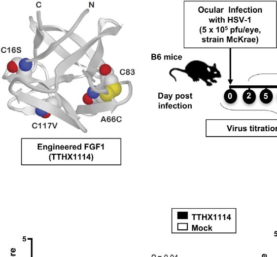
Dhanushkodi et al.                                                                                                                     FGF-1 Treatment for Ocular Herpes

                A                           B                                                     C

                D                                           E                                         F                         G

 FIGURE 1 | Effect of FGF-1 treatment on corneal disease during acute corneal HSV-1 infection: (A) Structure of FGF-1, an engineered FGF-1 known to aid in
 corneal epithelial wound healing. (B) Experimental plan to assess the effect of FGF-1 topical eye treatment in HSV-1 (McKrae) infected B6 mice is shown. Mice were
 mock treated/treated with FGF-1 (1.6 ng/eye, twice a day) from day1 post-infection with HSV-1 McKrae (5X10⁵/eye). Mice were scored at day 5, 7, 10,14, 21, 28 for
 pathological symptoms of Keratitis and blepharitis after infection with 5X 10⁵ pfu/eye of HSV-1 McKrae, mock/treated with FGF-1 from day1 p.i. Stromal keratitis was
 scored as 0- no disease; 1- cloudiness, some iris detail visible; 2- iris detail obscured; 3- cornea totally opaque; and 4- cornea perforation. Blepharitis was scored as
 0- no disease; 1- puffy eyelids; 2- puffy eyelids with some crusting; 3- eye swollen shut with severe crusting; and 4- eye completely swollen shut. Keratitis score and
 Blepharitis score in mice mock/treated with FGF-1 during corneal HSV-1 infection. (C) Representative eye pictures of mice mock treated/treated with FGF-1 from
 day1 after infection with HSV-1 McKrae. Graph showing corresponding keratitis (D) and blepharitis score (E) at day 7, 10 14 p.i. Data represent the mean score
 from experiment with a total of 10 mice per group. (F) Survival plot of mice mock treated/treated with FGF-1 (1.6 ng/eye) twice a day from day1 post-infection with
 HSV-1 McKrae (5X10⁵/eye). (G) Graph showing virus titre in eye swabs of TTHX1114 and mock treated mice, estimated by plaque assay. NS, not significant.

infected ocularly with 1 x 10⁵ or 5 x 10⁵ PFUs of HSV-1 (strain                            These results demonstrate that topical cornea treatment with
McKrae). Starting day one post-infection (PI) B6 mice received                          FGF-1 (TTHX1114) is associated with reduced primary corneal
daily topical ocular treatment with TTHX1114 twice daily (8 hour.                       keratopathy in the B6 mouse model of primary ocular herpes,
intervals) for 14 days (Figure 1B). The efficacy of TTHX1114 on                          independent of viral shedding or lymphangiogenesis.
primary corneal infection and disease was tested at an initial dose of
1.6 ng/eye, twice a day. The severity of primary stromal keratitis and                  FGF-1 Topical Ocular Treatment Reduced
blepharitis was monitored on days 2, 5, 7, 10, 14, 21 and 28 PI                         Recurrent Herpetic Disease in the Mouse
(Figure 1B). HSV-1 replication in cornea was also determined at 2,                      Model of UVB-Induced Virus Reactivation
5, 7, 10 days PI (Figure 1B). As shown in the representative corneal                    We next evaluated the effect of TTHX1114 on recurrent herpes
pictures in Figure 1C, compared to HSV-1 infected vehicle-treated                       infection and disease using the B6 mouse model of UVB induced
mice (lower panel), the infected and TTHX1114-treated mice (top                         reactivation (Figure 3A). In this model, the cornea of B6 mice
panel) showed a significant decrease of corneal herpetic disease. The                    were infected with HSV-1 with scarification and virus
most significant decrease in primary stromal keratitis was recorded                      reactivation was provoked at day 35 PI in latently infected
on days 14 and 21 (P = 0.04 and P = 0.02, respectively, Figure 1D).                     mice, using a 60 seconds corneal UV-B radiation, immediately
A significant decrease in blepharitis was recorded on days 7 and 14                      followed with topical treatment with TTHX1114 for two weeks.
(P = 0.02 and P = 0.04, respectively, Figure 1E). However, there was                    One group of mice was treated topically with TTHX1114 (n =
no significant effect observed with TTHX1114 on mouse survival                           26) from day 34 p.i for two weeks (two doses each day) while
following HSV-1 infection (Figure 1F). No significant effect of                          another group of mice was mock treated (control n = 26). The
TTHX1114 on corneal virus replication was detected (Figures 1F,                         efficacy of TTHX1114 on recurrent herpetic disease was tested at
G). The effect of TTHX1114 treatment was recorded at both high                          an initial dose of 1.6 ng/eye, twice a day on the severity of
dose 5 x 10⁵ PFUs and low 1 x 10⁵ dose of HSV-1, on blepharitis as                      recurrent stromal keratitis monitored daily for 30 post-UVB
early as day 5 post-treatment (Figure 2A) and on keratitis at day 7                     exposure (Figure 3A). HSV-1 reactivation in the cornea was also
post-treatment (Figure 2B). Immunohistochemistry and FACS                               determined 10 days PI (Figure 3A). As shown in the
analysis were carried out to assess if FGF-1 treatment can affect                       representative corneal pictures in Figure 3B, compared to
lymphangiogenesis and lymphocyte infiltration in HSV-1-infected                          HSV-1 infected non-treated mice (lower panel), the infected
mice. The significant reduction in primary corneal keratopathy                           and TTHX1114-treated mice (top panel) showed a significant
following TTHX1114 treatment was not associated with a reduction                        decrease of recurrent corneal herpetic disease. The most
in lymphangiogenesis (Supplemental Figure 1).                                           significant decrease in recurrent stromal keratitis was recorded

Frontiers in Immunology | www.frontiersin.org                                       4                                            May 2021 | Volume 12 | Article 673763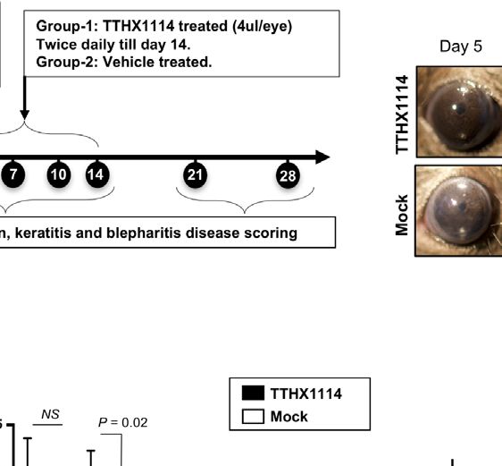
Dhanushkodi et al.                                                                                                                     FGF-1 Treatment for Ocular Herpes

                                    A                                                     B

 FIGURE 2 | Efficacy of an engineered FGF-1, TTHX1114, on the reduction of severity of primary corneal herpetic disease: Mice were treated, or mock treated, with
 TTHX1114 (1.6 ng/eye, twice a day) from day1 post-infection with HSV-1 McKrae (1 x 10⁵ and 5 x 10⁵ pfu/eye). Mice were scored for pathological symptoms of
 blepharitis and Keratitis after infection at day 5, 7, 10, 14, 21 and 28 -days post-infection. Stromal keratitis was scored as 0- no disease; 1- cloudiness, some iris
 detail visible; 2- iris detail obscured; 3- cornea totally opaque; and 4- cornea perforation. Blepharitis was scored as 0- no disease; 1- puffy eyelids; 2- puffy eyelids
 with some crusting; 3- eye swollen shut with severe crusting; and 4- eye completely swollen shut. Bar graph illustrating the percentage reduction in blepharitis (A)
 and keratitis (B) scores between mice infected with HSV-1 McKrae 1 x 10⁵ (white boxes) or 5 x 10⁵ pfu/eye (black boxes) untreated vs following treatment with
 TTHX1114. The reduction in blepharitis and keratitis score following treatment with TTHX1114 was calculated as percentage of mock treated group for each scoring
 day. In A, the efficacy of TTHX1114 in decreasing the blepharitis score at day 14 is identical in mice infected with a low (1 x 10⁵) or high (5 x 10⁵ pfu/eye) HSV-1
 McKrae strain titer. The reduction in blepharitis score is otherwise greater at all other time points in mice infected with HSV-1 McKrae 1 x 10⁵ compared to 5 x 10⁵
 pfu/eye. In contrast, the reduction in the keratitis score is comparable in mice infected with HSV-1 McKrae 1 x 10⁵ or to 5 x 10⁵ pfu/eye (B).

              A

                                                                                         C                                   D

              B

               E                                                 F                                                G                     H

 FIGURE 3 | Effect of FGF-1 treatment on recurrent ocular herpes mouse model of UVB-induced herpes reactivation: Wildtype B6 mice were infected with HSV-1
 (McKrae 5 x 10⁶ pfu/eye and at day 35 p.i, eyes were reactivated by exposure to UV-B radiation for one minute. One group of mice was mock treated (n=26) while
 another group was treated topically with FGF-1 (n=26) from day 34 p.i for two weeks (two doses each day). (A) Experimental plan for testing the effect of FGF-1 on
 mouse model of ocular herpes reactivation. (B) Representative mouse eye picture of herpes UV-B reactivated mice treated with FGF-1. (C) Cumulative number of
 eyes with reactivated disease in mock and FGF-1 treated mice group from day6 to day 28 post-reactivation is shown. (D) Graph showing cumulative disease score
 in mock and FGF-1 treated mice group from day6 to day 28 post-reactivation. (E) Percentage of disease occurrence in mock treated and FGF-1 treated mice group
 from day 2 to day 28 post-reactivation. Number above each bar represents the number of eyes showing disease out of 52 eyes. (F) Graph showing keratitis score
 (more than 2) in mock and FGF-1 treated at day10, day12 and day14 post-reactivation. (G) Duration of recurrent corneal herpetic disease in HSV-1 infected mice
 following treatment with FGF-1. The violin plot illustrates the distribution of disease duration post UV-B radiation in days between mock and TTHX1114 treated
 groups. Note the higher number of eyes displaying disease for more than 10 days in the mock group compared to TTHX1114 treated mice. Only eyes with disease
 score above 2 for both mock and treated groups were included in this analysis. (H) Graph showing cumulative number of eyes shedding virus in TTHX1114 treated
 and mock treated mice, estimated by viral plaque assay in eye swabs. NS, not significant.

Frontiers in Immunology | www.frontiersin.org                                        5                                            May 2021 | Volume 12 | Article 673763
Dhanushkodi et al.                                                                                                             FGF-1 Treatment for Ocular Herpes

on days 10 and 12 post-UVB-induced reactivation (P = 0.04 and                       determined its correlation with reduction of corneal keratopathy
P = 0.02, respectively, Figures 3C–F. Further comparison of the                     following TTHX1114 treatment in infected corneas. B6 mice were
distribution of recurrent herpetic disease duration, following                      infected with HSV-1 (McKrae 2 x 10⁵ PFU/eye) and then treated
UVB induced reactivation, between TTHX1114-treated and                              with TTHX1114 (1.6 ng/eye twice daily) or left untreated as controls
mock-treated showed a significantly higher number of eyes                            (mock). On days 2, 5, 8, 14 and 21, the corneas were harvested,
displaying recurrent herpetic disease for more than 10 days in                      pooled (6 corneas per group) and stained for total
the mock-treated compared to TTHX1114-treated group of                              CD45+CD11b+F4/80+ macrophages and analyzed by FACS assay
mice (Figures 3D, E, G). However, there was no significant                           using the gating strategy showed in Supplemental Figure 2. As
effect of TTHX1114 on virus shedding detected in the cornea                         shown in Figures 4A–C, similar frequencies of total
(Figure 3H).                                                                        CD45+CD11b+F4/80+ macrophages were detected in mouse
    These results demonstrate that topical cornea treatment with the                corneas treated or untreated with TTHX1114. We observed
FGF-1 (TTHX1114) is associated with reduced recurrent corneal                       decreased pro-inflammatory Ly6chighF4/80+CD11b+ macrophages
herpetic disease in the B6 mouse model of UVB induced                               on day7, day14 following TTHX1114 treatment (Figure 4C). In
reactivation independent of the level of virus shedding in the cornea.              addition, we observed a trend toward increased anti-inflammatory
                                                                                    CD206+F4/80+CD11b+ macrophages M2 on day7 following
                                                                                    TTHX1114 treatment (Figure 4C). These results correlate with a
The Reduction of Corneal Herpetic                                                   decreased inflammatory cytokine profile in mouse cornea upon
Keratopathy Following FGF-1 Treatment                                               TTHX1114 treatment during herpes infection (Figure 5). CD4 T
Was Associated With a Decrease of                                                   cells in the cornea did not show any difference in frequency and
Cornea-Resident Pro-Inflammatory M1                                                  activation as assessed by CD69 and GranzymeB expression.
Macrophages                                                                         Similarly, no significant effect of TTHX1114 treatment was
Since macrophages appeared to be an important inflammatory cell                      detected on the frequency and activation of cornea-resident CD4+
infiltrate in the corneas following ocular herpes infection (21–23),                 T cells in cornea (Figure 6). Thus, FGF-1 treatment did not affect
we next assessed the effect of TTHX1114 treatment on the                            the functional capability of CD4 and CD8 T cells at the site
infiltration and function of inflammatory immune cells and                            of infection.

              A                                                                                    B

              C

 FIGURE 4 | Effect of FGF-1 on the infiltration of M1/M2 macrophages in the cornea of HSV-1 infected B6 mice: FACS analysis was carried out to assess the effect
 of FGF-1 treatment on inflammatory immune cells infiltration in infected corneas. B6 mice infected with HSV-1 (McKrae 5X10⁵ PFU/eye) were treated withFGF-1
 (1.6ng/eye twice daily) and at day 2, 5, 7, 14 and 21, corneas were pooled (6 per group) and stained for FACS analysis. (A) Panel showing FACS plots for
 macrophages in mouse corneas. Graph (Right panel) showing corresponding average percentage of F4/80+CD11b+ macrophages in FGF-1 post ocular HSV-1
 infection. At day 7, 14 and 21 PI corneas were pooled (6 per group) and stained for FACS analysis. (B) FACS plot showing percentage of inflammatory Ly6chighF4/
 80+CD11b+ and Ly6cMedF4/80+CD11b+ macrophages in FGF-1 post ocular HSV-1 infection. Graph (lower panel) showing corresponding average percentage of
 Ly6chighF4/80+CD11b+ and M2 Ly6cMedF4/80+CD11b+ inflammatory macrophages in FGF-1 post ocular HSV-1 infection. (C) FACS plot showing percentage of M1
 (CD11c+F4/80+CD11b+) and M2 (CD206+F4/80+CD11b+) macrophages in FGF-1 post ocular HSV-1 infection. Graph (Right panel) showing corresponding average
 percentage of M1 (CD11c+F4/80+CD11b+) and M2 (CD206+F4/80+CD11b+) macrophages in FGF-1 post ocular HSV-1 infection.

Frontiers in Immunology | www.frontiersin.org                                   6                                         May 2021 | Volume 12 | Article 673763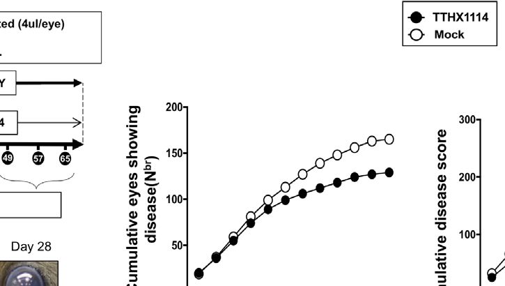
Dhanushkodi et al.                                                                                                                  FGF-1 Treatment for Ocular Herpes

    These results indicate that reduction of corneal keratopathy                      FGF-1 Treatment Skews Polarization of
following FGF-1 treatment was associated with an alteration in                        Human Monocyte Into M2 Macrophages
the ratio of cornea-resident M1/M2 macrophages infiltrating the                        That Produce Anti-Inflammatory
mouse cornea infected with HSV-1. However, there was no                               Cytokines/Chemokines
association with infiltration nor stimulation of cornea-resident                       Based on the mouse results above demonstrating the effect of
CD4+ and CD8+ T cells.                                                                TTHX1114 on cornea-resident M1/M2 macrophages, we next

 FIGURE 5 | Effect of FGF-1 on the production of inflammatory cytokine in mouse cornea following ocular HSV-1 infection: B6 mice infected with HSV-1 (McKrae
 5X10⁵ PFU/eye) were treated with FGF-1 (1.6ng/eye twice daily) and at day 2, 5, 7, 14 and 21, corneas were pooled (6 per group) and lysates were analyzed by
 luminex for inflammatory cytokine profile. Inflammatory cytokines (IFN-g, IL-1a, IL-2, IL-5, IL-12p40, IL-12p70, IL-15, IL-17a, IP-10, TNF-a), levels in the corneal
 lysates of herpes infected mice treated with TTHX1114 (black circle) compared with mock treated (open circle).

Frontiers in Immunology | www.frontiersin.org                                     7                                           May 2021 | Volume 12 | Article 673763
Dhanushkodi et al. FGF-1 Treatment for Ocular Herpes
A
B
FIGURE 6 | Effect of FGF-1 on lymphocyte activation in mouse cornea during HSV-1 infection: FACS analysis was carried out to assess the effect of FGF-1
treatment on CD4, CD8 T cells activation. B6 mice infected with HSV-1 (McKrae 5X10⁵ PFU/eye) were treated with FGF-1 (1.6ng/eye twice daily) and at day 8,
corneas were pooled (8 per group) and stained for FACS analysis. (A) Panel showing FACS plots for CD69 (activation marker), (cytotoxic granular protein) expression
in CD4+ T cells and CD8+ T cells in mouse corneas. (B) Graph showing corresponding average percentage of CD69+ CD4+, GzmB+ CD4+, CD69+ CD8+, GzmB
CD8+ T cells in cornea of B6 mice treated with FGF-1 post ocular HSV-1 infection. Statistical analysis carried out using student’s t test. NS, not significant.
determined whether this effect would be confirmed on human M1/ chemokines (i.e., CXCL10) produced by the monocytes-derived
M2 macrophages. Human unpolarized M0 macrophages were pro-inflammatory M1 macrophages.
generated from M-CSF-treated primary blood-derived monocytes These findings in humans confirm that FGF-1 treatment
and then either left untreated or treated with TTHX1114 at 0.5 and skews polarization of monocyte-derived macrophages into the
3 ng/mL respectively for 24 hours. The M1- and M2 macrophages anti-inflammatory M2 phenotype. Moreover, FGF-1 treatment
were subsequently generated following stimulation with either appeared to reduce production of anti-inflammatory mediators.
IFN-g or IL-4, respectively (Figures 7A, B, top panels). The
representative images of M1 and M2 macrophages untreated or
treated with TTHX1114 showed a different distribution and texture DISCUSSION
(Figures 7A, B, middle panels). Supplemental Figure 3 shows
expression of M1 and M2 polarization markers in human Currently, one of the major unmet clinical challenges remains
monocyte-derived macrophages. finding an effective medical treatment to offset vision-threatening
Following TTHX1114 treatment, we observed a significant inflammatory corneal herpetic disease. To our knowledge, this is the
reduction in the percentage of in vitro generated human first study to demonstrate that topical cornea treatment with FGF-1
monocyte-derived pro-inflammatory M1 macrophages has an anti-inflammatory role that reduces corneal keratopathy in a
expressing CD80, as detected by flow cytometry (Figure 7A, mouse model of primary and recurrent ocular herpes. The
lower two panels). Similarly, there was a significant reduction in decreased frequency and function of pro-inflammatory
the level of CD80 expressed on pro-inflammatory M1 macrophages M1 infiltrating the cornea was associated with
macrophages following TTHX1114 treatment as measured by reduced HSV-1-induced corneal immunopathology observed in
mean fluorescence intensity (MFI) (Figures 7A, lower two both primary and recurrent ocular herpes.
panels). In contrast to pro-inflammatory M1 macrophages, Fibroblast growth factor-1 (FGF-1), a naturally occurring
TTHX1114 treatment led to a significant increase in the protein, promotes tissue repair and regenerates corneal tissue
number of in vitro-generated human monocyte-derived anti- (19). The FGF family consists of a group of homologous growth-
inflammatory M2 macrophages expressing CD206, as detected promoting polypeptides that increase proliferation, angiogenesis,
by flow cytometry (Figure 7B, lower two panels). and wound healing (24–26). Several studies have shown modulation
Moreover, we investigated the signature of pro- and anti- of inflammatory responses by FGFs (25, 27–29). However, the role
inflammatory M1 and M2 cytokines secreted by M0/M1/M2 of FGF-1 on inflammation induced by herpes infection is not
human monocyte-derived macrophages following TTHX1114 currently known. Trefoil’s engineered FGF-1 TTHX1114 builds
treatment using the Luminex detection platform (Supplemental on the well-known activities of naturally-occurring (native) FGF-1
Figure 4). As shown in Figure 8, TTHX1114 treatment had an to enable its use as a pharmaceutical for corneal diseases. Native
overall trend in reduced production of pro-inflammatory cytokines FGF-1 is a potent stimulator of cell proliferation and migration, and
(i.e., IL1a, IL-2, IL-12, IL-15, IL17-a, TNF-a, CCL-5) and has cell protective properties, all key attributes for its use in corneal
Frontiers in Immunology | www.frontiersin.org 8 May 2021 | Volume 12 | Article 673763Dhanushkodi et al. FGF-1 Treatment for Ocular Herpes
A B
FIGURE 7 | Effect of FGF-1 on M1 and M2 polarization from human monocyte-derived macrophages: Unpolarized M0 macrophages were generated from M-CSF-
treated primary monocytes and treated with FGF-1 (0.5 and 3 ng/ml) for 24 hr. M1- and M2-polarized macrophages were then generated by stimulation with IFN-g
and IL-4, respectively. (A) Experimental plan as shown (top panel). Representative images of M1 and M2 macrophages treated with FGF-1 (0.5 and 3 ng/ml). CD80
and CD64 levels in M1 macrophages treated with FGF-1 were compared by flow cytometry. Dot plots and histograms depict the results obtained in one
representative donor; the grey histogram represent mock treated M1 macrophage, and the blue histogram represent the fluorescent profile of FGF-1 treated M1
macrophage stained with the indicated antibodies; the percentage and MFI (mean fluorescence intensity) of positive cells is indicated. The graph (lower panel)
represents mean results from at least three different donors. (B) Experimental plan as shown (top panel). CD163 and CD206 levels in M2 macrophages treated with
FGF-1 were compared by flow cytometry. Dot plots and histograms depict the results obtained in one representative donor; the grey histogram represent mock
treated M2 macrophage, and the blue histogram represent the fluorescent profile of FGF-1 treated M2 macrophage stained with the indicated antibodies; the
percentage and MFI (mean fluorescence intensity) of positive cells is indicated. The graph (lower panel) represents mean results from at least four different donors.
A B
C
FIGURE 8 | Effect of FGF-1 on cytokine secretion by human monocyte-derived M1 and M2 macrophages: Unpolarized M0 macrophages were generated from
M-CSF-stimulated primary monocytes and then treated with FGF-1 (0.5 and 3 ng/ml) for 24 hr. M1- and M2-polarized macrophages were then generated by
stimulation with IFN-g and IL-4, respectively. (A) M1 signature cytokine (IFN-g, IL-1a, IL-1b, IL-2, IL-5, IL-12p40, IL-12p70, IL-15, IL-17a, IP-10, TNF-a), levels in the
culture supernatants of M1 macrophage treated with FGF-1 and M2 signature cytokine (IL-4, Il-10, VEGF-a) levels in the culture supernatants of M2 macrophage
treated with FGF-1. (B) Correlation graph showing level of M1 cytokines with FGF-1 dose kinetics. (IL-1a, IL-1b, IL-12p40, IL-12p70, IL-15, Il-17a, TNF-a, CCL5).
(C) Correlation graph showing level of M2 cytokines with FGF-1 dose kinetics (IL-10, VEGF-a).
Frontiers in Immunology | www.frontiersin.org 9 May 2021 | Volume 12 | Article 673763Dhanushkodi et al. FGF-1 Treatment for Ocular Herpes disease treatment. The compound uniquely activates all seven forms orchestrators of the blinding immunoinflammatory lesion that of the FGF receptor, contributing to its potency. Unlike the represents an immunopathological response to HSV-1 infection. naturally-occurring FGF-1 with an extremely short half-life, Moreover, corneal herpetic lesions have an increased severity if TTHX1114 is much more stable making it more suitable for the regulatory Foxp3(+)CD4+ Treg response is compromised pharmaceutical use. from the onset of infection (32). Tregs beneficially influence HSV-1 infections of the cornea range in severity from minor the severity of ongoing tissue-damaging immune responses to transient discomfort to the blinding inflammatory disease herpes HSV-1 infection (32). This suggest that therapies, such as FGF-1, stromal keratitis (30). Here, we report a novel observation of boosting Treg function in the clinical phase hold promise for anti-inflammatory effect of an engineered FGF-1 (TTHX1114) controlling a lesion that is an important cause of human that healed both primary and recurrent corneal herpetic blindness. A potential effect of FGF-1 on cornea-resident immunopathology leading to transparency of cornea, which is Foxp3(+)CD4+ Treg must be determined during the ongoing essential for normal vision. This anti-inflammatory role of anti-inflammatory effect of FGF-1. engineered FGF-1 was associated with a decrease in the The underlying cellular and molecular mechanisms that led to frequency and function of pro-inflammatory M1 macrophages FGF-1 treatment decreasing HSV-1-induced corneal infiltrating the cornea and, in contrast, an increase in the immunopathology remain to be determined. HSV-1 infection frequency and function ofanti-inflammatory M2 macrophages of the cornea induces lymphangiogenesis that continues to infiltrating the cornea. These results agree with a previous report develop well beyond the resolution of infection. In this report, by Dr. Rouse that similarly demonstrated the inhibition of VEGF we discovered that topical cornea treatment with FGF-1 has an signaling with a Src Kinase inhibitor ameliorated stromal anti-inflammatory role that reduced corneal keratopathy in a keratitis (31) Although the anti-inflammatory role of FGF-1 is mouse model of primary and recurrent ocular herpes. The known in other disease conditions like renal diseases, its role in decrease in the frequency and function of cornea-resident pro- viral infection is not currently known. Nevertheless, it remains to inflammatory macrophages M1 was associated with reduced be determined: (i) whether the FGF-1 treatment can accelerate HSV-1-induced corneal immunopathology observed in both healing of primary and recurrent corneal herpetic disease in primary and recurrent ocular herpes following FGF-1 treatment. humans; and (ii) a potential role of other innate and adaptive Multiple pro-angiogenic factors, including FGF-1, are immune cells in the observed anti-inflammatory role FGF-1 in expressed within the cornea following virus clearance. Many HSV-1-induced immunopathology. angiogenic factors such as vascular endothelial growth factors are Our present study is the first to demonstrate a reduction of present in the cornea but their angiogenic activities are impeded corneal herpetic keratopathy following FGF-1 treatment by being bound to a soluble form of the VEGF receptors. It is associated with a decrease of cornea-resident pro-inflammatory likely that an imbalance between vascular endothelial growth macrophages M1. FGF-1 appeared to shift corneal-resident factors and their receptors present in the cornea occur after macrophages toward M2 phenotype. It remains to be ocular HSV-1 infection may cause prominent determined whether HSV-1 replication in M1 and M2 neovascularization, an essential step in the pathogenesis of the macrophages was lowered following FGF-1 treatment. vision-impairing lesion, stromal keratitis. However, the Moreover, we showed that the M1 macrophages expressed significant effect of FGF-1 on HSV-1-induced corneal significantly lower levels of HSV-1-induced pro-inflammatory immunopathology in the B6 mouse model of primary ocular cytokines and chemokines following FGF-1 treatment. Thus, herpes was independent of lymphangiogenesis. Indeed, the these findings shed significant light on a novel therapeutic immunohistochemistry and FACS analysis revealed that FGF-1 approach to reducing primary and recurrent corneal herpetic treatment did not affect lymphangiogenesis and lymphocyte disease by modulating both the phenotype and function of infiltration in HSV-1-infected mice. cornea-resident macrophages, which play a predominant role In conclusion, we report here that compared to HSV-1 in the corneas following ocular herpes infection. We therefore infected non-treated mice, the infected and FGF-1 treated mice suggest that inclusion of FGF-1 as a novel immunotherapeutic showed (i) an overall resistance to disease; (ii) a significant regimen against ocular herpes to skew cornea-resident decrease in primary stromal keratitis (days 5, 14, and 21) and macrophage development toward an anti-inflammatory M2 blepharitis (days 7 and 14); (iii) a significant decrease in disease phenotype, rather than a pro-inflammatory M1 phenotype. duration in herpes reactivation and (iv) a significant decrease in In the present report, the observed anti-inflammatory role corneal inflammatory macrophage. The effect of FGF-1 seen on FGF-1 in HSV-1-induced immunopathology was not associated mouse macrophages was conformed on human macrophage with a significant effect on the frequency and activation of total pointing to a potential clinical application. However, FGF-1 CD4+ and CD8+ T cells that infiltrate HSV-1-infected corneas. treatment did not affect the number and function of cornea The engineered FGF-1 (TTHX1114) affects the function and resident T cells nor virus corneal replication. Topical cornea frequency HSV-specific CD8+ T cells, but not HSV-specific treatment with eFGF-1 is associated with reduced corneal CD4+ T cells, that infiltrate HSV-1-infected corneas with a yet- keratopathy in a mouse model of primary ocular herpes. to-be determined mechanism. Thus, the effect of FGF-1 on Increased frequency and function of anti-inflammatory M2 cornea-resident HSV-specific CD8+ T cells remains to be macrophages was associated with reduced corneal keratopathy determined, since CD4 + T cells appear to be the main observed in the FGF-1 treated cornea. Frontiers in Immunology | www.frontiersin.org 10 May 2021 | Volume 12 | Article 673763
Dhanushkodi et al.                                                                                                                        FGF-1 Treatment for Ocular Herpes

DATA AVAILABILITY STATEMENT                                                              Design, Analyze data, Writing Manuscript), ED (Analyze
                                                                                         data, Writing Manuscript), LB (Analyze data, Writing
The original contributions presented in the study are included in                        Manuscript). All authors contributed to the article and
the article/Supplementary Material. Further inquiries can be                             approved the submitted version.
directed to the corresponding author.

                                                                                         FUNDING
ETHICS STATEMENT
                                                                                         The authors declare that this study received funding from Trefoil
Human protocols were approved by the University of California                            Therapeutics, Inc. The funder was not involved in the study design,
Irvine’s IRB committee (IRB-HS#_2020-5779). Written                                      collection, analysis, interpretation of data, the writing of this article
informed consent for participation was not required for this                             or the decision to submit it for publication. This work is supported
study in accordance with the national legislation and the                                by Public Health Service research grants EY019896, EY14900 and
institutional requirements. Animal protocols were approved by                            EY024618 from the National Eye Institutes (NEI) and AI150091,
the University of California Irvine’s institutional animal care and                      AI143348, AI147499, AI143326, AI138764, AI124911 and
use committee (IACUC #19-111).                                                           AI110902 from the National Institutes of Allergy and Infectious
                                                                                         Diseases (NIAID) to LM and from The Discovery Center for Eye
                                                                                         Research, and Research to Prevent Blindness.
AUTHOR CONTRIBUTIONS
ND (Experimental Design, Analyze data, Writing Manuscript),                              SUPPLEMENTARY MATERIAL
RS (Experimental Design, Analyze data, Writing Manuscript),
P-GC (Experimental Design, Analyze data, Writing                                         The Supplementary Material for this article can be found online at:
Manuscript), SP (Experimental Design, Analyze data, Writing                              https://www.frontiersin.org/articles/10.3389/fimmu.2021.673763/
Manuscript), SR (Experimental Design), DB (Experimental                                  full#supplementary-material

REFERENCES                                                                                     herpetic stromal keratitis. J Leukoc Biol (2005) 77:24–32. doi: 10.1189/
                                                                                               jlb.0904486
 1. Kuo T, Wang C, Badakhshan T, Chilukuri S, BenMohamed L. The challenges                9.    Dana MR, Qian Y, Hamrah P. Twenty-five-year panorama of corneal
    and opportunities for the development of a T-cell epitope-based herpes                      immunology: emerging concepts in the immunopathogenesis of microbial
    simplex vaccine. Vaccine (2014) 32:6733–45. doi: 10.1016/j.vaccine.                         keratitis, peripheral ulcerative keratitis, and corneal transplant rejection.
    2014.10.002                                                                                 Cornea (2000) 19:625–43. doi: 10.1097/00003226-200009000-00008
 2. Samandary S, Kridane-Miledi H, Sandoval JS, Choudhury Z, Langa-Vives F,              10.   Thomas J, Rouse BT. Immunopathogenesis of herpetic ocular disease.
    Spencer D, et al. Associations of HLA-A, HLA-B and HLA-C alleles frequency                 Immunol Res (1997) 16:375–86. doi: 10.1007/BF02786400
    with prevalence of herpes simplex virus infections and diseases across global        11.   Kumaraguru U, Davis I, Rouse BT. Chemokines and ocular pathology caused
    populations: implication for the development of an universal CD8+ T-cell                   by corneal infection with herpes simplex virus. J Neurovirol (1999) 5:42–7. doi:
    epitope-based vaccine. Hum Immunol (2014) 75:715–29. doi: 10.1016/                         10.3109/13550289909029744
    j.humimm.2014.04.016                                                                 12.   Chentoufi AA, Kritzer E, Tran MV, Dasgupta G, Lim CH, Yu DC, et al. The
 3. Chentoufi AA, Dervillez X, Rubbo PA, Kuo T, Zhang X, Nagot N, et al.                        herpes simplex virus 1 latency-associated transcript promotes functional
    Current trends in negative immuno-synergy between two sexually                             exhaustion of virus-specific CD8+ T cells in latently infected trigeminal
    transmitted infectious viruses: HIV-1 and HSV-1/2. Curr Trends Immunol                     ganglia: a novel immune evasion mechanism. J Virol (2011) 85:9127–38.
    (2012) 13:51–68.                                                                           doi: 10.1128/JVI.00587-11
 4. Dervillez X, Qureshi H, Chentoufi AA, Khan AA, Kritzer E, Yu DC, et al.               13.   HEDS. Acyclovir for the prevention of recurrent herpes simplex virus eye
    Asymptomatic HLA-A*02:01-restricted epitopes from herpes simplex virus                     disease. Herpetic Eye Disease Study Group. N Engl J Med (1998) 339:300–6.
    glycoprotein B preferentially recall polyfunctional CD8+ T cells from                      doi: 10.1056/NEJM199807303390503
    seropositive asymptomatic individuals and protect HLA transgenic mice                14.   Osorio Y, Cohen J, Ghiasi H. Improved protection from primary ocular HSV-
    against ocular herpes. J Immunol (2013) 191:5124–38. doi: 10.4049/                         1 infection and establishment of latency using multigenic DNA vaccines.
    jimmunol.1301415                                                                           Invest Ophthalmol Vis Sci (2004) 45:506–14. doi: 10.1167/iovs.03-0828
 5. Chentoufi AA, Zhang X, Lamberth K, Dasgupta G, Bettahi I, Nguyen A, et al.            15.   Lobo AM, Agelidis AM, Shukla D. Pathogenesis of herpes simplex keratitis:
    HLA-A*0201-restricted CD8+ cytotoxic T lymphocyte epitopes identified                       The host cell response and ocular surface sequelae to infection and
    from herpes simplex virus glycoprotein D. J Immunol (2008) 180:426–37. doi:                inflammation. Ocul Surf (2019) 17:40–9. doi: 10.1016/j.jtos.2018.10.002
    10.4049/jimmunol.180.1.426                                                           16.   Gurung HR, Carr MM, Bryant K, Chucair-Elliott AJ, Carr DJ. Fibroblast
 6. Zhang X, Dervillez X, Chentoufi AA, Badakhshan T, Bettahi I, Benmohamed                     growth factor-2 drives and maintains progressive corneal neovascularization
    L. Targeting the genital tract mucosa with a lipopeptide/recombinant                       following HSV-1 infection. Mucosal Immunol (2018) 11:172–85. doi: 10.1038/
    adenovirus prime/boost vaccine induces potent and long-lasting CD8+ T                      mi.2017.26
    cell immunity against herpes: importance of MyD88. J Immunol (2012)                  17.   Kim B, Lee S, Kaistha SD, Rouse BT. Application of FGF-2 to modulate
    189:4496–509. doi: 10.4049/jimmunol.1201121                                                herpetic stromal keratitis. Curr Eye Res (2006) 31:1021–8. doi: 10.1080/
 7. Liesegang TJ. Herpes simplex virus epidemiology and ocular importance.                     02713680601038824
    Cornea (2001) 20:1–13. doi: 10.1097/00003226-200101000-00001                         18.   Nesburn AB, Ramos TV, Zhu X, Asgarzadeh H, Nguyen V, BenMohamed L.
 8. Banerjee K, Biswas PS, Rouse BT. Elucidating the protective and pathologic T               Local and systemic B cell and Th1 responses induced following ocular
    cell species in the virus-induced corneal immunoinflammatory condition                      mucosal delivery of multiple epitopes of herpes simplex virus type 1

Frontiers in Immunology | www.frontiersin.org                                       11                                              May 2021 | Volume 12 | Article 673763
Dhanushkodi et al. FGF-1 Treatment for Ocular Herpes
glycoprotein D together with cytosine-phosphate-guanine adjuvant. Vaccine 28. Bennett MV, Garre JM, Orellana JA, Bukauskas FF, Nedergaard M, Saez JC.
(2005) 23:873–83. doi: 10.1016/j.vaccine.2004.08.019 Connexin and pannexin hemichannels in inflammatory responses of glia and
19. Eveleth D, Pizzuto S, Weant J, Jenkins-Eveleth J, Bradshaw RA. Proliferation neurons. Brain Res (2012) 1487:3–15. doi: 10.1016/j.brainres.2012.08.042
of Human Corneal Endothelia in Organ Culture Stimulated by Wounding and 29. Rusnati M, Presta M. Fibroblast growth factors/fibroblast growth factor
the Engineered Human Fibroblast Growth Factor 1 Derivative TTHX1114. receptors as targets for the development of anti-angiogenesis strategies.
J Ocul Pharmacol Ther (2020) 36:686–96. doi: 10.1089/jop.2019.0119 Curr Pharm Des (2007) 13:2025–44. doi: 10.2174/138161207781039689
20. Zhang X, Chentoufi AA, Dasgupta G, Nesburn AB, Wu M, Zhu X, et al. A 30. Rowe AM, Yun H, Treat BR, Kinchington PR, Hendricks RL. Subclinical Herpes
genital tract peptide epitope vaccine targeting TLR-2 efficiently induces local Simplex Virus Type 1 Infections Provide Site-Specific Resistance to an Unrelated
and systemic CD8+ T cells and protects against herpes simplex virus type 2 Pathogen. J Immunol (2017) 198:1706–17. doi: 10.4049/jimmunol.1601310
challenge. Mucosal Immunol (2009) 2:129–43. doi: 10.1038/mi.2008.81 31. Sharma S, Mulik S, Kumar N, Suryawanshi A, Rouse BT. An anti-
21. Park M, Richardson A, Pandzic E, Lobo EP, Lyons JG, Di Girolamo N. inflammatory role of VEGFR2/Src kinase inhibitor in herpes simplex virus
Peripheral (not central) corneal epithelia contribute to the closure of an 1-induced immunopathology. J Virol (2011) 85:5995–6007. doi: 10.1128/
annular debridement injury. Proc Natl Acad Sci USA (2019) 52:26633–43. doi: JVI.00034-11
10.1073/pnas.1912260116 32. Veiga-Parga T, Suryawanshi A, Mulik S, Gimenez F, Sharma S, Sparwasser T,
22. Chinnery HR, McMenamin PG, Dando SJ. Macrophage physiology in the eye. et al. On the role of regulatory T cells during viral-induced inflammatory
Pflugers Arch (2017) 469:501–15. doi: 10.1007/s00424-017-1947-5 lesions. J Immunol (2012) 189:5924–33. doi: 10.4049/jimmunol.1202322
23. Hamrah P, Dana MR. Corneal antigen-presenting cells. Chem Immunol
Allergy (2007) 92:58–70. doi: 10.1159/000099254
Conflict of Interest: ED and DB are employees and hold an equity interest in
24. Kumar S, Agrawal S, Maity S, AlRaawi Z, Al-Ameer M. Targeting Drugs
Trefoil Therapeutics, Inc. ED is an inventor on patents claiming TTHX1114.
Against Fibroblast Growth Factor(s)-Induced Cell Signaling. Curr Drug
Targets (2020) 22:214–40. doi: 10.2174/1389450121999201012201926 The remaining authors declare that the research was conducted in the absence of
25. Presta M, Andres G, Leali D, Dell’Era P, Ronca R. Inflammatory cells and any commercial or financial relationships that could be construed as a potential
chemokines sustain FGF2-induced angiogenesis. Eur Cytokine Netw (2009) conflict of interest.
20:39–50. doi: 10.1684/ecn.2009.0155
26. Barrientos S, Stojadinovic O, Golinko MS, Brem H, Tomic-Canic M. Growth Copyright © 2021 Dhanushkodi, Srivastava, Coulon, Prakash, Roy, Bagnol, David
factors and cytokines in wound healing. Wound Repair Regener (2008) and BenMohamed. This is an open-access article distributed under the terms of the
16:585–601. doi: 10.1111/j.1524-475X.2008.00410.x Creative Commons Attribution License (CC BY). The use, distribution or
27. Meyer M, Muller AK, Yang J, Sulcova J, Werner S. The role of chronic reproduction in other forums is permitted, provided the original author(s) and the
inflammation in cutaneous fibrosis: fibroblast growth factor receptor copyright owner(s) are credited and that the original publication in this journal is
deficiency in keratinocytes as an example. J Investig Dermatol Symp Proc cited, in accordance with accepted academic practice. No use, distribution or
(2011) 15:48–52. doi: 10.1038/jidsymp.2011.1 reproduction is permitted which does not comply with these terms.
Frontiers in Immunology | www.frontiersin.org 12 May 2021 | Volume 12 | Article 673763You can also read