Isolation and Characterization of a Porcine Transmissible Gastroenteritis Coronavirus in Northeast China
←
→
Page content transcription
If your browser does not render page correctly, please read the page content below
ORIGINAL RESEARCH
                                                                                                                                               published: 02 March 2021
                                                                                                                                         doi: 10.3389/fvets.2021.611721

                                             Isolation and Characterization of a
                                             Porcine Transmissible Gastroenteritis
                                             Coronavirus in Northeast China
                                             Dongwei Yuan 1,2 , Zihan Yan 1 , Mingyue Li 1 , Yi Wang 1 , Mingjun Su 1* and Dongbo Sun 1*
                                             1
                                              College of Animal Science and Veterinary Medicine, Heilongjiang Bayi Agricultural University, Daqing, China, 2 Daqing Center
                                             of Inspection and Testing for Agricultural Products Ministry of Agriculture, Daqing, China

                                             Transmissible gastroenteritis virus (TGEV) is a coronavirus (CoV) that is a major
                                             pathogenity of viral enteritis and diarrhea in suckling piglets, causing high morbidity
                                             and mortality. In this study, a TGEV strain HQ2016 was isolated from northeast
                                             China and characterized its genome sequence and pathogenicity. The phylogenetic
                                             analysis indicated that the TGEV HQ2016 strain was more similar to the TGEV Purdue
                                             cluster than to the Miller cluster. Both recombination and phylogenetic analysis based
                                             on each structural and non-structural gene revealed no recombination event in the
                          Edited by:
                                             HQ2016 strain. Experimental infection study using colostrum-deprived newborn piglets
                  Van Giap Nguyen,
        Vietnam National University of       successfully showed that the HQ2016 can cause clinical symptoms including anorexia
                 Agriculture, Vietnam        and yellow-to-whitish watery diarrhea, which are characteristics of TGE, in the inoculated
                       Reviewed by:          piglets 48 h post-inoculation. These results provide valuable information about the
                      Hye Kwon Kim,
        Chungbuk National University,        evolution of the porcine CoVs.
                          South Korea
                                             Keywords: transmissible gastroenteritis virus, virus isolate, phylogenetic analysis, pathogenicity, coronavirus
                      Ayako Miyazaki,
        National Agriculture and Food
        Research Organization, Japan
                                             INTRODUCTION
                  *Correspondence:
                          Mingjun Su
                                             Coronaviruses (CoVs) are the main etiological agents underlying outbreaks of porcine diarrhea,
                 mingjunsu@163.com
                        Dongbo Sun
                                             causing substantial economic losses (1). Transmissible gastroenteritis virus (TGEV) is a member
                dongbosun@126.com            of the family Coronaviridae that was first reported in 1946 in the USA (2). Since then, the disease
                                             always happened in swine-producing areas of the world (1, 3), and reported many times in China
                    Specialty section:       in recent years (4–8). Epidemiological investigations have shown that TGEV is often present in the
         This article was submitted to       spring and autumn in the northeast of China, sometimes in mixed infections with other diarrhea
        Veterinary Infectious Diseases,      virus, and caused viral enteritis and severe diarrhea in all ages of pigs, especially with high mortality
                a section of the journal     in suckling piglets (9, 10).
        Frontiers in Veterinary Science
                                                Transmissible gastroenteritis virus is an enveloped virus with a single-stranded, positive-
      Received: 29 September 2020            stranded RNA genome of ∼28.5-kb. The genome contains nine open reading frames (ORFs),
       Accepted: 05 February 2021            which encode four structural proteins and five non-structural proteins: the spike glycoprotein (S);
         Published: 02 March 2021
                                             envelope protein (E); membrane glycoprotein (M); nucleocapsid protein (N); replicases 1a and
                              Citation:      1b; ORF 3a and 3b proteins; and ORF 7 protein. The genes of TGEV are arranged in the order
   Yuan D, Yan Z, Li M, Wang Y, Su M         of 5′ -rep-S-3a-3b-E-M-N-ORF7-3′ (4–6). The mutation in the spikes protein may be an important
       and Sun D (2021) Isolation and
                                             indicator for evaluating the tropism and virulence of TGEV. The M protein is the main viral particle
         Characterization of a Porcine
         Transmissible Gastroenteritis
                                             membrane protein, which is mainly embedded in the lipid vesicle membrane and is connected to
      Coronavirus in Northeast China.        the capsule during assembly of the virus nucleocapsid. The E protein is a transmembrane protein,
            Front. Vet. Sci. 8:611721.       and the N protein is exists in the viral membrane. The ORF3 is composed of two open frames
     doi: 10.3389/fvets.2021.611721          ORF3a and ORF3b. ORF3a deletion is found in many TGEV strains and PRCV strain. The ORF7

Frontiers in Veterinary Science | www.frontiersin.org                               1                                             March 2021 | Volume 8 | Article 611721
Yuan et al.                                                                                          Transmissible Gastroenteritis Coronavirus in China

counteracts host-cell defenses and affects the persistence of              per dilution. The cells were then cultured continuously at 37◦ C
TGEV, and improves the survival rate of TGEV by negatively                 under 5% CO2 . The viral CPE was observed for 5–7 days. Tissue
regulating the downstream caspase-dependent apoptotic                      culture infective dose (TCID50 ) was determined with the Reed-
pathways (5, 6, 11, 12).                                                   Muench method (14) and expressed as TCID50 per milliliter.
   In this study, we isolated a TGEV from clinical samples
collected from farms in northeast China using PK15 cells,
                                                                           Indirect Immunofluorescence Assay
characterized its genome based on the whole-genome sequence,
                                                                           PK15 cells (1 × 10⁶ ) were seeded on six-well plates, cultured
and investigated its pathogenicity in colostrum-deprived
                                                                           overnight, and then infected with TGEV HQ2016 (passaged for
neonatal pigs in terms of a clinical assessment, viral shedding,
                                                                           10 times) at a multiplicity of infection (MOI) of 1.0. At 24 h
virus distribution, histopathological changes, and a mortality
                                                                           after inoculation, the cells were fixed with 4% paraformaldehyde
analysis. The results suggested that we have isolated porcine
                                                                           for 15 min and then per-meabilized with 0.2% Triton X-
enteric coronavirus TGEV HQ2016. The genetic characteristics
                                                                           100 for 15 min. The cells were then blocked with 5% skim
and pathogenicity of this virus provided valuable information
                                                                           milk, and incubated overnight at 4◦ C with a TGEV-specific
for the evolution of TGEV and will helpful research on the
                                                                           monoclonal antibody (5E8, supplied by Professor L. Feng,
molecular pathogenesis of TGEV.
                                                                           Harbin Veterinary Research Institute of the Chinese Academy
                                                                           of Agricultural Sciences, Harbin, China) diluted 1:1000. The
MATERIALS AND METHODS                                                      cells were washed three times with PBS and incubated with a
                                                                           secondary antibody (fluorescein-isothiocyanate-conjugated goat
Specimen Collection and Screening                                          anti-mouse IgG antibody, diluted 1:500) for 1 h at 37◦ C and then
In 2016, a total of 50 intestine samples from piglets were
                                                                           washed three times with PBS. The stained cells were visualized
collected from eight swine-raising farms in northeast China,
                                                                           with fluorescence microscopy (Leica DMi8, Germany).
in which the piglets showing watery diarrhea and dehydration
and as known that all sow without any diarrhea viral vaccine
inoculation. The intestinal samples were stored at −80◦ C. The             Electron Microscopic Assay
samples were homogenized and diluted with sterile phosphate-               Supernatants from plaque-purified TGEV HQ2016 (passaged
buffered saline (PBS). The suspensions were repeatedly frozen              for 8 times) infected cell cultures were concentrated by
and thawed three times, vortexed and clarified by centrifugation           ultracentrifugation method. The supernatants of the cell cultures
at 12,000 × g for 10 min at 4◦ C and the supernatants were filtered        were centrifuged first at 6,000 × g for 30 min at 4◦ C, and then at
through 0.22 µm filters (Millipore, Billerica, MA, USA). Semi-             60,000 × g for 2 h at 4◦ C. After ultracentrifugation, the samples
nest reverse transcription (RT)-PCR (13) was used to identify              were negatively stained with 2% ammonium molybdate and
the samples positive for TGEV, with two pairs of specific primers          adsorbed onto 300-mesh copper net for 2 min. The viral particles
(TGEV-N-F: GGTAGTCGTGGTG- CTAATAATGA; TGEV-N-                              were examined with an electron microscope (Hitachi H7500,
R1: CAGAATGCTAGACACAGATGGAA; TGEV-N-R2: GTT-                               Tokyo, Japan).
CTCTTCCAGGTGTGTTTGTT).
                                                                           Extraction of Viral RNA and Complete
Virus Isolation and Plaque Purification
PK15 cells (American Type Culture Collection [ATCC] CCL-33)                Genome Sequencing
were cultured in Dulbecco’s modified Eagle’s medium (DMEM;                 Culture supernatants from plaque-purified TGEV HQ2016
Hyclone, USA) supplemented with 10% fetal bovine serum (FBS;               (passaged for 8 times) infected cells were collected and used
Bovogen, Australia) at 37◦ C in a 5% CO2 incubator. Growth                 for preparation of viral RNA. Total RNA was extracted using
medium was removed from confluent monolayer cells; the cells               TRIzol Reagent (Invitrogen, Carlsbad, CA, USA), according to
were washed twice with DMEM and inoculated with a mixture                  the manufacturer’s instructions. The RNA samples were sent
of the supernatants of the positive tissue samples and DMEM                to testing company (Shanghai Probe Biotechnology Co., Ltd.)
containing 20 µg/ml trypsin (GIBCO, 1:250) at a ratio of 1:1.              to determined complete genomic sequence with the Illumina
After adsorption for 60 min at 37◦ C, the cells were washed                high-throughput deep sequencing platform (15).
with DMEM, and maintenance medium consisting of DMEM
supplemented with 10 µg/ml trypsin was added. The inoculated               Sequence Analysis
cell cultures were observed for CPE for 3–5 days, harvested, and           The sequences of TGEV reference strains used in this study were
blindly passaged for five times. The viruses in a CPE positive             obtained from GenBank, as shown in Table 1. The nucleotide
sample was cloned by repeating plaque purify three times and               and the amino acid sequences of TGEV HQ2016 strain were
designated as HQ2016.                                                      compared with the corresponding sequences of the TGEV
                                                                           strains in the GenBank database. The sequence was analyzed
Virus Titration With a Median Tissue                                       using the computer program MEGA version 6.0 (16) and
Culture Infective Dose Assay                                               DNASTAR (17). Nucleotide and amino acid sequence identities
PK15 cells were seeded on 96-well plates and cultured overnight.           were determined using the Clustal W program. To determine the
The collected TGEV HQ2016 (passaged for 10 times) was 10-fold              relationships between representative TGEV isolates and HQ2016
serially diluted, and used to inoculate cells, with eight replicates       strain, a phylogenetic tree based on the entire genome was

Frontiers in Veterinary Science | www.frontiersin.org                  2                                       March 2021 | Volume 8 | Article 611721
Yuan et al.                                                                                                    Transmissible Gastroenteritis Coronavirus in China

TABLE 1 | Information of the reference TGEV sequences used in this study in the database.

No.                            Isolate                        Collected year                      Country/Origin                      GenBank accession no.

1                               SHXB                                2013                              China                                  KP202848.1
2                           Purdue P115                             2009                               USA                                   DQ811788.1
3                           PUR46-MAD                                —                                 USA                                   AJ271965.2
4                               WH-1                                2011                              China                                  HQ462571.1
5                                AYU                                2009                              China                                  HM776941.1
6                              Puedue                                —                                 USA                                  NC_038861.1
7                                HX                                 2012                              China                                  KC962433.1
8                               HE-1                                2016                              China                                 KX083668.31
9                               SC-Y                                2006                              China                                  DQ443743.1
10                                Z                                 2006                               USA                                   KX900393.1
11                               HB                                 1988                               USA                                   KX900394.1
12                            Mex-145                               2018                               USA                                   KX900402.1
13                         Virulent Purdue                          1952                               USA                                   DQ811789.2
14                              AHHF                                2017                              China                                  KX499468.1
15                               TS                                 2016                              China                                  DQ201447.1
16                             JS2012                               2012                              China                                  KT696544.1
17                            Miller M6                             2009                               USA                                   DQ811785.1
18                          Attenuated H                            2009                              China                                  EU074218.2
19                               H16                                1973                              China                                  FJ755618.2
20                            HQ2016                                2016                              China                                  MT576083.1

constructed with the MEGA6.0 software through the neighbor-                          of the primers used were: forward, 5′ -AAACAACAGCAACGC
joining method. The reliability of the neighbor-joining tree was                     TCTCG-3′ ; reverse, 5′ -ATTGGCAACGAGGTCAGTGT-3′ . The
estimated by bootstrap analysis with 1,000 replicates.                               piglets in the two groups were sacrificed at 84 h after challenge.
                                                                                     At necropsy, fresh samples of duodenum, jejunum, ileum,
Recombination Analysis                                                               cecum, and colon were collected and fixed in 10% formalin
We used the RDP4 software, including RDP, Bootscan, and                              solution. The fresh samples were stored at −80◦ C before a
SiScan, for a recombination analysis to detect the probable                          viral RNA distribution analysis with quantitative RT-PCR (19),
parental isolates and recombination breakpoints of TGEV                              and formalin-fixed samples were used for histopathological and
HQ2016, with the default settings. The criteria used to detect                       immunohistochemical analyses. The mortality of the newborn
recombination and identify breakpoints were P < 10−6 and a                           piglets in each group was recorded daily.
recombination score >0.6 (18).
                                                                                     Statistical Analysis
Pathogenicity of TGEV HQ2016 in Newborn                                              The data, including the results of the clinical symptoms, fecal
Piglets                                                                              scores and viral load in which inoculated and control piglets,
We used 12 newborn piglets of both sexes without colostrum,                          were compared among the different groups by one-way repeated
who had not been exposed to TGEV before and no anti-TGEV                             measures ANOVA and the least significance difference (LSD).
antibodies. The newborn piglets were randomly allocated to the                       All data were processed and analyzed using SPSS21.0 Data
control group (n = 6) or the challenged group (n = 6). The                           Editor (SPSS Inc., Chicago, IL, USA). The results for the
piglets were fed a mixture of skim milk powder (Inner Mongolia                       comparisons among groups were considered different if ∗ P <
Yi Li Industrial Group Co., Ltd., China) and warm water. The                         0.05 or ∗∗ P< 0.01.
groups were separated by room and ventilation system within the
same facility. After acclimation for 1 day, the six piglets in the                   RESULTS
control group were orally administered 5 ml of DMEM and used
as the uninfected controls. The six piglets in the challenged group                  Virus Isolation and Identification
were orally administered 5 ml of DMEM containing 5 × 10⁶                             A total of 50 intestinal samples were collected from eight
TCID50 of TGEV HQ2016 (passaged for 10 times). All the piglets                       pig farms in northeast China. The piglets on these farms
were observed every 12 h for clinical signs of vomiting, diarrhea,                   suffered vomiting and diarrhea. TGEV was detected in 20%
lethargy, and altered temperature or body condition. Rectal swabs                    of the samples, and the positive samples were from six farms.
were collected from each piglet every 12 h and fecal consistency                     The supernatants of the TGEV-positive samples were used to
was scored. The grading standards for the clinical signs and                         inoculate PK15 cells, 6 of 10 positive samples were tested
fecal consistency are shown in Table 2. Fecal viral RNA shedding                     for virus isolation. Of which, three sample become positive
was detected with quantitative RT–PCR (19). The sequences                            CPE after five passages. No CPE was observed in control

Frontiers in Veterinary Science | www.frontiersin.org                            3                                       March 2021 | Volume 8 | Article 611721
Yuan et al.                                                                                               Transmissible Gastroenteritis Coronavirus in China

TABLE 2 | The grading standard for clinical symptom and feces of piglets.

Scores                             0                           1                       2                          3                               4

Clinical symptoms               Normal                  Slow movement,          Lies, spirit              Difficult to walk,             Difficulty standing,
                                                        normal appetite     languishes, loss of             dehydration                       dehydrated
                                                                                 appetite                                                   seriously and
                                                                                                                                              weight loss
Fecal consistency               Normal                     Soft feces           Liquid with solid          Watery feces                   Watery diarrhea
                                                                                feces admixture

PK-15 cells (Figure 1A). The CPE was characterized by cell                       TGEV HQ2016, it is alanine (Figure 3). Amino acids at 32, 72,
fusion, cell rounding and shrinkage, and the detachment of                       100, 184, 208, 218, 389, 403, 418, 487, 562, 590, 649, 675, 815,
the cells into the medium (Figures 1B,C). TGEV antigen was                       951, 1,109, and 1,234 of TGEV HQ2016 S protein are same to
identified in the cytoplasm of the virus inoculated PK-15 cells                  those of the Purdue subgroup strains, especially the three viruses
but not in mock inoculated cells by IFA using TGEV-specific                      from the United States, and HE-1, HX, AYU, WH-1, SHXB,
monoclonal antibody (Figures 1D,E). Coronavirus-like particles                   SC-Y from China, but differ from those of the Miller subgroup
with a diameter of 100 to 120 nm, similar to the size of TGEV                    strains (Figure 3). The structural proteins of E, M and N were
were identified in the culture supernatant of the virus inoculated               249-nt, 789-nt and 1,149-nt in length and predicted to encode
PK-15 cell by negative staining electron microscopy (Figure 1F).                 proteins of 82, 262, and 382 amino acids, respectively (Table 3),
The virus isolate was designated as TGEV HQ2016 strain                           and there was no deletions or insertions compared with other
hereafter. And then, the titer of TGEV HQ2016 reached 105.25                     TGEV reference strains.
TCID50 /0.1 ml at passage 10.                                                        The replicase genes contained ORF1a and ORF1b, which
                                                                                 were12,054-nt and 8,037-nt in length, predicted to encode
Complete Genomic Sequence of TGEV                                                proteins of 4,017 amino acids and a protein of 2,680 amino-
Strain HQ2016                                                                    acid, respectively (Table 3). There were a common 43-nt region
                                                                                 (nt 12,315–12,357) between ORF1a and ORF1b, and a “slippery
The genomic sequence of TGEV HQ2016 strain, determined
                                                                                 site” (5′ -UUUAAAC-3′ , nt 12,322–12,328) which allows the
with the illumina sequencing, platform was 28,571 nucleotides
                                                                                 ORF1a translation termination site to be bypassed and an
(nt) long, and the sequence was submitted to GenBank
                                                                                 additional ORF, ORF1b to be read. Nucleotide sequence analysis
under accession number MT576083, and exhibited the genomic
                                                                                 indicated that there were no major deletions or insertions
organization typical of all previously reported TGEV sequences,
                                                                                 presented in replicase genes both in any Purdue and Miller
which are arranged in the order of 5′ -rep-S-3a-3b-E-M-N-
                                                                                 TGEV strains. ORF3a and 3b of TGEV HQ2016 are216-nt
ORF7-3′ (4–6). The 5′ portion of the genome contains a 303-
                                                                                 and 735-nt in length, predicted to encode a protein of 71
nt untranslated region (UTR) which includes a potential short
                                                                                 amino acid and a protein of 244 amino acid, respectively
AUG-initiated ORF (nt 103–110), beginning with a Kozak
                                                                                 (Table 3). Previous research had demonstrated the presence
sequence (5′ -UCUAUGA-3′ ). The viral RNA-dependent RNA
                                                                                 of two deletions in the TGEV ORF3a/b gene in the Miller
replicase include ORF1a (nt 304–12,357) and ORF1b (nt 12,315–
                                                                                 subgroup (5), a 16-nt deletion and a 29-nt deletion were observed
20,357). Structural proteins encoding genes were S (nt 20,354–
                                                                                 in the strains of Miller subgroup in this study (Figure 2B),
24,697), E (nt 25,846–26,094), M (nt 26,105–26,893), and N (nt
                                                                                 but no deletions were detected in the ORF3a/b genes of
26,906–28,054), respectively. Non-structural protein encoding
                                                                                 TGEV HQ2016 and other Purdue strains. The ORF7 gene of
genes were ORF3a (nt 24,816–25,031), ORF3b (nt 25,125–
                                                                                 TGEV HQ2016 was 237-nt in length and predicted to encode
25,859), and ORF7 (nt 28,029–28,265), respectively. The 3′ end
                                                                                 a protein of 78 amino acid, which contains the common
of the genome contains a 275-nt untranslated sequence and a
                                                                                 PP1c-binding motif 5′ -RVIFLVI-3′ . No deletions or insertions
poly(A) tail. The octameric sequence 5′ -GGAAGAGC-3′ occurs
                                                                                 presented in ORF7 of TGEV HQ2016. The recombination
upstream from the poly(A) tail.
                                                                                 analysis showed that no recombination event has ever occurred
                                                                                 in TGEV HQ2016. Complete sequence alignment of 5′ and
Genomic Characteristics                                                          3′ -UTR regions, there was no deletions or insertions were
The S gene of TGEV HQ2016 was 4,344-nt in length, predicted                      found in strain HQ2016. The ORF initiated by short AUG
to a encode protein of 1,447 amino acids. A site of 6-nt deletion                beginning within the Kozak sequence (TCTATGA) in 5′ NTR
was observed in the S gene of TGEV HQ2016 at nt 1,123–                           regions, and the octameric sequence of “GGAAGAGC” at
1,128, which causes two amino acids shorter at this site than                    upstream of the 3′ end poly(A) tail, which could be found in
in strains of Virulent Purdue, AHHF, TS, Miller M6, JS2012,                      all strains.
Attenuated H, and H16 (Figure 2A). A other site of 3-nt deletion
was detected at nt 2,387–2,389 of the S gene in attenuated H,
H16, and AHHF, while it was not found in strain TGEV HQ2016                      Phylogenetic Tree and Homology Analysis
and other strains (Figure 2A). In the Virulent Purdue, Miller M6,                The complete genomic sequence of TGEV HQ2016 was
JS2012, and TS strains, amino acid 585 is serine, whereas in the                 compared with those of 19 TGEV reference strains. Phylogenetic

Frontiers in Veterinary Science | www.frontiersin.org                       4                                         March 2021 | Volume 8 | Article 611721
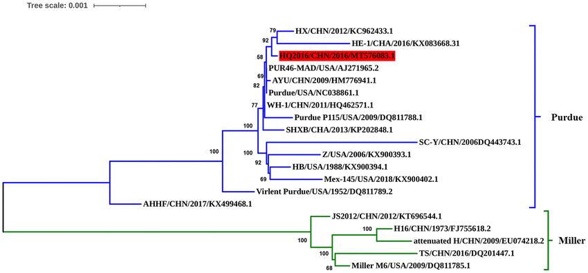
Yuan et al.                                                                                                            Transmissible Gastroenteritis Coronavirus in China

 FIGURE 1 | Isolation and identification of the TGEV HQ2016 strain. (A) Control (uninfected) PK-15 cells. (B) Cytopathic effect (CPE) induced by TGEV HQ2016 after
 infected 24 h in the PK-15 cell line. (C) Cytopathic effect (CPE) induced by TGEV HQ2016 after infected 36 h in the PK-15 cell line. (D) IFA identification of control
 (uninfected) PK15 cells. (E) IFA identification of TGEV HQ2016 infected PK15 cells. (F) Electron microscopy observation of TGEV HQ2016.

 FIGURE 2 | Visualization of genomic deletion regions in the 20 TGEV strains. (A) deletion regions of S gene. (B) deletion regions of ORF3ab gene.

trees based on the complete genome (Figure 4) divided the                                  To investigate the homology of TGEV HQ2016 with other
TGEV strains into the Purdue and Miller genotypes (5). The                              TGEVs, the nucleotide and predicted amino acid sequences of
TGEV HQ2016 strain clustered in the Purdue subgroup, together                           structural proteins and non-structural proteins were compared
with SHXB, Purdue, Purdue P115, PUR46-MAD, WH-1, AYU,                                   (Table 4). The results shown that structural proteins (S, E, M,
SC-Y, HX, HE-1, Z, HB, Mex145, Virulent Purdue, and AHHF,                               N) and non-structural proteins (replicases 1a and 1b, ORF
whereas the Miller subgroup included TS, JS2012, Miller M6,                             3a and 3b, ORF 7) of TGEV HQ2016 shared greater identity
Attenuated H, and H16. Thus, TGEV strain HQ2016 is closely                              with Purdue strains (Table 4), identity of predicted amino acid
related to the Purdue strains and more distantly to the Miller                          sequence identity in ORF1a was 98.7–100%, in ORF1b was 98.6–
strains. The strains of Purdue subgroup appear to share a                               100%, in S protein was 97.1–100%, in ORF3a was 88.3–100%,
common ancestor.                                                                        in ORF3b was 97.1–100%, in E protein was 91.5–98.8%, in M

Frontiers in Veterinary Science | www.frontiersin.org                               5                                             March 2021 | Volume 8 | Article 611721
Yuan et al.                                                                                                      Transmissible Gastroenteritis Coronavirus in China

 FIGURE 3 | Alignment of partial deduced amino acid sequence of S protein compared with strain TGEV HQ2016. (N) indicates amino acid 585, (⋆) indicates 6-nt
 deletion in the S gene, (•) indicates amino acids of the Purdue subgroup strains include TGEV HQ2016 are different from those of Miller subgroups strains.

protein was 97.3–99.6%, in N protein was 98.2–100%, in ORF7                        (jejunum and ileum) of the TGEV-HQ2016-challenged piglets.
was 93.6–100%.                                                                     The whole intestinal tracts, in which yellow watery contents had
                                                                                   accumulated, were transparent, thin walled, and gas distended.
Clinical Signs in TGEV HQ2016 Inoculated                                           No lesions were observed in any other organs of the TGEV
Piglets                                                                            HQ2016 inoculated piglets or in the organs in the negative
To evaluate the pathogenicity of TGEV HQ2016 in piglets,                           control piglets, indicating that the intestinal tract is the target
12 newborn piglets were used without colostrum. The piglets                        organ of TGEV infection. In a microscopic examination, villus
were active and fleshy before inoculation, with normal fecal                       atrophy, degenerate mucosal epithelial cells, and necrosis were
consistency. Mild diarrhea and loss of appetite were observed                      observed in both the jejunum and ileum tissues of the TGEV
in the piglets of the TGEV HQ2016 inoculated group after 12 h.                     HQ2016 inoculated piglets, but not in those of the control piglets,
Severe depression, loss of appetite, vomiting, and yellow and                      as shown in Figure 6. An immunohistochemical examination
white watery diarrhea appeared in the TGEV HQ2016 inoculated                       showed TGEV antigen in the cytoplasm of the epithelial cells in
group after 48 h. After 72 h, all the piglets in TGEV HQ2016                       the atrophied villi of the segments of jejunum and ileum tissues
inoculated group suffered watery diarrhea and were seriously                       from the piglets inoculated with TGEV HQ2016, but no reactivity
dehydrated. None of the piglets inoculated with TGEV HQ2016                        in either the jejunal or ileal tissues of the control group, as shown
died within the 84 h of the experimental period, and the control                   in Figure 6.
piglets showed no vomiting or diarrhea. The body temperatures,
body weight changes, clinical symptoms, and fecal scores of                        Viral Loads in Fecal Samples and Intestinal
both groups are shown in Figure 5. The body temperatures and                       Tissues of TGEV HQ2016 Inoculated Piglets
body weight changes were significantly lower in the piglets of                     Because TGEV caused diarrhea and intestinal damage in the
the TGEV HQ2016 inoculated group after 72 h. The clinical                          newborn piglets, we collected rectal swabs and intestinal samples
symptoms and fecal scores increased continuously for 24 h after                    from them to investigate the viral shedding in the TGEV
TGEV HQ2016 inoculated and differed significantly from those                       HQ2016 inoculated piglets. White and yellow watery feces
in the control group.                                                              were present in the TGEV HQ2016 inoculated piglets from
                                                                                   48 h after virus challenged. As shown in Figure 7, the TGEV
Histopathological Observations                                                     viral RNA was detected with quantitative RT-PCR (19). The
All the piglets were sacrificed after virus challenged 84 h.                       TGEV levels in the fecal samples were 5–10 log10 RNA copies/g
Pathological changes were mainly observed in the intestinal tracts                 at 12–84 hpi, indicating that TGEV HQ2016 infected and

Frontiers in Veterinary Science | www.frontiersin.org                          6                                           March 2021 | Volume 8 | Article 611721
Yuan et al.                                                                                                             Transmissible Gastroenteritis Coronavirus in China

TABLE 3 | Length of amino acids in the predicted structural and non-structural proteins of TGEV strains.

Strain                      ORF1a              ORF1b                S             ORF3a              ORF3b               E             M              N             ORF7

SHXB                         4017               2678              1447               71                244              82            262            382              78
Purdue P115                  4017               2678              1447               71                244              82            262            382              78
PUR46-MAD                    4017               2678              1447               71                244              82            262            382              78
WH-1                         4017               2678              1447               71                244              82            262            382              78
AYU                          4017               2678              1447               71                244              82            262            382              78
Purdue                       4017               2678              1447               71                244              82            262            382              78
HX                           4017               2678              1447               71                244              82            262            382              78
HE-1                         4017               2678              1447               71                244              82            262            382              78
SC-Y                         4017               2678              1447               71                244              82            262            382              78
Z                            4017               2678              1447               71                244              82            262            382              78
HB                           4017               2678              1447               71                244              82            262            382              78
Mex145                       4017               2678              1447               71                244              82            262            382              78
Virulent Purdue              4017               2678              1449               71                244              82            262            382              78
AHHF                         4017               2678              1448               71                244              82            262            382              78
TS                           4017               2678              1449               65                244              82            262            382              78
JS2012                       4017               2678              1449               65                244              82            262            382              78
Miller M6                    4017               2678              1449               65                244              82            262            382              78
Attenuated H                 4017               2678              1448               65                244              82            262            382              78
H16                          4017               2678              1448               65                244              82            262            382              78
HQ2016                       4017               2678              1447               71                244              82            262            382              78

    FIGURE 4 | Phylogenetic analysis of the complete genome sequences of the strain HQ2016, other TGEV reference strains. TGEV HQ2016 belongs to the Purdue
    cluster of TGEV, not the Miller cluster. Complete genome were aligned used Clustal W program which have trimed both 3′ and 5′ ends gaps between TGEV genomes
    Phylogenetic tree was constructed using the neighbor-joining method with the MEGA 6.0 program. The optimal tree with the sum of branch length = 0.02540989 is
    shown. The percentage of replicate trees in which the associated taxa clustered together in the bootstrap test (1,000 replicates) are shown next to the branches. The
    tree is drawn to scale, with branch lengths in the same units as those of the evolutionary distances used to infer the phylogenetic tree. The evolutionary distances
    were computed using the Tajima-Nei method.

reproduced in these challenged piglets. At the end of the                                 84 hpi, the viral level was highest in the jejunum (7.21 ± 0.11
challenge experiment, samples of duodenum, jejunum, ileum,                                log10 RNA copies/g), and then (in decreasing order) in the
caecum, and colon were collected for viral RNA detection. At                              ileum (6.51 ± 0.31 log10 RNA copies/g), cecum (6.28 ± 0.39

Frontiers in Veterinary Science | www.frontiersin.org                                7                                             March 2021 | Volume 8 | Article 611721
Yuan et al. Transmissible Gastroenteritis Coronavirus in China
TABLE 4 | Nucleotide and amino acid sequence identities (%) of TGEV HQ2016 strain compared with other 19 TGEV strains.
ORF1a ORF1b S ORF3a ORF3b E M N ORF7
SHXB 99.9/99.9 100.0/100.0 100.0/100.0 100.0/100.0 99.9/99.6 99.2/97.6 99.7/99.2 99.9/99.7 99.3/97.4
Purdue P115 99.9/99.9 100.0/100.0 99.9/99.9 100.0/100.0 99.9/99.6 99.6/98.8 99.9/99.6 99.9/99.7 100.0/100.0
PUR46-MAD 100.0/100.0 100.0/100.0 100.0/100.0 100.0/100.0 100.0/100.0 99.6/98.8 99.9/99.6 100.0/100.0 100.0/100.0
WH-1 100.0/100.0 100.0/100.0 100.0/100.0 100.0/100.0 99.9/99.6 99.6/98.8 99.9/99.6 100.0/100.0 100.0/100.0
AYU 99.9/99.9 100.0/100.0 100.0/100.0 100.0/100.0 100.0/100.0 99.6/98.8 99.7/99.2 100.0/100.0 100.0/100.0
Purdue 100.0/100.0 100.0/100.0 100.0/100.0 100.0/100.0 100.0/100.0 99.6/98.8 99.9/99.6 100.0/100.0 100.0/100.0
HX 99.9/99.9 100.0/100.0 99.9/99.9 100.0/100.0 100.0/100.0 99.6/98.8 100.0/100.0 100.0/100.0 100.0/100.0
HE-1 99.9/99.7 99.8/99.7 99.9/99.8 100.0/100.0 100.0/100.0 98.8/98.8 99.5/98.5 99.9/99.7 99.8/98.7
SC-Y 99.5/99.2 99.8/99.8 99.7/99.5 100.0/100.0 99.9/99.6 99.6/98.8 99.7/99.2 99.9/99.7 100.0/100.0
Z 99.9/99.8 99.9/99.9 99.6/99.0 99.1/98.6 99.9/99.6 99.2/98.8 99.7/99.2 99.8/99.7 100.0/100.0
HB 99.9/99.9 100.0/100.0 99.7/99.4 100.0/100.0 99.9/99.6 99.6/98.8 99.7/99.2 100.0/100.0 100.0/100.0
Mex145 99.9/99.8 99.9/99.9 99.7/99.2 99.5/98.6 99.9/99.6 99.2/98.8 99.7/99.2 99.9/99.7 100.0/100.0
Virulent Purdue 99.9/99.7 100.0/100.0 99.5/99.1 99.5/98.6 99.7/99.2 99.2/97.6 99.7/99.2 99.7/99.7 100.0/100.0
AHHF 99.5/99.5 100.0/100.0 98.9/98.6 100.0/100.0 99.9/99.6 99.6/98.8 99.7/99.2 100.0/100.0 100.0/100.0
TS 98.8/98.7 99.0/98.6 98.3/98.1 87.0/89.5 98.5/96.3 98.4/95.1 98.0/96.9 98.1/98.2 96.8/93.6
JS2012 99.0/99.1 99.0/99.7 98.6/98.3 88.0/88.7 98.8/97.1 98.4/95.1 98.2/97.7 98.2/98.4 96.8/93.6
Miller M6 99.0/99.1 99.1/99.6 98.3/97.1 88.0/88.3 98.9/97.5 98.0/93.9 98.2/97.7 98.2/98.4 96.6/93.6
Attenuated H 98.9/98.9 99.0/99.6 98.0/97.7 87.5/88.7 98.8/97.1 96.8/91.5 98.1/97.3 98.1/98.4 96.8/93.6
H16 98.9/98.9 99.0/99.6 98.2/97.9 88.0/88.7 98.9/97.5 97.6/93.9 98.1/97.3 98.2/98.4 96.8/93.6
FIGURE 5 | The clinical symptom in the piglets. (A) The temperature changes in different groups. (B) The body weight changes in different groups. (C) The clinical
symptom scores in different groups. (D) The fecal scores in different groups. Data are shown as mean standard (*p < 0.05, **p < 0.01).
Frontiers in Veterinary Science | www.frontiersin.org 8 March 2021 | Volume 8 | Article 611721Yuan et al. Transmissible Gastroenteritis Coronavirus in China FIGURE 6 | Pathological changes and IHC assays of TGEV HQ2016-inoculated piglets. (A,B) H.E staining for jejunum and ileum tissue section of control piglets. (C,D) H.E staining for jejunum and ileum tissue section of TGEV HQ2016 challenged piglets. Villus atrophy, degenerate mucosal epithelial cells, and necrosis. (E,F) IHC assays for jejunum and ileum tissue section of control piglets. (G,H) IHC assays for jejunum and ileum tissue section of TGEV HQ2016 challenged piglets. Positive cells presented in the epithelial cells in the atrophied villi of the segments of jejunal and ileal tissues from the piglets. Frontiers in Veterinary Science | www.frontiersin.org 9 March 2021 | Volume 8 | Article 611721
Yuan et al.                                                                                                          Transmissible Gastroenteritis Coronavirus in China

 FIGURE 7 | Reproduction of watery diarrhea and viral shedding in newborn piglets inoculated with TGEV HQ2016 via oral feeding. (A) Quantification of viral RNA levels
 of fecal samples of piglets inoculated with TGEV HQ2016. (B) Quantification of viral RNA levels in intestine tissues of piglets at 84 h inoculated with TGEV HQ2016.

log10 RNA copies/g), colon (6.23 ± 0.55 log10 RNA copies/g),                           ileum, caecum and colon, which is similarity with the report
and duodenum (5.09 ± 0.61 log10 RNA copies/g). These results                           previously (5), but there was no obviously pathological changes
confirm that TGEV HQ2016 infected the piglets and invaded                              and TGEV antigen presence in caecum and colon epithelial
their intestinal tissues.                                                              cells (which is not shown in the results of this study), this
                                                                                       result suggested that caecum and colon contained virus but
                                                                                       epithelial cells had not yet been infected. Virus-positive epithelial
DISCUSSION                                                                             cells and presence of virus in intestines indicated that TGEV
                                                                                       HQ2016 prefers to infect small intestinal epithelial cells and
TGEV is an enteropathic coronavirus that infects pigs, and                             replicate, caused pathological changes in the small intestinal
was first reported in the USA in the 1940s, after which spread                         epithelial cells, and then necrotic epithelial cells released the
throughout the world (1–3). TGEV causes significant diarrhea,                          virus into the intestinal contents, and finally excreted through
vomiting, and dehydration in suckling piglets, with a high                             the large intestines. This finding may provide a proof for
mortality rate (10). In recent years, mixed infections of TGEV                         the study of host cell infection and transmission mechanism
with other swine diarrhea virus have occurred frequently, causing                      in coronavirus.
serious economic losses in the pig industry (1). In this study, a                         Traditional TGEVs can be divided into two clusters, the
natural strain of TGEV, HQ2016, was successfully isolated from                         Purdue and Miller groups (4, 5, 7, 12, 21). In this study,
piglets intestinal samples, which collected from swine-raising                         we sequenced the entire genome of TGEV HQ2016, and a
farms in northeast China. In the farms, sows did not receive any                       phylogenetic analysis placed TGEV HQ2016 in the Purdue
vaccination for preventing diarrhea and piglets developed clinical                     cluster, indicating that it is more distantly evolutionarily
symptoms including vomiting, diarrhea, rapid weight loss and                           related to the Miller cluster. Additionally, sequence alignment
dehydration. After experimental infection, piglets showed the                          result showed two large deletions in ORF3a/3b that occur
characteristic clinical symptoms (diarrhea and vomiting) of TGE                        in the strains of the Miller cluster are not found in TGEV
from 12 h after TGEV HQ2016 inoculated until the end of the                            HQ2016 or the Purdue cluster, this may be considered to
experiment. A histopathological analysis showed villous atrophy,                       a marker of distinguishing the Purdue and Miller cluster
together with mucosal epithelial cells degeneration and necrosis,                      of TGEV. Phylogenetic analysis shown that TGEV HQ2016
in the jejunum and ileum, and virus-positive cells were present                        is closely related to with strains PUR46-MAD, Purdue,
in the villous epithelial cells in the jejunum and ileum by IHC.                       WH-1, AYU, which have the same ancestor, and this is
These results demonstrate that TGEV HQ2016 was replicated                              consistent with the results of homology comparison. Nucleotide
and had pathogenicity in enterocyte, is a natural, transmissible,                      and predicted amino-acid sequence homology comparison
enteric pathogenic porcine coronavirus. Viral nucleic acid of                          shown the structural and non-structural proteins of TGEV
TGEV was detected on rectal swabs as early as 12 h after                               HQ2016 is very similar to PUR46-MAD, Purdue, AYU
viral challenge, which indicated that virus infected the intestine                     and WH-1. These data suggest that TGEV HQ2016 might
and released to intestinal content, as described previously in                         be had the same origin with WH-1 and AYU strains in
infections with TGEV (6, 20). At 84 h of TGEV HQ2016                                   China and more similar with Purdue and PUR46-MAD
inoculated, we found a high level of viral RNA in jejunum,                             from USA.

Frontiers in Veterinary Science | www.frontiersin.org                             10                                            March 2021 | Volume 8 | Article 611721
Yuan et al. Transmissible Gastroenteritis Coronavirus in China
The 5′ - and 3′ -UTRs of CoVs are critically important for acid mutations in S gene might reduce the virulence of TGEV
viral replication and transcription (5, 22, 23). The “slippery” HQ2016 through the highly passage, this need to be confirmed
heptanucleotide sequence and a pseudoknot structure are in future studies. This hypothesis needs to be confirmed in future
both critical for viral RNA synthesis and are involved in studies and facilitate the development of an attenuated vaccine
ribosomal frame shifting (24). A complete sequence analysis for TGEV.
indicated that no deletions or insertions are present in the In conclusion, a epidemical strain of TGEV, HQ2016, was
5′ - or 3′ -UTR regions of TGEV HQ2016, and that it contains isolated from swine-raising farms in northeast China. Typical
both the slippery sequence and pseudoknot structure. These clinical signs, pathologic alterations and histological changes
sequence data suggest that the replication and transcription associated with TGE were observed in piglets inoculated with the
mechanisms of TGEV HQ2016 are conserved, as reported TGEV HQ2016 strain. Phylogenetic analysis of whole genome,
previously (5, 21, 25). nucleotide and amino acid sequence homology analysis of the
CoVs attach to their host cells via the S protein, which is the structural proteins and non-structural proteins indicated that
major immunogenic protein of the virus and stimulate the host TGEV HQ2016 belongs to the Purdue cluster, and it might be had
to produce antibodies with neutralizing activity (26). There are the same origin with WH-1 and AYU strain in China and more
at least four main antigenic sites on the S protein, designated A, similar with Purdue strains from USA. These results provide
B, C, and D (4, 27, 28). The A/B sites (amino acids 506–706) are essential information for further understanding the evolution of
the major antigenic sites and have been mapped. Single-amino- TGEV and will facilitate future investigations into the molecular
acid changes in the S protein might affect its antigenicity or pathogenesis of TGEV.
virulence (4–6). A mutation at amino acid 585 in the main major
antigenic sites A/B of the S protein of TGEV HQ2016 causes DATA AVAILABILITY STATEMENT
a serine to alanine change, which also occurs in the PUR46-
MAD, Purdue, Purdue P115, WH-1, AYU, HX, HE-1, SHXB, The datasets generated in this study can be found in online
SC-Y, Z, HB, Mex145, AHHF, H16, and Attenuated H strains, repositories. The names of the repository/repositories and
but not in the JS2012, Miller M6, TS, or Virulent Purdue strains. accession number(s) can be found below: https://www.ncbi.nlm.
This mutation may significantly influence receptor binding or nih.gov/genbank/, MT576083.
the virus interactions with neutralizing antibodies, significantly
affecting their antigenicity, this is also considered to be a marker ETHICS STATEMENT
of attenuation (6). There was a 6-nt deletion detected in the
TGEV HQ2016 S gene, as in the rest of the Purdue cluster, except The animal study was reviewed and approved by Animal
for the Virulent Purdue and AHHF strains. A 6-nt deletion (nt Experiment Ethical Committee of Heilongjiang Bayi
1,123-1,128) in the S gene was considered a trait of the TGEV Agricultural University.
strains in the Purdue cluster (5). This 6-nt deletion in the S gene
was also considered to play a role in viral attenuation (6). The AUTHOR CONTRIBUTIONS
S gene is also a hypervariable region in the TGEV genome, and
amino acids 32, 72, 100, 184, 208, 218, 389, 403, 418, 487, 562, DY: formal analysis and writing—original draft. ZY:
590, 649, 675, 815, 951, 1,109, and 1,234 of TGEV HQ2016 are methodology and validation. ML: methodology. YW: data
identical among the viruses in the Purdue cluster, but differ from curation. MS: writing and picture editing. DS: supervision.
those in the Miller cluster. These changes of amino acid in S All authors contributed to the article and approved the
gene may be related to the changes of virus virulence, which submitted version.
needs to be discussed in follow-up research. Except for S gene,
ORF3a/3b genes were considered to affect the variation between FUNDING
attenuated and virulent strains (12). However, there are some
uncertainties about the effects of deletions in TGEV ORF3a/3b This work was supported by the National Key Research
on viral virulence (1, 28–31). In our study, homology analysis and Development Program of China (2017YFD0501604-5),
shown that HQ2016 and attenuated strains PUR46-MAD (4, 32) Natural Science Foundation of Heilongjiang Province of
had highly identity. PUR46-MAD was generally considered an China (C2018049) and Postdoctoral Science Foundation of
attenuated strain of TGEV, which derivative of Purdue P115, Heilongjiang Province of China (LBH-Z19220).
and both were derived from the strain virulent Purdue after
highly passage in cell culture (4, 12, 25, 32, 33). TGEV HQ2016 ACKNOWLEDGMENTS
used in our infected experiment was only 10th passage in cell
culture. Therefore, we think that the virulence of HQ2016 might We also thank International Science Editing for editing the
be reduced by highly passage in cell culture in the future, as English text of a draft of this manuscript (http://www.
previously reported for PUR46-MAD. 6-nt deletion or amino internationalscienceediting.com).
Frontiers in Veterinary Science | www.frontiersin.org 11 March 2021 | Volume 8 | Article 611721Yuan et al. Transmissible Gastroenteritis Coronavirus in China
REFERENCES 20. Kim B, Chae C. Experimental infection of piglets with transmissible
gastroenteritis virus: a comparison of three strains (Korean, Purdue and
1. Zuniga S, Pascual-Iglesias A, Sanchez CM, Sola I, Enjuanes L.Virulence factors Miller). J Comp Pathol. (2002) 126:30–7. doi: 10.1053/jcpa.2001.0517
in porcine coronaviruses and vaccine design. Virus Res. (2016) 226:142–51. 21. Hu WW, Yu QH, Zhu LQ, Liu HF, Zhao SS, Gao Q, et al. Complete genomic
doi: 10.1016/j.virusres.2016.07.003 sequence of the coronavirus transmissible gastroenteritis virus SHXB isolated
2. Doyle LP, Hutchings LM. A transmissible gastroenteritis in pigs. J Am Vet Med in China. Arch Virol. (2014) 159:2295–302. doi: 10.1007/s00705-014-2080-9
Assoc. (1946) 108:257–9. 22. Ritchie DB, Foster DA, Woodside MT. Programmed-1 frameshifting
3. Xue R, Tian Y, Zhang Y, Zhang M, Tian F, Ma J, et al. Efficacy efficiency correlates with RNA pseudoknot conformational plasticity, not
and immunogenicity of a live L. acidophilus expressing SAD epitope of resistance to mechanical unfolding. Proc Natl Acad Sci USA. (2012)
transmissible gastroenteritis virus as an oral vaccine. Acta Virol. (2019) 109:16167–72. doi: 10.1073/pnas.1204114109
63:301–8. doi: 10.4149/av_2019_310 23. Sola I, Almazan F, Zuniga S, Luis E. Continuous and discontinuous
4. Hu XL, Li NN, Tian ZG, Yin X, Qu LD, Qu JJ. Molecular characterization and RNA synthesis in coronaviruses. Annu ReV Virol. (2015) 2:265–88.
phylogenetic analysis of transmissible gastroenteritis virus HX strain isolated doi: 10.1146/annurev-virology-100114-055218
from China. BMC Vet Res. (2015) 21:72–9. doi: 10.1186/s12917-015-0387-8 24. Sanchez CM, Gebauer F, Sune C, Mendez A, Dopazo J, Enjuanes L. Genetic
5. Zhang X, Zhu YN, Zhu XD, Shi HY, Chen JF, Shi D, et al. Identification evolution and tropism of transmissible gastroenteritis coronaviruses. Virology.
of a natural recombinant transmissible gastroenteritis virus between (1992) 190:92–105. doi: 10.1016/0042-6822(92)91195-Z
Purdue and Miller clusters in China. Emerg Microbes Infect. (2017) 6:e74. 25. Penzes Z, Gonzalez JM, Calvo E, Izeta A, Smerdou C, Mendez A, et al.
doi: 10.1038/emi.2017.62 Complete genome sequence of transmissible gastroenteritis coronavirus
6. Guo RL, Fan BB, Chang XJ, Zhou JZ, Zhao YX, Shi DY, et al. PUR46-MAD clone and evolution of the purdue virus cluster. Virus Genes.
Characterization and evaluation of the pathogenicity of a natural recombinant (2001) 23:105–18. doi: 10.1023/A:1011147832586
transmissible gastroenteritis virus in China. Virology. (2020) 545:24–32. 26. Godet M, Grosclaude J, Delmas B, Laude H. Major receptor-binding and
doi: 10.1016/j.virol.2020.03.001 neutralization determinants are located within the same domain of the
7. Li JQ, Cheng J, Lan X, Li XR, Li W, Yin XP, et al. Complete transmissible gastroenteritis virus (coronavirus) spike protein. J Virol. (1994)
genomic sequence of transmissible gastroenteritis virus TS and 3’ end 68:8008–16. doi: 10.1128/JVI.68.12.8008-8016.1994
sequence characterization following cell culture. Virol Sin. (2010) 25:213–24. 27. Brian DA, Baric RS. Coronavirus genome structure and replication. Curr Top
doi: 10.1007/s12250-010-3108-2 Microbiol Immunol. (2005) 287:1–30. doi: 10.1007/3-540-26765-4_1
8. Hou Y, Yue X, Cai X, Wang S, Liu Y, Yuan C, et al. Complete genome of 28. Sanchez CM, Pascual-Iglesias A, Sola I, Sonia Zuniga S, Enjuanes L.
transmissible gastroenteritis virus AYU strain isolated in Shanghai, China. J Minimum determinants of transmissible gastroenteritis virus enteric tropism
Virol. (2012) 86:11935. doi: 10.1128/JVI.01839-12 are located in the N-terminus of spike protein. Pathogens. (2019) 9:2.
9. Zhang X, Zhu Y, Zhu X, Chen J, Shi H, Shi D, et al. ORF3a deletion in doi: 10.3390/pathogens9010002
field strains of porcine-transmissible gastroenteritis virus in China: a hint 29. Balint A, Farsang A, Zadori Z, Hornyak A, Dencso L, Almazan F, et al.
of association with porcine respiratory coronavirus. Transbound Emerg Dis. Molecular characterization of feline infectious peritonitis virus strain DF-
(2017) 64:698–702. doi: 10.1111/tbed.12634 2 and studies of the role of ORF3abc in viral cell tropism. J Virol. (2012)
10. Xia L, Yang YH, Wang JL, Jing YC, Yang Q. Impact of TGEV infection on the 86:6258–67. doi: 10.1128/JVI.00189-12
pig small intestine. J Virol. (2018) 15:102–9. doi: 10.1186/s12985-018-1012-9 30. Kim L, Hayes J, Lewis P, Parwani AV, Chang KO, Saif LJ. Molecular
11. Vaughn EM, Halbur PG, Paul PS. Sequence comparison of porcine respiratory characterization and pathogenesis of transmissible gastroenteritis
coronavirus isolates reveals heterogeneity in the S, 3, and 3-1genes. J Virol. coronavirus (TGEV) and porcine respiratory coronavirus (PRCV) field
(1995) 69:3176–84. doi: 10.1128/JVI.69.5.3176-3184.1995 isolates co-circulating in a swine herd. Arch Virol. (2000) 145:1133–47.
12. Zhang X, Hasoksuz M, Spiro D, Halpin R, Wang S, Stollar S, et al. Complete doi: 10.1007/s007050070114
genomic sequences, a key residue in the spike protein and deletions in 31. Sola I, Alonso S, Zuniga S, Balasch M, Plana-Duran J, Enjuanes
nonstructural protein 3b of US strains of the virulent and attenuated L. Engineering the transmissible gastroenteritis virus genome as an
coronaviruses, transmissible gastroenteritis virus and porcine respiratory expression vector inducing lactogenic immunity. J Virol. (2003) 77:4357–69.
coronavirus. Virology. (2007) 358:424–35. doi: 10.1016/j.virol.2006.08.051 doi: 10.1128/JVI.77.7.4357-4369.2003
13. Sun B, Li MY, Lin SY, Shen GN, Mao RF, Yan ZH, et al. 32. Reguera J, Santiago C, Mudgal G, Ordono D, Enjuanes L, Casasnovas JM.
Establishment and application of semi-nest RT-PCR for detection Structural bases of coronavirus attachment to host aminopeptidase N and
of transmissible gastroenteritis virus. Chin Vet Sci. (2020) 50:556–62. its inhibition by neutralizing antibodies. PLoS Pathog. (2012) 8:e1002859.
doi: 10.16656/j.issn.1673-4696.2020.0062 doi: 10.1371/journal.ppat.1002859
14. Reed LJ, Muench HA. Simple method of estimating fifty percent Endpoints. 33. Sanchez CM, Izeta A, Sanchez-Morgado JM, Alonso S, Sola I, Balasch M, et al.
Am J Hygiene. (1937) 27:493–7. doi: 10.1093/oxfordjournals.aje.a118408 Targeted recombination demonstrates that the spike gene of transmissible
15. Huang B, Jennison A, Whiley D, McMahon J, Hewitson G, Graham R, et al. gastroenteritis coronavirus is a determinant of its enteric tropism and
Illumina sequencing of clinical samples for virus detection in a public health virulence. J Virol. (1999) 73:7607–18. doi: 10.1128/JVI.73.9.7607-7618.1999
laboratory. Sci Rep. (2019) 9:5409. doi: 10.1038/s41598-019-41830-w
16. Tamura K, Stecher G, Peterson D, Filipski A, Kumar S. MEGA6: molecular Conflict of Interest: The authors declare that the research was conducted in the
evolutionary genetics analysis version 6.0. Mol Biol Evol. (2013) 30:2725–9. absence of any commercial or financial relationships that could be construed as a
doi: 10.1093/molbev/mst197 potential conflict of interest.
17. Burland TG. DNASTAR’s Lasergene sequence analysis software. Methods Mol
Biol. (2000) 132:71–91. doi: 10.1385/1-59259-192-2:71 Copyright © 2021 Yuan, Yan, Li, Wang, Su and Sun. This is an open-access article
18. Martin DP, Murrell B, Golden M, Khoosal A, Muhire B. RDP4: detection distributed under the terms of the Creative Commons Attribution License (CC BY).
and analysis of recombination patterns in virus genomes. Virus Evol. (2015) The use, distribution or reproduction in other forums is permitted, provided the
1:vev003. doi: 10.1093/ve/vev003 original author(s) and the copyright owner(s) are credited and that the original
19. Yuan DW, Yan ZH, Shen GN, Wang Y, Li MY. Establishment of qRT- PCR publication in this journal is cited, in accordance with accepted academic practice.
for detection of transmissible gastroenteritis virus. Chin J Vet Sci. (2020) No use, distribution or reproduction is permitted which does not comply with these
40:1913–7. doi: 10.16303/j.cnki.1005-4545.2020.10.04 terms.
Frontiers in Veterinary Science | www.frontiersin.org 12 March 2021 | Volume 8 | Article 611721You can also read