Illuminating the cells: transient transformation of citrus to study gene functions and organelle activities related to fruit quality - Nature
←
→
Page content transcription
If your browser does not render page correctly, please read the page content below

Gong et al. Horticulture Research (2021)8:175
                                    https://doi.org/10.1038/s41438-021-00611-1
                                                                                                                                                                                         Horticulture Research
                                                                                                                                                                                                     www.nature.com/hortres

                                     METHOD                                                                                                                                                     Open Access

                                    Illuminating the cells: transient transformation of
                                    citrus to study gene functions and organelle
                                    activities related to fruit quality
                                    Jinli Gong1,2,3, Zhen Tian1,2,3, Xiaolu Qu1, Qiunan Meng1,2,3, Yajie Guan1,2,3, Ping Liu4, Chuanwu Chen4, Xiuxin Deng                                                                           1
                                                                                                                                                                                                                                      ,
                                    Wenwu Guo1, Yunjiang Cheng1,2 and Pengwei Wang 1,2,3 ✉

                                      Abstract
                                      Although multiple microscopic techniques have been applied to horticultural research, few studies of individual
                                      organelles in living fruit cells have been reported to date. In this paper, we established an efficient system for the
                                      transient transformation of citrus fruits using an Agrobacterium-mediated method. Kumquat (Fortunella crassifolia
                                      Swingle) was used; it exhibits higher transformation efficiency than all citrus fruits that have been tested and a
                                      prolonged-expression window. Fruits were transformed with fluorescent reporters, and confocal microscopy and live-
                                      cell imaging were used to study their localization and dynamics. Moreover, various pH sensors targeting different
                                      subcellular compartments were expressed, and the local pH environments in cells from different plant tissues were
                                      compared. The results indicated that vacuoles are most likely the main organelles that contribute to the low pH of
                                      citrus fruits. In summary, our method is effective for studying various membrane trafficking events, protein localization,
                                      and cell physiology in fruit and can provide new insight into fruit biology research.
                  1234567890():,;
1234567890():,;
                  1234567890():,;
1234567890():,;

                                    Introduction                                                                                        their heterogeneous expression, such a system is not ideal
                                       Citrus is one of the most important and highest-yielding                                         for studying genes that are specifically expressed in fruits.
                                    fruits in the world, and identifying genes associated with                                          Although transient transformation techniques have been
                                    desirable traits is important for the sustainable development                                       tested in fruits, such as strawberry3,4, apple5,6, and tomato7,
                                    of citrus production. However, due to its long juvenile                                             none of these techniques proved to be suitable for studying
                                    phase and diverse genetic background1, obtaining trans-                                             subcellular activities and cell physiology.
                                    genic citrus plants is difficult and time-consuming. There-                                             Conventionally, cell biological studies in perennial woody
                                    fore, developing an effective method for the transient                                              plants and fruits have relied on squashing, sectioning, or
                                    transformation of citrus species is important for gene                                              enzyme-mediated degradation of the cell wall to gain
                                    function characterizations and high-throughput screening.                                           access to the inner compartments. Therefore, conclusions
                                    The Agrobacterium-mediated infiltration of tobacco leaves                                            are inevitably obtained from observations of fixed plant
                                    is a commonly used method to test gene function in vivo or                                          tissue, which sometimes does not reflect what truly occurs
                                    study protein subcellular localization2. However, because of                                        in planta. For example, the endomembrane compartment is
                                                                                                                                        constantly moving and remodeling. It contains the plasma
                                                                                                                                        membrane (PM), endoplasmic reticulum (ER), Golgi
                                    Correspondence: Pengwei Wang (wangpengwei@mail.hzau.edu.cn)
                                    1                                                                                                   apparatus, and vacuoles, which play vital roles in protein
                                     Key Laboratory of Horticultural Plant Biology (Ministry of Education), College
                                    of Horticulture and Forestry Science, Huazhong Agricultural University, 430070                      secretion, storage, and degradation8–10. These functional
                                    Wuhan, China
                                    2
                                                                                                                                        compartments need to maintain a suitable environment;
                                     National R&D Centre for Citrus Preservation, Huazhong Agricultural University,
                                                                                                                                        any alterations of their structure, redox potential, pH
                                    430070 Wuhan, China
                                    Full list of author information is available at the end of the article
                                    These authors contributed equally: Jinli Gong, Zhen Tian

                                    © The Author(s) 2021
                                                        Open Access This article is licensed under a Creative Commons Attribution 4.0 International License, which permits use, sharing, adaptation, distribution and reproduction
                                                        in any medium or format, as long as you give appropriate credit to the original author(s) and the source, provide a link to the Creative Commons license, and indicate if
                                    changes were made. The images or other third party material in this article are included in the article’s Creative Commons license, unless indicated otherwise in a credit line to the material. If
                                    material is not included in the article’s Creative Commons license and your intended use is not permitted by statutory regulation or exceeds the permitted use, you will need to obtain
                                    permission directly from the copyright holder. To view a copy of this license, visit http://creativecommons.org/licenses/by/4.0/.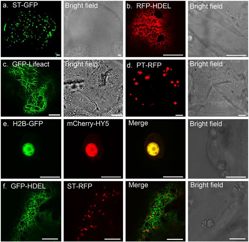
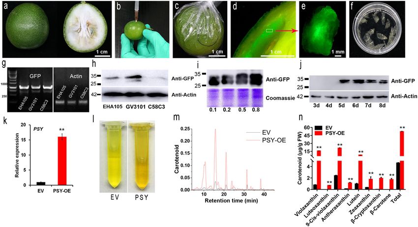
Gong et al. Horticulture Research (2021)8:175                                                                                                 Page 2 of 9

 Fig. 1 Schematic illustration of different steps in citrus fruit transient expression and detection. a A representative image of kumquat fruits
 used for injection. b Infiltration procedure: Agrobacterium solution (supplemented with p19) was injected into the epicarp at a depth of 0.1–0.3 cm.
 With gentle pressure on the plunger, the liquid spread into the fruit tissues. Each fruit was injected with ~0.4 ml Agrobacterium suspension. c Fruits
 were stored at room temperature for 2 days before being covered with cling film to prevent dehydration. d–e Dissected sections expressing GFP
 fusion proteins. f Cellulase treatment of citrus tissue for microscopic observation. g–h PCR and western blot analysis of GFP-HDEL expression in
 kumquat transformed with Agrobacterium strains EHA105, GV3101, and C58C3. i Western blot analysis of kumquat expressing GFP-HDEL at different
 bacterial (GV3101) densities. j Kumquat fruit expressing GFP-HDEL at 3 d, 4 d, 5 d, 6 d, 7 d, and 8 d after injection; actin was used as a reference. k
 Relative expression of the PSY gene in kumquat pulp after infiltration. EV, empty vector. l–m Carotenoid extracts were measured using pulp injected
 with EV (black) and PSY (red). n Carotenoid content in pulp infiltrated with PSY and EV. The data are presented as the mean ± SD of 3 independent
 replicates. Analysis of variance was performed to evaluate the significant differences based on the Student’s t-test at the significance levels of P < 0.05
 (*) and P < 0.01 (**). Considering individual variation, all data presented here were generated from at least 15 fruits, with a minimum of 3 repeats

gradient, or lipid composition may lead to severe devel-                         Here, kumquat fruits (Fortunella crassifolia Swingle) were
opmental defects and affect cell viability11,12.                               selected for Agrobacterium-mediated transient expression.
  However, most of the studies on plant cell biology have                      The kumquat is closely related to most major citrus culti-
been carried out in model systems; little is known about                       vars whose genomic information is widely available. It is
horticultural plants and fruits, which have pleiotropic fea-                   also characterized by its high sugar content, thin skin, and
tures that are absent in Arabidopsis and crop species. For                     relatively few juice cells; these characteristics provide a good
example, the local pH of every organelle is tightly controlled                 environment for infiltration and infection by Agrobacter-
and is critical to its function. Organelle pH can be measured                  ium. The feasibility of this method was demonstrated by
in vivo using a GFP-derived ratiometric pH sensor                              expressing different organelle markers to study protein
(pHluorin) in tobacco and Arabidopsis13,14. For fruits, pH                     localization, as well as by expressing pHluorin-derived
homeostasis not only affects the cell physiological envir-                     reporters to measure the pH of specific compartments of
onment but is also relevant to fruit acidity and quality.                      citrus cells. In addition, our results demonstrate that this
Despite its importance, little is known about the pH                           technique is efficient not only for cell biology studies but
environment within most intracellular compartments of                          also for the functional characterization of genes related to
fruit cells. For example, fruits of nonacidic Faris lemon had                  fruit physiology and quality.
average pH of 5.8–5.9, whereas those of acidic Faris lemon
was 4.0, and those of Frost Lisbon lemon were 3.5–3.615.                       Results and discussion
Therefore, there must be specific pathways regulating pH in                     Agrobacterium-mediated transformation of citrus fruits
different fruit varieties. To understand this biological pro-                    The transformation of citrus fruits was carried out in
cess, it is essential to measure and monitor local pH at the                   kumquat (Fig. 1a), which is one of the major citrus cul-
subcellular level.                                                             tivars in southern China16. Kumquat blooms four times a
Gong et al. Horticulture Research (2021)8:175                                                                          Page 3 of 9

year, and fresh fruits are available from November to               to test the gene function of a particular metabolic pathway
March of the next year. With good preharvest manage-                could be useful for high-throughput assays. Here, phy-
ment and postharvest storage conditions, the period of              toene synthase (PSY), which regulates the most critical
kumquat availability can be further extended. For the               step in carotenoid synthesis17,18, was selected as an
transformation experiment, fruits at the green ripening             example. The results indicated that the transient over-
stage (~120–180 days after flowering) were used, and they            expression of PSY resulted in significantly higher
were infiltrated with Agrobacterium tumefaciens carrying             expression of PSY at the mRNA level compared to the
an expression vector (35S promoter-driven). The bacterial           control fruits (Fig. 1k), concomitant with carotenoid
solution was gently injected into the epicarp at a depth of         accumulation (Fig. 1l–n). In addition, with a protein tag
0.1–0.3 cm (Fig. 1b). The mixture could distribute to an            (e.g., GFP, HA, Myc), immunoprecipitation (Co-IP) or
area with a radius of 0.8–1.3 cm from the injection point,          chromatin immunoprecipitation (ChIP-seq) could be
as indicated using a red dye (Supplementary Fig. S1). The           performed to screen possible interacting patterners of
infiltrated fruits were stored at room temperature for               citrus protein in its native environment. This is one of the
~2 days and wrapped with cling film to prevent dehy-                 advantages of our system over tobacco expression.
dration (Fig. 1c). Approximately 5 days after injection,
fluorescent signals were clearly visible near the site of            Live cell imaging of citrus fruit cells
injection (Fig. 1d–e). We tested this method using dif-                The use of GFP and its derivatives is a landmark in cell
ferent citrus cultivars, including orange, pomelo, and              biology; it can be fused with genes of interest to study their
mandarin (Supplementary Fig. S2) and found that kum-                subcellular localization, dynamics, and protein–protein
quat constantly produced good signals and high trans-               interactions8. To study subcellular activities and protein
formation efficiency. In addition, fluorescent signals could          localization in living citrus fruits, infiltrated fruits were
be detected in different tissues of the kumquat, including          prescreened using a stereomicroscope to identify the
the flavedo, albedo, juice sacs, and partition, into which           successfully transformed area. Tissues with strong fluor-
the Agrobacterium solution penetrated (Supplementary                escence signals were then hand-sliced into small pieces
Fig. S3).                                                           and transferred to a slide for observation. Alternatively,
  Moreover, we tested different Agrobacterium tumefaciens           samples could be incubated in a buffer containing cellulose
strains (e.g., EHA105, GV3101, and C58C3), all of which             and macerozyme for 1–2 h to partially digest the cell wall
carried a construct expressing GFP-HDEL (an ER marker).             (Fig. 1f). The main purpose of this additional step was to
Western blot and PCR results demonstrated that GFP was              release the cells from the tissue to facilitate observation.
expressed in all three experiments, and GV3101-mediated             Prolonged digestion can result in the production of pro-
transformation produced the highest amount of protein               toplasts, but it is normally unnecessary; ideally, it is good
(Fig. 1g–h). The optical density (OD600) of Agrobacterium           to keep any disruptions of this process to a minimum. In
also affects the level of protein expression (Fig. 1i), and it is   addition, it is crucial to minimize mechanical damage and
necessary to optimize the best OD600 for a new construct            external pressure during the sectioning and imaging of
before the actual study. If potential overexpression artifacts      fruit tissues.
are a major concern, using the lowest OD600 that produces              Protein localization is one of the key factors involved in
a strong enough signal is recommended. Otherwise, an                understanding protein function in vivo. Here, we infil-
OD600 of 0.5–0.8 can be used to provide maximum                     trated various organelle markers tagged with GFP/RFP,
expression. Moreover, it is also important to keep the              such as ST-GFP for the Golgi apparatus (Fig. 2a), RFP-
injection volume between 0.2 and 0.5 ml for each fruit, as          HDEL for the ER network (Fig. 2b), GFP-Lifeact for the
overdoses of infiltration buffer may result in unexpected            actin cytoskeletons (Fig. 2c), and PT-RFP for the plastids
rotting. In most experiments, protein expression became             (Fig. 2d). Confocal microscopy suggested that their loca-
detectable 4–5 days after infiltration (Fig. 1j). Protein            lization in citrus fruits was consistent with the results
expression could even be detected up to one month after             from N. benthamiana leaf epidermal cells (Supplementary
infiltration if the fruits were kept under appropriate con-          Fig. S5). Next, we infiltrated different markers at the same
ditions (Supplementary Fig. S4). This prolonged-expression          time to study protein colocalization. For example, H2B-
pattern was identified in most constructs in our study and           GFP and mCherry-HY5 (elongated hypocotyl5, a light-
could potentially provide a wide time window for certain            responsive transcription factor) were co-infiltrated and
downstream analyses.                                                shown to be colocalized in the nucleus (Fig. 2e). Most
                                                                    organelles and membrane structures can physically
Using the transient expression method for gene                      interact with each other to form a nexus in eukaryotic
characterization                                                    cells19,20; one of the best examples of this is the ER-Golgi
  Fruits may have unique metabolic pathways that are                interface21. In our study, we coexpressed GFP-HDEL with
absent in other plant tissues. Therefore, an efficient assay         ST-RFP to label the ER network and Golgi apparatus, and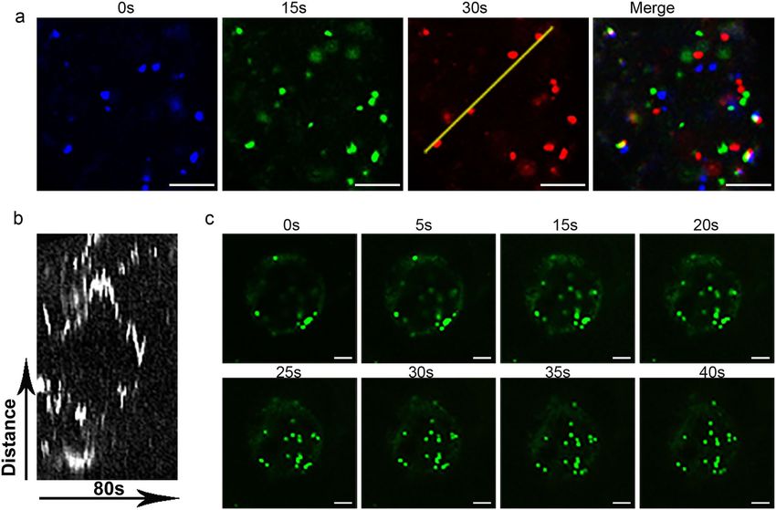
Gong et al. Horticulture Research (2021)8:175                                                                                               Page 4 of 9

 Fig. 2 Transient expression of fluorescent fusion proteins in citrus fruit cells. All constructs were infiltrated at OD600 = 0.8, and images were
 taken 5 days after infiltration. a–d Representative images of fluorescent protein fusions localized to the Golgi apparatus (ST-GFP), ER (RFP-HDEL), actin
 cytoskeleton (GFP-Lifeact), and plastids (PT-RFP). e Transient expression of mCherry-HY5 (a transcription factor) localized to the nucleus that was
 colabeled with H2B-GFP (a nuclear marker). f Coexpression of GFP-HDEL and ST-RFP identified the close association between the ER network and
 Golgi bodies in kumquat fruit cells. Scale bar, 10 μm

the results indicated a close association between these                       pHluorin-derived fluorescence ratio imaging demonstrates
structures (Fig. 2f). This finding is consistent with other                    the local pH environment at the organelle level
studies in higher plants22.                                                     The total acid level is an important measure of fruit
  It is worth pointing out that the structures of the ER and                  quality. As the fruit matures, the acidity of the juice
actin cytoskeleton are normally sensitive to mechanical                       increases, and the pH decreases. Recent developments in
disruption. Their structures remained intact in our                           pH-sensitive fluorescent sensors have provided tools to
experiment, indicating that the manipulation throughout                       measure the local pH of different subcellular compart-
the process caused little disruption to the cells. The Golgi                  ments in vivo13. For example, pHluorin is a GFP derivative
apparatus is the compartment that regulates protein                           that can be activated differentially at 405 nm and 488 nm
sorting and secretion23. In our study, ST-GFP-labeled                         lasers depending on the surrounding proton concentra-
Golgi bodies exhibited rapid movement, as indicated by                        tion. A higher 405/488 ratio is expected under basic
our time series images and kymograph (Fig. 3). This result                    conditions, while a lower 405/488 ratio indicates a more
further indicated that live-cell imaging and advanced light                   acidic environment. In this study, we used this method to
microscopy techniques (e.g., BiFC, FRAP, FRET, and ratio                      measure the local pH environment of the apoplast (PM-
imaging) could potentially be applied to study subcellar                      Apo), cytoplasm (Cyto-pH), ER (pH-HDEL) network, and
activities related to fruit physiology.                                       vacuole (BCECF-AM).
Gong et al. Horticulture Research (2021)8:175                                                                                          Page 5 of 9

 Fig. 3 Live-cell imaging of ST-GFP reveals the dynamic movement of the Golgi bodies in citrus fruit cells. a Time series of ST-GFP-labeled
 Golgi bodies over 30 s. The images representing the different time points are pseudocolored: blue (0 s), green (15 s), and red (30 s). The merged
 picture with separated signals indicates that the puncta are mobile. b A kymograph was generated along the yellow line, demonstrating the
 movement of these structures. c Time-lapse images of ST-GFP-labeled Golgi bodies over 40 s in kumquat cells. Scale bar, 5 μm

  First, His-tagged pHluorin was expressed in E. coli and                  similar to those in previous studies, indicating that this
purified using nickel-agarose beads under nondenaturing                     system works well.
conditions. These agarose beads (attached to the pHluorin                    Next, the same studies were performed in citrus. Fruits
protein) were used for calibration at a specific pH, and the                were transformed as mentioned previously, while citrus
fluorescence emission ratio at 405/488 nm was measured.                     calli were transformed using protoplasts by a PEG-
A series of measurements were performed under different                    mediated method. Surprisingly, the measured pH
pH conditions, and the data were plotted against pH as a                   showed a similar value for the apoplast and small differ-
sigmoidal curve24 (Boltzmann function, Fig. 4a).                           ences in the cytoplasm and ER lumen among leaves, calli,
  The generated sigmoidal curve was then used to cal-                      and fruit (Fig. 4). Since significantly different pH values
culate the intracellular pH in the following experiments.                  were found in crude tissue extracts (Fig. 4s), we suspect
However, pHluorin is not ideal for measuring any pH                        that this variation may have been caused by the vacuole.
below 5, as its signal tends to be quenched under extre-                   To test this hypothesis, the vacuoles were stained with
mely acidic conditions. To overcome this problem,                          BCECF-AM, and the luminal measurements showed that
another fluorescent dye, BCECF-AM, was used in addi-                        kumquat fruits exhibited the lowest vacuolar pH (5.2 ±
tion to measuring the pH within the vacuole. Using var-                    0.4), significantly lower than the 5.7 in the callus and the
ious pH buffers (pH = 3–6), the calibration curve was                      6.0 in N. benthamiana leaves (Fig. 4r).
calculated as the fluorescence ratio of BCECF-AM at                           The unique pH value of each compartment is essential
488 nm/448 nm (Fig. 4b).                                                   for protein sorting, as the binding or loading of cargo may
  Before applying these pH sensors to citrus, we verified                   rely on the formation of hydrogen bonds with their
their subcellular localization in N. benthamiana leaves                    receptors, and this process is pH sensitive. Our results
and measured their pH environment as described by                          show that every organelle in the different plants has a
Martinière et al.13. Using our calibration curves, the pH of               signature pH (Fig. 5). Most compartments in fruit cells
the apoplast, cytoplasm, ER network, and vacuole in leaf                   exhibit pH values similar to those of leaves and calli;
cells was measured to be 6.4 ± 0.2, 7.4 ± 0.5, 7.3 ± 0.5, and              however, pH variation is mainly observed in vacuoles. It is
6.0 ± 0.6, respectively (Fig. 4). These results are very                   known that the pH value decreases along the membrane
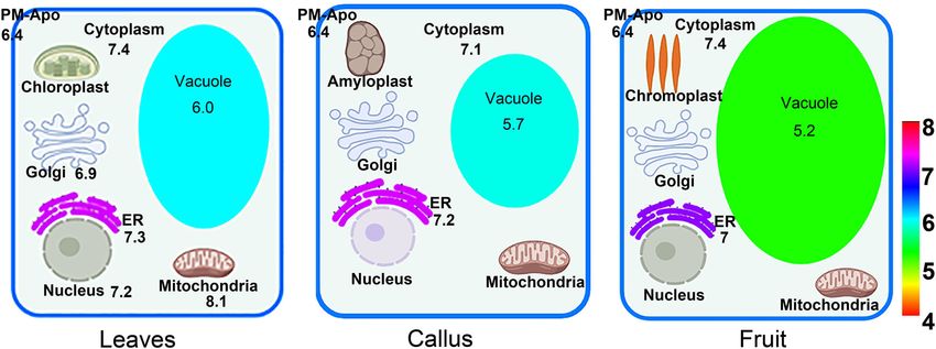
Gong et al. Horticulture Research (2021)8:175                                                                                                           Page 6 of 9

 Fig. 4 Measurement of the local pH value of different endomembrane compartments. Measurement of pH was performed in N. benthamiana
 leaf epidermal cells, citrus callus, and kumquat fruit cells transiently expressing pHluorin sensors (PM-Apo, PM facing the apoplast side, Cyto-pH for
 cytoplasm, pH-HDEL for ER), and the dye BCECF-AM for the vacuole. a In vitro calibration of pHluorin. Fluorescence emission ratios (405 nm/488 nm)
 of purified pHluorin protein in a buffer with a calibrated pH between 5.0 and 8.5. The equation applied for curve fitting is y = 0.0644e0.3827x, with R² =
 0.9895. b Images of BCECF-AM-labeled vacuoles in various designated pH buffers. The calibration curve for BCECF-AM in a pH range from 3 to 6 was
 fitted using the equation y = 0.1542x + 0.3505, with R² = 0.9477. c–e Representative image of PM-Apo in N. benthamiana leaves (c), citrus callus (d),
 and kumquat fruits (e). f–h Transient expression of Cyto-pH in N. benthamiana leaves (f), citrus callus (j) and kumquat fruits (h). i–k Transient
 expression of pH-HDEL in N. benthamiana leaves (i), citrus callus (j), and kumquat fruits (k). All images in c–k were taken with the same confocal
 settings as the image (a). Scale bars, 10 μm. l–n Representative images of BCECF-AM staining of the vacuole in N. benthamiana leaves (l), citrus callus
 (m), and kumquat fruits (n). Images were taken using the same confocal settings as the image (b). Scale bars, 10 μm. o–r Comparison of the
 measured pH of PM-Apo (o), cytoplasm (p), ER (q), and vacuole (r) in N. benthamiana leaf cells, citrus callus, and kumquat fruit cells. PM-Apo, pH =
 6.4 ± 0.2, 6.4 ± 1.0, 6.4 ± 0.6 in N. benthamiana leaf cells, citrus callus and kumquat fruit cells, respectively; Cyto, pH = 7.4 ± 0.5, 7.1 ± 0.3, 7.4 ± 0.2 in N.
 benthamiana leaf cells, citrus callus and kumquat fruit cells, respectively; ER, pH = 7.3 ± 0.5, 7.2 ± 0.4, 7.0 ± 0.3 in N. benthamiana leaf cells, citrus callus
 and kumquat fruit cells, respectively; Vacuole, pH = 6.0 ± 0.6, 5.7 ± 0.4, 5.2 ± 0.4 in N. benthamiana leaf cells, citrus callus, and kumquat fruit cells,
 respectively; n > 50; Significant differences were calculated using the Duncan test, α = 0.05. s Comparison of the total pH of crude extracts in N.
 benthamiana leaves and kumquat fruit. Student’s t-test at the significance level of P < 0.01 (**)
Gong et al. Horticulture Research (2021)8:175                                                                                        Page 7 of 9

 Fig. 5 Comparison of the pH values of intracellular compartments in leaves, calli, and fruit cells. The pH values for the mitochondria, nucleus
 and Golgi in tobacco leaves were reported previously, and the other pH measurements were generated from this study

trafficking pathway (ER-Golgi-PVC/MVB-Vacuole); pro-                         Agrobacterium transformed with a gene construct was
ton pumps that differentially localize to these compart-                  grown overnight in liquid LB medium to an O.D. of
ments may work together to achieve such a pH gradient.                    0.8–1.0. Then, they were resuspended to a final O.D. of
However, for fruit varieties with high acid contents,                     0.8 in liquid injection medium containing 0.05 M MES,
unique vacuolar proton pumps or vacuole biogenesis                        2 mM Na3PO4, 0.5% (m/v) D-glucose, and 0.1 mM
pathways must be available to generate high proton gra-                   acetylsyringone.
dients and create an extremely low pH environment25.
  In summary, we studied citrus fruit cells at the sub-                   Analysis of gene and protein expression
cellular level by using a series of fluorescent organelle                    Quantitative analysis of PSY expression levels was per-
markers and pH reporters. Different sensors (e.g., roGFP                  formed with a Roche LightCycler 480 system using the 2×
for redox potential26) could also be applied to study the                 LightCycler 480 SYBR Green master mix (Roche) and a
communication between distinct intracellular compart-                     three-step program. The primers were listed in Supple-
ments and specific metabolite (e.g., citrate27) accumula-                  mental Table 1. A total amount of 30 μg protein was used
tion at the organelle level in fruit cells. Future studies                for western blot analysis and detected by either GFP or RFP
using this method could potentially be useful to reveal the               antibodies (Biorbyt) at a dilution of 1:5000 or 1:10,000,
function and regulatory mechanisms of a particular                        respectively. Goat anti-mouse HRP (Biorbyt) was used as
organelle during fruit development or postharvest.                        the secondary antibody at a dilution ratio of 1:10,000. The
                                                                          fluorescent signals were detected using a chemilumines-
Materials and methods                                                     cent gel imaging system (Amersham Imager 600).
Plant material
  Kumquat (F. crassifolia Swingle) fruits were collected                  HPLC analysis for carotenoid metabolites
from Rong’an and Guilin city, Guangxi Province, China.                      Carotenoid extraction and quantitation were performed
They were sterilized with 2% sodium hypochlorite solu-                    as described previously28. Carotenoid metabolites were
tion for 2 min and rinsed thoroughly in water before                      analyzed by high-performance liquid chromatography
Agrobacterium injection.                                                  (HPLC), which was performed in a Waters liquid chro-
                                                                          matography system as described previously29. The car-
Plasmid construction and Agrobacterium infiltration                        otenoids were identified by their characteristic absorption
  RNA was extracted from citrus fruits using TRIzol reagent               spectra and retention times based on the literature and
according to the HiPure HP Plant RNA Mini Kit (Magen),                    standards purchased from CaroteNature (Lupsingen,
and single-stranded cDNA was synthesized by a cDNA                        Switzerland). The carotenoids were quantified using cali-
Synthesis Kit (Ferment). The PSY and HY5 coding sequences                 bration curves at 450 nm.
were PCR amplified from the cDNA library of kumquat fruit
and cloned using the pCR™8/GW/TOPO TA Cloning Kit                         Light microscopy
(Invitrogen). Recombinase reaction was performed using                      Citrus tissues injected with the Agrobacterium suspen-
Gateway LR Clonase II Enzyme Mix (Invitrogen) according                   sion were gently placed onto microscope slides and
to the manufacturer’s instructions in order to insert the gene            immediately observed with a stereomicroscope (SZX7;
into the pMDC43 plasmid for overexpression.                               Olympus) equipped with a DP70 camera. For confocal
Gong et al. Horticulture Research (2021)8:175 Page 8 of 9
microscopy, a disposable sharp blade was used to quickly et al.32 with a 505–550 nm emission bandwidth of the
cut the expressed tissue as thinly as possible on micro- PMT and excitation at 488 or 448 nm. The total pH was
scope slides dipped in water droplets for direct observa- calculated by measuring the juice of the leaf and fruit
tion. To avoid damage to fruit cells during ultrathin tissues with a pH meter (ST3100, OHAUS).
sectioning, alternatively, the samples were digested for
Acknowledgements
~1–2 h in a filter-sterilized enzymatic solution containing We thank Anne Osterrieder (Oxford, UK) for proofreading the manuscript. We
1.5% cellulase R10, 0.4% macerozyme R1, 0.4 M mannitol, thank Nadine Paris (Montpellier, France) for providing the various pH sensors
20 mM KCl, 20 mM MES (pH = 5.8), and 10 mM CaCl2. used in this study. We thank Haoliang Wan, Meiyan Shi, and Miao Zhang
(Huazhong Agricultural University) for their help in this work. This work was
The samples were mounted on microscope slides, very supported by the National Key Research and Development Program
carefully sealed with nail polish, and imaged as described (2019YFD1000103) and NSFC grants (no. 31772281, 91854102) to P.W.
previously30. All fluorescent protein markers used in this
study are listed in Supplemental Table 2. Author details
1
Key Laboratory of Horticultural Plant Biology (Ministry of Education), College
of Horticulture and Forestry Science, Huazhong Agricultural University, 430070
Protoplast preparation and transfection Wuhan, China. 2National R&D Centre for Citrus Preservation, Huazhong
Citrus unshiu callus “Guoqing No. 1” grown for Agricultural University, 430070 Wuhan, China. 3Interdisciplinary Sciences
Research Institute, Huazhong Agricultural University, 430070 Wuhan, China.
~20 days was selected and carefully clamped into a vessel 4
Guangxi Academy of Specialty Crops/Guangxi Engineering Research Center of
containing the enzymatic solution (same as above) with Citrus Breeding and Culture, 541004 Guilin, China
tweezers. Then, enzymatic hydrolysis was induced by
Author contributions
shaking the mixture slightly overnight under dark con- P.W. and J.G. designed the experiments; J.G. and Z.T. performed the
ditions to obtain enough protoplasts. Protoplast trans- experiments; X.Q., Q.M., Y.G., P.L., and C.C. helped with the experiments; W.G.
fection was performed as recommended by Yoo et al.31. and X.D. provided citrus materials and provided valuable suggestions
throughout the study; P.W. and J.G. wrote the manuscript; and all authors read
the final manuscript.
Calibration of pH sensors
Escherichia coli (BL21) was grown to OD600 = 0.8 at Competing interests
37 °C, and 0.1 mM IPTG was added to the bacterial The authors declare no competing interests.
solution to induce the expression of the target protein
overnight. Then, the bacteria were collected by cen- Supplementary information The online version contains supplementary
trifugation, washed three times with water, and sonicated material available at https://doi.org/10.1038/s41438-021-00611-1.
in phosphate buffer. The bacterial lysate was centrifuged
at 1000 rpm and 4 °C for 1 h, and the supernatant was Received: 11 January 2021 Revised: 14 April 2021 Accepted: 4 May 2021
collected and filtered through a 0.45 μm filter. Next, the
target protein was purified using Ni-NTA His Bind Resin
following the manufacturer’s instructions (Biotech,
References
Wuhan, China). The recombinant pHluorin was diluted in 1. Peña, L. & Navarro, L. Transgenic citrus. In: Biotechnology in Agriculture And
50 mM MES-KOH buffers with different pH values. Using Forestry: Transgenic Trees. (ed. Bajaj Y. P. S.) 39–53 (Springer Verlag, Berlin, 1999).
a confocal microscope (SP8; Leica), fluorescence signals at 2. Sparkes, I. A., Runions, J., Kearns, A. & Hawes, C. Rapid, transient expression of
fluorescent fusion proteins in tobacco plants and generation of stably
emission wavelengths of 505–550 nm were collected with transformed plants. Nat. Protoc. 1, 2019–2025 (2006).
excitation wavelengths of 405 or 488 nm. The pH profile 3. Agius, F., Amaya, I., Botella, M. A. & Valpuesta, V. Functional analysis of
was expressed by converting the grayscale ratio image into homologous and heterologous promoters in strawberry fruits using transient
expression. J. Exp. Bot. 56, 37–46 (2005).
pseudocolor using ImageJ. All data are shown as the 4. Hoffmann, T., Kalinowski, G. & Schwab, W. RNAi-induced silencing of gene
means and standard deviations from at least 10 images for expression in strawberry fruit (Fragaria x ananassa) by agroinfiltration: a rapid
each indicated pH value. All curve fittings were performed assay for gene function analysis. Plant J. 48, 818–826 (2006).
5. An, J.-P. et al. EIN3-LIKE1, MYB1, and ETHYLENE RESPONSE FACTOR3 act in a
using Origin 9.0 SR1(OriginLab Corp., Northhampton, regulatory loop that synergistically modulates ethylene biosynthesis and
MA, USA). anthocyanin accumulation. Plant Physiol. 178, 808–823 (2018).
6. Li, T. et al. Apple (Malus domestica) MdERF2 negatively affects ethylene bio-
synthesis during fruit ripening by suppressing MdACS1 transcription. Plant J.
Measurement of pH 88, 735–748 (2016).
To measure the pH of the specific pHluorin-targeted 7. Orzaez, D., Mirabel, S., Wieland, W. H. & Granell, A. Agroinjection of tomato
compartment, samples were excited at 405 or 488 nm as fruits. A tool for rapid functional analysis of transgenes directly in fruit. Plant
Physiol. 140, 3–11 (2006).
described above. The parameters of the images taken with 8. Hawes, C., Boevink, P., Roberts, A. & Brandizzi, F. GFP enlightens the study of
CLSM were described by Martinière et al.13, and the pH endomembrane dynamics in plant cells. Plant Biosyst. 135, 3–12 (2001).
value was extrapolated from the sigmoidal function 9. Robinson, D. G., Brandizzi, F., Hawes, C. & Nakano, A. Vesicles versus tubes: is
endoplasmic reticulum-golgi transport in plants fundamentally different from
obtained from the calibration curve in vitro. The vacuolar other eukaryotes? Plant Physiol. 168, 393–406 (2015).
pH was measured with the cell-permeant and pH- 10. Wang, P. W. & Hussey, P. J. Interactions between plant endomembrane sys-
sensitive fluorescent dye BCECF-AM according to Tang tems and the actin cytoskeleton. Front. Plant Sci. 6, 422 (2015).Gong et al. Horticulture Research (2021)8:175 Page 9 of 9
11. Boutte, Y. & Moreau, P. Modulation of endomembranes morphodynamics in 22. Sparkes, I. A., Ketelaar, T., de Ruijter, N. C. A. & Hawes, C. Grab a golgi: laser
the secretory/retrograde pathways depends on lipid diversity. Curr. Opin. Plant trapping of golgi bodies reveals in vivo interactions with the endoplasmic
Biol. 22, 22–29 (2014). reticulum. Traffic 10, 567–571 (2009).
12. Chen, J., Stefano, G., Brandizzi, F. & Zheng, H. Q. Arabidopsis RHD3 mediates 23. Boevink, P. et al. Stacks on tracks: the plant Golgi apparatus traffics on an actin/
the generation of the tubular ER network and is required for Golgi distribution ER network. Plant J. 15, 441–447 (1998).
and motility in plant cells. J. Cell Sci. 124, 2241–2252 (2011). 24. Schulte, A., Lorenzen, I., Bottcher, M. & Plieth, C. A novel fluorescent pH probe
13. Martiniere, A. et al. In vivo intracellular pH measurements in tobacco and for expression in plants. Plant Methods 2, 7 (2006).
Arabidopsis reveal an unexpected pH gradient in the endomembrane system. 25. Strazzer, P. et al. Hyperacidification of Citrus fruits by a vacuolar proton-
Plant Cell 25, 4028–4043 (2013). pumping P-ATPase complex. Nat. Commun. 10, 744 (2019).
14. Shen, J. B. et al. Organelle pH in the Arabidopsis endomembrane system. Mol. 26. Wang, P. et al. KMS1 and KMS2, two plant endoplasmic reticulum proteins
Plant 6, 1419–1437 (2013). involved in the early secretory pathway. Plant J. 66, 613–628 (2011).
15. Aprile, A. et al. Expression of the H plus -ATPase AHA10 proton pump is 27. Hu, Y. et al. A novel “off-on” fluorescent probe based on carbon nitride
associated with citric acid accumulation in lemon juice sac cells. Funct. Integr. nanoribbons for the detection of citrate anion and live cell imaging. Sensors
Genomics 11, 551–563 (2011). 18, 1163 (2018).
16. Zhu, M. et al. A comprehensive proteomic analysis of elaioplasts from citrus 28. Cao, H. B. et al. Comprehending crystalline beta-carotene accumulation by
fruits reveals insights into elaioplast biogenesis and function. Hortic. Res. 5, 6 comparing engineered cell models and the natural carotenoid-rich system of
(2018). citrus. J. Exp. Bot. 63, 4403–4417 (2012).
17. Fraser, P. D., Enfissi, E. M. A. & Bramley, P. M. Genetic engineering of 29. Lu, S. W. et al. The citrus transcription factor CsMADS6 modulates carotenoid
carotenoid formation in tomato fruit and the potential application of metabolism by directly regulating carotenogenic genes. Plant Physiol. 176,
systems and synthetic biology approaches. Arch. Biochem. Biophys. 483, 2657–2676 (2018).
196–204 (2009). 30. Wang, P. W. & Hussey, P. J. NETWORKED 3B: a novel protein in the actin
18. Paine, J. A. et al. Improving the nutritional value of Golden Rice through cytoskeleton-endoplasmic reticulum interaction. J. Exp. Bot. 68, 1441–1450
increased pro-vitamin A content. Nat. Biotechnol. 23, 482–487 (2005). (2017).
19. Stefano, G., Hawes, C. & Brandizzi, F. ER - the key to the highway. Curr. Opin. 31. Yoo, S. D., Cho, Y. H. & Sheen, J. Arabidopsis mesophyll protoplasts: a versatile
Plant Biol. 22, 30–38 (2014). cell system for transient gene expression analysis. Nat. Protoc. 2, 1565–1572
20. Wang, P. W., Hawes, C. & Hussey, P. J. Plant endoplasmic reticulum-plasma (2007).
membrane contact sites. Trends Plant Sci. 22, 289–297 (2017). 32. Tang, R.-J. et al. Tonoplast calcium sensors CBL2 and CBL3 control plant
21. Hawes, C., Osterrieder, A., Hummel, E. & Sparkes, I. The plant ER-golgi interface. growth and ion homeostasis through regulating V-ATPase activity in Arabi-
Traffic 9, 1571–1580 (2008). dopsis. Cell Res. 22, 1650–1665 (2012).You can also read