Mitogen-Inducible Gene 6 Inhibits Angiogenesis by Binding to SHC1 and Suppressing Its Phosphorylation - Frontiers
←
→
Page content transcription
If your browser does not render page correctly, please read the page content below
ORIGINAL RESEARCH
                                                                                                                                     published: 22 February 2021
                                                                                                                                   doi: 10.3389/fcell.2021.634242

                                            Mitogen-Inducible Gene 6 Inhibits
                                            Angiogenesis by Binding to SHC1
                                            and Suppressing Its Phosphorylation
                                            Lixian Liu, Liying Xing, Rongyuan Chen, Jianing Zhang, Yuye Huang, Lijuan Huang,
                                            Bingbing Xie, Xiangrong Ren, Shasha Wang, Haiqing Kuang, Xianchai Lin, Anil Kumar,
                                            Jong Kyong Kim, Chunsik Lee* and Xuri Li*
                                            State Key Laboratory of Ophthalmology, Zhongshan Ophthalmic Center, Sun Yat-sen University, Guangzhou, China

                                            The mitogen-inducible gene 6 (MIG6) is an adaptor protein widely expressed in vascular
                                            endothelial cells. However, it remains unknown thus far whether it plays a role in
                                            angiogenesis. Here, using comprehensive in vitro and in vivo model systems, we unveil
                          Edited by:
                                            a potent anti-angiogenic effect of MIG6 in retinal development and neovascularization
                             Bin Ren,       and the underlying molecular and cellular mechanisms. Loss of function assays using
University of Alabama at Birmingham,
                                            genetic deletion of Mig6 or siRNA knockdown increased angiogenesis in vivo and in
                        United States
                                            vitro, while MIG6 overexpression suppressed pathological angiogenesis. Moreover, we
                      Reviewed by:
                         Jack Lawler,       identified the cellular target of MIG6 by revealing its direct inhibitory effect on vascular
Beth Israel Deaconess Medical Center        endothelial cells (ECs). Mechanistically, we found that the anti-angiogenic effect of
         and Harvard Medical School,
                       United States
                                            MIG6 is fulfilled by binding to SHC1 and inhibiting its phosphorylation. Indeed, SHC1
                           Qiulun Lu,       knockdown markedly diminished the effect of MIG6 on ECs. Thus, our findings show
     Nanjing Medical University, China
                                            that MIG6 is a potent endogenous inhibitor of angiogenesis that may have therapeutic
                            Biao Yan,
              Fudan University, China       value in anti-angiogenic therapy.
                   *Correspondence:         Keywords: MIG6, angiogenesis, SHC1, endothelial cell, ocular neovascularization
                                Xuri Li
               lixr6@mail.sysu.edu.cn
                          Chunsik Lee
            chunsik@mail.sysu.edu.cn
                                            INTRODUCTION
                                            The blood vessel network is vital for both normal physiology and numerous diseases. Blood
                     Specialty section:
                                            vessels not only transport oxygen and nutrient to tissues and cells required for their functions
          This article was submitted to
                               Signaling,
                                            and maintenance, they also have unique functions by serving as a regulator of vascular tone, organ
                 a section of the journal   development, immunity, and blood-organ communication (Karaman et al., 2018; Li and Carmeliet,
   Frontiers in Cell and Developmental      2018). However, uncontrolled growth of new blood vessel can result in life-threatening pathologies,
                                 Biology    such as cancer and many other neovascular diseases (Apte et al., 2019; Li et al., 2019). Angiogenesis
       Received: 27 November 2020           therefore needs to be tightly controlled to avoid overproduction of blood vessels. Currently, the
        Accepted: 05 February 2021          majority of the studies have focused on angiogenic factors. Much knowledge in this aspect has been
       Published: 22 February 2021          gained and anti-angiogenic therapies targeting angiogenic factors, such as VEGF, have been used to
                              Citation:     treat patients with neovascular diseases (Apte et al., 2019). However, despite the great success, drug
      Liu L, Xing L, Chen R, Zhang J,       resistance can develop over time, suggesting the involvement of multiple factors and pathways,
     Huang Y, Huang L, Xie B, Ren X,        such as genetic instability of tumor cells that up-regulates pro-angiogenic factors to overcome the
   Wang S, Kuang H, Lin X, Kumar A,         inhibitor (Ribatti, 2016; Haibe et al., 2020). Given the presence of diverse and abundant angiogenic
       Kim JK, Lee C and Li X (2021)
                                            factors, endogenous anti-angiogenic factors would be constantly required to keep the angiogenic
    Mitogen-Inducible Gene 6 Inhibits
Angiogenesis by Binding to SHC1 and
                                            factors in check but less is known in this aspect. Indeed, decreased or the lack of the expression of
     Suppressing Its Phosphorylation.       anti-angiogenic factors may often be the reason of pathological neovascularization (Murugeswari
      Front. Cell Dev. Biol. 9:634242.      et al., 2008; Zhang et al., 2020). Studying such endogenous anti-angiogenic molecules is therefore
      doi: 10.3389/fcell.2021.634242        of critical importance.

Frontiers in Cell and Developmental Biology | www.frontiersin.org                1                                       February 2021 | Volume 9 | Article 634242
Liu et al.                                                                                       Anti-angiogenic Effects of MIG6 by Binding to SHC1

    MIG6 is a ubiquitously expressed cytoplasmic adaptor protein          retinal neovascularization areas were analyzed using whole
that modulates many cell surface receptors (Zhang and Vande               mount retinae stained with IB4-Alexa 488 (I21411, Invitrogen,
Woude, 2007; Sasaki et al., 2018). MIG6 is involved in diverse            Waltham, MA, USA). Neovascularization areas were outlined
biological events, including suppressing the epidermal growth             and quantified as the percentage of the total area of the retina
factor receptor (EGFR) pathway (Ferby et al., 2006; Zhang and             using ImageJ (NIH, Bethesda, MD, USA).
Vande Woude, 2007; Park et al., 2015). However, MIG6 has many
EGFR-independent functions. For example, MIG6 induces cell                Isolation and Culture of Murine Primary
cycle arrest in an EGFR-independent manner (Sasaki et al., 2018).         Vascular Endothelial Cells
MIG6 is widely expressed in various types of cells and tissues (Jin       Murine primary endothelial cells were isolated from lungs
et al., 2007; Zhang and Vande Woude, 2007; Anastasi et al., 2016),        of 6-week old mice. Lungs were harvested and digested
including the vascular system (Jin et al., 2009; Lee et al., 2014).       with collagenase type I (17100017, ThermoFisher Scientific,
The expression of MIG6 is rapidly induced by various growth               Waltham, MA, USA). The digested tissues were pelleted and
factors and cellular stresses under pathological conditions (Zhang        resuspended in PBS with 0.1% BSA and incubated with rat
and Vande Woude, 2007). However, it remains thus far unknown              anti-mouse CD31 conjugated Dynabeads at room temperature
whether it plays a role in angiogenesis.                                  for 15 min. The bead-bound cells were recovered using a
    Here, we utilized both knockout mice and cultured endothelial         magnetic separator and resuspended in complete culture
cells and examined the effect of MIG6 on angiogenesis. We found           medium (DMEM containing 20% FBS, supplemented with
that MIG6 is a potent endogenous inhibitor of angiogenesis.               100 µg/ml heparin, 100 µg/ml ECGS, non-essential amino acids,
Genetic deletion of Mig6 increased retinal angiogenesis in                L-glutamine and antibiotics). Primary ECs within 3 passages were
mice, and its overexpression inhibited hypoxia-induced retinal            used for experiments.
neovascularization. Overexpression of MIG6 reduced aortic and
choroidal microvessel growth, and suppressed endothelial cell
proliferation, migration and sprouting. Mechanistically, we show
                                                                          siRNA Knockdown and Adenovirus
that the anti-angiogenic effect of MIG6 is exerted by inhibiting          Infection of Endothelial Cells
SHC1 signaling. Our thus findings reveal a new function for               For siRNA knockdown, ECs were transfected with human MIG6
MIG6 as an endogenous inhibitor of angiogenesis, which may                siRNA (5′ -CUACACUUUCUGAUUUCAA-3′ ) (Liu et al., 2012),
have implications in anti-angiogenic therapy.                             human SHC1 siRNA (5′ CUACUUGGUUCGGUACAUGGG-
                                                                          3′ ) (Lundgren et al., 2006), or non-targeting scrambled negative
                                                                          control (Ribobio, Guangzhou, China) using ESCORT III (L3037,
MATERIALS AND METHODS                                                     Sigma). For adenoviral infection, ECs were infected with Ad-
Mice                                                                      MIG6 or Ad-GFP at an MOI of 10 for 48 h.
All animal experiments were approved by the Animal Use
and Care Committee of Zhongshan Ophthalmic Center, Sun                    Antibodies
Yat-sen University (2015-098). The Mig6 knockout mice were                Antibodies used in Western blots were as the following: anti-β-
kindly provided by Dr. George Vande Woude at Van Andel                    actin (A5316, Sigma), anti-tubulin (T6734, Sigma), anti-MIG6
Institute (Grand Rapids, MI, USA). Mig6 knockout mice used for            (WH0054206M1, Sigma), anti-pTyr239/240 SHC1 (2434, Cell
experiments were bred on C57Bl/6J background for more than                Signaling), anti-SHC1 (610878, BD Bioscience), anti-phospho-
six generations and littermates were used for experiments.                p44/42 MAPK (9101, Cell Signaling), anti-p44/42 MAPK (9102,
                                                                          Cell Signaling), anti-GST (2625, Cell Signaling), and anti-HA
Cell Culture and Reagents                                                 tag (A01244, GenScript). Immunoreactivity was detected using
The primary human retinal endothelial cells (HREC) were                   horseradish-peroxidase (HRP)-conjugated secondary antibody
purchased from Angio-Proteomie (Boston, MA, USA) and                      (RAG0072 for anti-rabbit, GAM0072 for anti-mouse, 1:5,000
cultured in endothelial cell medium (ScienCell Research,                  dilution, Multi Sciences, Hangzhou, China).
Carlsbad, CA, USA) containing endothelial cell growth
supplement (ECGS), 5% FCS, and penicillin/streptomycin.                   Construction of SHC1 Deletion Mutants
HREC within 8 passages were used for experiments.                         The cDNA encoding human SHC1 in pcDNA 3.1 (+) vector was
                                                                          obtained from GenScript (Piscataway, NJ, USA) and subcloned
Retinopathy of Prematurity (ROP) Mouse                                    into a pLV-3xHA vector (Inovogen, Chongqing, China). The
Model                                                                     deletion mutants of SHC1 1PTB (deletion of amino acids
C57Bl6J mice at post-natal day 7 (P7) were exposed to 75%                 30–210 corresponding to protein tyrosine binding domain),
oxygen for 5 days, after which the mice were intravitreally               1Pro-rich (deletion of amino acids 300–366 corresponding
injected with 1 µl of adenovirus expressing GFP (Ad-GFP,                  to proline-rich domain), and 1SH2 (deletion of amino
CV10001, 1.0 × 10¹³ pfu/ml, Vigene Biosciences, Rockville,                acids 377–469 corresponding to SH2 domain) were generated
MD, USA) or adenovirus expressing human MIG6 (Ad-                         using a Quickchange site-directed mutagenesis kit (Agilent
MIG6, VH894726, 1.0 × 10¹³ pfu/ml, Vigene Biosciences,                    Technologies) according to the manufacturer’s instructions. The
Rockville, MD, USA), and then returned to normoxia for                    sequences of the oligonucleotides used to generate Shc1 mutants
additional 5 days. Mouse eyes were collected at P17 and the               are as follows:

Frontiers in Cell and Developmental Biology | www.frontiersin.org     2                                   February 2021 | Volume 9 | Article 634242
Liu et al.                                                                                        Anti-angiogenic Effects of MIG6 by Binding to SHC1

(1) 1PTB                                                                 HRECs were suspended in serum-free endothelial medium after
    CCGGACTCAGATCTCGAATT (forward)                                       treatment with siMIG6 or siMIG6 + siSHC1 and subsequently
    TACCCGGTAGAATTATCTAGGGATC (reverse)                                  plated at 10,000 cells/100 µl/well on top of Matrigel in triplicates.
(2) 1Pro-rich                                                            After 6 h, the formation of tube-like structures was taken and
    ATCCAGAAGTCCGCAAACAGTCGGTGTCCATGG                                    analyzed using ImageJ.
    CTGAG (forward)
    CTGTTTGCGGACTTCTGGA (reverse)                                        EC Spheroid Sprouting Assay
(3) 1SH2                                                                 3.2 × 10⁵ of HRECs (800 cells/spheroid) were trypsinized and
    CTGAGCAGCTCCGAGGGGAGGAGCGGAAACTGTC                                   suspended in EC medium (ScienCell Research) containing
    TAGAGG (forward)                                                     20% methylcellulose (Methocel, M0512, Sigma). 100 µl/well of
    CTCCCCTCGGAGCTG (reverse)                                            HRECs were in a round-bottom 96-well plate and incubated
                                                                         overnight for spheroid formation. EC spheroids were
The deletion mutants were verified by sequencing and expressed
                                                                         resuspended in collagen I solution (800 µl of collagen, 200
in 293T cells by transfection using Fugene 6 (Promega, Madison,
                                                                         µl of 10x M199 media, 240 µl of 0.25N of NaOH, 200 µl 0.2M
WI, USA). Two days after transfection, pull-down assays were
                                                                         of HEPES, 360 µl of H2 O) and added 50 µl collagen solution in
performed to examine the binding of MIG6.
                                                                         a 96-well plate and incubate at 37◦ C for 20 min. EC spheroids
                                                                         were centrifuged at 300 × g and resuspended in media after
Purification of GST Fusion Protein and                                   collecting from a 96-well plate. 80% Methocel (80% Methocel +
Pull-Down Assays                                                         20% FBS) with collagen I solution were mixed and resuspended
The glutathione S-transferase (GST) fusion MIG6 protein                  EC spheroids and transfer to a 96-well plate. After incubation at
was generated by expressing human full-length MIG6 cDNA                  37◦ C for 15 min, serum-free media was added on top of the gel.
subcloned into a pGEX-4T-1 (GE Healthcare Life Sciences) in              After 24 h, the images were obtained and the sprouts and their
E. coli. For GST-Mig6 pull-down assay, 2 µg of GST-MIG6                  total length were analyzed using an ImageJ.
fusion protein was added to 30 µl of glutathione magnetic beads
(L00327, GenScript) and incubated at 4◦ C for 2 h. The protein-          Aortic Ring Assay
bound beads were then washed three times with RIPA buffer                Aorta from Mig6 knockout mice and wild-type littermates were
(R0010, Solarbio, Beijing, China) and incubated with the cell            excised and the surrounding tissues were removed. After aortic
lysates of 293T cells transfected with SHC1 plasmids at 4◦ C for         rings were cut into pieces (1 mm in length) and were placed on
overnight. The beads were then washed with RIPA buffer and               the top of growth factor-reduced Matrigel (354230, Corning) in
subjected to Western blot analysis.                                      24-well plates and incubated for 7 days. Images of individual
                                                                         explants were obtained using a phase-contrast microscope, and
Co-immunoprecipitation of MIG6 and                                       converted to binary mode using a low-pass filter and threshold
SHC1                                                                     transformation using ImageJ. The areas of sprouting microvessels
HRECs were homogenized in RIPA lysis buffer with a protease              were quantified using ImageJ.
and phosphatase inhibitor tablet (88668, Thermo Fisher
Scientific) following EGF (50 ng/ml, PeproTech) stimulation              Choroid Sprouting Assay
for 30 min. The cell lysates were incubated with an anti-                Mouse eyes were enucleated from wild-type and Mig6 knockout
MIG6 antibody (sc-46167, Santa Cruz Biotechnology) for                   mice. After the cornea and the lens were removed, the choroid-
overnight at 4◦ C and precipitated using immobilized protein             scleral complex was separated from the retina and cut into ∼1 ×
A/G plus-agarose (sc-2003, Santa Cruz Biotechnology).                    1 mm pieces. The choroid-scleral complex was placed in growth
Immunoprecipitated protein complexes were subjected to                   factor-reduced Matrigel (354230, Corning) for a week. The areas
Western blot analysis.                                                   of sprouting microvessels were quantified using ImageJ.

Cell Proliferation and Viability Assay                                   Immunofluorescence Staining and Analysis
Cell proliferation was determined using a Click-iT EdU Imaging           of Mouse Retinal Vasculature
Kit with Alexa Fluor 594 (C10086, Invitrogen) according to the           The mouse retinal vascularized area of wild-type and Mig6
manufacturer’s instruction. Images were obtained and analyzed            knockout mice at P5 was stained using Alexa fluor 488-
by ImageJ. For cell viability, 5 × 10³ cells of MIG6-knockdown or        conjugated IB4 (I21411, Invitrogen) and anti-Erg (ab92513,
MIG6-overexpressing HRECs were seeded in 96-well plates. The             Abcam) antibody. DAPI (D3571, Thermo Fisher Scientific)
cells were incubated for 24 h and viable cells were assessed using       was used for nuclear staining. Images were acquired and the
an MTT (3-(4,5-dimethylthiazol-2-yl)-2,5-diphenyltetrazolium             vascularized retinal areas quantified using ImageJ.
bromide, CT02, Sigma) method.
                                                                         Transwell Cell Migration Assay
Tube Formation Assay                                                     HRECs (7.5 × 10⁴ ) in serum-free media were seeded to the
96-well plates were coated with 50 µl/well of growth factor              upper chamber of the transwell system with 8 µm pore size (3422,
reduced Matrigel (356230, BD Biosciences). The plates were               Corning) for 24 h after 2 days infection with Ad-Control or Ad-
incubated at 37◦ C for 30 min to allow Matrigel to polymerize. The       MIG6. Each 50 ng/ml of either EGF (PeproTech) or VEGFA

Frontiers in Cell and Developmental Biology | www.frontiersin.org    3                                     February 2021 | Volume 9 | Article 634242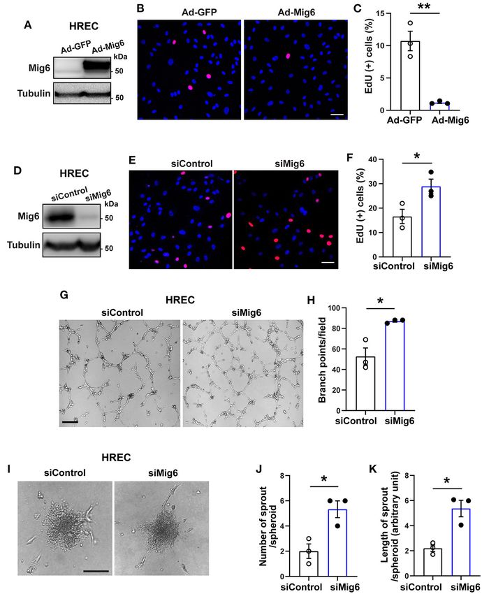
Liu et al.                                                                                                              Anti-angiogenic Effects of MIG6 by Binding to SHC1

(100-20, PeproTech) was added in the bottom chamber. After                                Statistical Analysis
24 h, the bottom chamber was stained with DAPI (Thermo Fisher                             Comparisons between two groups were analyzed using an
Scientific) and images were taken to analyze. The number of                               unpaired or paired Student’s t-test (two-tailed) using GraphPad
migrated cells per area was quantified using ImageJ.                                      Prism (GraphPad Software, La Jolla, CA, USA). Data are

  FIGURE 1 | Increased retinal angiogenesis in Mig6 deficient mice. (A,B) Representative images of whole mount retinae of wild-type (WT) and Mig6−/− mice at P5
  stained for IB4 (green). (C) Quantification of retinal blood vessel densities stained with IB4 in WT and Mig6−/− mice (n = 5 fields per retina). (D,E) Higher magnification
  of dotted regions in whole mount retinae of WT (D) and Mig6−/− (E) mice. (F) IB4 staining (green) in (D) and (E) showing more vascular branch points in the retinae of
  Mig6−/− mice at P5 (n = 5 fields per retina). (G) Whole mount retinae co-stained for ERG (red) and IB4 (white) to label endothelial cells in WT and Mig6−/− mice at P5.
  (H) Quantification of ERG+ ECs in the front of retinal vascular plexus (n = 3 fields per retina) in WT and Mig6−/− mice at P5. (I) Images of retinal blood vessel tip cells
  and their filopodia extensions at the angiogenic front of WT and Mig6−/− retinae at P5. (J) Quantification of the number of retinal blood vessel tip cells per field (n = 3
  fields per retina) in WT and Mig6−/− mice at P5. (K) Measurement of the length of filopodia of tip cells per field (n = 3 fields per retina). Scale bars: 50 µm for (A) and
  (I); 100 µm for (G). Data represent mean ± SEM. * p < 0.05, ** p < 0.01 (two-tailed paired Student’s t-test).

Frontiers in Cell and Developmental Biology | www.frontiersin.org                     4                                            February 2021 | Volume 9 | Article 634242
Liu et al.                                                                                                            Anti-angiogenic Effects of MIG6 by Binding to SHC1

  FIGURE 2 | MIG6 suppresses microvessel outgrowth. (A) Mouse aortic rings from WT or Mig6−/− aorta incubated for 7 days. (B) Quantification of vascular sprouting
  area per aortic ring in (A). n = 4 aortic rings per group. (C) Mouse aortic rings infected with Ad-GFP or Ad-MIG6 and incubated for 7 days. (D) Quantification of
                                                                                                                                                              (Continued)

Frontiers in Cell and Developmental Biology | www.frontiersin.org                   5                                           February 2021 | Volume 9 | Article 634242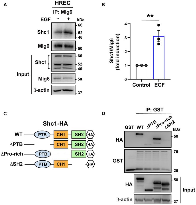
Liu et al. Anti-angiogenic Effects of MIG6 by Binding to SHC1
FIGURE 2 | vascular sprouting area per aortic ring in (C). n = 3 aortic rings per group. (E) Mouse choroidal tissues of WT or Mig6−/− mice incubated for 7 days. (F)
Quantification of vascular sprouting area per choroid tissue in (E). n = 3 choroids per group. (G) Mouse choroidal tissues infected with Ad-GFP or Ad-MIG6 and
incubated for 7 days. (H) Quantification of vascular sprouting area per choroid tissue in (G). n = 3 choroids per group. Scale bars: 500 µm for (A); 250 µm for (C,E,G).
The data are shown as the mean ± SEM from at least three independent experiments. * p < 0.05, ** p < 0.01 (two-tailed paired Student’s t-test).
presented as mean ± SEM. p < 0.05 was considered apart from the retina, MIG6 also inhibits angiogenesis in other
statistically significant. tissues, such as in the aorta and choroids.
RESULTS MIG6 Inhibits Endothelial Cell Proliferation,
Migration, and Tube Formation
MIG6 Is Expressed in Various Types of Although MIG6 is expressed in various types of endothelial cells
Vascular Endothelial Cells (ECs) From (Supplementary Table 1) (Jin et al., 2009; Lee et al., 2014), it
Different Human Organs remains thus far unknown whether it regulates EC functions.
To obtain insight into the expression of MIG6 in vascular We therefore investigated into this using both gain- and loss-of-
endothelial cells, we searched the public database Human Protein function assays. An EdU incorporation assay revealed that Mig6
Atlas (www.proteinatlas.org), in which the expression levels of overexpression (Figure 3A) markedly inhibited the proliferation
MIG6 are derived from published single cell RNA sequencing of human retinal endothelial cells (HREC) (Figures 3B,C), while
studies. We found that MIG6 is expressed in ECs from various MIG6 knockdown (Figure 3D) increased HREC proliferation
tissues/organs, including skin, liver, prostate, lung, heart muscle, (Figures 3E,F). This finding was further supported by an MTT
eye, placenta, and testis (Supplementary Table 1), indicating a assay showing that MIG6 knockdown increased and MIG6
potential effect of MIG6 on ECs. overexpression decreased HREC proliferation, respectively
(Supplementary Figures 1B,C). Moreover, an HREC migration
Genetic Deletion of Mig6 Increases Retinal assay showed that overexpression of MIG6 inhibited HREC
Angiogenesis migration (Supplementary Figures 2A–C), and MIG6
MIG6 has also been shown to be expressed in mouse vascular knockdown enhanced HREC tube formation (Figures 3G,H).
system (Zhang and Vande Woude, 2007; Jin et al., 2009). Furthermore, an HREC spheroid assay revealed that MIG6
However, it remains unknown thus far whether it plays a knockdown markedly increased the number and length of
role in angiogenesis. To address this, we evaluated vascular EC sprouts in HREC (Figures 3I–K). Thus, multiple assays
formation in the retinae of Mig6 knockout mice. Isolectin B4 showed that MIG6 overexpression inhibits HREC proliferation,
(IB4) staining showed that Mig6 knockout mice displayed higher migration, and tube formation.
retinal blood vessel density and branch points at postnatal day
5 (P5) (Figures 1A–F), while no difference was found in total MIG6 Inhibits Ischemia-Induced Retinal
retinal areas (Supplementary Figure 1A). Immunofluorescence Neovascularization
staining of ERG, an endothelial cell (EC) nuclear marker, revealed Led by our observation on the anti-angiogenic effect of
a higher number of ECs in the retinae of Mig6 knockout mice MIG6, we further tested whether MIG6 could inhibit
(Figures 1G,H). Notably, more tip cells and longer filopodia pathological neovascularization using a mouse model of
extensions of tip cells, known to be critical for angiogenesis retinopathy of prematurity (ROP) (Connor et al., 2009)
(Gerhardt et al., 2003; Ochsenbein et al., 2016), were found in the (Figure 4A). Adenoviruses encoding MIG6 were intravitreally
retinae of Mig6 knockout mice (Figures 1I–K). Together, these injected at P12 to overexpress MIG6, with Ad-GFP as a
findings show that loss of Mig6 increased retinal angiogenesis. control (Supplementary Figure 3). After 5 days, the retinae
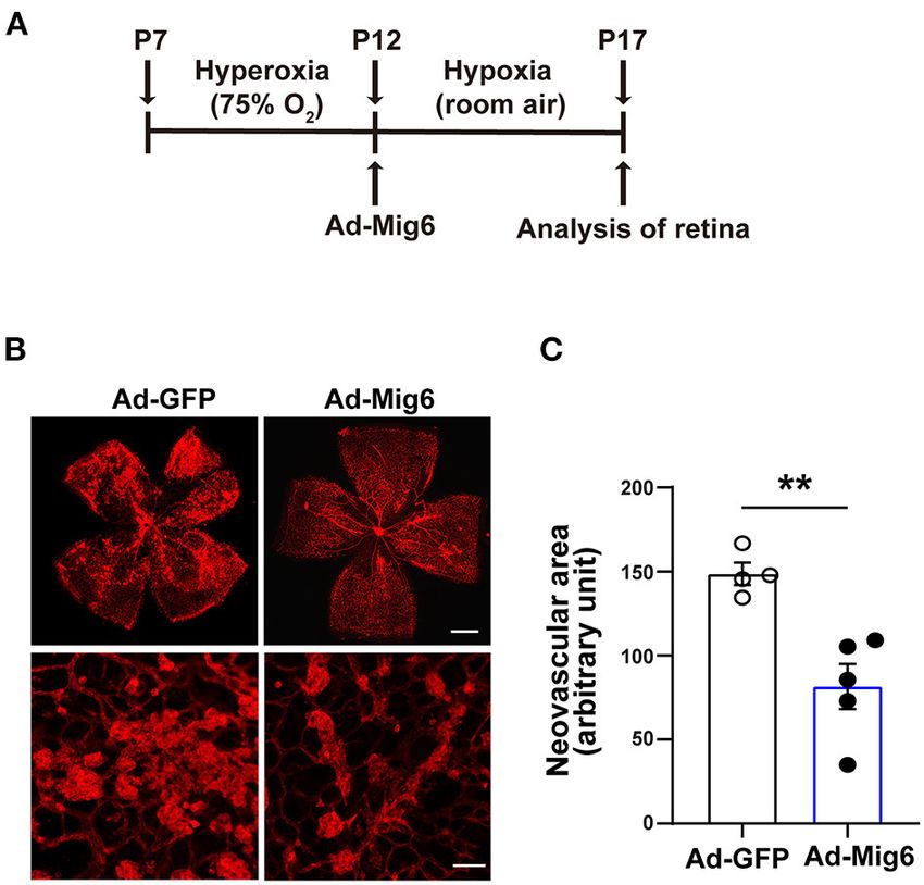
were collected to analyze neovascularization. The retinae
MIG6 Inhibits Aortic and Choroidal treated with Ad-MIG6 displayed less neovascularization and
Microvessel Growth fewer neovascular tufts compared with the Ad-GFP retinae
We next investigated whether MIG6 affected angiogenesis (Figures 4B,C), demonstrating that MIG6 overexpression
in other tissues. An aortic ring assay showed that gene inhibits retinal neovascularization.
deletion of Mig6 significantly increased microvessel growth
(Figures 2A,B), while MIG6 overexpression by adenovirus MIG6 Binds to SHC1
markedly inhibited microvessel sprouting (Figures 2C,D). SHC1 has a central role in the signaling of many tyrosine
Choroidal neovascularization is a devastating pathology that kinases (Zheng et al., 2013; Ahn et al., 2017) and binds to
can cause blindness (Shao et al., 2013). We therefore tested the pY[I/E/Y/L][X][I/L/M] motif (X representing any of the
whether MIG6 affected choroidal angiogenesis using a choroidal 20 amino acids), which is found in MIG6 (YYLL: 394Tyr-
sprouting assay. We found that Mig6 deficient choroids gave 397Leu) (Wills and Jones, 2012; Suen et al., 2013). We therefore
rise to more microvessels than those of wild type (WT) choroids tested whether it binds to MIG6. A co-immunoprecipitation
(Figures 2E,F), whereas MIG6 overexpression significantly assay revealed that SHC1 formed complex with MIG6 in
inhibited choroidal microvessel sprouting (Figures 2G,H). Thus, HREC, which was augmented by EGF treatment (Figures 5A,B).
Frontiers in Cell and Developmental Biology | www.frontiersin.org 6 February 2021 | Volume 9 | Article 634242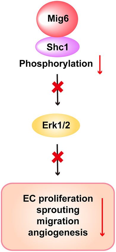
Liu et al.                                                                                                            Anti-angiogenic Effects of MIG6 by Binding to SHC1

  FIGURE 3 | MIG6 inhibits endothelial cell proliferation, migration, tube formation, and sprouting. (A) Western blot for MIG6 overexpression in HRECs treated with
  Ad-MIG6. (B) Representative images for EdU incorporation in MIG6-overexpressing HRECs. (C) Bar graphs represent the mean ± SEM of % EdU+ cells per field in
  (B). (D) Western blot for MIG6 expression in MIG6 knockdown HRECs. (E) Images showing EdU incorporation in MIG6 knockdown HRECs. (F) Bar graphs represent
  the mean ± SEM of % EdU+ cells per field in (E). (G) Tube formation in HRECs upon control (siControl) or MIG6 siRNA (siMIG6)-mediated knockdown. (H)
  Quantification of the number of the branch points per field in (G). (I) Representative images of EC spheroids using siMIG6 HREC for 48 h. (J) Quantification of the
  number of sprouts per EC spheroid in (I). (K) Quantification of the total sprout length per EC spheroid in (I). Scale bars: 50 µm for (B,E); 100 µm for (G); 500 µm for
  (I). The data are shown as mean ± SEM from three independent experiments. * p < 0.05, ** p < 0.01 (two-tailed paired Student’s t-test).

To determine the domain of the SHC1 bound by MIG6,                                      MIG6 Inhibits SHC1 Phosphorylation
GST-conjugated MIG6 fusion protein (GST-MIG6) and the                                   It is known that phosphorylation of SHC1 is critical in
truncated mutants of SHC1 protein were produced (Figure 5C,                             promoting cell proliferation, migration, and survival (Zheng
Supplementary Figures 4A,B). The GST-MIG6 protein bound                                 et al., 2013; Ahn et al., 2017; Wright et al., 2019). We therefore
to the full-length and deletion mutant of SHC1 lacking the                              examined whether MIG6 affected SHC1 phosphorylation.
proline-rich domain (deletion of amino acids 300–366) but not                           We found that gene deletion of Mig6 increased SHC1
to the mutants lacking PTB (deletion of amino acids 30–210)                             phosphorylation at both the basal level and after EGF stimulation
or SH2 domain (deletion of amino acids 377–469) (Figure 5D),                            in primary mouse lung ECs (Figures 6A,B). Moreover, MIG6
suggesting that the PTB and SH2 domains of SHC1 are critical                            overexpression decreased SHC1 phosphorylation in HRECs
for MIG6 binding.                                                                       (Figures 6C,D), while opposite effects were observed after MIG6

Frontiers in Cell and Developmental Biology | www.frontiersin.org                   7                                           February 2021 | Volume 9 | Article 634242
Liu et al.                                                                                                      Anti-angiogenic Effects of MIG6 by Binding to SHC1

                                                                                        about such endogenous anti-angiogenic factors as opposed to
                                                                                        the pro-angiogenic factors. In this study, we identified MIG6 as
                                                                                        a potent endogenous inhibitor of angiogenesis by investigating
                                                                                        the functions of MIG6 in multiple experimental systems.
                                                                                        Furthermore, we reveal the molecular mechanism underlying
                                                                                        the anti-angiogenic functions of MIG6, which implicates the
                                                                                        inhibition of SHC1 signaling driven by MIG6 binding-mediated
                                                                                        inhibition of SHC1 phosphorylation.
                                                                                            MIG6      is    widely    expressed    in     vascular    cells
                                                                                        (Supplementary Table 1) (Jin et al., 2009; Lee et al., 2014). Yet,
                                                                                        little is known whether MIG6 functions in them. Here, we found
                                                                                        Mig6 knockout mice displayed increased blood vessel density
                                                                                        and number of branch points in the retinae, demonstrating an
                                                                                        anti-angiogenic effect of MIG6 in retinal vascularization. Indeed,
                                                                                        in vitro, MIG6 inhibits EC proliferation, viability and sprouting.
                                                                                        It remains unclear thus far whether MIG6 regulates pathological
                                                                                        neovascularization. We found in this work that overexpression
                                                                                        of MIG6 suppressed retinal neovascularization in a mouse model
                                                                                        of retinopathy of prematurity, providing evidence for a role of
                                                                                        MIG6 in pathological neovascularization. It has been shown that
                                                                                        Mig6 deficiency led to endometrial hyperplasia (Jin et al., 2007)
  FIGURE 4 | MIG6 inhibits pathological angiogenesis. (A) Timeline of the
  retinopathy of prematurity model. P7 neonatal mice were exposed to                    and neointimal hyperplasia (Lee et al., 2014), thus raising the
  hyperoxia for 5 days. Intravitreal injection of adenovirus expressing GFP or          possibility that MIG6 deficiency-induced angiogenesis might
  MIG6 was executed at P12. Mice were then returned to room air until P17. (B)          contribute to these pathological conditions. Further studies are
  Images of whole mount retinae at P17 stained with IB4-Alexa 594 (top). High           needed to verify this.
  magnification images of neovascular tufts in the retinae infected with Ad-GFP
                                                                                            Our findings of the anti-angiogenic effects of MIG6 present
  or Ad-MIG6 (bottom). (C) Quantification of neovascular areas in retinal whole
  mounts in (B). n = 4 fields per Ad-GFP treated retina, n = 5 fields per Ad-Mig6       different observations from another gene knockout study,
  treated retina. Scale bars: 300 µm for the top panel of (B); 50 µm for the            which reported the opposite roles of MIG6 in angiogenesis by
  bottom panel of (B). The data are shown as mean ± SEM. ** p < 0.01                    showing that neovascularization is reduced compared with wild-
  (two-tailed paired Student’s t-test).                                                 type lungs, and pro-angiogenic factors, including VEGF-A, are
                                                                                        downregulated at P3 in Mig6 knockout lungs (Jin et al., 2009).
                                                                                        At least one of the reasons for this discrepancy might be due
                                                                                        to differential expression of MIG6 in different tissues during
knockdown (Supplementary Figures 5A–D). These data thus
                                                                                        development (Jin et al., 2009). Additionally, in our current work,
demonstrate that MIG6 has a critical suppressive effect on
                                                                                        the Mig6 knockout mice used were bred on C57Bl/6J background
SHC1 phosphorylation.
                                                                                        for more than six generations. In the Jin et al. (2009) study,
   We next verified whether SHC1 played a role in modulating
                                                                                        however, it was not clearly indicated whether C57Bl6 strain
MIG6 function. We found that SHC1 knockdown by siRNA
                                                                                        was used. In addition, the Mig6 knockout mice used in the Jin
markedly reduced MIG6 knockdown-induced tube formation in
                                                                                        et al. study were produced by crossing Mig6fl/fl with Rosa26-Cre-
HRECs (Figures 6E,F), demonstrating that SHC1 is required for
                                                                                        ERT2, which was a different targeting strategy compared with
the inhibitory effect of MIG6 on angiogenesis. Moreover, at a
                                                                                        that of our knockout mice, which is global knockout without any
molecular level, we found that SHC1 knockdown also abolished
                                                                                        Cre recombination.
siMIG6-induced ERK1/2 activation in the presence of EGF in
                                                                                            However, in line with our findings, vascular smooth muscle
HREC (Figures 6G,H). Taken together, our data show that MIG6
                                                                                        cells (SMCs) in SMC-specific Mig6 conditional knockout mice
has a potent anti-angiogenic effect by binding to SHC1 and
                                                                                        displayed an increased cell migration and proliferation (Lee
inhibiting its phosphorylation (Figure 7).
                                                                                        et al., 2014). Due to this SMC phenotype, it cannot rule out
                                                                                        the possibility that some of the effect of MIG6 deletion on
DISCUSSION                                                                              angiogenesis could be secondary to SMC defect, if any. It indeed
                                                                                        has been reported that the EC-SMC interplay affects collective EC
Uncontrolled growth of blood vessels can result in many                                 movements driving capillary elongation in the aortic ring assay
devastating neovascular diseases. Therefore, a tight regulation of                      (Arima et al., 2011). On the other hand, our HREC proliferation,
angiogenesis is essential to prevent overgrowth of blood vessels                        tube formation and spheroid assays showed a direct effect of
and consequential exacerbation or development of neovascular                            MIG6 on them. Also, ERG (an EC marker) staining showed
diseases. Given the presence diverse and abundant angiogenic                            more ERG+ cells in Mig6 KO mice, indicating that MIG6 has
factors, naturally occurring endogenous anti-angiogenic factors                         a direct effect on ECs. Moreover, MIG6 activities vary since it
would be critical to counteract excessive pro-angiogenic activities                     interacts with a wide range of receptor tyrosine kinases (RTK),
to maintain vascular homeostasis. Yet, currently, less is known                         such as c-Met, FGFR2, and PDGFR (Pante et al., 2005; Zhang

Frontiers in Cell and Developmental Biology | www.frontiersin.org                   8                                    February 2021 | Volume 9 | Article 634242
Liu et al. Anti-angiogenic Effects of MIG6 by Binding to SHC1
FIGURE 5 | MIG6 binds to SHC1 through PTB and SH2 domains. (A) Immunoprecipitation (IP) followed by Western blot showing binding of MIG6 with SHC1 in
HRECs, which was further augmented by EGF (50 ng/ml) treatment. (B) MIG6 binding to SHC1 was analyzed by densitometry and normalized by total MIG6. Fold
induction relative to the control is shown as the mean ± SEM from three independent experiments. ** p < 0.01 (two-tailed paired Student’s t-test). (C) Schematic
representation of SHC1 deletion mutants. The full-length (WT) and the truncated deletion mutants of SHC1 were tagged with HA in their C-terminus. The truncated
mutants of SHC1 lack the PTB domain (1PTB: deletion of amino acids 30–210), proline-rich domain (1Pro-rich: deletion of amino acids 300–366) in CH1 (collagen
homology 1) region, and the SH2 domain (1SH2: deletion of amino acids 377–469). (D) Association of MIG6 with the truncated mutants of SHC1 was determined by
GST pull-down assay, showing that MIG6 binding is mediated by the PTB and SH2 domains in SHC1.
and Vande Woude, 2007; Borad et al., 2014; Migliore et al., effect of MIG6 on SHC1 phosphorylation. Importantly, SHC1
2018). Furthermore, a recent study showed that Akt is a novel knockdown largely abolished MIG6 depletion-induced EC tube
binding partner of MIG6 to modulate its activation in several formation and the increased ERK1/2 activation by EGF in ECs,
types of cancer cells expressing a low level of EGFR (Cairns et al., suggesting that the anti-angiogenic function of MIG6 is mediated
2018). As such, by interacting with different signaling molecules by its suppressive effect on SHC1. Indeed, SHC1 has been shown
depending on their expression status, MIG6 may appear to be to be pro-angiogenic by promoting EC proliferation, survival
multi-functional in different cell types or tissues. and blood vessel maturation (Saucier et al., 2004; Sweet et al.,
The signaling pathway of MIG6 is poorly understood thus 2012). Noteworthy, SHC1 has a critical role in inducing VEGF
far. In this study, we found that MIG6 forms complex with expression (Saucier et al., 2004) and enhancing the activities
SHC1, an intracellular adaptor protein that is highly expressed of several angiogenic pathways, including VEGFR2 (Lai and
in the vascular system (Lai and Pawson, 2000; Sweet et al., 2012). Pawson, 2000; Sweet et al., 2012), raising the question whether
Moreover, we show that the PTB and SH2 domains in SHC1 are MIG6 affects the angiogenic activities of the VEGFA-VEGFR2
critical regions for MIG6 binding, which leads to the inhibition of pathway. Future studies are needed to address this.
the SHC1 downstream signaling. Gene deletion of Mig6 increased In summary, we demonstrate that MIG6 deficiency increases
SHC1 phosphorylation in ECs, demonstrating the inhibitory angiogenesis both in vivo and in vitro. We also show that MIG6
Frontiers in Cell and Developmental Biology | www.frontiersin.org 9 February 2021 | Volume 9 | Article 634242Liu et al. Anti-angiogenic Effects of MIG6 by Binding to SHC1
FIGURE 6 | MIG6 has a potent anti-angiogenic effect by inhibiting SHC1 phosphorylation and the subsequent ERK1/2 activation. (A) Western blots showing that
gene deletion of Mig6 increases SHC1 phosphorylation in mouse primary lung ECs at the baseline level and in the presence of EGF. (B) Tyrosine phosphorylation of
(Continued)
Frontiers in Cell and Developmental Biology | www.frontiersin.org 10 February 2021 | Volume 9 | Article 634242Liu et al. Anti-angiogenic Effects of MIG6 by Binding to SHC1
FIGURE 6 | SHC1 (pTyr239/240 SHC1) was analyzed by densitometry and normalized by total SHC1. Fold induction relative to the control is shown as the mean ±
SEM from three independent experiments. (C) Western blots showing that overexpression of MIG6 decreases SHC1 phosphorylation in HRECs at the baseline level
and in the presence of EGF. (D) Tyrosine phosphorylation of SHC1 (pTyr239/240 SHC1) was analyzed by densitometry and normalized by total SHC1. Fold induction
relative to the control is shown as the mean ± SEM from three independent experiments. (E) Images of tube formation assay showing that SHC1 knockdown
abolished siMIG6-induced tube formation of HRECs. (F) Quantification of the branch points per field in (E). The graph is shown as the mean ± SEM from three
independent experiments. (G) Western blots showing that SHC1 knockdown abolished siMIG6-induced ERK1/2 phosphorylation in HRECs. (H) Phosphorylation of
ERK1/2 was analyzed by densitometry and normalized by total ERK1/2. Three isoforms of SHC1 are indicated by the arrowhead (A,C). Fold induction relative to the
control is shown as the mean ± SEM from three independent experiments. * p < 0.05, ** p < 0.01, *** p < 0.001 (two-tailed paired Student’s t-test).
DATA AVAILABILITY STATEMENT
The original contributions presented in the study are included
in the article/Supplementary Material, further inquiries can be
directed to the corresponding authors.
ETHICS STATEMENT
The animal study was reviewed and approved by Animal Use
and Care Committee of Zhongshan Ophthalmic Center, Sun
Yat-sen University.
AUTHOR CONTRIBUTIONS
LL and LX designed and performed experiments, analyzed data,
and wrote a part of the manuscript. RC, YH, JZ, LH, BX, XR,
SW, HK, and XLin performed experiments and analyzed data.
AK and JK provided critical experimental tools and suggestions.
CL and XLi designed and supervised experiments, analyzed data,
and wrote the manuscript. All authors contributed to the article
and approved the submitted version.
FUNDING
FIGURE 7 | Scheme of the proposed function of MIG6 and the underlying
mechanism in endothelial cells. Shown is the proposed working model This study is supported by the State Key Laboratory of
displaying that MIG6 restrains SHC1 signaling pathway by binding to it and
Ophthalmology (SKLO), Zhongshan Ophthalmic Center (ZOC)
inhibiting its phosphorylation, leading to reduced ERK1/2 activation and EC
proliferation and migration, thus resulting in the inhibition of angiogenesis.
at the Sun Yat-sen University, the National Natural Science
Foundation of China (81670855), and a Key Program of
Guangzhou Scientific Research Plan (201804020010).
antagonizes SHC1 signaling to inhibit angiogenesis. Our results SUPPLEMENTARY MATERIAL
demonstrate that the signaling axis of MIG6 and SHC1 plays a
critical role in keeping angiogenesis balanced. Our work provides The Supplementary Material for this article can be found
new insights into the pathogenesis of neovascular diseases, and online at: https://www.frontiersin.org/articles/10.3389/fcell.2021.
may have therapeutic implications in anti-angiogenic therapy. 634242/full#supplementary-material
REFERENCES Apte, R. S., Chen, D. S., and Ferrara, N. (2019). VEGF in signaling
and disease: beyond discovery and development. Cell 176, 1248–1264.
Ahn, R., Sabourin, V., Bolt, A. M., Hébert, S., Totten, S., De Jay, N., et al. doi: 10.1016/j.cell.2019.01.021
(2017). The Shc1 adaptor simultaneously balances Stat1 and Stat3 activity Arima, S., Nishiyama, K., Ko, T., Arima, Y., Hakozaki, Y., Sugihara,
to promote breast cancer immune suppression. Nat. Commun. 8:14638. K., et al. (2011). Angiogenic morphogenesis driven by dynamic and
doi: 10.1038/ncomms14638 heterogeneous collective endothelial cell movement. Development 138,
Anastasi, S., Lamberti, D., Alemà, S., and Segatto, O. (2016). Regulation of the 4763–4776. doi: 10.1242/dev.068023
ErbB network by the MIG6 feedback loop in physiology, tumor suppression Borad, M. J., Champion, M. D., Egan, J. B., Liang, W. S., Fonseca, R.,
and responses to oncogene-targeted therapeutics. Semin. Cell Dev. Biol. 50, Bryce, A. H., et al. (2014). Integrated genomic characterization reveals
115–124. doi: 10.1016/j.semcdb.2015.10.001 novel, therapeutically relevant drug targets in FGFR and EGFR pathways
Frontiers in Cell and Developmental Biology | www.frontiersin.org 11 February 2021 | Volume 9 | Article 634242Liu et al. Anti-angiogenic Effects of MIG6 by Binding to SHC1
in sporadic intrahepatic cholangiocarcinoma. PLoS Genet. 10:e1004135. filopodia formation by blood vascular tip cells. Development 143, 589–594.
doi: 10.1371/journal.pgen.1004135 doi: 10.1242/dev.127670
Cairns, J., Fridley, B. L., Jenkins, G. D., Zhuang, Y., Yu, J., and Wang, L. (2018). Pante, G., Thompson, J., Lamballe, F., Iwata, T., Ferby, I., Barr, F. A., et al.
Differential roles of ERRFI1 in EGFR and AKT pathway regulation affect cancer (2005). Mitogen-inducible gene 6 is an endogenous inhibitor of HGF/Met-
proliferation. EMBO Rep. 19:e44767. doi: 10.15252/embr.201744767 induced cell migration and neurite growth. J. Cell Biol. 171, 337–348.
Connor, K. M., Krah, N. M., Dennison, R. J., Aderman, C. M., Chen, J., Guerin, K. doi: 10.1083/jcb.200502013
I., et al. (2009). Quantification of oxygen-induced retinopathy in the mouse: a Park, E., Kim, N., Ficarro, S. B., Zhang, Y., Lee, B. I., Cho, A., et al. (2015). Structure
model of vessel loss, vessel regrowth and pathological angiogenesis. Nat. Protoc. and mechanism of activity-based inhibition of the EGF receptor by Mig6. Nat.
4, 1565–1573. doi: 10.1038/nprot.2009.187 Struct. Mol. Biol. 22, 703–711. doi: 10.1038/nsmb.3074
Ferby, I., Reschke, M., Kudlacek, O., Knyazev, P., Pantè, G., Amann, K., Ribatti, D. (2016). Tumor refractoriness to anti-VEGF therapy. Oncotarget 7,
et al. (2006). Mig6 is a negative regulator of EGF receptor-mediated 46668–46677. doi: 10.18632/oncotarget.8694
skin morphogenesis and tumor formation. Nat. Med. 12, 568–573. Sasaki, M., Terabayashi, T., Weiss, S. M., and Ferby, I. (2018). The tumor
doi: 10.1038/nm1401 suppressor MIG6 controls mitotic progression and the G2/M DNA damage
Gerhardt, H., Golding, M., Fruttiger, M., Ruhrberg, C., Lundkvist, A., Abramsson, checkpoint by stabilizing the WEE1 kinase. Cell Rep. 24, 1278–1289.
A., et al. (2003). VEGF guides angiogenic sprouting utilizing endothelial tip cell doi: 10.1016/j.celrep.2018.06.064
filopodia. J. Cell Biol. 161, 1163–1177. doi: 10.1083/jcb.200302047 Saucier, C., Khoury, H., Lai, K. M., Peschard, P., Dankort, D., Naujokas, M. A., et al.
Haibe, Y., Kreidieh, M., El Hajj, H., Khalifeh, I., Mukherji, D., Temraz, S., et al. (2004). The Shc adaptor protein is critical for VEGF induction by Met/HGF and
(2020). Resistance mechanisms to anti-angiogenic therapies in cancer. Front. ErbB2 receptors and for early onset of tumor angiogenesis. Proc. Natl. Acad. Sci.
Oncol. 10:221. doi: 10.3389/fonc.2020.00221 U. S. A. 101, 2345–2350. doi: 10.1073/pnas.0308065101
Jin, N., Cho, S. N., Raso, M. G., Wistuba, I., Smith, Y., Yang, Y., et al. (2009). Mig-6 Shao, Z., Friedlander, M., Hurst, C. G., Cui, Z., Pei, D. T., Evans, L. P., et al. (2013).
is required for appropriate lung development and to ensure normal adult lung Choroid sprouting assay: an ex vivo model of microvascular angiogenesis. PLoS
homeostasis. Development 136, 3347–3356. doi: 10.1242/dev.032979 ONE 8:e69552. doi: 10.1371/journal.pone.0069552
Jin, N., Gilbert, J. L., Broaddus, R. R., Demayo, F. J., and Jeong, J. W. Suen, K. M., Lin, C. C., George, R., Melo, F. A., Biggs, E. R., Ahmed, Z.,
(2007). Generation of a Mig-6 conditional null allele. Genesis 45, 716–721. et al. (2013). Interaction with Shc prevents aberrant Erk activation in
doi: 10.1002/dvg.20348 the absence of extracellular stimuli. Nat. Struct. Mol. Biol. 20, 620–627.
Karaman, S., Leppänen, V. M., and Alitalo, K. (2018). Vascular endothelial growth doi: 10.1038/nsmb.2557
factor signaling in development and disease. Development 145:dev151019. Sweet, D. T., Chen, Z., Wiley, D. M., Bautch, V. L., and Tzima, E.
doi: 10.1242/dev.151019 (2012). The adaptor protein Shc integrates growth factor and ECM
Lai, K. M., and Pawson, T. (2000). The ShcA phosphotyrosine docking protein signaling during postnatal angiogenesis. Blood 119, 1946–1955.
sensitizes cardiovascular signaling in the mouse embryo. Genes. Dev. 14, doi: 10.1182/blood-2011-10-384560
1132–1145. doi: 10.1101/gad.14.9.1132 Wills, M. K., and Jones, N. (2012). Teaching an old dogma new tricks: twenty years
Lee, J. H., Choung, S., Kim, J. M., Lee, J. U., Kim, K. S., Kim, H. J., et al. (2014). of Shc adaptor signalling. Biochem. J. 447, 1–16. doi: 10.1042/BJ20120769
Mig-6 gene knockout induces neointimal hyperplasia in the vascular smooth Wright, K. D., Miller, B. S., El-Meanawy, S., Tsaih, S. W., Banerjee, A., Geurts,
muscle cell. Dis. Markers 2014:549054. doi: 10.1155/2014/549054 A. M., et al. (2019). The p52 isoform of SHC1 is a key driver of breast cancer
Li, X., and Carmeliet, P. (2018). Targeting angiogenic metabolism in disease. initiation. Breast Cancer Res. 21:74. doi: 10.1186/s13058-019-1155-7
Science 359, 1335–1336. doi: 10.1126/science.aar5557 Zhang, K., Li, M., Yin, L., Fu, G., and Liu, Z. (2020). Role of thrombospondin-1
Li, X., Sun, X., and Carmeliet, P. (2019). Hallmarks of endothelial cell metabolism and thrombospondin-2 in cardiovascular diseases (Review). Int. J. Mol. Med.
in health and disease. Cell Metab. 30, 414–433. doi: 10.1016/j.cmet.2019.08.011 45, 1275–1293. doi: 10.3892/ijmm.2020.4507
Liu, N., Matsumoto, M., Kitagawa, K., Kotake, Y., Suzuki, S., Shirasawa, Zhang, Y. W., and Vande Woude, G. F. (2007). Mig-6, signal transduction,
S., et al. (2012). Chk1 phosphorylates the tumour suppressor Mig-6, stress response and cancer. Cell Cycle 6, 507–513. doi: 10.4161/cc.6.
regulating the activation of EGF signalling. EMBO J. 31, 2365–2377. 5.3928
doi: 10.1038/emboj.2012.88 Zheng, Y., Zhang, C., Croucher, D. R., Soliman, M. A., St.-Denis, N.,
Lundgren, T. K., Scott, R. P., Smith, M., Pawson, T., and Ernfors, P. (2006). Pasculescu, A., et al. (2013). Temporal regulation of EGF signalling networks
Engineering the recruitment of phosphotyrosine binding domain-containing by the scaffold protein Shc1. Nature 499, 166–171. doi: 10.1038/nature
adaptor proteins reveals distinct roles for RET receptor-mediated cell survival. 12308
J. Biol. Chem. 281, 29886–29896. doi: 10.1074/jbc.M600473200
Migliore, C., Morando, E., Ghiso, E., Anastasi, S., Leoni, V. P., Apicella, M., Conflict of Interest: The authors declare that the research was conducted in the
et al. (2018). miR-205 mediates adaptive resistance to MET inhibition via absence of any commercial or financial relationships that could be construed as a
ERRFI1 targeting and raised EGFR signaling. EMBO Mol. Med. 10:e8746. potential conflict of interest.
doi: 10.15252/emmm.201708746
Murugeswari, P., Shukla, D., Rajendran, A., Kim, R., Namperumalsamy, Copyright © 2021 Liu, Xing, Chen, Zhang, Huang, Huang, Xie, Ren, Wang, Kuang,
P., and Muthukkaruppan, V. (2008). Proinflammatory cytokines and Lin, Kumar, Kim, Lee and Li. This is an open-access article distributed under the
angiogenic and anti-angiogenic factors in vitreous of patients with terms of the Creative Commons Attribution License (CC BY). The use, distribution
proliferative diabetic retinopathy and eales’ disease. Retina 28, 817–824. or reproduction in other forums is permitted, provided the original author(s) and
doi: 10.1097/IAE.0b013e31816576d5 the copyright owner(s) are credited and that the original publication in this journal
Ochsenbein, A. M., Karaman, S., Proulx, S. T., Berchtold, M., Jurisic, G., is cited, in accordance with accepted academic practice. No use, distribution or
Stoeckli, E. T., et al. (2016). Endothelial cell-derived semaphorin 3A inhibits reproduction is permitted which does not comply with these terms.
Frontiers in Cell and Developmental Biology | www.frontiersin.org 12 February 2021 | Volume 9 | Article 634242You can also read