Morphological and structural study of seed pericarp of Opuntia ficus-indica prickly pear fruits - Nopal Tunisie
←
→
Page content transcription
If your browser does not render page correctly, please read the page content below
Available online at www.sciencedirect.com

                                                    Carbohydrate Polymers 72 (2008) 102–112
                                                                                                                         www.elsevier.com/locate/carbpol

                       Morphological and structural study of seed pericarp
                           of Opuntia ficus-indica prickly pear fruits
                                        a,1,*
               Youssef Habibi                   , Laurent Heux a, Mostafa Mahrouz b, Michel R. Vignon                                       a

     a
         Centre de Recherches sur les Macromolécules Végétales (CERMAV-CNRS), affiliated with Joseph Fourier University, and member of Institut de
                                        Chimie Moléculaire de Grenoble (ICMG), BP 53, 38041 Grenoble Cedex 9, France
                b
                  Unité de chimie agro-alimentaire, Faculté des Sciences Semlalia (Université Cadi Ayyad), BP 2390, Marrakech, Maroc, France

                                     Received 27 May 2007; received in revised form 5 July 2007; accepted 24 July 2007
                                                             Available online 2 August 2007

Abstract

    The morphological study of pericarp of Opuntia ficus-indica (OFI) seeds showed that the cells were mainly made up of spindle-shaped
sclerenchyma fibers. The chemical composition of the pericarp revealed a significant amount of polysaccharides, with cellulose (35%) and
xylan (27%). The structure of xylan and cellulose, both in isolated form and as a component of seed pericarp of OFI were studied by
X-ray and CP/MAS 13C NMR spectroscopy. The supramolecular structure of xylan is very sensitive to the surrounding environment,
in particular to the presence of water and of cellulose fibers. The cellulose fibers presented X-ray diagrams typical of secondary wall
cellulose but they were sensitive toward NaOH since they started to be converted into cellulose II at a NaOH concentration as low
as 8%. In seed pericarp, cellulose fibers interact with xylan polymers, causing these to adopt a conformation different to the one observed
for xylan both in dry or hydrated form, suggesting that xylans were probably present as composites with cellulose fibers.
 2007 Elsevier Ltd. All rights reserved.

Keywords: Cellulose; CP/MAS; Morphology; Opuntia ficus-indica; Seed; X-ray; Xylan

1. Introduction                                                                 curonoxylan free from arabinose substituents dominates in
                                                                                hardwoods (Stephen, 1983; Whistler & Chen, 1991; Wilkie,
   Cellulose is the most abundant biopolymer on earth and                       1979).
the major component in the cell walls of wood and terres-                          Strong interactions are present between xylan and cellu-
trial plants. In contrast to cellulose, which is a homopoly-                    lose as evidenced by the fact that the synthesis and deposi-
saccharide, hemicelluloses which also occur in the cell walls                   tion of xylan are intimately linked with cellulose during the
are heteropolysaccharides. Xylans are the most abundant                         assembly of the cell wall. The xylan retention phenomenon
of the hemicelluloses found in the cell walls of plants, of                     has been explained by co-crystallization of xylan segments
which they can constitute more than 30% of the dry weight.                      with cellulose and by the formation of strong xylan–cellu-
Xylans are characterized by a b-(1 fi 4)-D-Xylp backbone                         lose hydrogen bonds. Several researchers have observed
to which arabinosyl, glucuronic acid, and acetyl substitu-                      the strong tendency of xylans to self-associate (Brett,
ents can be attached. The main xylan in softwoods is                            2000; Joseleau, Comtat, & Ruel, 1992; Mora, Ruel, Com-
arabino-4-O-methylglucuronoxylan, while 4-O-methyl glu-                         tat, & Joseleau, 1986).
                                                                                   The structures of cellulose in wood and pulp as well as
 *
                                                                                interaction between cellulose and hemicelluloses have been
   Corresponding author. Tel.: +33 476826961; fax: +33 476826933.               extensively studied by CP/MAS 13C NMR spectroscopy
   E-mail address: Youssef.Habibi@efpg.inpg.fr (Y. Habibi).
 1
   Present address: Ecole Française de Papèterie et des Industries
                                                                                (Hult, Larsson, & Iversen, 2000; Larsson, Hult, Wickholm,
Graphiques (EFPG-INPG), BP 65, 38402, St-Martin d’Hères Cedex,                 Pettersson, & Iversen, 1999; Larsson, Wickholm, &
France.                                                                         Iversen, 1997; Lindgren, Edlund, & Iversen, 1995; Maunu,

0144-8617/$ - see front matter  2007 Elsevier Ltd. All rights reserved.
doi:10.1016/j.carbpol.2007.07.032
Y. Habibi et al. / Carbohydrate Polymers 72 (2008) 102–112 103
Liitiä, Kauliomäki, Hortling, & Sundquist, 2000; Newman, cracked in an analytical grinder for a few minutes and
1998; Newman & Hemmingson, 1990; Newman, Hem- the pericarp was recovered after sieving on 60 mesh sieve.
mingson, & Suckling, 1993; Van der Hart & Atalla, 1984;
Wickholm, Larsson, & Iversen, 1998). The most important 2.2. Analytical methods
progress in this context is reported by Larsson in a recent
review (Larsson, 2004). Neutral sugars were analyzed, after H2SO4 hydrolysis,
Opuntia ficus-indica is a tropical or subtropical plant, by Gas Liquid Chromatography (GLC) as their corre-
which belongs to the Cactaceae family and is mainly used sponding alditol acetates, using a Packard and Becker 417
for fruit production (De Cortazar & Nobel, 1992). Because instrument coupled to a Hewlett-Packard 3380 A integra-
of its high adaptation to the harsh desert environment and tor. Glass columns (3 mm · 2 m) packed with 3% SP 2340
its different applications, the Opuntia ficus-indica fruit, on Chromosorb W-AW DMCS (100–120 mesh), or 3%
commonly known as prickly pear, is an important and OV 17 on the same support were used. The lignin analysis
abundant potential raw material for the Moroccan indus- was achieved according to the TAPPI standard T222-03-75.
try. Within the last decade, prickly pear fruits have become
an important crop in the semi-arid lands of Morocco, 2.3. Isolation and purification
where they play a strategic role in subsistence agriculture.
Efforts are currently made to develop the fruit production The scheme of extraction and purification of xylan and
and to find new applications in the food industries. Several cellulose from pericarp seed of Opuntia ficus-indica is given
bilateral projects and local programs have promoted the in Fig. 2. Defatted seed pericarp was treated by water
establishment of Opuntia ficus-indica to orient the utiliza- (2 · 1 h at 100C) and a sodium chlorite treatment accord-
tion of the plant for multiple purposes. A better under- ing to Wise et al. (Wise, Murphy, & D’Addieco, 1946) was
standing of their chemical composition could allow performed to remove residual protein and lignin. The
opening new opportunities for both food and non-food bleached residues were treated, respectively, by 2%, 4%
applications of this abundant resource. In the present and 6% NaOH aqueous solutions. All extracts were neu-
study, we explored the morphological organization of cells tralized (pH 5–6) and the precipitates were recovered by
in seed pericarp of Opuntia ficus-indica prickly pear fruit. centrifugation. The crude extracted fractions were purified
We used X-ray and CP/MAS 13C NMR spectroscopy to by solubilization in 1% NaOH solution and precipitation
characterize the structure of xylan and cellulose fibers in with Fehling solution according to Jones and Stoodley
both isolated form and as component of pericarp seed of (Jones & Stoodley, 1965).
OFI. For cellulose residue purification, the resulting residue
IV is treated according to two methods: (i) Alkaline extrac-
2. Experimental tion: residue IV was extracted sequentially by 8% and 12%
NaOH solution at 80 C for 2 h to give Residue V-A and
2.1. Materials Residue VI-A, respectively. (ii) Mechanical treatment com-
bined with an alkaline extraction: residue IV at 1% wt con-
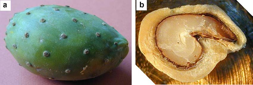
Fresh mature prickly pear fruits of Opuntia ficus-indica centration in water was disintegrated for 15 min in a
(Fig. 1a) were collected from an experimental station plan- Waring Blender operated at full speed where a final tem-
tation located in the vicinity of Marrakech (Morocco). The perature of 60 C was reached. The suspension was then
harvested fruits were washed, carefully hand-peeled and homogenized by 15 passes through a Manton Gaulin labo-
the pulp was mixed for a few minutes in a mixer grinder. ratory homogenizer operated at 500 bars at a temperature
The seeds were recovered from the resulting pulp juice by that was controlled at 80 C to give the homogenized resi-
straining through a metallic strainer and cleaned by several due noted Residue IV-G. This residue was then treated by
washings with distilled water. After drying, they were 6% NaOH solution to give a final residue (Residue V-G).
Fig. 1. (a) Opuntia ficus-indica fruit, (b) cross-section of seed from Opuntia ficus-indica fruit.104 Y. Habibi et al. / Carbohydrate Polymers 72 (2008) 102–112
Pericarp powder
Ethanol- Toluene (62-38), 24h
Defatted pericarp
NaOCl2 , pH=4.8, 70ºC, 1h (× 2)
Residue I
NaOH 2%, 80ºC, 2h (× 2)
Residue II Extract I
NaOCl2 , pH=4.8, 70ºC, 1h (× 2) Neutralization AcOH 20%
NaOH 4%, 80ºC, 2h (× 2) WIX-I
Residue III Extract II
Neutralization AcOH 20%
NaOCl2 , pH=4.8, 70ºC, 1h (× 2)
NaOH 6%, 80ºC, 2h (× 2) WIX-II
Residue IV Extract III
Neutralization AcOH 20%
Manton Gaulin WIX-III
NaOH 8%, 80ºC, 2h
Homogenization
Residue IV-G Residue V-A
NaOH 6%, 80ºC, 2h NaOH 12%, 80ºC, 2h
Residue V-G Residue VI-A
Fig. 2. Scheme of fractionation and purification of xylan and cellulose from pericarp seed of OFI.
13
2.4. CP/MAS C solid state NMR spectroscopy 2.6. Scanning electron microscopy
The NMR experiments were performed on a Bruker Seeds were embedded in LR White resin (hard mixture;
Avance spectrometer (13C frequency of 100 MHz), using London Resin Co., Woking, UK) polymerized for 24 h at
proton dipolar decoupling (DD), magic angle spinning 50 C. Ultra-thin (250 nm) longitudinal and transversal
(MAS) and cross-polarization (CP). CP transfer was cross-sections were cut with a microtome (MTX RMC
achieved using a ramped amplitude sequence (RAMPCP) Ultrotome) and fixed in a freshly prepared mixture of
for an optimized total contact time of 2 ms. The spinning 0.3% glutaraldehyde, 2% paraformaldehyde in 0.05 M
speed was set at 6 kHz, sweep width 50,000 Hz, recycle phosphate buffer pH 7–7.2. Samples were dehydrated
delay 4 s. An average number of 10,000 scans was through a graded series of ethanol. Before viewing, the
acquired for each spectrum. The various xylan samples samples were sputtered with gold–palladium alloy in a
were examined under different conditions either dried or JEOL JFC sputterer. The observations were made with a
analyzed wet in the presence of liquid water. Each cellu- JEOL JMS-6100 SEM operating at an accelerating voltage
lose residue sample was analyzed in the presence of ranging from 5 to 8 kV and in secondary electron mode.
liquid water. The 13C chemical shifts were measured rel-
ative to carbon chemical shift of glycine carboxyl group 2.7. Optical microscopy
(176.03 ppm).
Observations of the cellulose fiber residues were
2.5. X-ray diffraction achieved with a Zeiss Axiophot 2 optical microscope oper-
ated in Nomarski contrast, equipped with a camera and
The X-ray diagrams were recorded on a Warhus flat film controlled by computer.
vacuum X-ray camera mounted on a Philips PW 1720 X-
ray generator operated at 20 mA and 30 kV. X-ray mea- 2.8. Determination of molecular weight
surements were made on dry xylan powder or hydrated
xylan inserted and sealed in thin wall glass capillaries and The purified and freeze-dried xylans were dissolved in
on films obtained by water evaporation of the suspensions DMSO. The solutions were filtered directly into light scat-
for cellulose residues. tering cells through 0.2 lm nylon membranes (Pall GelmanY. Habibi et al. / Carbohydrate Polymers 72 (2008) 102–112 105
Laboratory). The concentration of the solutions for each different tissues, the endosperm (E) and the pericarp (P), as
sample was chosen so that the experiment could be carried shown in Figs. 1b and 3a. In a whole Opuntia ficus-indica
out under dilute regime conditions. Static light scattering prickly pear fruit, the amount of seed is important, and
(SLS) experiments were performed with a spectrometer can vary from 30% to 40% on a dry weight basis. The peri-
equipped with an argon ion laser (Spectra Physics, model carp corresponded to 90–95% of the whole seed. The con-
2020, k = 488 nm) and fitted with a variable-angle detec- stituents and chemical composition of the pericarp are
tion system operated by a stepping motor (ALV, Langen- given in Table 1.
Germany Instruments). The sample temperature was set An important amount of lignin (20 wt%), as well as fats
at 25 ± 0.1 C, and the scattered intensity was measured and waxes (8 wt%) are observed. The minerals and protein
through a band-pass filter (488 nm) and a 200-lm pinhole content are low (2.5 wt%), and the main constituents are
with a photomultiplier tube (ALV). polysaccharides (62 wt%), including cellulose (35 wt%).
The morphological study carried out by scanning elec-
3. Results and discussion tron microscopy showed that the cells of pericarp seed of
Opuntia ficus-indica were mainly made up of spindle-shaped
3.1. Morphological and chemical analysis sclerenchyma fibers organized into two distinct orientations:
P1 and P2 (Fig. 3b, c, and d). These tissues are typical con-
The micrographs of transversal cross-section of seeds of stituent of the secondary walls. We can also observe some
Opuntia ficus-indica showed that the seed consisted of two spiral conducting vessels in simple helix (Fig. 3e and f).
Fig. 3. SEM micrographs of transverse sections of: (a) an Opuntia ficus-indica seed; (b) the pericarp of the seed with the two orientations P1 and P2; (c)
and (d) enlargement of P1 and P2, respectively; (e) and (f) longitudinal and transversal cross-sections, respectively, of spiral conducting vessels of P2.106 Y. Habibi et al. / Carbohydrate Polymers 72 (2008) 102–112
Table 1 Table 3
Chemical composition of pericarp seed of OFI Sugars content of different treated cellulose fibers from pericarp seed of
Opuntia ficus-indica
Constituents Dry wt%
Residue Sugarsa
Ash 2.5
Fat and wax 8 Xylose Glucose
Lignin 20 Defatted pericarp 42.7 47.1
Protein 1.5
Residue IV 19.5 80.5
Other polysaccharides 27
Residue V-A 10.6 89.4
Cellulose 35
Residue VI-A 1.0 99.0
Residue IV-G 18.8 82.2
Residue V-G 1.2 98.8
In Table 2 we reported the results of sugar analysis of a
Expressed in relative weight percentages.
the whole seed and of the pericarp. Sugar composition
showed a predominance of xylose (42.7%) and glucose
(47.1%) residues which indicated that the pericarp seed reported in Table 3, indicated that 80% of the xylan could
consisted of a natural xylan–cellulose composite. be extracted by 2, 4, and 6% NaOH extractions.
Cellulosic residues were analyzed by CP/MAS 13C
3.2. Extraction of xylan content and purification of cellulose NMR and we reported in Fig. 4 the spectra of defatted res-
fibers idue and of different alkali treated residues. We noticed the
presence, in addition to characteristic signals of cellulose,
Xylan can be extracted directly from wood with aque- of several signals in the spectrum of the defatted pericarp
ous alkali, although their alkali-extractability cannot be at 20.94, 56.06, and 173.21 ppm. The two signals at 20.94
complete because xylans were differently associated physi- and 173.21 ppm disappeared after the first alkali treatment
cally and chemically with lignin and cellulose. A higher suggesting that they were removed during the treatment.
efficiency of extraction is reached with higher alkali con- This observation suggested that they corresponded to ace-
centrations, but high concentrations of sodium hydroxide tate groups carried by the xylan. It indicates that the native
can cause mercerization of cellulose. Mercerization pro- xylans of seed pericarp are partially acetylated. After
cess involves swelling of native cellulose I fibers in concen- bleaching treatment, the signals at 56.06, and between
trated sodium hydroxide with formation of cellulose– 112 and 152 ppm disappeared indicating that they corre-
NaOH complexes. These complexes can recrystallize in sponded more likely to lignin residues. The spectrum of
an anti-parallel manner to form the energetically favorable residue IV corresponds roughly to cellulose-rich polysac-
cellulose II polymorph after removal of the swelling agent charide. Nevertheless, the sugars analysis showed that it
with water (O’Sullivan, 1997). In addition, it has been contained an important amount of xylan (19.5%), which
demonstrated that the mercerization of cellulose fibers required purification. Thereafter, we were interested in
depends on their origin and their morphology. As an the purification of this cellulosic fraction.
example, mercerization of secondary wall cellulose such In order to purify cellulose fibers from pericarp seed of
as cotton linters does not occur if the concentration of Opuntia ficus-indica, we studied two extraction methods as
NaOH is below 10% (w/w) (Lindgren et al., 1995) and shown in Fig. 1: (i) entirely alkaline treatments and (ii) a
starts at 9% NaOH (w/w) in the case of primary wall cel- combination of Manton Gaulin mechanical treatment
lulose extracted from sugar beet pulp (Dinand, Vignon, and alkaline extractions. A study was achieved to check
Chanzy, & Heux, 2002). Other alternative methods, such the effect of alkaline concentrations and optimize the xylan
as enzymatic degradation, chemical acidic hydrolysis, or extraction and cellulose purification.
thermo-mechanical treatments, are used to remove xylan
and purify cellulose. 3.3. Alkaline treatment of seed pericarp of OFI
The seed pericarp of Opuntia ficus-indica is composed
essentially of xylan and cellulose. The extraction of xylan Before purification of cellulose fibers from pericarp seed
content is carried out by alkaline treatments as show in of OFI, we were interested by their mercerization. We
Fig. 2. The efficiency of the extraction is controlled by sug- determined by X-rays and CP/MAS 13C NMR spectros-
ars dosage and CP/MAS 13C NMR. Sugars analysis, copy the alkali concentration threshold where the cellulose
I fi cellulose II transformation starts. After washing and
drying, the X-ray investigation of the various alkali treated
Table 2
samples gave typical variations in the diffraction patterns.
Sugar composition of the seed and of seed pericarp
This is illustrated in Fig. 5, where the four patterns corre-
Component Uronic acida Neutral sugarsa
sponded to samples treated with different alkali concentra-
Rha Glc Gal Ara Xyl Man tion. The patterns in Fig. 5a–d are those of residues
Whole seed – 0.6 40.6 1.0 3.1 44.8 1.0 treated, respectively, with 2%, 6%, 8%, and 12% NaOH.
Pericarp 9 – 47.1 – 1.3 42.7 0 The pattern in Fig. 5b has the same features as the one
a
Expressed in relative weight percentages. in Fig. 5a, but with a slightly better resolution. BothY. Habibi et al. / Carbohydrate Polymers 72 (2008) 102–112 107
a
b
c
d
e
180 160 140 120 100 80 60 40 20
ppm
Fig. 4. CP/MAS 13C NMR spectra of: (a) defatted pericarp; (b) aq. 2% NaOH – Residue II; (c) aq. 6% NaOH – Residue IV; (d) aq. 8% NaOH – Residue
V-A; (e) aq. 6% NaOH – Gaulin Residue V-G.
d-spacing of 0.258 nm, (ii) two stronger and slightly
broader rings at d = 0.43 and 0.40 nm, the ring at
0.40 nm being more intense than the one at 0.43 nm, and
(iii) a broad ring of medium intensity centered at 0.57 nm.
The sample treated with 8% NaOH solution gave a dia-
gram (pattern Fig. 5c) that differed somewhat from the pat-
terns in Fig. 5a and b. The pattern 5c presented four
diffraction rings, located at nearly the same position as
those in 5a and 5b, but with different relative intensities.
Indeed, in 5c the rings at 0.43 and 0.40 nm have practically
the same strong intensity, the one at 0.43 nm being some-
what more intense than the one at 0.40 nm. On the other
hand, the sharp ring at 0.258 nm and the broad one at
0.57 nm became extremely weak. This pattern demon-
strated clearly the cellulose I fi cellulose II conversion.
The pattern 5d was different from the other three patterns.
Fig. 5. X-ray diffraction patterns of alkali-treated pericarp seed of It showed only two rings at d-spacing of 0.40 and 0.43 nm,
Opuntia ficus-indica: (a) aq. 2% NaOH, (b) aq. 6% NaOH, (c) aq. 8% the ring at 0.43 nm being slightly stronger than the one at
NaOH, and (d) aq. 12% NaOH. 0.40 nm. This pattern corresponded to mercerized cellulose
II.
Fig. 5a and b are typical cellulose I patterns. These patterns According to the X-ray diagrams, the cellulose I fi cel-
consist of four diffraction rings: (i) a sharp weak ring at a lulose II transformation of cellulose fibers from seed108 Y. Habibi et al. / Carbohydrate Polymers 72 (2008) 102–112
pericarp of OFI starts at 8% NaOH concentration, which is As shown in Table 3, the xylose content of 6% NaOH
unusual in the case of secondary wall cellulose fibers. treated residue (residue IV) is important (19.5%), demon-
The various alkali-treated residues were also analyzed strating the presence of an important amount of residual
by CP–MAS 13C NMR spectroscopy and the spectra of xylan. No variation in the sugar composition was observed
various residues are shown in Fig. 4. The spectrum 4-C pre- after the mechanical treatment (Residue IV-G). However,
sented the characteristic signals of native cellulose (Atalla, the sugar composition of the cellulosic residue was exten-
Gast, Sindorf, Bartuska, & Maciel, 1980; Earl & Vander- sively modified after the last alkaline treatment (residue
Hart, 1980). In the spectrum treated with 8% NaOH aque- V-G). The glucose was the dominant constituent (98.1%)
ous solution (Fig. 3d), the apparition of some signals, of residue V-G showing that the removal of residual xylan
particularly the doublets near 104.80 and 107.02 ppm was facilitated by the mechanical treatment which allowed
together with the doublet near 62.86 and 62.40 ppm, dem- a better accessibility. The mechanical treatment induced a
onstrated clearly the onset of cellulose conversion. The better access to fibers’ surface and allowed the extraction
concomitant decrease of the cellulose I doublet centered of the residual xylan (20%), without mercerization of cellu-
near 64.95 ppm was also observed. The percentage of cellu- lose as demonstrated by X-ray experiments (not shown) as
lose II increased steadily with the concentration in NaOH well as CP/MAS 13C NMR (Fig. 4e). Indeed, the CP/MAS
13
to reach 100% conversion with the sample treated with C NMR spectrum presented the characteristic signals of
12% NaOH (spectra not showed). These results confirmed native cellulose.
that mercerization of pericarp seed cellulose started with These results showed the efficiency of the mechanical–
8% NaOH. chemical combined treatments used for purification of cel-
lulose from pericarp seed of OFI.
3.4. Mechanical treatment of seed pericarp of OFI
3.5. Ratio of Ia and Ib cellulose content and degree of
We chose to combine alkaline extraction with mechani- cellulose crystallinity
cal treatment in order to purify cellulose from seed pericarp
of OFI. This mechanical treatment makes it possible to Solid state NMR was generally used to investigate the
individualize fibers in microfibrils and to increase their ratio of cellulose Ia and Ib in cellulose species. In particular
accessibility and their alkaline extraction without cellulose the C-4 region was used for this analysis, the signals from
mercerization. ordered and less ordered regions being well separated. The
The residue IV, according to the preceding extraction signals between 86 and 92 ppm corresponded to C-4 of the
scheme (Fig. 2), was treated in a Manton Gaulin homoge- highly ordered cellulose crystallite, whereas the broader
nizer. The homogenized residue, noted Residue IV-G, was upfield signal between 79 and 86 ppm were assigned to
then treated another time by 6% alkaline aqueous solution the C-4 of disordered cellulose as well as to the less ordered
to give Residue V-G. The mechanical treatment and purifi- cellulose chains of the crystallite surfaces (Van der Hart &
cation were controlled by microscopy, sugar analysis and Atalla, 1984; Wickholm et al., 1998). Larsson used spectral
also by CP/MAS 13C NMR. fitting for the C-4 region of cotton cellulose and assigned
Fig. 6 showed the optical micrograph of residue IV three Lorentzian lines for the signals from cellulose Ia,
before and after mechanical treatment in a Manton Gaulin I(a+b), and Ib and four Gaussian lines for the signals from
homogenizer. These images indicated that the fibers have para-cristalline cellulose, inaccessible fibril surfaces, and
been completely disrupted during the homogenization two accessible fibril surfaces.
treatment and the homogenized residue consisted of disen- The ratio of Ia and Ib cellulose polymorphs and the
crusted cellulose fibers. degree of cellulose crystallinity in the case of seed pericarp
Fig. 6. Optical micrographs in Nomarski contrast of disencrusted cells (Residue IV): (a) before and (b) after homogenization.Y. Habibi et al. / Carbohydrate Polymers 72 (2008) 102–112 109
of OFI were determined using the areas of the crystalline
and amorphous C-4 signals according to Larsson fitting.
The results indicated that cellulose of seed pericarp was
constituted of 73% of cellulose Ia and 27% of cellulose Ib
with a degree of crystallinity near to 60%.
3.6. Characterization and organization of extracted xylan
The xylan content of pericarp seed of OFI was charac-
terized essentially by high resolution 1H and 13C NMR
spectroscopy in our previous work (Habibi, Mahrouz, &
Vignon, 2002). We distinguished two types of xylans from
pericarp seed of OFI. The water soluble xylans are charac-
terized by a molar ratio of xylose to uronic acid varying
from 8/1 to 12/1. The water insoluble xylans which are in
larger amount, have a low uronic acid content (varying
from 26/1 to 65/1 Xyl/UA). Fig. 7. X-ray diffraction patterns of WIX-II from pericarp seed of OFI (a)
dried form; (b) hydrated form.
In the present work, we are interested by the water insol-
uble xylans (WIX) which were therefore isolated by
sequential alkaline extractions with aqueous 2%, 4%, and X-ray diffraction patterns of xylan extracted with aqueous
6% (w/w) NaOH solutions, according to Habibi et al. 4% NaOH (fraction WIX-II) in dry and hydrated form. We
(Habibi et al., 2002) with minor modifications as reported can observe that hydration caused fundamental change in
in Fig. 2. X-ray diffraction pattern showing a re-organization of
The sugar composition and average molecular weight of xylan under hydration. The X-ray pattern of dry xylan
each insoluble xylan were reported in Table 4. We noticed shows one broad diffraction ring centered at 0.416 nm indi-
that the molar ratio of xylose to uronic acid as well as cating that xylan does not present any organization. After
molecular weight of xylan fractions increased with the hydration we noticed the apparition of several diffraction
NaOH concentration. The average molecular weight ran- rings at 0.790, 0.698, 0.531, 0.460, 0.391, 0.355, 0.309,
ged from 11,250 g/mol for WIX-I, and 13,500 g/mol and and 0.280 nm which corroborated with values published
24,000 g/mol for WIX-III which correspond to degree of by Horio and Imamura (1964) and Nieduszynski and Mar-
polymerization values of approximately 75, 90, and 160 chessault (1972). No difference is observed between the var-
for WIX-I, WIX-II, and WIX-III, respectively. These val- ious xylan fractions, what is due probably to their low
ues corroborated the weight-average molecular weight uronic acid content. The behavior observed suggests that
reported earlier for xylan extracted from plants holocellu- water induces a helical conformation and re-organization
lose with aqueous alkaline solutions (Ebringerova & Hei- of the xylan polysaccharide. This phenomenon has already
nze, 2000). been reported for several helix-forming polysaccharides,
We were interested in the organization of xylan and cel- such as b-(1 fi 3)-linked D-glucans (Pelosi et al., 2003; Sai-
lulose, both in isolated form and as a component of seed to, Tabeta, Yokoi, & Erata, 1987; Saito, Yokoi, &
pericarp of OFI. Xylan and cellulose residues were studied Yoshioka, 1989), b-(1 fi 3)-linked D-xylans (Lahaye, Ron-
essentially by X-ray and CP/MAS spectroscopy. deau-Mouro, Deniaud, & Buleon, 2003; Saito, Yamada,
Xylan type polysaccharides are not thought to be crys- Yoshioka, Shibata, & Erata, 1991), and amylases (Chee-
talline in situ in the wood cell wall, but these polymers tham & Tao, 1998).
can crystallize under certain conditions (Chanzy, Dube, This phenomenon was also observed by CP/MAS 13C
& Marchessault, 1979; Nieduszynski & Marchessault, NMR spectroscopy. Hydration of plant cell wall materials
1972; Roelofsen, 1954). The backbone of xylan in crystal- (Rondeau-Mouro, Crepeau, & Lahaye, 2003), xylan
line form in aqueous solution has a three-fold, left-handed (Lahaye et al., 2003; Saito et al., 1991), and galactans (Sai-
conformation. In Fig. 7 we reported, as an example, the to, Yokoi, & Yamada, 1990) or other polysaccharides, such
Table 4
w ) of water insoluble xylans from seed pericarp
Yields, sugar composition and average molecular weight (M
Xylan fraction Yields Sugarsa w (·103) (g mol 1)
M
Uronic acid Glucose Arabinose Xylose
WIX-I 5.1 11.5 Tr – 88.3 11.25
WIX-II 2.2 5.9 1.1 – 93.0 13.50
WIX-III 7.0 2.5 Tr – 98.2 24
a
Expressed in relative weight percentages.110 Y. Habibi et al. / Carbohydrate Polymers 72 (2008) 102–112
as starch (Cheetham & Tao, 1998; Tanner, Ring, Whittam, ferent alkali treated residues and of a sample of extracted
& Belton, 1987) and b-glucans (Fyfe et al., 1984; Saito xylan. As first interesting observation, the signal at
et al., 1987, 1989, 1991; Saito, Yoshioka, Yokoi, & Yam- 101.4 ppm, corresponding to C-1 of hydrated xylan, was
ada, 1990; Stipanovic, Giammatteo, & Robie, 1985) has not observed in the spectra of the different residues spe-
also been reported to increase the CP/MAS 13C NMR sig- cially defatted residue and 2% NaOH treated residue (Res-
nal resolution. The CP/MAS 13C NMR spectra of WIX-I idue II), whereas these residues are rich in xylan.
xylan fraction in the dry and hydrated states are shown Apparently, the structure of xylan in pericarp residues dif-
in Fig. 8a and b, respectively, and that of WIX-III xylan fered from that of hydrated xylan.
fraction at hydrated form is reported in Fig. 8c. We can Furthermore, we noticed a continuous decrease of the
notice that hydration of xylan induced a marked increase signal at 81.9 ppm with the progression of xylan extraction.
in the signal resolution in the CP/MAS NMR spectrum, However this signal is not present in the spectrum of
demonstrating the re-organization of the xylan chains hydrated xylan and dry xylan. This signal was assigned
under hydration. The spectrum of the dry sample showed to xylan interacting with cellulose; probably xylan presents
the three broad unresolved peaks. These peaks are cen- as co-aggregates with cellulose fibril aggregates.
tered toward 101.9, 74.4, and 63.5 ppm and are assigned, These observations were already reported by Wickholm
respectively, to C-1 (C-2, C-3, C-4), and C-5 of b-(1 fi 4)- et al. (1998), Larsson et al. (1999), Teleman, Larsson, and
linked D-xylose residues. Hydratation of xylan from peri- Iversen (2001), as well as Liitiae et al. (2003). They con-
carp of OFI markedly improved the resolution of the sig- cluded that the xylan adopted, in the presence of the cellu-
nals and affected also the chemical shifts of broad lose, an organization in co-aggregations. This organization
resonances, particularly of C-1 (101.4 ppm) and C-5 is different to the one adopted by isolated xylan either in
(62.83 ppm). Also no difference is observed between the dry, hydrated form or in the presence of other substances
spectra of various xylan fractions both in dried or (such as methanol, isopropanol). This change in conforma-
hydrated states. tion induced in the CP/MAS NMR spectrum principally a
shift toward the weak fields of the signals assigned to C-4
3.7. Organization and interaction of xylan–cellulose in seed and C-1 of the xylose residues. The shift of C-4 signal from
pericarp 74.38 ppm for hydrated xylan to 81.87 ppm for xylan-com-
posite in pericarp indicated a large difference in the molec-
The organization of xylan–cellulose composite in seed ular environment of the xylan. The xylan backbone itself
percicarp of OFI was also studied. Different residues have imposed certain minimal structural constraints, but in fact
been examined by CP/MAS 13C NMR in hydrated state. the interactions between xylan and cellulose chains deter-
We reported in Fig. 9 the spectra of defatted residue, of dif- mined the final conformation.
a
b
c
110 100 90 80 70 60
(ppm)
Fig. 8. 13C CP/MAS NMR spectra of xylan extracted from pericarp seed of OFI: (a) WIX-I dried form; (b) WIX-I hydrated form and (c) WIX-III
hydrated form.Y. Habibi et al. / Carbohydrate Polymers 72 (2008) 102–112 111
a
b
c
11 0 100 90 80 70 60
(ppm)
Fig. 9. CP/MAS 13C NMR spectra of xylan or treated pericarp seed of OFI. (a) Aq. 6% NaOH – Residue IV; (b) aq. 6% NaOH – Gaulin Residue V-G; (c)
6% NaOH extracted xylan.
4. Conclusion thanks are given to Danielle Dupeyre (CERMAV-CNRS)
for her help in SEM observations.
The study of pericarp of Opuntia ficus-indica (OFI) seeds
showed that it is mainly made up of spindle-shaped scleren- References
chyma fibers and consisted of a natural xylan–cellulose–lig-
nin composite. After lignin elimination, the xylan and Atalla, R. H., Gast, J. C., Sindorf, D. W., Bartuska, V. J., & Maciel, G. E.
(1980). Carbon-13 NMR spectra of cellulose polymorphs. Journal of
cellulose were studied by X-ray and CP/MAS 13C NMR
the American Chemical Society, 102(9), 3249–3251.
spectroscopy. The water induced a better organization of Brett, C. T. (2000). Cellulose microfibrils in plants: Biosynthesis, depo-
supramolecular structure of isolated xylan. The scleren- sition, and integration into the cell wall. International Review of
chyma cellulose fibers are typical of secondary wall cellu- Cytology, 199, 161–199.
lose, with polymorphs ratios of 73% Ia and 27% Ib and Chanzy, H., Dube, M., & Marchessault, R. H. (1979). Structural
of crystallinity near to 60%, but they were sensitive toward polymorphism of (1 fi 4)-b-D-xylan. Polymer, 20, 1037–1039.
Cheetham, N. W. H., & Tao, L. (1998). Solid state NMR studies on the
NaOH because they started to be converted into cellulose structural and conformational properties of natural maize starches.
II at NaOH concentration as low as 8%. As component Carbohydrate Polymers, 36(4), 285–292.
of seed pericarp, cellulose fibers interact with xylan poly- De Cortazar, V. G., & Nobel, P. S. (1992). Biomass and fruit
mers, causing these to adopt a conformation different to production for the prickly pear cactus, Opuntia ficus-indica.
the one observed for xylan both in dry or hydrated form. Journal of the American Society for Horticultural Science, 117(4),
558–562.
Dinand, E., Vignon, M. R., Chanzy, H., & Heux, L. (2002). Mercerization
of primary wall cellulose and its implication for the conversion of
Acknowledgements
cellulose I fi cellulose II. Cellulose, 9(1), 7–18.
Earl, W. L., & VanderHart, D. L. (1980). High resolution, magic angle
We acknowledge the financial help of the Comité Mixte sampling spinning carbon-13 NMR of solid cellulose I. Journal of the
Franco-Marocain (Action Intégrée 236/SVS/00). Special American Chemical Society, 102(9), 3251–3252.112 Y. Habibi et al. / Carbohydrate Polymers 72 (2008) 102–112
Ebringerova, A., & Heinze, T. (2000). Xylan and xylan derivatives: thesized in vitro by enzymes from Saprolegnia monoica. Comparison
Biopolymers with valuable properties. 1. Naturally occurring xylans with a corresponding in vitro product from blackberry (Rubus
structures, isolation procedures and properties. Macromolecular Rapid fruticosus). Biochemistry, 42(20), 6264–6274.
Communication, 21(9), 542–556. Roelofsen, P. A. (1954). Some notes on xylan crystals. Biochima et
Fyfe, C. A., Stephenson, P. J., Taylor, M. G., Bluhm, T. L., Deslandes, Y., Biophysica Acta, 13, 592–593.
& Marchessault, R. H. (1984). Hydration effects in the carbon-13 CP/ Rondeau-Mouro, C., Crepeau, M. J., & Lahaye, M. (2003). Appli-
MAS NMR spectra of solid (1 fi 3)-b-D-glucans. Macromolecules, cation of CP-MAS and liquid-like solid-state NMR experiments
17(3), 501–502. for the study of the ripening-associated cell wall changes in
Habibi, Y., Mahrouz, M., & Vignon, M. R. (2002). Isolation and structure tomato. International Journal of Biological Macromolecules, 31(4–
of D-xylans from pericarp seeds of Opuntia ficus-indica prickly pear 5), 235–244.
fruits. Carbohydrate Research, 337(17), 1593–1598. Saito, H., Tabeta, R., Yokoi, M., & Erata, T. (1987). A high-resolution
Horio, M., & Imamura, R. (1964). Crystallographic study of xylan from solid-state carbon-13 NMR study of the secondary structure of linear
wood. Journal Polymer of Science Part A, 2, 627–644. (1 fi 3)-b-D-glucans: a conformational elucidation of noncrystalline
Hult, E. L., Larsson, P. T., & Iversen, T. (2000). A comparative CP/ and crystalline forms by means of conformation-dependent carbon-13
MAS13 C NMR study of cellulose structure in spruce wood and kraft chemical shifts. Bulletin of the Chemical Society of Japan, 60(12),
pulp. Cellulose, 7, 35–55. 4259–4266.
Jones, J. K. N., & Stoodley, R. J. (1965). Fractionation using copper Saito, H., Yamada, J., Yoshioka, Y., Shibata, Y., & Erata, T. (1991).
complexes. Methods of Carbohydrate Chemistry, 5, 36–38. Evidence of three distinct conformations – single chain, single helix,
Joseleau, J. P., Comtat, J., Ruel, K. (1992). Chemical structure of xylans and triple helix – of (1 fi 3)-b-D-xylan in the solid and intact frond of
and their interaction in the plant cell walls. Progress in Biotechnol- green algae as studied by 13C NMR spectroscopy. Biopolymers, 31(8),
ogy(Xylans Xylanases), 7, 1–15. 933–940.
Lahaye, M., Rondeau-Mouro, C., Deniaud, E., & Buleon, A. (2003). Saito, H., Yokoi, M., & Yamada, J. (1990). Hydration–dehydration
Solid-state13 C NMR spectroscopy studies of xylans in the cell wall of induced conformational changes of agarose, and kappa- and iota-
Palmaria palmata (L. Kuntze, Rhodophyta). Carbohydrate Research, carrageenans as studied by high-resolution solid-state 13C-nuclear
338(15), 1559–1569. magnetic resonance spectroscopy. Carbohydrate Research, 199(1),
Larsson, P. T. (2004). Interaction between cellulose I and hemicelluloses 1–10.
studied by spectral fitting of CP/MAS13 C NMR spectra. ACS Saito, H., Yokoi, M., & Yoshioka, Y. (1989). Effect of hydration on
Symposium Series, 254–268. conformational change or stabilization of (1 fi 3)-b-D-glucans of
Larsson, P. T., Hult, E. L., Wickholm, K., Pettersson, E., & Iversen, T. various chain lengths in the solid state as studied by high-resolution
(1999). CP/MAS13 C NMR spectroscopy applied to structure and solid-state carbon-13 NMR spectroscopy. Macromolecules, 22(10),
interaction studies on cellulose I. Solid State Nuclear Magnetic 3892–3898.
Resonance, 15, 31–40. Saito, H., Yoshioka, Y., Yokoi, M., & Yamada, J. (1990). Distinct
Larsson, P. T., Wickholm, K., & Iversen, T. (1997). A CP/MAS13 C NMR gelation mechanism between linear and branched (1 fi 3)-b-D-glucans
investigation of molecular ordering in celluloses. Carbohydrate as revealed by high-resolution solid-state carbon-13 NMR. Biopoly-
Research, 302, 19–25. mers, 29(14), 1689–1698.
Liitiae, T., Maunu, S., Hortling, B., Tamminen, T., Pekkala, O., & Stephen, A.M. (1983). Other plant polysaccharides. In G.O. Aspinall. The
Varhimo, A. (2003). Cellulose crystallinity and ordering of hemicel- Polysaccharides, 98–193.
luloses in pine and birch pulps as revealed by solid-state NMR Stipanovic, A. J., Giammatteo, P. J., & Robie, S. B. (1985). Cross-
spectroscopic methods. Cellulose, 10(4), 307–316. polarization/magic-angle spinning carbon-13 NMR of (1 fi 6)-b-D-
Lindgren, T., Edlund, U., & Iversen, T. (1995). A multivariate charac- glucan (pustulan): Mechanism of gelation. Biopolymers, 24(12),
terization of crystal transformations of cellulose. Cellulose, 2, 273–288. 2333–2343.
Maunu, S., Liitiä, T., Kauliomäki, S., Hortling, B., & Sundquist, J. (2000). Tanner, S. F., Ring, S. G., Whittam, M. A., & Belton, P. S. (1987). High
13
C CPMAS NMR investigations of cellulose polymorphs in different resolution solid state carbon-13 NMR study of some a(1 fi 4) linked
pulps. Cellulose, 7, 147–159. glucans: The influence of water on structure and spectra. International
Mora, F., Ruel, K., Comtat, J., & Joseleau, J. P. (1986). Aspect of native Journal of Biological Macromolecules, 9(4), 219–224.
and redeposited xylans at the surface of cellulose microfibrils. Teleman, A., Larsson, P. T., & Iversen, T. (2001). On the accessibility and
Holzforschung, 40(2), 85–91. structure of xylan in birch kraft pulp. Cellulose, 8(3), 209–215.
Newman, R. H. (1998). Evidence for assignment of13C NMR signals to Van der Hart, D. L., & Atalla, R. H. (1984). Studies of microstructures in
cellulose crystallite surfaces in wood, pulp and isolated celluloses. native celluloses using solid-state 13C NMR. Macromolecules, 17,
Holzforschung, 52, 157–159. 1465–1472.
Newman, R. H., & Hemmingson, J. A. (1990). Determination of the Whistler, R. L., & Chen, C. C. (1991). Hemicelluloses. International Fiber
degree of cellulose crystallinity in wood by carbon-13 nuclear magnetic Science Technology, 11, 287–319.
resonance spectroscopy. Holzforschung, 44, 351–355. Wickholm, K., Larsson, P. T., & Iversen, T. (1998). Noncrystalline forms
Newman, R. H., Hemmingson, J. A., & Suckling, I. D. (1993). Carbon-13 in cellulose I by CP/MAS spectroscopy. Carbohydrate Research, 312,
nuclear magnetic resonance studies of kraft pulping. Holzforschung, 123–129.
47, 234–238. Wilkie, K. C. B. (1979). The hemicelluloses of grasses and cereals.
Nieduszynski, I., & Marchessault, R. H. (1972). Structure of b-D-(1 fi 4)- Advances in Carbohydrate Chemistry and Biochemistry, 36,
xylan hydrate. Biopolymers, 11, 1335–1344. 215–264.
O’Sullivan, A. C. (1997). Cellulose: The structure slowly unravels. Wise, L. E., Murphy, M., & D’Addieco, A. A. (1946). Chlorite
Cellulose, 4, 173–207. holocellulose, its fractionation and bearing on summative wood
Pelosi, L., Imai, T., Chanzy, H., Heux, L., Buhler, E., & Bulone, V. (2003). analysis and on studies on the hemicelluloses. Paper Trade Journal,
Structural and morphological diversity of (1 fi 3)-b-D-Glucans syn- 122(2), 35–43.You can also read