Shiny wing scales cause spec(tac)ular camouflage of the angled sunbeam butterfly, Curetis acuta
←
→
Page content transcription
If your browser does not render page correctly, please read the page content below
bs_bs_banner

Biological Journal of the Linnean Society, 2013, 109, 279–289. With 8 figures

Shiny wing scales cause spec(tac)ular camouflage of the
angled sunbeam butterfly, Curetis acuta
BODO D. WILTS1*, PRIMOŽ PIRIH2,3, KENTARO ARIKAWA4 and
DOEKELE G. STAVENGA1
1
  Computational Physics, Zernike Institute for Advanced Materials, University of Groningen,
Groningen NL-9747AG, The Netherlands
2
  Department of Biology, Faculty of Biotechnical Sciences, University of Ljubljana, Ljubljana SI-1000,
Slovenia
3
  Department of Materials and Metallurgy, Faculty of Natural Sciences and Engineering, University
of Ljubljana, Ljubljana,SI-1000, Slovenia
4
  Laboratory of Neuroethology, Sokendai-Hayama (The Graduate University for Advanced Studies),
Hayama 240-0193, Japan

Received 12 December 2012; revised 16 January 2013; accepted for publication 16 January 2013

The angled sunbeam butterfly, Curetis acuta (Lycaenidae), is a distinctly sexually dimorphic lycaenid butterfly from
Asia. The dorsal wings of female and male butterflies have a similar pattern, with a large white area in the female
and an orange area in the male, framed within brown–black margins. The ventral wings of both sexes are silvery
white, which is caused by stacks of overlapping, non-pigmented, and specular-reflecting scales. With oblique
illumination, the reflected light of the ventral wings is strongly polarized. We show that the silvery reflection
facilitates camouflage in a shaded, foliaceous environment. The ecological function of the silvery reflection is
presumably two-fold: for intraspecific signalling in flight, and for reducing predation risk at rest and during
hibernation. © 2013 The Linnean Society of London, Biological Journal of the Linnean Society, 2013, 109, 279–289.

ADDITIONAL KEYWORDS: camouflage – iridescence – Lycaenidae – polarization – scattering.

                   INTRODUCTION                                 a dull, inconspicuous colour, which then serves to
                                                                camouflage the butterfly and decrease the risk of
A tapestry of numerous small scales imbricates the
                                                                predation: e.g. the ventral sides of I. io are brown–
wings of butterflies, often causing strikingly colourful
                                                                black, presumably rendering the butterfly inconspi-
patterns (Nijhout, 1991). In many species, the pat-
                                                                cuous against the background when they rest
terns differ strongly between the dorsal (upper) and
                                                                or hibernate. In the green hairstreak butterfly,
the ventral (under) wing sides, because of opposite
                                                                Callophrys rubi, the colour of the ventral wings
biological functions. The dorsal wings, which are
                                                                matches that of plant leaves (Michielsen & Stavenga,
exposed in active butterflies, for instance during
                                                                2008; Michielsen, De Raedt & Stavenga, 2010;
flight, are often brightly coloured. These markings
                                                                Schröder-Turk et al., 2011).
function in intra- and interspecific signalling: for
                                                                  The colour of the wing scales can be mainly pigmen-
example, in the radiant blue male Morpho butterflies
                                                                tary (as in I. io), as a result of pigments deposited in
and in the elaborately coloured peacock butterfly,
                                                                the wing scale structures, or can have a structural
Inachis io (Nijhout, 1991; Srinivasarao, 1999;
                                                                basis (as in C. rubi), when the structure of the scales
Kinoshita, 2008). In most butterflies, the ventral
                                                                have periodicities in the nanometer range. Butterfly
wings, exposed when the butterflies are at rest, have
                                                                wing scales commonly consist of two layers: a flat basal
                                                                lamina and a structured upper lamina. The two layers
                                                                are joined by pillar-like trabeculae (Ghiradella, 1984;
*Corresponding author. E-mail: b.d.wilts@rug.nl                 Ghiradella, 1998; Ghiradella, 2010). When the upper

© 2013 The Linnean Society of London, Biological Journal of the Linnean Society, 2013, 109, 279–289                279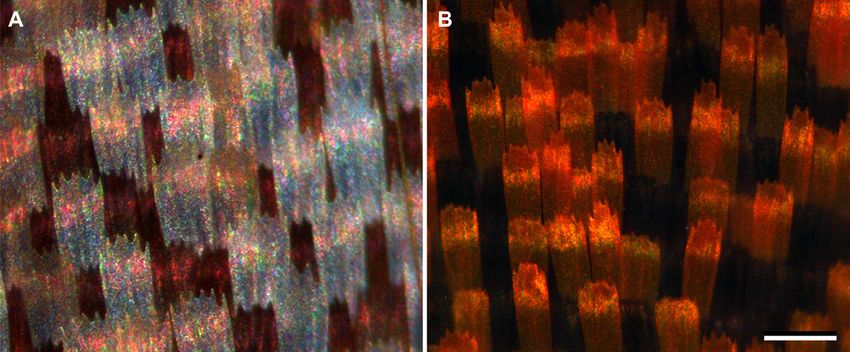
280    B. D. WILTS ET AL.

lamina is irregularly structured, the scale acts as a         angle-dependent reflectance measurements. We
diffuser, and, in the absence of a light-absorbing            discuss the biological function of the silvery-white
pigment, thus becomes a white light reflector                 coloration as an adaptive coloration strategy, serving
(Stavenga et al., 2004; Morehouse, Vukusic &                  for camouflage in a foliaceous environment and as an
Rutowski, 2007). For example, the wing scales of white        intraspecific signal during patrolling flights.
pierids (cabbage butterflies) have highly elaborate,
granular structures, which effectively scatter broad-
band light (Giraldo, Yoshioka & Stavenga, 2008).                         MATERIAL AND METHODS
These granules contain a pterin pigment, leucopterin                            ANIMALS
(Yagi, 1954), which absorbs light in the ultraviolet
                                                              Specimens of Curetis acuta Moore, 1877 (Lycaenidae)
wavelength range (invisible for humans, but visible
                                                              were captured near Sokendai, Shonan Village, Kana-
for insects). Xanthopterin and erythropterin, other
                                                              gawa Prefecture, Japan. Micrographs of wing patches
pterins common in pierid butterflies, also absorb in the
                                                              were taken with a Zeiss Universal Microscope (Carl
blue and green wavelength ranges, respectively, and
                                                              Zeiss AG, Oberkochen, Germany) and an Olympus
thus the wings have a yellow, orange, or red colour
                                                              SZX16 stereomicroscope (Olympus, Tokyo, Japan),
(Wijnen, Leertouwer & Stavenga, 2007).
                                                              equipped with Kappa DX-40 (Kappa Optronics
   Structural coloration occurs when periodic struc-
                                                              GmbH, Gleichen, Germany) and Olympus DP70
tures enhance light reflection in a specific wavelength
                                                              digital cameras, respectively.
range by constructive light interference and suppress
light reflection at other wavelengths by destructive
interference (Vukusic & Sambles, 2003; Kinoshita,
                                                                           SCANNING    AND TRANSMISSION
Yoshioka & Miyazaki, 2008). The periodicity can be
                                                                              ELECTRON MICROSCOPY
one-dimensional, as in the multilayers of the blue-
coloured wing scales of various lycaenids (Wilts,             The ultrastructure of the wing scales was investi-
Leertouwer & Stavenga, 2009), or three-dimensional,           gated with an XL-30 ESEM scanning electron micro-
as in the gyroid-type photonic crystals in the green-         scope (Philips, Eindhoven, Netherlands). Prior to
coloured wing scales of the papilionid Parides                imaging, the wing scales were sputtered with palla-
sesostris (Land, 1972; Vukusic & Sambles, 2003;               dium. For transmission electron microscopy (TEM) of
Michielsen & Stavenga, 2008; Wilts et al., 2012a). In         the scales, wing parts were prefixed in 2% parafor-
many butterfly species the structural colours are             maldehyde and 2.5% glutaraldehyde in 0.1 mol l-1
tuned by filtering pigments: that is, pigmentary and          sodium cacodylate buffer (CB, pH 7.3) for ~45 min,
structural colorations are combined, for instance in          then post-fixed in 2% osmium tetroxide in 0.1 M CB
the green scales of P. sesostris (Wilts et al., 2012a) and    for 2 h at room temperature (25 °C), and further
in the purple wing tip scales of the male pierid Colotis      block-stained in 2% uranyl acetate in 50% EtOH for
regina (Wilts, Pirih & Stavenga, 2011).                       1 h. After dehydrating with a graded series of ethanol
   Structural coloration commonly refers to reflections       and infiltration with propylene oxide, the tissues were
in a restricted wavelength band. Some structurally            embedded in Spurr’s resin. The tissues were cut into
coloured butterfly species, however, have wing scales         40–50-nm ultrathin sections, which were observed
reflecting broadband light, for example the nymphalid         using a Hitachi H7650 (Tokyo, Japan) transmission
Argyrophorus argenteus, where the wings are fully             electron microscope.
covered by silvery reflecting scales (Vukusic, Kelly &
Hooper, 2009). Silvery reflecting wing scales also
occur in patches at the ventral wings of several fri-                              SPECTROMETRY
tillary butterflies (Simonsen, 2007; Giraldo, 2008).          Reflectance spectra of the wings were measured with
   Another prominent example is the angled sunbeam            a bifurcated probe (Avantes FCR-7UV200; Avantes,
butterfly Curetis acuta, which is a widespread Asian          Eerbeek, the Netherlands) and with an angle-
butterfly (Eliot, 1990). In central Japan, its flying         dependent reflectance measurement (ARM) set-up,
period is between May and November, with a peak in            using an Avaspec 2048-2 CCD detector array spec-
the autumn on warm and sunny days. The ventral                trometer. The procedures were performed as
wings of both sexes of C. acuta are almost fully              described previously (Pirih, Wilts & Stavenga, 2011;
covered by silvery coloured scales, but the coloration        Stavenga et al., 2011). Transmittance spectra of single
of the dorsal wings is sexually dimorphic (see Fig. 1).       scales were measured with a microspectrophotometer
To unravel the spectral and spatial reflection proper-        (MSP), consisting of a Leitz Ortholux microscope,
ties of C. acuta wings and wing scales, we have               with an Olympus 20¥ objective (NA 0.46), connected
applied a variety of optical methods: among others            with the Avantes spectrometer. The reference was a
microspectrophotometry, imaging scatterometry, and            diffuse white reflectance tile (Avantes WS-2).

                      © 2013 The Linnean Society of London, Biological Journal of the Linnean Society, 2013, 109, 279–289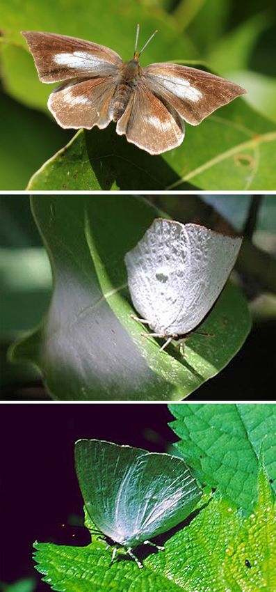
SHINY CAMOUFLAGE OF CURETIS ACUTA                       281

Figure 1. Sexual dichromatism of the angled sunbeam butterfly, Curetis acuta, and reflectance spectra. A, dorsal side
of the female C. acuta with large, white-coloured areas on the fore- and hindwing, framed by brown–black borders. B,
dorsal side of the male, with large, orange–red-coloured areas. C, brilliant-white ventral side of the female. (The ventral
wing side of the male is identical.) D, reflectance spectra measured with a bifurcated probe of the numbered wing areas
in (A–C). Scale bar: 1 cm.

               IMAGING   SCATTEROMETRY                         sets. The dorsal fore- and hindwings in both sexes are
For investigating the spatial reflection characteristics       framed within brown–black margins, whereas the
of the wing scales, we performed imaging scatterom-            central areas are white in the female and orange in
etry (Stavenga et al., 2009). An isolated, single scale        the male (Fig. 1A, B). The ventral wings of both
or a wing patch was glued at the pulled tip of a glass         females and males are silvery white (Fig. 1C). We
micropipette, and subsequently positioned at the first         measured the reflectance spectra from the various
focal point of the ellipsoidal mirror of the imaging           wing areas with a bifurcated probe (Fig. 1D) and from
scatterometer. The object was illuminated with a               single wing scales with a microspectrophotometer.
narrow (or alternatively wide) aperture, focused light           The white areas of the female dorsal wings contain
beam, and the spatial distribution of the far-field            a majority of silvery-whitish scales, intermingled with
scattered light was then monitored. A flake of mag-            brown–black scales (Fig. 2A), with the ratio of the two
nesium oxide served as a white diffuse reference               scale types depending on the location. The orange
object (for further procedural details, see Vukusic &          areas of the male dorsal wings contain orange–red
Stavenga, 2009; Wilts et al., 2009).                           scales, mixed with brown–black scales (Fig. 2B). The
                                                               latter scale type exclusively populates the brown wing
                                                               margins. These scales are clearly coloured by melanin
                                                               pigment, as is evident from the sharp rise in reflect-
                       RESULTS
                                                               ance with increasing wavelength (Fig. 1D, curve 1).
     SEXUAL    DICHROMATISM OF      CURETIS   ACUTA              The reflectance of the white areas in the female
Both sexes of C. acuta have a wingspan of about 4 cm           dorsal wings (Fig. 1D, curve 2) is low in the UV range,
(Fig. 1). The dorsal wings of females and males have           with a minimum at ~280 nm, possibly because of
similar patterns, but are composed of different colour         absorption by uric acid (Wijnen et al., 2007) and/or

© 2013 The Linnean Society of London, Biological Journal of the Linnean Society, 2013, 109, 279–289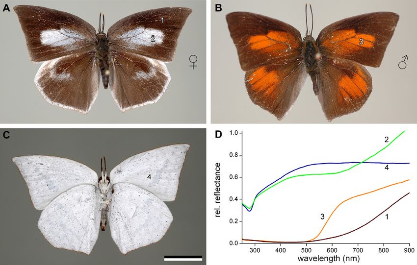
282 B. D. WILTS ET AL.
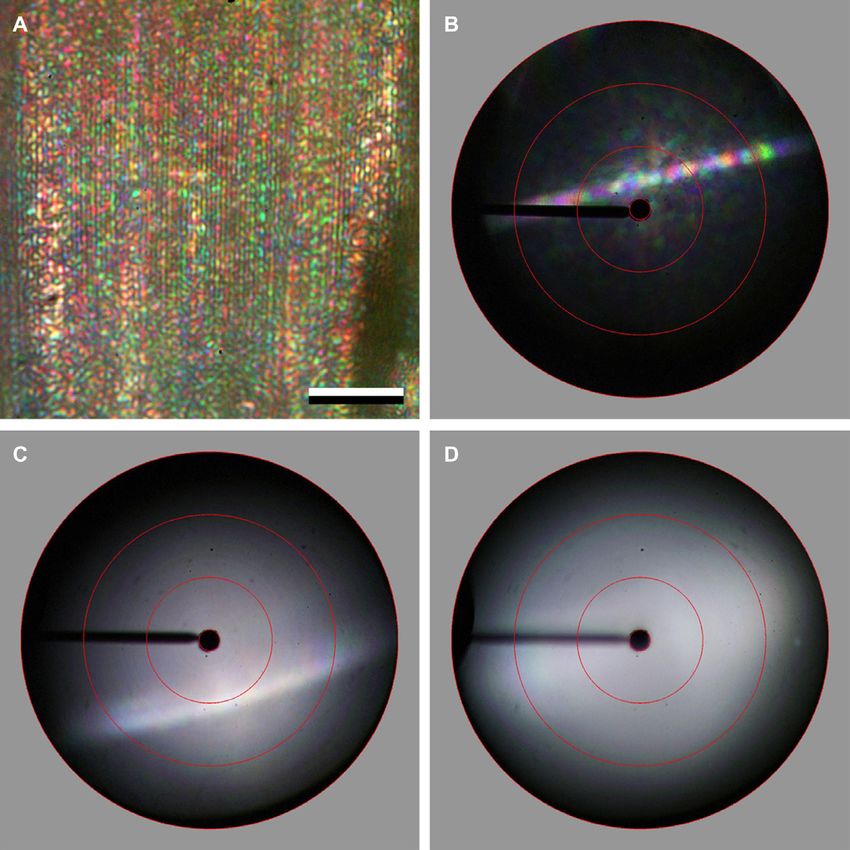
Figure 2. Epi-illumination light microscopy of Curetis acuta wing patches. A, an area in the female dorsal hindwing
showing silvery-white and brown–black scales. B, an area of the male dorsal forewing showing orange–red and black
scales. Scale bar: 100 mm.
proteins (Pace et al., 1995); the reflectance is high in
the visible wavelength range, and rises further above
600 nm. The latter rise is the result of melanin-
containing brown–black scales (Fig. 2A). The reflect-
ance of the orange areas in the male dorsal wings
(Fig. 1D, curve 3) is very low in the short wavelength
range, up to 500 nm, evidently because of a pigment
that absorbs light in the violet–blue wavelength
range, and then rises in two phases, between 500 and
600 nm, because of the decreasing absorption of the
blue-absorbing pigment in the orange–red scales, and
again above 600 nm because of the scattering caused
by the melanin-containing brown–black scales
(Fig. 2B).
The reflectance spectrum of the ventral wings of
both females and males is broadband, with a Figure 3. Absorbance spectra of single orange–red and
minimum in the UV range, very similar as that brown–black scales in immersion, deduced from transmis-
measured in the white scales of the female dorsal sion measurements with a microspectrophotometer.
wings (Fig. 1D, curve 4). In the visible wavelength
range, the reflectance of the ventral wings exceeds spectral transmission on single scales immersed in a
that of the white areas of the female’s dorsal wings, fluid with refractive index 1.56; this value closely
because of stacking in the white scales. Below the matches the refractive index of chitin, the material of
stacks of white scales there are some black scales, but butterfly scales (Leertouwer, Wilts & Stavenga, 2011).
their contribution to the overall reflectance remains Figure 3 shows the absorbance spectra obtained. The
minor. Measurements with an integrating sphere (not pigment in the brown–black scales has a broad
shown) yield reflectances of about 0.5 because of the absorbance spectrum, very reminiscent of melanin
large number of up to six overlapping scales, similar (Stavenga et al., 2012a). The pigment in the orange–
to pierid butterflies (Stavenga, Giraldo & Hoenders, red scales absorbs maximally at ~455 nm, and there-
2006; Wilts et al., 2011). fore the pigment is presumably a mixture of different
The reflectance spectra of the silvery-white areas ommochromes (Nijhout, 1997).
indicate the absence of a pigment absorbing light in
the visible wavelength range, but the orange–red and ANATOMY OF THE WING SCALES
brown–black scales obviously do contain pigments. Visual inspection showed that the dorsal and ventral
We characterized these pigments by measuring the wing sides not only strongly differ in coloration, but
© 2013 The Linnean Society of London, Biological Journal of the Linnean Society, 2013, 109, 279–289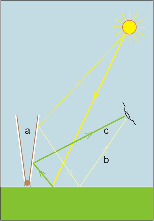
SHINY CAMOUFLAGE OF CURETIS ACUTA                     283

                                                                  The orange–red scales have the standard, basic
                                                               structure of butterfly wing scales (Fig. 4A). The upper
                                                               lamina is formed by numerous parallel ridges, with
                                                               spacing of about 1.3 mm, and connecting cross-ribs,
                                                               with spacing of about 0.6 mm. The windows in
                                                               between the cross-ribs are open, and trabeculae
                                                               support the upper lamina onto the more or less
                                                               flat, continuous lower lamina (Ghiradella, 1989;
                                                               Ghiradella, 2010). The silvery scales of the ventral
                                                               wings have a different structure (Fig. 4B). The dis-
                                                               tance between the ridges is ~1.8 mm, and that
                                                               between the just-visible cross-ribs is ~3 mm. The
                                                               windows are virtually fully closed, except for some
                                                               small, irregular perforations. Transmission electron
                                                               microscopy revealed that the silvery scales are slight
                                                               modifications of the standard butterfly wing scale.
                                                               The upper lamina is characteristically marked by
                                                               pronounced ridges, and the lower lamina is basically
                                                               a thin film (Fig. 4C). The trabeculae connecting the
                                                               upper and lower laminae are irregularly spaced.

                                                                 SPECTRAL    AND SPATIAL SCATTERING PROPERTIES
                                                                               OF THE SILVERY SCALES
                                                               The nearly flat laminae of the silvery scales suggest
                                                               that these scales act as thin-film reflectors. A scale of
                                                               the ventral wing of C. acuta observed with an epi-
                                                               illumination microscope, using a high-power objec-
                                                               tive, reveals a colourful, speckled image (Fig. 5A). The
                                                               local speckles average out at low magnification
                                                               (Fig. 2A), merging into the silvery-white colour
                                                               observed overall (Fig. 1C).
                                                                  We investigated the spatial scattering properties of
                                                               the silvery scales by imaging scatterometry (Fig. 5B–
                                                               D). Figure 5B presents a scatterogram of a single
                                                               white scale, oriented nearly perpendicular to the hori-
                                                               zontal axis of the scatterometer: the ridge direction
Figure 4. Ultrastructure of the wing scales. A, scanning       made a small angle with the vertical axis. The light
electron micrograph of an orange–red scale of the male         source was a narrow-aperture (5°) white-light beam,
dorsal wing, showing the basic bauplan of scales: ridges       and the illuminated area was a spot with diameter of
connected by cross-ribs; pigment is immersed throughout        10 mm. The resulting scattering pattern is a coloured
the scale material. B, scanning electron micrograph of         line perpendicular to the ridges. It is caused by dif-
a silvery scale from the ventral wing of a female: the         fraction at the grating created by the prominent scale
windows in between the ridges and cross-ribs are closed,       ridges, which interrupt the plane of the reflecting
except for a few perforations. C, transmission electron        window panes (Kinoshita, 2008; Kinoshita et al.,
microscopy of a cross section of a silvery scale. Scale        2008; Stavenga et al., 2009). Figure 5C shows the
bar: 2 mm.                                                     scatter diagram when a wing patch area of 140 mm in
                                                               diameter (approximately three to four wing scales)
also in spatial reflection properties. Whereas the             was illuminated with the same narrow-aperture
dorsal wings reflect light rather diffusely, the ventral       beam. The resulting scatter pattern is a broadened
wings are rather specular. This indicated a very               white line on top of a white, diffuse background. The
different fine structure of the scales, and thus               broad white line is the average of the diffraction lines
we performed scanning electron microscopy on the               created by the individual scales. By opening the aper-
orange–red scales of the male dorsal wings, and                ture, so that the illuminating light source becomes
on the silvery scales of the female ventral wings              hemispherical, a diffuse, hemispherical scatter
(Fig. 4A, B).                                                  pattern was obtained (Fig. 5D).

© 2013 The Linnean Society of London, Biological Journal of the Linnean Society, 2013, 109, 279–289
284 B. D. WILTS ET AL.
Figure 5. Epi-illumination light microcopy and imaging scatterometry of silvery wing scales. A, a scale of the ventral
wing observed with an epi-illumination light microscope. Scale bar: 20 mm. B, scatterogram resulting from a narrow
aperture axial light beam illuminating a small part of a single scale. A diffraction pattern emerges perpendicular to the
ridges, which are slightly oriented obliquely. C, off-axis illumination on several scales yields a more diffuse scatter pattern.
D, illumination by a hemispherical light source results in a diffuse scatter pattern.
ANGLE- AND POLARIZATION-DEPENDENT polarization-dependent analysis of the reflectance of
REFLECTANCE the whole wing (see also Fig. S1). Figure 6 shows the
angle-dependent reflectance measured for incident
The ventral wing scales consist of reflecting plates, linearly polarized light, i.e. transverse electric (TE, or
suggesting that at large angles of light incidence s) and transverse magnetic (TM, or p) polarized light,
the reflected light may become strongly polarized. averaged over the wavelength range of 300–700 nm.
We therefore performed a detailed angle- and The plane of light incidence, which contains the
© 2013 The Linnean Society of London, Biological Journal of the Linnean Society, 2013, 109, 279–289SHINY CAMOUFLAGE OF CURETIS ACUTA 285
central areas in the males and white central areas in
the females (Figs 1, 2). The displayed colours are the
result of pigments and/or wing scale structuring. In a
study on the swordtail butterfly Graphium sarpedon
we found that the white scales of G. sarpedon have
the standard papilionid scale structure (Stavenga
et al., 2012b), which is a slight modification of the
basic butterfly scale structure (Ghiradella, 2010). The
standard papilionid scale structure results in diffuse
scattering (Wilts et al., 2012b), and the same occurs
with the orange–red scales of the male C. acuta,
which have the basic nymphalid butterfly scale struc-
ture (Fig. 4A).
The ventral wings of both females and males are
silvery white, and are essentially identical. The
Figure 6. Angle-dependent reflectance measured from a silvery scales somewhat resemble the glass scales of
ventral wing of a male Curetis acuta for transverse electric G. sarpedon (Stavenga et al., 2012b), which have fully
(TE) and transverse magnetic (TM) polarized light aver- closed, flat windows and act as strong polarizing
aged over the wavelength range of 300–800 nm. The plane reflectors for large angles of light incidence. The
of light incidence was normal to the wing and parallel with silvery scales of the ventral wings of C. acuta inves-
the scale ridges. The angle of light incidence, determined tigated here similarly act as polarizers; however, dif-
by the angle of the fibre delivering the illumination, was fusely scattered light on the prominent ridges and the
varied in steps of 5°, and the angle of the detection fibre window perforations of the stacked silvery scales
was varied mirrorwise. The reflectance of TE-polarized creates a substantial background (compare Fig. 5C
light increases with increasing angle; the reflectance of with Fig. 6), which reduces the polarization contrast
TM-polarized light decreases with increasing angle, but (see Fig. S1) as well as the specularity of the silver
goes through a minimum at ~50° (see blue arrows), as reflectors.
expected for a mirror-like dielectric surface. The minimum The chitin layers of the scale laminae of G. sarpe-
reflectance of the TM-polarized light is not zero, because of don each have a thickness of ~200 nm (for similar
a background of diffusely scattered light. values of other butterflies, see Ghiradella, 1998). Like
the glass scales of G. sarpedon, the membranes in the
closed windows of Figure 4B will act as optical thin
incident light rays, was set perpendicular to the wing
films. Thin films have coloured reflections, which
and parallel with the scale ridges. The angle of illu-
rapidly change in colour upon slight changes in thick-
mination, delivered by a focused light fibre, was
ness. This is the reason why the silvery scales of
varied in steps of 5°, and the angle of the detection
C. acuta, when observed with an epi-illumination
fibre was varied mirror-wise. The reflectance of
microscope, create a colourful, speckled image. It
TE-polarized light increases monotonically with
shows that the local thickness of the scale cuticle
increasing angle; the reflectance of TM-polarized light
varies (Fig. 5A). The overall optical behaviour of the
decreases with an increasing angle of incidence up to
glass scales of G. sarpedon and the silvery scales of
~55°, but above this angle, the reflectance rises again.
C. acuta is quite different. Whereas the reflections of
This behaviour resembles that of an ideal, flat chitin
the glass scales of G. sarpedon are strongly coloured
layer, where the reflectance of TM-polarized light goes
and polarized overall, depending on the angle of illu-
to zero at Brewster’s angle (see the blue arrows in
mination and observation, the specular reflections of
Fig. 6). The substantial background reflectance of
the silvery scales of C. acuta are whitish and only a
~0.45 is presumably caused by incoherent scattering
little polarized. The latter is the result of additive
induced by irregularities in the scale structures and
colour mixing of light reflected by multiple small
in the scale stacking (indicated in Fig. 6 by the
domains of different thin-film thicknesses (see
dashed green line).
Figs 4C, 5A).
DISCUSSION THE FUNCTION OF THE SILVERY SCALES – NOT TO
COMPARISON OF THE SCALES OF CURETIS ACUTA BE SEEN IN THE SHADE
WITH THOSE OF GRAPHIUM SARPEDON Whereas the dorsal wing sides are displayed only
Female and male C. acuta differ substantially in the when the animals are active (Fig. 7A), the ventral
coloration of the dorsal wings, which have orange wing sides are exposed when they are at rest (Fig. 7B,
© 2013 The Linnean Society of London, Biological Journal of the Linnean Society, 2013, 109, 279–289286 B. D. WILTS ET AL.
A
B
Figure 8. Diagram of light reflected from a Curetis acuta
butterfly resting on a leaf. The letters (a, b, and c) refer to
the panels presented in Figure 7.
(Figs 7C, 8); however, when the butterflies are illumi-
C nated by direct sun, their distinct, silvery-white
reflections start to contrast with the surrounding
leaves (Fig. 7B). Interestingly, the vegetation of the
coastal regions of central Japan contains numerous
sclerophyllous plants with waxy leaves (e.g. the Japa-
nese oak Quercus acuta), which produce green reflec-
tions with white specularity when exposed in the sun.
In this environment the silvery-white ventral side of
C. acuta appears to blend with the background, even
when exposed to direct sunlight. Indeed, when chased
C. acuta flee into dense sclerophyllous shrubs and sit
on the twigs, where they are virtually impossible to
detect from the background, both under overcast con-
ditions and under clear skies. To a human observer,
Figure 7. Curetis acuta perching on leaves. A, a female
exposing the dorsal wings. B, exposed ventral wings,
the resting butterflies resemble a half-eaten dead
showing a distinct silvery-white reflection resulting from leaf, both in shape and in colour. As C. acuta survives
direct illumination by the sun. C, dorsal wings observed the winter in the imago form, the individuals conceiv-
from an angle substantially deviating from the mirror angle ably increase their chance of survival by actively
of the sun, so that the wing reflections are the result of light searching for evergreen trees and shrubs with waxy
emerging from the leaves (cropped photo by Oleg Kosterin. vegetation before they start hibernation (and possibly
the white colour helps for hiding in times of snow).
The sparse dark scales present on the ventral wings
C). When the butterflies rest on the leaves of shrubs may well add to the blending of the coloration with
and trees in the shade or under overcast skies, the the twigs and dead leaves.
ventral wing sides assume a green colour, thus The white coloration of the ventral wings may in
serving as camouflage in a shaded environment addition function in intraspecific signalling. When in
© 2013 The Linnean Society of London, Biological Journal of the Linnean Society, 2013, 109, 279–289SHINY CAMOUFLAGE OF CURETIS ACUTA 287
flight, at up to 10 m above the ground, C. acuta functions, acting for display when the butterflies are
flashes when reflecting direct sunlight, especially active and for camouflage when the butterflies are at
against clear blue skies. In males this flashing signal rest.
may be enhanced by the black and orange of the
dorsal wings, which are visible during the wingflap.
We conclude that the silvery-white coloration of ACKNOWLEDGEMENTS
C. acuta, a combination of diffuse and specular
reflectance, serves both to conceal and to reveal. We thank two anonymous reviewers for their helpful
Given that C. acuta has a broad geographical dis- comments and Oleg Kosterin for his kind permission
tribution, ranging from India to North Japan, its to use his photograph. This study was financially
forest habitats are likely to be very diverse. Further- supported by the Air Force Office of Scientific
more, other species of the genus Curetis, which com- Research/European Office of Aerospace Research and
prises 16 species, are likely to live in even more Development (AFOSR/EOARD grant FA8655-08-1–
diverse environments (from the Philippines to the 3012), by the JSPS Grant-in-Aid for Scientific
Himalayas), yet they all have silvery-white ventral Research no. 21247009, and by the MAFF (Ministry
wings (Eliot, 1990). It would therefore be very inter- of Agriculture, Forestry and Fisheries of Japan) grant
esting to learn how well the silvery-white camouflage (Elucidation of biological mechanisms of photore-
performs in environments with different optical prop- sponse and development of advanced technologies uti-
erties, and what is the primary evolutionary drive to lizing light) no. INSECT-1101.
become silvery white, i.e. for camouflaging or for
signalling purposes.
REFERENCES
SILVER COLORATION IN NATURE Denton EJ. 1970. On the organization of reflecting surfaces
in some marine animals. Philosophical Transactions of the
A metallic silver coloration occurs frequently in the Royal Society of London B 258: 285–313.
animal kingdom, and is mostly caused by multilay- Denton EJ, Gilpin-Brown JB, Wright PG. 1972. The
ered structures in the outermost organismal layers. A angular distribution of the light produced by some mesope-
silvery reflection is often used for camouflage as it lagic fish in relation to their camouflage. Proceedings of the
allows blending in with the environment with diffuse Royal Society B 182: 145–158.
illumination from the surroundings (see above). This Eliot JN. 1990. Notes on the genus Curetis Hübner (Lepi-
is the case in some butterfly pupae (Steinbrecht, doptera, Lycaenidae). Tyô to Ga 41: 201–225.
1985), for example. Also, many fish and cephalopods Ghiradella H. 1984. Structure of iridescent lepidopteran
use a silver coloration as a ubiquitous camou- scales: variations on several themes. Annals of the Entomo-
flage strategy, i.e. by reducing contrast with the sur- logical Society of America 77: 637–645.
rounding light conditions (Denton, 1970; Denton, Ghiradella H. 1989. Structure and development of iridescent
Gilpin-Brown & Wright, 1972; Mäthger et al., 2009; butterfly scales: lattices and laminae. Journal of Morphol-
Holt et al., 2011; Jordan, Partidge & Roberts, 2012). ogy 202: 69–88.
Silver reflectors can, for example, be found in the skin Ghiradella H. 1998. Hairs, bristles, and scales. In: Locke M,
of herrings and sardines (Jordan et al., 2012), and ed. Microscopic anatomy of invertebrates, Vol 11A: Insecta.
around the eyes of squids and cephalopods (Mäthger New York: Wiley-Liss, 257–287.
et al., 2009; Holt et al., 2011). In fish, the silver reflec- Ghiradella H. 2010. Insect cuticular surface modifications:
tors are made of multilayers, consisting of a large scales and other structural formations. Advances in Insect
Physiology 38: 135–180.
stack of layers (of more than ten), which is in contrast
Giraldo MA. 2008. Butterfly wing scales: pigmentation and
to C. acuta where two air-separated layers of chitin
structural properties. PhD thesis, Groningen.
create the silvery appearance. Fish reflectors can
Giraldo MA, Yoshioka S, Stavenga DG. 2008. Far field
therefore be specialized for high reflectance and low
scattering pattern of differently structured butterfly scales.
polarization at all angles of light incidence (Jordan Journal of Comparative Physiology A 194: 201–207.
et al., 2012). In other animals, like birds, butterflies, Holt AL, Sweeney AM, Johnsen S, Morse DE. 2011. A
and beetles, silver coloration is often employed for a highly distributed Bragg stack with unique geometry pro-
contrastful, highly visible display (Vukusic et al., vides effective camouflage for Loliginid squid eyes. Journal
2009). In the bird of paradise Parotia lawesii (Lawes’ of the Royal Society Interface 8: 1386–1399.
parotia), silver-coloured occipital feathers act as a Jordan TM, Partidge JC, Roberts NW. 2012. Non-
unique visual cue in the mating ritual (Laman & polarizing broadband multilayer reflectors in fish. Nature
Scholes, 2012; D. G. Stavenga, H. L. Leertouwer, Photonics 6: 759–763.
D. Osorio & B. D. Wilts, unpubl. data). Curetis acuta Kinoshita S. 2008. Structural colors in the realm of nature.
may be unique in that the silvery wings combine two Singapore: World Scientific.
© 2013 The Linnean Society of London, Biological Journal of the Linnean Society, 2013, 109, 279–289288 B. D. WILTS ET AL.
Kinoshita S, Yoshioka S, Miyazaki J. 2008. Physics of the wings of pierid butterflies. Optics Express 14: 4880–
structural colors. Reports on Progress in Physics 71: 076401. 4890.
Laman T, Scholes E. 2012. Birds of paradise: revealing the Stavenga DG, Leertouwer HL, Hariyama T, De Raedt H,
world’s most extraordinary birds. Washington: National Wilts BD. 2012a. Sexual dichromatism of the damselfly
Geographic. Calopteryx japonica caused by a melanin-chitin multilayer
Land M. 1972. The physics and biology of animal reflectors. in the male wing veins. PLoS ONE 7: e49743.
Progress in Biophysics and Molecular Biology 24: 75–106. Stavenga DG, Mashushita A, Arikawa K, Leertouwer HL,
Leertouwer HL, Wilts BD, Stavenga DG. 2011. Refractive Wilts BD. 2012b. Glass scales on the wing of the swordtail
index and dispersion of butterfly scale chitin and bird butterfly Graphium sarpedon act as thin film polarizing
feather keratin measured by interference microscopy. Optics reflectors. Journal of Experimental Biology 215: 657–662.
Express 19: 24061–24066. Stavenga DG, Leertouwer HL, Pirih P, Wehling MF.
Mäthger LM, Denton EJ, Marshall NJ, Hanlon RT. 2009. 2009. Imaging scatterometry of butterfly wing scales. Optics
Mechanisms and behavioural functions of structural colora- Express 17: 193–202.
tion in cephalopods. Journal of the Royal Society Interface 6: Stavenga DG, Stowe S, Siebke K, Zeil J, Arikawa K.
Suppl 2: S149–S163. 2004. Butterfly wing colours: scale beads make white pierid
Michielsen K, De Raedt H, Stavenga DG. 2010. Reflectiv- wings brighter. Proceedings of the Royal Society B 271:
ity of the gyroid biophotonic crystals in the ventral wing 1577–1584.
scales of the Green Hairstreak butterfly, Callophrys rubi. Stavenga DG, Wilts BD, Leertouwer HL, Hariyama T.
Journal of the Royal Society Interface 7: 765–771. 2011. Polarized iridescence of the multilayered elytra of the
Michielsen K, Stavenga DG. 2008. Gyroid cuticular struc- Japanese Jewel Beetle, Chrysochroa fulgidissima. Philo-
tures in butterfly wing scales: biological photonic crystals. sophical Transactions of the Royal Society B 366: 709–723.
Journal of the Royal Society Interface 5: 85–94. Steinbrecht RA. 1985. Fine structure and development of
Morehouse NI, Vukusic P, Rutowski R. 2007. Pterin the silver and golden cuticle in butterfly pupae. Tissue and
pigment granules are responsible for both broadband light Cell 17: 745–762.
scattering and wavelength selective absorption in the wing Vukusic P, Kelly R, Hooper I. 2009. A biological sub-micron
scales of pierid butterflies. Proceedings of the Royal Society thickness optical broadband reflector characterized using
B 274: 359–366. both light and microwaves. Journal of the Royal Society
Nijhout HF. 1991. The development and evolution of butterfly Interface 6: Suppl 2: S193–S201.
wing patterns. Washington: Smithsonian Institution Press. Vukusic P, Sambles JR. 2003. Photonic structures in
Nijhout HF. 1997. Ommochrome pigmentation of the linea biology. Nature 424: 852–855.
and rosa seasonal forms of Precis coenia (Lepidoptera: Nym- Vukusic P, Stavenga DG. 2009. Physical methods for inves-
phalidae). Archives of Insect Biochemistry and Physiology tigating structural colours in biological systems. Journal of
36: 215–222. the Royal Society Interface 6: Suppl 2: S133–S148.
Pace CN, Vajdos F, Fee L, Grimsley G, Gray T. 1995. How Wijnen B, Leertouwer HL, Stavenga DG. 2007. Colors and
to measure and predict the molar absorption coefficient of a pterin pigmentation of pierid butterfly wings. Journal of
protein. Protein Science 4: 2411–2423. Insect Physiology 53: 1206–1217.
Pirih P, Wilts BD, Stavenga DG. 2011. Spatial reflection Wilts BD, Leertouwer HL, Stavenga DG. 2009. Imaging
patterns of iridescent pierid butterfly wings and the depend- scatterometry and microspectrophotometry of lycaenid but-
ence of visibility on scale curvature. Journal of Comparative terfly wing scales with perforated multilayers. Journal of
Physiology A 197: 987–997. the Royal Society Interface 6: Suppl 2: S193–S202.
Schröder-Turk GE, Wickham S, Averdunk H, Brink F, Wilts BD, Michielsen K, De Raedt H, Stavenga DG.
Fitz Gerald JD, Poladian L, Large MC, Hyde ST. 2011. 2012a. Iridescence and spectral filtering of the gyroid-type
The chiral structure of porous chitin within the wing-scales photonic crystals in Parides sesostris wing scales. Interface
of Callophrys rubi. Journal of Structural Biology 174: 290– Focus 2: 681–687.
295. Wilts BD, Pirih P, Stavenga DG. 2011. Spectral reflectance
Simonsen TJ. 2007. Comparative morphology and evolution- properties of iridescent pierid butterfly wings. Journal of
ary aspects of the reflective under wing scale-pattern in Comparative Physiology A 197: 693–702.
Fritillary butterflies (Nymphalidae: Argynnini). Zoolo- Wilts BD, Trzeciak TM, Vukusic P, Stavenga DG. 2012b.
gischer Anzeiger 246: 1–10. Papiliochrome II pigment reduces the angle dependency of
Srinivasarao M. 1999. Nano-optics in the biological world: structural wing colouration in nireus group papilionids.
beetles, butterflies, birds and moths. Chemical Reviews 99: Journal of Experimental Biology 215: 795–805.
1935–1961. Yagi N. 1954. Note of electron microscope research on pterin
Stavenga DG, Giraldo MA, Hoenders BJ. 2006. Reflect- pigment in the scales of pierid butterflies. Annotationes
ance and transmittance of light scattering scales stacked on Zoologicae Japonensis 27: 113–114.
© 2013 The Linnean Society of London, Biological Journal of the Linnean Society, 2013, 109, 279–289SHINY CAMOUFLAGE OF CURETIS ACUTA 289
SUPPORTING INFORMATION
Additional Supporting Information may be found in the online version of this article at the publisher’s web-site:
Figure S1. Spatial reflectance maps for (A) unpolarised, (B) TE-(s)-polarized and (C) TM-(p)-polarized light
measured of the ventral wing of a female Curetis acuta. The polarization contrast, i.e. the s/p reflectance ratio
map is shown in (D). The polarization contrast is highest under large angle of illumination and detection. Black
dots represent measurement points used for the interpolation. The procedure and analysis of these plots is as
explained in Pirih et al. (2011).
© 2013 The Linnean Society of London, Biological Journal of the Linnean Society, 2013, 109, 279–289You can also read