SARS-COV-2 OMICRON HAS EXTENSIVE BUT INCOMPLETE ESCAPE OF PFIZER BNT162B2 ELICITED NEUTRALIZATION AND REQUIRES ACE2 FOR INFECTION
←
→
Page content transcription
If your browser does not render page correctly, please read the page content below
SARS-CoV-2 Omicron has extensive but incomplete escape of Pfizer
BNT162b2 elicited neutralization and requires ACE2 for infection

Sandile Cele1,2, Laurelle Jackson1, ,David S. Khoury3, Khadija Khan1,2, Thandeka Moyo-Gwete4,5 ,
Houriiyah Tegally6,7, James Emmanuel San6, Deborah Cromer3, Cathrine Scheepers4,5, Daniel
Amoako4, Farina Karim1,2, Mallory Bernstein1, Gila Lustig8, Derseree Archary8,9, Muneerah Smith10,
Yashica Ganga1, Zesuliwe Jule1, Kajal Reedoy1, Shi-Hsia Hwa1,11, Jennifer Giandhari6, Jonathan M.
Blackburn10,12, Bernadett I. Gosnell13, Salim S. Abdool Karim8,14, Willem Hanekom1,11, NGS-SA§,
COMMIT-KZN Team§§, Anne von Gottberg4,5, Jinal Bhiman4,5, Richard J. Lessells6,7,8, Mahomed-Yunus
S. Moosa13, Miles P. Davenport3, Tulio de Oliveira6,7,8,15, Penny L. Moore4,5,8,12, Alex Sigal1,2,16*
1
 Africa Health Research Institute, Durban, South Africa. 2School of Laboratory Medicine and Medical
Sciences, University of KwaZulu-Natal, Durban, South Africa. 3Kirby Institute, University of New South
Wales, Sydney, Australia. 4National Institute for Communicable Diseases of the National Health
Laboratory Service, Johannesburg, South Africa. 5MRC Antibody Immunity Research Unit, School of
Pathology, Faculty of Health Sciences, University of the Witwatersrand, Johannesburg, South Africa.
6
 KwaZulu-Natal Research Innovation and Sequencing Platform, Durban, South Africa. 7Centre for
Epidemic Response and Innovation, School of Data Science and Computational Thinking,
Stellenbosch University, Stellenbosch, South Africa. 8Centre for the AIDS Programme of Research in
South Africa, Durban, South Africa. 9Department of Medical Microbiology, University of KwaZulu-
Natal, Durban, South Africa 10Department of Integrative Biomedical Sciences, Faculty of Health
Sciences, University of Cape Town, Cape Town, South Africa. 11Division of Infection and Immunity,
University College London, London, UK. 12Institute of Infectious Disease and Molecular Medicine,
University of Cape Town, Cape Town, South Africa.13Department of Infectious Diseases, Nelson R.
Mandela School of Clinical Medicine, University of KwaZulu-Natal, Durban, South Africa.
14
 Department of Epidemiology, Mailman School of Public Health, Columbia University, New York, NY,
United States 15Department of Global Health, University of Washington, Seattle, USA. 16Max Planck
Institute for Infection Biology, Berlin, Germany.
* Corresponding author. Email: alex.sigal@ahri.org

The emergence of SARS-CoV-2 Omicron, first identified in Botswana and South Africa, may
compromise vaccine effectiveness and the ability of antibodies triggered by previous infection to
protect against re-infection (1). Here we investigated whether Omicron escapes antibody
neutralization in South Africans, either previously SARS-CoV-2 infected or uninfected, who were
vaccinated with Pfizer BNT162b2. We also investigated if Omicron requires the ACE2 receptor to
infect cells. We isolated and sequence confirmed live Omicron virus from an infected person in
South Africa and compared plasma neutralization of this virus relative to an ancestral SARS-CoV-2
strain with the D614G mutation, observing that Omicron still required ACE2 to infect. For
neutralization, blood samples were taken soon after vaccination, so that vaccine elicited
neutralization was close to peak. Neutralization capacity of the D614G virus was much higher in
infected and vaccinated versus vaccinated only participants but both groups had 22-fold Omicron
escape from vaccine elicited neutralization. Previously infected and vaccinated individuals had
residual neutralization predicted to confer 73% protection from symptomatic Omicron infection,
while those without previous infection were predicted to retain only about 35%. Both groups were
predicted to have substantial protection from severe disease. These data support the notion that
high neutralization capacity elicited by a combination of infection and vaccination, and possibly
boosting, could maintain reasonable effectiveness against Omicron. A waning neutralization
response is likely to decrease vaccine effectiveness below these estimates. However, since
protection from severe disease requires lower neutralization levels and involves T cell immunity,
such protection may be maintained.

The emergence of the Omicron variant (https://www.nicd.ac.za/wp-
content/uploads/2021/11/Update-of-SA-sequencing-data-from-GISAID-26-Nov_Final.pdf) of SARS-
CoV-2 in November 2021 in South Africa and Botswana has raised concerns that, based on the large
number of mutations in the spike protein and elsewhere on the virus
(https://covdb.stanford.edu/page/mutation-viewer/#omicron), this variant will have considerable
escape from vaccine elicited immunity. Furthermore, several mutations in the receptor binding
domain and S2 are predicted to impact transmissibility and affinity for human ACE2 (hACE2).
We previously engineered a human lung cell line (H1299-ACE2, Fig S1) to over-express the hACE2
receptor (2). Here we used it here to both isolate Omicron and test neutralization (Materials and
methods). Isolation of the Omicron virus was done using two passages in H1299-ACE2, with the second
passage a coculture of infected H1299-ACE2 with the Vero E6 African green monkey kidney cell line.
Sequencing of the isolated virus confirmed it was the Omicron variant bearing the R346K mutation.
There were no in vitro introduced mutations as majority or minority variants (Table S1). H1299-ACE2
cells were similar to Vero E6 in the formation of infection foci in a live virus infection with ancestral
D614G and Beta variant viruses but were considerably more sensitive relative to unmodified Vero E6
(Fig S2A-B). Infection by cell-free Omicron of unmodified Vero E6 cells was poor (Fig S2C) and we were
unable to use cell-free Omicron infection in Vero E6 cells to generate a useable virus stock (Fig S2D).
We first tested the isolated virus on the H1299-ACE2 and H1299 parental cells and observed that
Omicron infected the hACE2-expressing cells in a concentration dependent manner but did not infect
the parental H1299, indicating that hACE2 is required for Omicron entry (Fig. 1A-B). We then tested
the ability of plasma from BNT162b2 vaccinated study participants to neutralize Omicron versus
ancestral D614G virus in a live virus neutralization assay. We tested plasma samples from 19
participants (Table S2, S3), with 6 having no previous record of SARS-CoV-2 infection nor detectable
SARS-CoV-2 nucleocapsid antibodies indicative of previous infection. Samples from a later timepoint
were available for two of the vaccinated only participants (Table S3) and these were also tested. The
previously infected and vaccinated participants were infected with either ancestral strains or the Delta
variant (Table S3). To quantify neutralization in the live virus neutralization assay, we calculated the
focus reduction neutralization test (FRNT50) value, which is the inverse of the plasma dilution required
for 50% reduction in infection focus number.
Consistent with previous studies (3-5), we observed that previously infected and vaccinated
individuals had higher neutralization capacity of ancestral virus relative to vaccinated only participants
(Fig 1C). For all participants, the ability to neutralize Omicron was lower than ancestral virus (Fig 1C).
Geometric mean titer (GMT) FRNT50 for all participants declined from 1963 to 89, a 22-fold drop (Fig
1D, 95% CI 16-30). The fold drop was 22-fold both in individuals who were previously infected and
vaccinated (95% CI 16-34) and vaccinated only (95% CI 15-32) despite the higher neutralization
capacity of the vaccinated and previously infected participants (Fig 1D). We compared these results
with neutralization of the Beta variant (2, 6-12) using Beta and ancestral virus infection of H1299-ACE2
(Fig S3A) and Vero E6 (Fig S3B) cells. Fold-drop relative to the ancestral D614G virus was 4.3 for H1299-
ACE2 and 5.0 for Vero E6. These two cell lines therefore gave similar results and showed approximately
4 to 5-fold greater escape of Omicron relative to Beta.
The higher residual Omicron neutralization capacity in previously infected vaccinated participants is
expected to translate to higher vaccine efficacy (13, 14). We therefore calculated the predicted
vaccine efficacy for protecting against symptomatic infection and severe disease using a previously
developed model which fits neutralization values to vaccine efficacy data from randomized control
trials (13, 14). A limitation of this approach for predicting vaccine efficacy against severe disease is
that, although a relationship between neutralization and protection from severe outcomes has been
demonstrated, there is considerably more uncertainty in this estimate given lower numbers of severe
cases in the randomized control trials. Furthermore, protection against severe outcomes may vary
depending on how the pathogenicity of Omicron infections compares to other SARS-CoV-2 variants.
Using this model, we estimate a vaccine efficacy for preventing Omicron symptomatic infection of 73%
(95% CI 58-83%) for vaccinated and previously infected individuals and 35% (95% CI 20-50%) for
vaccinated only participants, essentially compromising the ability of the vaccine to protect against
infection in the latter group (Fig 1E). Efficacy against severe disease is predicted to be 95% (95% CI 81-
99%) for vaccinated and previously infected individuals and 77% (95% CI 42-93%) for vaccinated only
(Fig 1E). For the vaccinated only group, confidence intervals are wide but indicate substantial
protection.
Shortly after we released results, several other groups reported results (15) including Pfizer- BioNTech
(https://www.businesswire.com/news/home/20211208005542/en/). These results mirror ours, with
large fold-drops in neutralization of Omicron versus ancestral virus. Interestingly, the Pfizer- BioNTech
study reports that boosting greatly reduces Omicron escape. We do not see such a qualitative effect
in the vaccinated previously infected participants in this study and instead we observe that samples
with high neutralization have very similar fold-drops to samples with lower neutralization.
Limitations of this study include the inaccuracy inherent in predicting protection from severe disease.
Also, the R346K substitution in the isolate, a putative escape mutation (16) which may confer
additional antibody resistance (https://jbloomlab.github.io/SARS2_RBD_Ab_escape_maps/escape-
calc/), is not found in the majority of Omicron genomes. Importantly, the timing of sample collection
soon after vaccination (Table S1, S2) does not account for the waning of neutralization capacity (17,
18) which will reduce vaccine efficacy estimates. However, while T cell immunity against Omicron has
yet to be measured, such responses have thus far maintained activity against Beta and Delta, and will
likely contribute to protection from severe disease caused by Omicron infection (19, 20) and may
counterbalance the effects of waning neutralization. While there may be other explanations for lower
pathogenicity in an emerging variant (21), the incomplete escape of Omicron from neutralization may
predict that the frequency of severe disease in an Omicron infection wave would be less than in
previous infection waves where immunity to SARS-CoV-2 at the population level was lower.
Materials and methods
Ethical statement
Blood samples were obtained from hospitalized adults with PCR-confirmed SARS-CoV-2 infection
and/or vaccinated individuals who were enrolled in a prospective cohort study approved by the
Biomedical Research Ethics Committee at the University of KwaZulu–Natal (reference
BREC/00001275/2020). Use of residual swab sample was approved by the University of the
Witwatersrand Human Research Ethics Committee (HREC) (ref. M210752).
Whole-genome sequencing, genome assembly and phylogenetic analysis
cDNA synthesis was performed on the extracted RNA using random primers followed by gene-specific
multiplex PCR using the ARTIC V.3 protocol (https://www.protocols.io/view/covid-19-artic-v3-
illumina-library-construction-an-bibtkann). In brief, extracted RNA was converted to cDNA using the
Superscript IV First Strand synthesis system (Life Technologies) and random hexamer primers. SARS-
CoV-2 whole-genome amplification was performed by multiplex PCR using primers designed using
Primal Scheme (http://primal.zibraproject.org/) to generate 400-bp amplicons with an overlap of 70
bp that covers the 30 kb SARS-CoV-2 genome. PCR products were cleaned up using AmpureXP
purification beads (Beckman Coulter) and quantified using the Qubit dsDNA High Sensitivity assay on
the Qubit 4.0 instrument (Life Technologies). We then used the Illumina Nextera Flex DNA Library Prep
kit according to the manufacturer’s protocol to prepare indexed paired-end libraries of genomic DNA.
Sequencing libraries were normalized to 4 nM, pooled and denatured with 0.2 N sodium acetate.
Then, a 12-pM sample library was spiked with 1% PhiX (a PhiX Control v.3 adaptor-ligated library was
used as a control). We sequenced libraries on a 500-cycle v.2 MiSeq Reagent Kit on the Illumina MiSeq
instrument (Illumina). We assembled paired-end fastq reads using Genome Detective 1.126
(https://www.genomedetective.com) and the Coronavirus Typing Tool. We polished the initial
assembly obtained from Genome Detective by aligning mapped reads to the reference sequences and
filtering out low-quality mutations using the bcftools 1.7-2 mpileup method. Mutations were
confirmed visually with BAM files using Geneious software (Biomatters). P2 stock was sequenced and
confirmed Omicron with the following substitutions:
E:T9I,M:D3G,M:Q19E,M:A63T,N:P13L,N:R203K,N:G204R,ORF1a:K856R,ORF1a:L2084I,ORF1a:A2710T,
ORF1a:T3255I,ORF1a:P3395H,ORF1a:I3758V,ORF1b:P314L,ORF1b:I1566V,ORF9b:P10S,S:A67V,S:T95I
,S:Y145D,S:L212I,S:G339D,S:R346K,S:S371L,S:S373P,S:S375F,S:K417N,S:N440K,S:G446S,S:S477N,S:T4
78K,S:E484A,S:Q493R,S:G496S,S:Q498R,S:N501Y,S:Y505H,S:T547K,S:D614G,S:H655Y,S:N679K,S:P681
H,S:N764K,S:D796Y,S:N856K,S:Q954H,S:N969K,S:L981F. Deletions: N:E31-,N:R32-,N:S33-
,ORF1a:S2083-,ORF1a:L3674-,ORF1a:S3675-,ORF1a:G3676-,ORF9b:E27-,ORF9b:N28-,ORF9b:A29-
,S:H69-,S:V70-,S:G142-,S:V143-,S:Y144-,S:N211-.Sequence was deposited in GISAID, accession:
EPI_ISL_7358094.
SARS-CoV-2 nucleocapsid enzyme-linked immunosorbent assay (ELISA)
2 μg/ml nucleocapsid protein (Biotech Africa; Catalogue number: BA25-P was used to coat 96-well,
high-binding plates and incubated overnight at 4°C. The plates were incubated in a blocking buffer
consisting of 5% skimmed milk powder, 0.05% Tween 20, 1x PBS. Plasma samples were diluted to a
1:100 dilution in a blocking buffer and added to the plates. IgG secondary antibody was diluted to
1:3000 in blocking buffer and added to the plates followed by TMB substrate (Thermo Fisher
Scientific). Upon stopping the reaction with 1 M H2SO4, absorbance was measured at a 450 nm
wavelength.
Cells
Vero E6 cells (ATCC CRL-1586, obtained from Cellonex in South Africa) were propagated in complete
DMEM with 10% fetal bovine serum (Hylone) containing 1% each of HEPES, sodium pyruvate, L-
glutamine and nonessential amino acids (Sigma-Aldrich). Vero E6 cells were passaged every 3–4 days.
The H1299-E3 cell line for first-passage SARS-CoV-2 expansion, derived as described in (2), was
propagated in complete RPMI with 10% fetal bovine serum containing 1% each of HEPES, sodium
pyruvate, L-glutamine and nonessential amino acids. H1299 cells were passaged every second day.
Cell lines have not been authenticated. The cell lines have been tested for mycoplasma contamination
and are mycoplasma negative.
Virus expansion
All work with live virus was performed in Biosafety Level 3 containment using protocols for SARS-CoV-
2 approved by the AHRI Biosafety Committee. ACE2-expressing H1299-E3 cells were seeded at 4.5 ×
105 cells in a 6 well plate well and incubated for 18–20 h. After one DPBS wash, the sub-confluent cell
monolayer was inoculated with 500 μL universal transport medium diluted 1:1 with growth medium
filtered through a 0.45-μm filter. Cells were incubated for 1 h. Wells were then filled with 3 mL
complete growth medium. After 4 days of infection (completion of passage 1 (P1)), cells were
trypsinized, centrifuged at 300 rcf for 3 min and resuspended in 4 mL growth medium. Then 2 mL was
added to Vero E6 cells that had been seeded at 2 × 10⁵ cells per mL, 5mL total, 18–20 h earlier in a T25
flask (approximately 1:8 donor-to-target cell dilution ratio) for cell-to-cell infection. The coculture of
ACE2-expressing H1299-E3 and Vero E6 cells was incubated for 1 h and the flask was then filled with
7 mL of complete growth medium and incubated for 4 days. The viral supernatant (passage 2 (P2)
stock) was used for experiments. Further optimization of the viral outgrowth protocol used for
subsequent omicron isolates showed that addition of 4 mL instead of 2 mL of infected H1299-E3 cells
to Vero E6 cells that had been seeded at 2 × 10⁵ cells per mL, 20 mL total, 18–20 h earlier in a T75 flask
gave P2 stocks with substantially higher titers.
Live virus neutralization assay
H1299-E3 cells were plated in a 96-well plate (Corning) at 30,000 cells per well 1 day pre-infection.
Plasma was separated from EDTA-anticoagulated blood by centrifugation at 500 rcf for 10 min and
stored at −80 °C. Aliquots of plasma samples were heat-inactivated at 56 °C for 30 min and clarified by
centrifugation at 10,000 rcf for 5 min. Virus stocks were used at approximately 50-100 focus-forming
units per microwell and added to diluted plasma. Antibody–virus mixtures were incubated for 1 h at
37 °C, 5% CO2. Cells were infected with 100 μL of the virus–antibody mixtures for 1 h, then 100 μL of
a 1X RPMI 1640 (Sigma-Aldrich, R6504), 1.5% carboxymethylcellulose (Sigma-Aldrich, C4888) overlay
was added without removing the inoculum. Cells were fixed 18 h post-infection using 4% PFA (Sigma-
Aldrich) for 20 min. Foci were stained with a rabbit anti-spike monoclonal antibody (BS-R2B12,
GenScript A02058) at 0.5 μg/mL in a permeabilization buffer containing 0.1% saponin (Sigma-Aldrich),
0.1% BSA (Sigma-Aldrich) and 0.05% Tween-20 (Sigma-Aldrich) in PBS. Plates were incubated with
primary antibody overnight at 4 °C, then washed with wash buffer containing 0.05% Tween-20 in PBS.
Secondary goat anti-rabbit horseradish peroxidase (Abcam ab205718) antibody was added at 1 μg/mL
and incubated for 2 h at room temperature with shaking. TrueBlue peroxidase substrate (SeraCare
5510-0030) was then added at 50 μL per well and incubated for 20 min at room temperature. Plates
were imaged in an ELISPOT instrument with built-in image analysis (C.T.L).
Statistics and fitting
All statistics and fitting were performed using MATLAB v.2019b. Neutralization data were fit to
Tx=1/1+(D/ID50).
Here Tx is the number of foci normalized to the number of foci in the absence of plasma on the same
plate at dilution D and ID50 is the plasma dilution giving 50% neutralization. FRNT50 = 1/ID50. Values
of FRNT50
Previously, the fold reduction in neutralization was shown to correlate and predict vaccine efficacy
against symptomatic and severe infection with ancestral SARS-CoV-2 infection (14), and more recently
with variants of concern (13). The methods used in these previous studies were used here to estimate
the vaccine efficacy against Omicron based on the loss of neutralization observed in this study. Briefly,
vaccine efficacy (VE) was estimated based on the (log10) mean neutralization titer as a fold of the
mean convalescent titer reported for BNT162b2 in phase 1/2 trials ( ), and the (log10) fold drop in
neutralization titer to Omicron ( ) using the equation:
 ∞
 1
 ( , ) = ∫ ( , − , ) .
 −∞ 1 + − ( − 50 )
Here, is the probability density function of a normal distribution with mean − and standard
deviation , and and 50 are the parameters of the logistic function relating neutralization to
protection. All parameters excluding were estimated previously as, = log10 2.4, = 0.46, = 3
and 50 = log10 0.2 for symptomatic infection and 50 = log10 0.03 for severe disease (14).

Acknowledgements
This study was supported by the Bill and Melinda Gates award INV-018944 (AS), National Institutes of
Health award R01 AI138546 (AS), and South African Medical Research Council awards (AS, TdO, PLM)
and the UK Foreign, Commonwealth and Development Office and Wellcome Trust (Grant no
221003/Z/20/Z, PLM). PLM is also supported by the South African Research Chairs Initiative of the
Department of Science and Innovation and the NRF (Grant No 98341). DSK, DC, and MPD are
supported by NHMRC (Australia) Fellowship / Investigator grants. The funders had no role in study
design, data collection and analysis, decision to publish, or preparation of the manuscript.

References
1. Pulliam JRC, van Schalkwyk C, Govender N, von Gottberg A, Cohen C, Groome MJ, Dushoff J,
Mlisana K, Moultrie H. Increased risk of SARS-CoV-2 reinfection associated with emergence of the
Omicron variant in South Africa. medRxiv. 2021:2021.11.11.21266068.
2. Cele S, Gazy I, Jackson L, Hwa SH, Tegally H, Lustig G, Giandhari J, Pillay S, Wilkinson E,
Naidoo Y, Karim F, Ganga Y, Khan K, Bernstein M, Balazs AB, Gosnell BI, Hanekom W, Moosa MS,
Network for Genomic Surveillance in South A, Team C-K, Lessells RJ, de Oliveira T, Sigal A. Escape of
SARS-CoV-2 501Y.V2 from neutralization by convalescent plasma. Nature. 2021;593(7857):142-6.
3. Keeton R, Richardson SI, Moyo-Gwete T, Hermanus T, Tincho MB, Benede N, Manamela NP,
Baguma R, Makhado Z, Ngomti A, Motlou T, Mennen M, Chinhoyi L, Skelem S, Maboreke H, Doolabh
D, Iranzadeh A, Otter AD, Brooks T, Noursadeghi M, Moon JC, Grifoni A, Weiskopf D, Sette A,
Blackburn J, Hsiao NY, Williamson C, Riou C, Goga A, Garrett N, Bekker LG, Gray G, Ntusi NAB, Moore
PL, Burgers WA. Prior infection with SARS-CoV-2 boosts and broadens Ad26.COV2.S immunogenicity
in a variant-dependent manner. Cell Host Microbe. 2021;29(11):1611-9 e5.
4. Stamatatos L, Czartoski J, Wan Y-H, Homad LJ, Rubin V, Glantz H, Neradilek M, Seydoux E,
Jennewein MF, MacCamy AJ. mRNA vaccination boosts cross-variant neutralizing antibodies elicited
by SARS-CoV-2 infection. American Association for the Advancement of Science; 2021.
5. Ebinger JE, Fert-Bober J, Printsev I, Wu M, Sun N, Prostko JC, Frias EC, Stewart JL, Van Eyk JE,
Braun JG, Cheng S, Sobhani K. Antibody responses to the BNT162b2 mRNA vaccine in individuals
previously infected with SARS-CoV-2. Nature Medicine. 2021;27(6):981-4.
6. Tegally H, Wilkinson E, Giovanetti M, Iranzadeh A, Fonseca V, Giandhari J, Doolabh D, Pillay
S, San EJ, Msomi N, Mlisana K, von Gottberg A, Walaza S, Allam M, Ismail A, Mohale T, Glass AJ,
Engelbrecht S, Van Zyl G, Preiser W, Petruccione F, Sigal A, Hardie D, Marais G, Hsiao NY, Korsman S,
Davies MA, Tyers L, Mudau I, York D, Maslo C, Goedhals D, Abrahams S, Laguda-Akingba O,
Alisoltani-Dehkordi A, Godzik A, Wibmer CK, Sewell BT, Lourenco J, Alcantara LCJ, Kosakovsky Pond
SL, Weaver S, Martin D, Lessells RJ, Bhiman JN, Williamson C, de Oliveira T. Detection of a SARS-CoV-
2 variant of concern in South Africa. Nature. 2021;592(7854):438-43.
7. Garcia-Beltran WF, Lam EC, Denis KS, Nitido AD, Garcia ZH, Hauser BM, Feldman J, Pavlovic
MN, Gregory DJ, Poznansky MC. Multiple SARS-CoV-2 variants escape neutralization by vaccine-
induced humoral immunity. Cell. 2021;184(9):2372-83. e9.
8. Wibmer CK, Ayres F, Hermanus T, Madzivhandila M, Kgagudi P, Oosthuysen B, Lambson BE,
de Oliveira T, Vermeulen M, van der Berg K, Rossouw T, Boswell M, Ueckermann V, Meiring S, von
Gottberg A, Cohen C, Morris L, Bhiman JN, Moore PL. SARS-CoV-2 501Y.V2 escapes neutralization by
South African COVID-19 donor plasma. Nat Med. 2021;27(4):622-5.
9. Zhou D, Dejnirattisai W, Supasa P, Liu C, Mentzer AJ, Ginn HM, Zhao Y, Duyvesteyn HME,
Tuekprakhon A, Nutalai R, Wang B, Paesen GC, Lopez-Camacho C, Slon-Campos J, Hallis B, Coombes
N, Bewley K, Charlton S, Walter TS, Skelly D, Lumley SF, Dold C, Levin R, Dong T, Pollard AJ, Knight JC,
Crook D, Lambe T, Clutterbuck E, Bibi S, Flaxman A, Bittaye M, Belij-Rammerstorfer S, Gilbert S,
James W, Carroll MW, Klenerman P, Barnes E, Dunachie SJ, Fry EE, Mongkolsapaya J, Ren J, Stuart DI,
Screaton GR. Evidence of escape of SARS-CoV-2 variant B.1.351 from natural and vaccine-induced
sera. Cell. 2021;184(9):2348-61 e6.
10. Planas D, Bruel T, Grzelak L, Guivel-Benhassine F, Staropoli I, Porrot F, Planchais C,
Buchrieser J, Rajah MM, Bishop E, Albert M, Donati F, Prot M, Behillil S, Enouf V, Maquart M, Smati-
Lafarge M, Varon E, Schortgen F, Yahyaoui L, Gonzalez M, De Seze J, Pere H, Veyer D, Seve A, Simon-
Loriere E, Fafi-Kremer S, Stefic K, Mouquet H, Hocqueloux L, van der Werf S, Prazuck T, Schwartz O.
Sensitivity of infectious SARS-CoV-2 B.1.1.7 and B.1.351 variants to neutralizing antibodies. Nat Med.
2021;27(5):917-24.
11. Wang P, Nair MS, Liu L, Iketani S, Luo Y, Guo Y, Wang M, Yu J, Zhang B, Kwong PD, Graham
BS, Mascola JR, Chang JY, Yin MT, Sobieszczyk M, Kyratsous CA, Shapiro L, Sheng Z, Huang Y, Ho DD.
Antibody resistance of SARS-CoV-2 variants B.1.351 and B.1.1.7. Nature. 2021;593(7857):130-5.
12. Wang Z, Schmidt F, Weisblum Y, Muecksch F, Barnes CO, Finkin S, Schaefer-Babajew D,
Cipolla M, Gaebler C, Lieberman JA, Oliveira TY, Yang Z, Abernathy ME, Huey-Tubman KE, Hurley A,
Turroja M, West KA, Gordon K, Millard KG, Ramos V, Da Silva J, Xu J, Colbert RA, Patel R, Dizon J,
Unson-O'Brien C, Shimeliovich I, Gazumyan A, Caskey M, Bjorkman PJ, Casellas R, Hatziioannou T,
Bieniasz PD, Nussenzweig MC. mRNA vaccine-elicited antibodies to SARS-CoV-2 and circulating
variants. Nature. 2021;592(7855):616-22.
13. Cromer D, Steain M, Reynaldi A, Schlub TE, Wheatley AK, Juno JA, Kent SJ, Triccas JA, Khoury
DS, Davenport MP. Neutralising antibody titres as predictors of protection against SARS-CoV-2
variants and the impact of boosting: a meta-analysis. The Lancet Microbe. 2021.
14. Khoury DS, Cromer D, Reynaldi A, Schlub TE, Wheatley AK, Juno JA, Subbarao K, Kent SJ,
Triccas JA, Davenport MP. Neutralizing antibody levels are highly predictive of immune protection
from symptomatic SARS-CoV-2 infection. Nature medicine. 2021:1-7.
15. Wilhelm A, Widera M, Grikscheit K, Toptan T, Schenk B, Pallas C, Metzler M, Kohmer N,
Hoehl S, Helfritz FA, Wolf T, Goetsch U, Ciesek S. Reduced Neutralization of SARS-CoV-2 Omicron
Variant by Vaccine Sera and monoclonal antibodies. medRxiv. 2021:2021.12.07.21267432.
16. Weisblum Y, Schmidt F, Zhang F, DaSilva J, Poston D, Lorenzi JC, Muecksch F, Rutkowska M,
Hoffmann H-H, Michailidis E. Escape from neutralizing antibodies by SARS-CoV-2 spike protein
variants. Elife. 2020;9:e61312.
17. Goldberg Y, Mandel M, Bar-On YM, Bodenheimer O, Freedman L, Haas EJ, Milo R, Alroy-Preis
S, Ash N, Huppert A. Waning immunity of the BNT162b2 vaccine: A nationwide study from Israel.
medRxiv. 2021:2021.08.24.21262423.
18. Chemaitelly H, Tang P, Hasan MR, AlMukdad S, Yassine HM, Benslimane FM, Al Khatib HA,
Coyle P, Ayoub HH, Al Kanaani Z. Waning of BNT162b2 vaccine protection against SARS-CoV-2
infection in Qatar. New England Journal of Medicine. 2021.
19. Keeton R, Richardson SI, Moyo-Gwete T, Hermanus T, Tincho MB, Benede N, Manamela NP,
Baguma R, Makhado Z, Ngomti A. Prior infection with SARS-CoV-2 boosts and broadens Ad26. COV2.
S immunogenicity in a variant dependent manner. medRxiv. 2021.
20. Tarke A, Sidney J, Methot N, Yu ED, Zhang Y, Dan JM, Goodwin B, Rubiro P, Sutherland A,
Wang E. Impact of SARS-CoV-2 variants on the total CD4+ and CD8+ T cell reactivity in infected or
vaccinated individuals. Cell Reports Medicine. 2021;2(7):100355.
21. Cele S, Karim F, Lustig G, San JE, Hermanus T, Tegally H, Snyman J, Moyo-Gwete T, Wilkinson
E, Bernstein M, Khan K, Hwa S-H, Tilles SW, Singh L, Giandhari J, Mthabela N, Mazibuko M, Ganga Y,
Gosnell BI, Karim SA, Hanekom W, Van Voorhis WC, Ndung’u T, Team C-K, Lessells RJ, Moore PL,
Moosa M-YS, de Oliveira T, Sigal A. SARS-CoV-2 evolved during advanced HIV disease
immunosuppression has Beta-like escape of vaccine and Delta infection elicited immunity. medRxiv.
2021:2021.09.14.21263564.
A H1299-ACE2 B
 1:10 1:20 1:40 1:80 1:160 1:320 150

 100

 Foci
 H1299 parental
 1:10 1:20 1:40 1:80 1:160 1:320 50

 0
 0.1 0.01
 Dilution

 All 2xBNT162b2+prev. infect. 2xBNT162b2 only
C D E Infection Severe
 35 100
 1963 89

 Predicted vaccine efficacy (%)
 30
 105 80
 ****
 25
 104
 Fold-change

 60
FRNT50

 20
 103
 15 40
 102
 10
 101 20
 5

 100 0 0
 D614G Omicron

Figure 1: ACE2 dependence and neutralization of the Omicron variant by Pfizer BNT162b2
elicited immunity (A) Images of infection foci in titration of live SARS-CoV-2 Omicron virus on H1299-ACE2
and H1299 parental cells. Numbers above well images denote viral stock dilution. Scale bar is 2mm. (B)
Quantified number of foci as a function of Omicron virus stock dilution. Mean and standard deviation of 6
replicates from 2 independent experiments. (C) Neutralization of Omicron virus compared to D614G ancestral
virus by plasma from participants vaccinated with BNT162b2 and previously SARS-CoV-2 infected (green) or
vaccinated only (orange). Numbers in black above each virus strain are geometric mean titers (GMT) of the
reciprocal plasma dilution (FRNT50) resulting in 50% reduction in the number of infection foci. Red horizontal
line denotes most concentrated plasma used. 21 samples were tested from 19 participants in 2 independent
experiments, where n=6 were vaccinated only and n=13 were vaccinated and previously infected. p=4.8 ×10⁻⁵
as determined by the Wilcoxon rank sum test. (D) Geometric means and 95% confidence intervals of fold-changes
between participant FRNT50 values for ancestral D614G versus Omicron neutralization. Purple denotes all
participants, green denotes vaccinated previously infected and orange denotes vaccinated only. (E) Prediction of
vaccine efficacy against symptomatic infection (left) and severe disease (right) based on Omicron neutralization
results.

 1
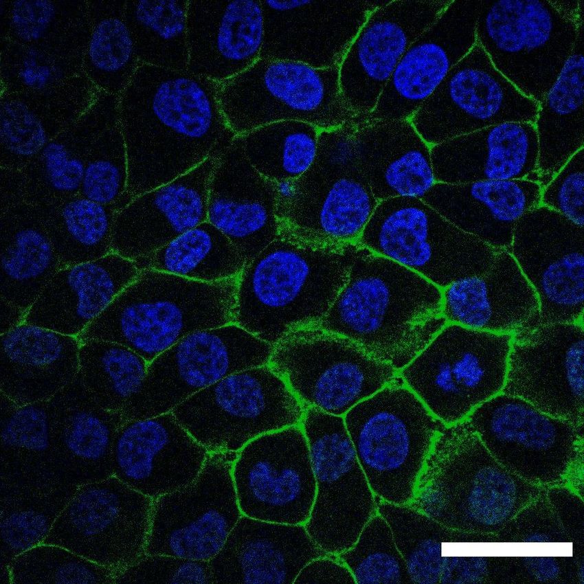
A Serial dilution to 0.5 cell/well
 Lentivirus 384 well plate
 w/ ACE2 (3 weeks incubation)

 Infection

 ACE2

 Duplicate plates
 H1299 Infect w/ spike
 Confirm w/ spike
 cells
 pseudotyped lentivirus X pseudotyped
 mCherry (575/20nm) lentivirus
 Expansion plate Infection plate

 Expansion

 YFP (530/30nm)
 B H1299 H1299-ACE2
 Uninfected Pseudovirus infection Pseudovirus infection
 0.006% 0.005% 30.8%
 mCherry (616nm)

 YFP (530nm)

 C ACE2-
 Phase AlexFluor555 DAPI Merged

Figure S1: Generation of H1299-ACE2 clonal cell line. (A) The H1299 uman non-small cell lung carci-
noma cell line with YFP labelled histone H2AZ was spinfected with the pHAGE2-EF1a-Int-ACE2 lentivector.
Cells were single cell cloned by limiting dilution in a 384-well plate. Clones were expanded into duplicate 96-
well plates, where one plate was used to select infectable clones based on mCherry signal from infection with
SARS-CoV-2 mCherry expressing spike pseudotyped lentivirus. Clones were chosen based on infectability and
expanded from the non-infected replicate 96-well plate. (B) Flow cytometry of SARS-CoV-2 mCherry express-
ing spike pseudotyped lentivirus infection in H1299-ACE2 cells. (C) Images of H1299-ACE2 cells stained with
anti-ACE2 antibody and DAPI. Note membrane localization of ACE2. Scale bar is 50µm.

 2
A Ancestral virus B Beta virus
 1:5 1:50 1:500 1:5000 1:50000 1:5 1:50 1:500 1:5000 1:50000

 H1299-ACE2
H1299-ACE2
Vero E6

 Vero E6
C Omicron virus D Omicron virus (1:25)
 1:25 1:50 1:100 1:200 1:400 P2 stock P3 stock
H1299-ACE2

 H1299-ACE2
Vero E6

 Vero E6

Figure S2: Comparison of SARS-CoV-2 infection in H1299-ACE2 and Vero E6 cells. Both H1299-
ACE2 and Vero E6 cells were infected with the same viral stock in the same experiment with D614G virus (A)
or Beta virus (B) and a focus forming assay was performed. (C) Focus forming assay with stock of Omicron
virus isolate on H1299-ACE2 and Vero E6 cells. (D) Comparison of passage 2 (P2) and passage 3 (P3) stock,
where P3 stock was generated by infection of 1 mL of cell-free P2 stock in 20 mL of Vero E6 cells seeded at 2 ×
105 cells per mL and incubated over 4 days. Numbers above well images denote viral stock dilution. Scale bar
is 2mm.

 3
A B

 H1299-ACE2 Vero E6
 FRNT50

 FRNT50

 D614G Beta D614G Beta

Figure S3: Neutralization of the Beta variant by Pfizer BNT162b2 elicited immunity. Neutralization
of the Beta variant virus compared to D614G ancestral virus in H1299-ACE2 (A) or Vero E6 cells (B) in
participants vaccinated with BNT162b2 and infected by SARS-CoV-2 (green) or vaccinated only (orange).
Numbers in black above each virus strain are geometric mean titers (GMT) of the reciprocal plasma dilution
(FRNT50) resulting in 50% reduction in the number of infection foci. Numbers in red denote fold-change in
GMT between virus strain on the left and the virus strain on the right of each panel. Red horizontal line denotes
most concentrated plasma used. Samples were tested from n=19 participants, where n=6 were vaccinated only
and n=13 were vaccinated and previously infected. p=0.006 for both (A) and (B) as determined by the Wilcoxon
rank sum test.

 4
Table S1: Codon frequency table
This table shows the amino acid change, the nucleotide position of the genome, codon change and the frequency of the
 codon on the assembled genome.
Amino Acid Nucleotide Change Codon(s) Change K032623_N67
Change
A67V 21762C>T 21761 GCT - 0
 GCT>GTT GTT - 133
*H69_V70del 21766_21771delACATGT 21766_21771ACATGT ACATGT - 0
 >del del - 123
T95I 21846C>T 21845 ACT - 0
 ACT>ATT ATT - 164
*G142D 21987_21989delGTG 21987_21989GTG GTG - 0
 >del del - 432
*V143_Y145del 21990_21995delTTTATT 21990_21995TTTATT TTTATT - 0
 >del del - 432
*L212I 22194_22196delATT 22194_22196ATT ATT - 0
 >del del - 146
*R214_D215 22204_22205insGAGCCAGAA 22204_22205GAGCCAGAA WT - 37
 >ins insGAGCCAGAA - 74
G339D 22578G>A 22577 GGT - 0
 GGT>GAT GAT - 255
R346K 22599G>A 22598 AGA - 1
 AGA>AAA AAA - 250
S371L 22674C>T 22674 TCC - 0
 TCC>CTC CTC - 152
S373P 22679T>C 22679 TCA - 3
 TCA>CCA CCA - 166
S375F 22686C>T 22685 TCC - 0
 TCC>TTC TTC - 160
S477N 22992G>A 22991 AGC - 0
 AGC>AAC AAC - 54
T478K 22995C>A 22994 ACA - 0
 ACA>AAA AAA - 59
E484A 23013A>C 23012 GAA - 0
 GAA>GCA GCA - 110
Q493R 23040A>G 23039 CAA - 0
 CAA>CGA CGA - 128
G496S 23048G>A 23048 GGT - 0
 GGT>AGT AGT - 150
Q498R 23055A>G 23054 CAA - 1
 CAA>CGA CGA - 144
N501Y 23063A>T 23063 AAT - 0
 AAT>TAT TAT - 209
Y505H 23075T>C 23075 TAC - 1
 TAC>CAC CAC - 261
T547K 23202C>A 23201 ACA - 0
 ACA>AAA AAA - 777
D614G 23403A>G 23402 GAT - 1
 GAT>GGT GGT - 1803
H655Y 23525C>T 23525 CAT - 3
 CAT>TAT TAT - 1639
N679K 23599T>G 23597 AAT - 1
 AAT>AAG AAG - 682
P681H 23604C>A 23603 CCT - 0
 CCT>CAT CAT - 535
Q954H 24424A>T 24422 CAA - 1
 CAA>CAT CAT - 753
N969K 24469T>A 24467 AAT - 0
 AAT>AAA AAA - 1692
L981F 24503C>T 24503 CTT - 0
 CTT>TTT TTT - 1797
*Only deletions or insertion where the adjacent codon was preserved where counted; WT - Wild Type i.e reads without
the insertion.

 5
Table S2: Summary Table of Participants
 All Vaccinated only Infected and vaccinated
 Number of Participants 19 6 13
 Age (years) 52 (39-67) 54 (36-71) 51 (45-63)
 Days post-vaccination 26 (14-33) 14.5 (8.5-37.5) 28 (18-32)
 Days post-infection 379 (127-468)
 Days post-infection to vaccination 353 (114-444)
 Date range of symptom onset Jun 2020 – Jul 2021
 Male sex 7 2 5
All values are median (IQR) and inclusive of all samples used (early and late timepoints for 2 participants).

 6
Table S3: Participant information per sample
 Date symptom
 Days post Days between diagnostic onset or Infecting
 Sample Participant Age Sex vaccination swab and sample diagnostic test virus*
 1 2029 69 F 10 - - -
 2 2030 71 M 10 - - -
 3 2030 71 M 45 - - -
 4 2032 35 M 14 - - -
 5 2033 74 F 10 - - -
 6 2033 74 F 48 - - -
 7 2035 39 F 10 - - -
 8 2003 36 F 33 - - -
 9 0011 45 F 14 458 Jul-2020 Ancestral
 10 0014 66 F 63 468 Jul-2020 Ancestral
 11 0043 26 F 31 487 Aug-2020 Ancestral
 12 0053 22 M 37 493 Jul-2020 Ancestral
 13 0057 67 F 28 378 Jul-2020 Ancestral
 14 0067 63 M 26 379 Jul-2020 Ancestral
 15 0069 48 F 32 479 Aug-2020 Ancestral
 16 0106 51 M 30 370 Sep-2020 Ancestral
 17 0001 43 F 22 456** Jun-2020** Ancestral/Delta
 18 0109 47 M 18 83 Jul-2021*** Delta
 19 0110 75 M 37 8 Jul-2021 Delta
 20 0149 59 F 13 127 Jul-2021*** Delta
 21 0171 60 F 14 103 Jul-2021 Delta
*Determined by infection wave in South Africa. First infection wave (April-October 2020) consisted of ancestral strains with the D614G
mutation. Third infection wave (April-October 2021) was dominated by the Delta variant. **Participant reinfected during Delta infection
wave, sample is taken 3 months post-recovery of Delta infection. Asymptomatic during reinfection. ***Asymptomatic.

 7
You can also read