Specialized structures on the border between rhizocephalan parasites and their host's nervous system reveal potential sites for host-parasite ...
←
→
Page content transcription
If your browser does not render page correctly, please read the page content below
www.nature.com/scientificreports

            OPEN           Specialized structures on the
                           border between rhizocephalan
                           parasites and their host’s nervous
                           system reveal potential sites for
                           host-parasite interactions
                           A. Miroliubov1*, I. Borisenko         2
                                                                     , M. Nesterenko3, A. Lianguzova3, S. Ilyutkin3, N. Lapshin3 &
                           A. Dobrovolskij3
                           Rhizocephalan barnacles are a unique group of endoparasitic crustaceans. In their extreme adaptation
                           to endoparasitism, rhizocephalan adults have lost almost all features of their free-living relatives but
                           acquired an outstanding degree of control over the body of their hosts (mostly decapods). The subtle
                           influence exercised by rhizocephalans on the physiology, morphology and behaviour of their hosts is
                           a vivid example of the most intimate host-parasite interactions but their mechanisms are very poorly
                           known. In this study we examined the morphology and the adaptive ultrastructure of the organs
                           invading the nervous system of the host in two rhizocephalan species from the families Peltogastridae,
                           (Peltogaster paguri) and Peltogasterellidae (Peltogasterella gracilis). We found two essentially different
                           types of structures involved in interactions of these two rhizocephalans with the nervous system of their
                           hosts: modified rhizocephalan rootlets lying inside the ganglia and the neural fibres of the host enlacing
                           the trophic rootlets of the parasites. We suggest that both these structures may be highly specialized
                           tools allowing the parasite to interact with the host on the humoral level via neuromediators,
                           hormones, attractants and trophic factors.

                           Rhizocephala, or rhizocephalan barnacles, are a group of parasitic crustaceans that have lost almost all morpho-
                           logical characteristics of their free-living relatives due to adaptation to an endoparasitic lifestyle. These animals
                           have a modified life cycle1–4 and a unique ontogeny resulting in a heavily transformed adult living as an endopar-
                           asite within other crustaceans, mostly decapods. In the life cycle of rhizocephalans, the adult organism originates
                           from a group of poorly differentiated cells (“vermigon”) injected into the host by the larvae. Thus, the adult body
                           of a rhizocephalan is newly formed and does not inherit any larval organs. It consists of two functional parts: an
                           interna, which is a system of ramifying feeding rootlets spanning the body of the host, and an externa, which is a
                           structure containing the reproductive system5–9.
                               All parasites influence their hosts at different levels (community, population, physiology of individual host)
                           but the degree of influence varies. Here we will focus on the effect that a peculiar type of parasite (rhizocephalan
                           barnacle) has on its host. Most parasites oppress their hosts by deriving nutrients from them but do not manip-
                           ulate them. At the same time, some highly specialized parasites are known to modify physiology, morphology
                           and behaviour of their hosts. This is characteristic of some trematodes, cestodes, nematodes and acanthoce-
                           phalans10–19. Some metacercaria of trematodes are able to change the behavior of infected fish by modifying the
                           concentration of monoamines in the brain20. Larvae of acanthocephalans induce serotonergic imbalance in a
                           nervous system of their crustacean intermediate hosts21. Nematomorph hairworms alter the normal functions
                           of their host’s (grasshopper) nervous system by the excretion of some special signal molecules22. However, not

                           1
                            Zoological Institute RAS, Laboratory of parasitic worms and protists, Universitetskaya emb., 1, Saint-Petersburg,
                           199034, Russia. 2Saint-Petersburg State University, Department of Embryology, Universitetskaya Emb., 7/9,
                           Saint Petersburg, 199034, Russia. 3Saint-Petersburg State University, Department of Invertebrate Zoology,
                           Universitetskaya Emb., 7/9, Saint Petersburg, 199034, Russia. *email: alexmiroliubov@gmail.com

Scientific Reports |   (2020) 10:1128 | https://doi.org/10.1038/s41598-020-58175-4                                                            1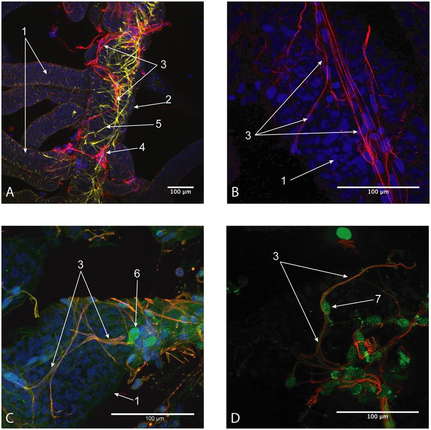
www.nature.com/scientificreports/                                                                www.nature.com/scientificreports

                           Figure 1. (A,C) – Histological sections of goblet-shaped organs (Peltogaster paguri) penetrating neural
                           ganglion of the host (Pagurus pubescens). (В) – Scheme of goblet shaped organ of Peltogaster paguri. (D) –
                           Histological cross section of the common trophic rootlet (Peltogaster paguri) in the hemocoel of the host
                           (Pagurus pubescens). 1-rootlet associated with neural ganglion, 2-central lumen, 3-part of rootlet inside neural
                           tissue, 4-extracellular matrix between layers of cells, 5-outer layer of cells, 6-inner layer of cells, 7-outer surface
                           of goblet shaped organ (green), 8-inner surface of goblet shaped organ (red), 9-common cuticle on the surface
                           of a rootlet, 10-layer of epithelial cells, 11-layer of axial cells, 12-ganglion envelope.

                           all taxa of parasites are well studied in terms of host-parasite manipulation. There are very few data about rhizo-
                           cephalans, which are quite exceptional in their ability to take control over the host’s body.
                                Rhizocephalan barnacles show the highest level of integration with their hosts among metazoan parasites23
                           and the closest host-parasite interactions. They influence the moulting cycle, general physiology, morphology and
                           behaviour of their hosts6,23–30. A vivid example of this influence is the feminization of the infested males6,23,29,31.
                           Rhizocephalans are considered as parasitic castrators suppressing the reproductive system of the host6,32 but
                           in contrast to some other parasitic castrators such as trematodes, which destroy the gonads of the host, rhizo-
                           cephalan barnacles suppress gametogenesis. In addition, some of them take control over the moulting cycle and
                           suppress moulting. This is important for their survival because moulting could result in damage or loss of the
                           rhizocephalan externa, an organ of sexual reproduction positioned outside the host’s body6,23,25–28,33.
                                Infested male crabs demonstrate behaviour typical of females releasing larvae. However, the parasite forces
                           the host to disseminate its own nauplii. The infestation by rhizocephalans reduces the level of aggressiveness and
                           general activity of the infected crabs which might be considered a sort of manipulation also aimed at protecting
                           the externa from damage34–38. At the same time, the voracity of the infected individuals increases39.
                                The mechanisms of control that rhizocephalans exercise over their hosts are still enigmatic. They may be
                           expected to be highly species-specific and diverse. There is evidence that rootlets of the interna penetrate the
                           neural ganglia within the ventral nervous cord of the host40–42. This is consistent with the significant metabolic
                           and behavioural changes that infected crabs start to exhibit involving hormonal balance, respiration rate, hiding
                           behavior, food consumption rate, aggressiveness and many others34–39. One may conjecture that there should be
                           significant differences in histological structure and ultrastructure of the specialized rootlets invading the neural
                           tissue and the trophic rootlets located within the body cavity of the host6,26,27,29,40–42. However, these differences
                           have never been the focus of investigation.
                                In this study, we examined the morphology and adaptive ultrastructure of the organs invading the nerv-
                           ous system of the host in two rhizocephalan species, Peltogaster paguri from the family Peltogastridae and
                           Peltogasterella gracilis from new established family Peltogasterellidae43. The aim of this study was to make a first
                           attempt at revealing the structural mechanisms of interactions between rhizocephalans and their hosts. This study
                           is going to be followed by biochemical and molecular investigations. In a broader context, our study is a first step
                           towards understanding the integrative role of host nervous system for the parasite.

Scientific Reports |   (2020) 10:1128 | https://doi.org/10.1038/s41598-020-58175-4                                                               2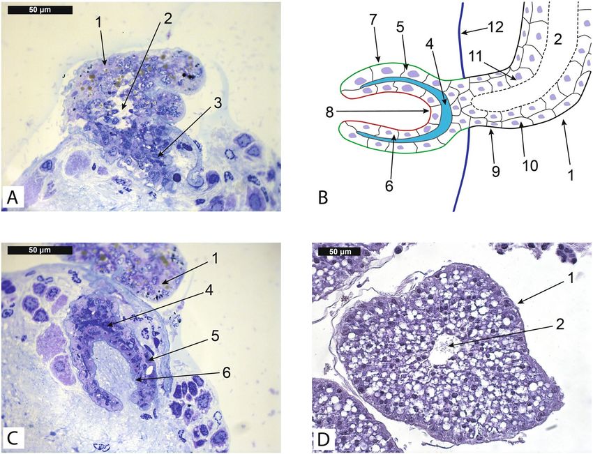
www.nature.com/scientificreports/                                                               www.nature.com/scientificreports

                           Figure 2. (A,B) – Ultrastructure of the cuticle on the internal surface of goblet-shaped organ of Peltogaster
                           paguri, (C) – Ultrastructure of the cuticle of common trophic rootlet of Peltogaster paguri, (D) – Extracellular
                           matrix between two layers of cells in goblet-shaped organ. (TEM) 1-cuticle on the internal surface of goblet-
                           shaped organ, 2-cell of the parasite, 3-neural tissue of the host, 4-microville beneath the inner cuticle of goblet-
                           shaped organ, 5-epithelial cell, 6-homogenous layer of cuticle, 7-microprojections of electron-dense layer of
                           cuticle, 8-microville of epithelial cells, 9- extracellular matrix between two layers of cells in goblet-shaped organ.

                           Results
                           Sections revealed that some roots of the interna of Peltogater paguri (Peltogastridae fam.) were associated with
                           the first and the second abdominal neural ganglia of the host. It could be seen at histological sections that these
                           roots penetrated the ganglion’s envelope (Fig. 1A,B) and that their distal parts were located in the mass of the
                           neural tissue. The distal parts of these roots were modified into goblet-shaped structures with the funnel opening
                           at the tip (Fig. 1C), which we will refer to as goblet-shaped organs. The average size of these organs in longitudinal
                           direction was about 100 ± 10 μm. The tissue organization in goblet-shaped organs differed considerably from
                           that in common trophic roots located in the body cavity of the host (Fig. 1D). The wall of goblet-shaped organs
                           consisted of two layers of cells (an outer and an inner layer) and a layer of dark extracellular matrix containing
                           dark round bodies (Figs. 1C, 2D).
                               The ultrastructure of the cuticle and the underlying cells in goblet-shaped organs also differed from that in
                           trophic roots. The cuticle of a trophic root consisted of a delicate electron-dense outer layer and a thick homo-
                           geneous electron-translucent inner layer. The surface of the cuticle facing the host in most areas bore a zone of
                           microprojections—extensions of the delicate electron-dense layer (Fig. 2C). The cuticle covering goblet-shaped
                           organs differed on the inner and the outer surface of this organ. Inside the funnel (that is, on the inner surface of
                           the goblet-shaped organ) it consisted of one electron dense layer with sparse microprojections (Fig. 2A,B) located
                           between the neural fibres of the host. The apical surface of epithelial cells bore numerous microvilli arranged in a
                           compact manner so the space between them was represented by small channels (Fig. 2B). In contrast, epithelial
                           cells in the trophic roots formed apical microvilli with a wide subcuticular space (Fig. 2C). Whereas, the cuticle

Scientific Reports |   (2020) 10:1128 | https://doi.org/10.1038/s41598-020-58175-4                                                              3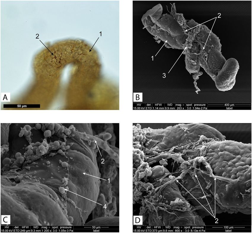
www.nature.com/scientificreports/                                                               www.nature.com/scientificreports

                           Figure 3. (A–D) – Rootlets of Peltogasterella gracilis inside neural ganglia of the host. (A,D – histological
                           sections, B,C – semithin sections) 1-ganglion’s envelope, 2-goblet-shaped organ, 3-rootlets, 4-follicle like
                           structure on the tip of growing rootlet, 5-rootlet penetrating the ganglion’s envelope, 6-neural tissue of the host,
                           7- vesicles with homogenous content inside cells of goblet-shaped organ (boarders of the vesicles pointed by the
                           red line).

                           on the outer surface of the goblet-shaped organs lacked any microprojections, and there were no microvilli on the
                           apical surface of the underlying cells. The cells of goblet-shaped organs contained numerous mitochondria and
                           an abundant endoplasmic reticulum.
                               Goblet-shaped organs of Peltogasterella gracilis (Peltogasterellidae fam.) had a similar gross morphol-
                           ogy as those of Peltogaster paguri but differed somewhat in tissue organization and ultrastructure. In case of
                           Peltogasterella gracilis goblet-shaped organs were much more abundant in the neural ganglia of the host than
                           in case of Peltogaster paguri (Fig. 3A). Goblet-shaped organs of Peltogasterella gracilis were about twice smaller
                           than those of Peltogaster paguri. Their average size in longitudinal direction was about 50 ± 10 μm. The wall of
                           these organs in Peltogasterella gracilis consisted of a single layer of mainly polygonal cells (Fig. 3B,C). The cuticle
                           of goblet-shaped organs in Peltogasterella gracilis consisted of two layers: a thin electron dense outer layer and
                           a homogenous inner layer (Fig. 4A), which was similar to the cuticle structure in trophic roots (Fig. 4B). The
                           surface of the cuticle inside the funnel of the goblet-shaped organs bore numerous projections lying between
                           the elements of the host’s neural tissue but they were much thicker than the microprojections of trophic roots in
                           Peltogaster paguri. Cells underlying the cuticle bore numerous long microvilli (Fig. 5C). On the outer surface of
                           goblet-shaped organs, the cuticle also consisted of two layers and bore small sparse microprojections (Fig. 4C).
                               Large membrane bound vesicles with homogenous content were clearly seen in the cells of goblet-shaped
                           organs of Peltogasterella gracilis (Fig. 3C), while the cells of Peltogaster paguri lacked any such structures. These
                           vesicles were swollen cisterns of endoplasmic reticulum (Fig. 5A). We also observed empty vesicles located close
                           to the endoplasmic reticulum (Fig. 5A), which were probably large vesicles with excreted content.
                               The effect on the host’s neural tissue was quite different in the two studied species. Despite the close contact
                           with the parasite, the neural tissue of the host inside the funnel of goblet-shaped organs in Peltogaster paguri
                           looked unmodified (Fig. 2A,B). Neural cells processes, synaptic structures, mitochondria and transport vesicles
                           could be seen in cells located inside the funnel of the goblet-shaped organs. The fine structure of neural cells
                           inside the funnel of goblet-shaped organs was indistinguishable of that of the cells outside. Inside the funnel
                           of goblet-shaped organs in Peltogasterella gracilis the neural tissue of the host underwent changes resembling
                           degeneration. The whole volume of the funnel of goblet-shaped organ was filled with some electron dense struc-
                           tures and membrane bodies similar to the structures characteristic of lysosomal autophagy, only few remnants of
                           normal neural fibres could be rarely seen (Fig. 5D).
                               Besides goblet-shaped organs, roots with follicle-like structures on the tip were observed inside the host’s neu-
                           ral ganglia (Fig. 3D). These structures looked similar to the follicles on the tips of common trophic roots23. The

Scientific Reports |   (2020) 10:1128 | https://doi.org/10.1038/s41598-020-58175-4                                                              4
www.nature.com/scientificreports/ www.nature.com/scientificreports
Figure 4. (A) – Ultrastructure of the cuticle on the internal surface of goblet-shaped organ of Peltogasterella
gracilis, (B) – Ultrastructure of the cuticle of common trophic rootlet of Peltogasterella gracilis, (C) –
Ultrastructure of the cuticle on the outer surface of goblet-shaped organ of Peltogasterella gracilis, (D) –
longitudinal section through the opening of funnel of goblet-shaped organ of Peltogasterella gracilis. (TEM
1- cuticle, 2-cuticular micro projections, 3-microwilli beneath cuticle, 4 neural tissue of the host, 5- cuticle of
common trophic rootlet, 6-cells of parasite, 7-body cavity of the host, 8- cuticle on the outer surface of goblet-
shaped organ, 9-neural tissue of the host out of the goblet-shaped organ, 10-modified neural tissue inside
goblet-shaped organ.
wall of these structures consisted of a layer of columnar cells with oval nuclei as well as the common follicles on
the tips of trophic roots. These follicles, observed in both species studied, seemed to be growing parts of the roots.
We discovered that the trophic roots of Peltogaster paguri were enlaced by processes of the host cells. These
processes, clearly seen as fibres on histological sections and SEM images (Fig. 6B–D), formed a network on the
surface of trophic roots. The processes were α-tubulin-positive (Fig. 7A–D), while the cell bodies were serotonin-
or FMRF-amid-positive (Fig. 7C,D), indicating the neuronal origin of these cellular structures. Host cells forming
these processes resembled typical peripheral neurons in shape. In addition to antibody stainings and silver stain-
ing of rootlets of Polyascus polygenea revealed nervous network enlancing the rootlet (Fig. 6A).
Discussion
In this study, we described two different types of structures involved in direct contact between rhizocephalans and
the nervous system of their hosts: rhizocephalan rootlets modified into goblet-shaped organs and neural fibres of
the host entwining the trophic internae. The presence of these structures might reflect the structural mechanism
which facilitates interactions between rhizocephalans and their hosts.
In particular, we showed that ganglia-residing goblet-shaped organs in Peltogaster paguri and Peltogasterella
gracilis differed from the common trophic roots in shape, ultrastructure and tissue organization. We hypothesize
that they also have different functions. We did not find any synaptic contacts between the host tissue and the cells
Scientific Reports | (2020) 10:1128 | https://doi.org/10.1038/s41598-020-58175-4 5www.nature.com/scientificreports/ www.nature.com/scientificreports
Figure 5. (A,B) – Bloated cisterns of endoplasmic reticulum in the cells of goblet-shaped organs of
Peltogasterella gracilis, (C) – Ultrastructure of the cuticle on the internal surface of goblet-shaped organ of
Peltogasterella gracilis, (D) – Ultrastructure of the modified neural tissue in the funnel of goblet-shaped organs
of Peltogasterella gracilis. 1-bloated cisterns of endoplasmic reticulum, 2-endoplasmic reticulum, 3-cuticle
on the internal surface of goblet-shaped organ, 4-neural tissue of the host, 5-microwille beneath the cuticle,
6-neural fiber of the host, 7-cells of goblet shaped organ.
of goblet-shaped organs, which agrees with the fact that we never observed the nervous system inside parasitic
interna44. This suggests that the interaction with the host nervous system may be mediated by soluble molecules
produced by non-neural but nevertheless specialized tissue of the parasite. The ultrastructural features of the cells
of goblet-shaped organs such as numerous microvilli beneath the cuticle, numerous mitochondria and abundant
endoplasmic reticulum indicate high levels of local cellular synthesis and transport. We suggest that with the use
of goblet-shaped organs, the parasite might inject specific substances such as neurotransmitters or neurohor-
mones directly into the host’s neurons and could receive some other substances or neural-born signals from the
host. That said, we point out that the permeability of the parasite’s cuticle for different molecules is unknown26.
The rest of the interna is located in the body cavity of the host and surrounded by the hemolymph and is con-
sidered to have a trophic function6,26. At the same time, the specific shape, tissue organisation and ultrastructure
of goblet-shaped organs in comparison to the potentially trophic part of interna might indicate that they have a
different set of functions. We suggest that goblet-shaped organs, which are located inside the neural tissue of the
host and demonstrate an organization specialized for synthesis and transport, might also play a key role in mod-
ifying the physiological status, moulting cycle and behaviour of the host.
We observed specific differences in histology and ultrastructure of goblet-shaped organs in Peltogaster paguri
and similar organs in Peltogasterella gracilis. The influence on the integrity of the host’s neural tissue also dif-
fered between these two species at the morphological level. Peltogasterella gracilis caused significant changes to
the neural tissue of the host inside the funnel of goblet-shaped organs, whereas the neural tissue of the host of
Peltogaster paguri seemed unmodified. The reason of these differences remains to be revealed. However, despite
Scientific Reports | (2020) 10:1128 | https://doi.org/10.1038/s41598-020-58175-4 6www.nature.com/scientificreports/ www.nature.com/scientificreports
Figure 6. (A)- Cells stained with silver nitrate on the surface of the roots of Polyascus polygenea. (whole mount)
(B–D)- host tissue on the surface of the roots of Peltogaster paguri. (SEM) 1-root, 2-fibers of host’s tissue,
3-groups of cells associated with fibers, 4-separate cells on the surface of the root.
the differences in the ultrastructure and tissue organisation of the goblet-shaped organs in the two studied species,
we hypothesize that they are functionally analogous because their gross morphology and localisation are quite
similar. Morphological examination of the goblet-shaped organs of both species described above was repeated
in a number of specimens and all the examined parasites were sexually mature so we can confirm that there were
differences between these two species.
Goblet-shaped organs are located inside of the neural ganglia of the host but it is unknown how they ger-
minate and penetrate the ganglion’s envelope. We speculate that the parasitic follicles observed inside the host’s
ganglia (Fig. 3D) are, in fact, a stage of the developing goblet-shaped organs responsible for penetrating the gan-
glia and their connective tissue sheaths. These follicles might excrete factors diluting the ganglial envelope and
develop into mature goblet-shaped organs after penetration.
Penetration of interna rootlets into the ganglia of ventral nervous cord was shown in several studies. According
to Nielsen (1970)40, some roots of Peltogaster paguri and Peltogasterella sulcata were associated with the posterior
part of thoracic ganglion and the next three abdominal ganglia. The rootlets penetrated the ganglial envelope
and were located in the neural tissue. Structures similar to goblet-shaped organs can be clearly seen in the figures
in that article (Nielsen, 1970, p. 26) but were not described there. Similar structures were also found inside the
ganglia of crabs infested by sacculinid parasites41. Unfortunately, they were described only at the histological
level. However, the fact that representatives of other families have similar organs points to the common trends of
interactions between parasitic barnacles and the nervous system of their hosts.
The phenomenon of nervous fibres enlacing the trophic roots of the interna discovered in our study is intrigu-
ing and suggested many further questions. This is another site of direct contact of the parasite with the nervous
system of the host and its role may be different from that of goblet shaped organ. We suggest that the parasite
might induce the growth of neurons by emitting some analogues of the host’s neuronal attractants, which force
neurons to grow on the surface of the roots and to enlace them. The purpose of this is unclear but, as a further
conjecture, this site might serve for direct host-parasite interactions.
To conclude, rhizocephalan barnacles of these both species (Peltogaster paguri and Peltogasterella gracilis)
have at least two distinct types of direct contact with the neural system of the host: goblet shaped organs and the
Scientific Reports | (2020) 10:1128 | https://doi.org/10.1038/s41598-020-58175-4 7www.nature.com/scientificreports/ www.nature.com/scientificreports
Figure 7. (A,B) – Main trunk and rootlet of Peltogaster paguri with neural fibers of the host (Confocal Z-stack),
(C) – Neural fibers on the surface of rootlet of Polyascus. Polygenea (Sacculinidae fam.) (Confocal Z-stack), (D)
– Neuron of the host on the surface of rootlet (Peltogaster paguri) (Confocal Z-stack). (Red-α-tubulin, Yellow-
phalloidin, Blue-dapi, Green-Serotonin/FMRF-amid). 1-rootlets, 2-main trunk, 3-neural fibers of the host,
4-groups of cell bodies associated with neural fibers, 5-muscles, 6-body of neuron stained with FMRF-amide,
7- body of neuron stained with Serotonin.
neural fibres entwining the rootlets. Both of these structures may be highly specialized tools allowing the parasite
to interact with the host. Molecular mechanisms for these interactions might be a promising direction of future
studies. Interaction with the nervous system of the host is all the more fascinating as rhizocephalan interna has
never been shown to have its own nervous system44,45. At the same time, its muscular system is well-developed45
but it is unclear how it is innervated. While neural tissue is absent in parasite body it is still unknown where mus-
cular contraction originate and how they are transmitted.
Materials and Methods
Hermit crabs Pagurus pubescens (Krøyer, 1838) (17 specimens) infected with the parasite Peltogaster paguri
(Rathke, 1842) were collected at the White Sea (Educational and research station “Belomorskaia” of St Petersburg
State University) (N: 66.308210, E: 33.627816) in the summer of 2016 and 2017. Hermit crabs Pagurus ochoten-
sis (Brandt, 1851) and Pagurus pectinatus (Stimpson 1858) (15 specimens) infected with Peltogasterella gracilis
(Boschma, 1927) and crabs Hemigrapsus sanguineus (De Haan 1835) infected with Polyascus polygenea (Lützen,
J. & T. Takahashi 1997) (10 specimens) were collected at the Sea of Japan (Marine biological station “Vostok” of
Institute of Marine Biology of the Russian Academy of Sciences) (N: 42.893720, E: 132.732755). All collected
parasites were adults with fully developed externas. The infected hosts were dissected and the ventral nerve cords
with the roots of rhizocephalan parasites were removed.
Scientific Reports | (2020) 10:1128 | https://doi.org/10.1038/s41598-020-58175-4 8www.nature.com/scientificreports/ www.nature.com/scientificreports
Immunocytochemistry (ICC). For immunocytochemical visualization the internae of the rhizocephalans
were fixed with 4% paraformaldehyde (PFA; Sigma-Aldrich) in phosphate-buffered saline (PBS; Fluka) at 4 °C
overnight. Before immunocytochemical staining, the fixed material was rinsed with PBT several times in the
course of 24 hours (PBS + 0.1% Triton-X100; Sigma-Aldrich). The specimens were blocked with 1% bovine serum
albumin in PBT for an hour and incubated with primary antibodies diluted in blocking solution overnight at
4 °C. The primary antibodies used were anti-tyrosine α-tubulin (mouse monoclonal, 1:2,000; Sigma Cat #T9028)
and anti-acetylated α-tubulin (mouse monoclonal, 1:2,000; Sigma Cat #T6793). After incubation with primary
antibodies, the samples were washed four times (for 3 hours each time) in PBT, and incubated for 12 hours at
4 °C with a 1:1000 dilution of Donkey Anti-Mouse IgG Antibodies labelled with Alexa Fluor 488 (Molecular
Probes, Cat #A21202). The material was then rinsed three times for 10 min each time in PBS, stained with the
DAPI nuclei stain (1 ug/ml; Sigma) in PBS for an hour, rinsed with PBS and mounted in DABCO-glycerol. The
specimens were examined using the confocal laser scanning microscopes Leica TCS SPE in the Resource Centre
“Microscopy and Microanalysis” of Research park of St State University. The images were processed using ImageJ
software (FiJi).
Histology and light microscopy. The dissected internae were fixed with Bouin solution. Paraffin sections
(5 μm thick) were made using standard histological methods on the Leica RM-2265 microtome and stained with
hematoxylin-eosin. The sections were examined under a Leica DM2500 microscope, photos were taken with a
Nikon DS-Fi1 camera and processed with ImageJ software (FiJi).
Interna of Polyascus polygenea (fam. Polyascidae) isolated from the host was rinsed in fresh water and incu-
bated in silver nitrate solution for 24 hours, after that whole specimen was mount on slides. This method is used
to visualize the nervous system.
Transmission and scanning electron microscopy. For transmission electron microscopy samples
were fixed overnight at 4 °C in 2.5% glutaraldehyde in phosphate buffer with addition of Sucrose (pH 7.4; 750
mOsM), and postfixed in 1% OsO4 in the same buffer (an hour at room temperature). After washing with the
same buffer, the specimens were dehydrated through an ethanol series and acetone and embedded in Epon-812
embedding media (Fluka). Semithin (1 μm thick) and ultra-thin (60–80 nm) sections were cut with a Leica EM
UC7 ultratome. Semi-thin sections were stained with methylene blue and studied under Leica DM2500 micro-
scope. Ultra-thin sections were stained with uranyl acetate followed by Reynolds lead nitrate46 and examined
under transmission electron microscope JEOL JEM 1400 in Resource Centres “Chromas” and “Molecular and
Cell Technologies” of the Research Park of St Petersburg State University. Specimens for SEM were dehydrated in
ethanol series and acetone, critical point-dried in Hitachi critical point dryer HCP- 2, mounted on stubs, coated
with platinum using Giko IB-5 Ion coater, and viewed under FEI Quanta 250 scanning electron microscope in
“Taxon” Research Resource Center (http://www.ckp-rf.ru/ckp/3038/) of the Zoological Institute of the Russian
Academy of Sciences.
Received: 13 May 2019; Accepted: 13 January 2020;
Published: xx xx xxxx
References
1. Glenner, H. Cypris Metamorphosis, Injection and Earliest Internal Development of the Rhizocephalan Loxothylacus panopaei
(Gissler). Crustacea: Cirripedia: Rhizocephala: Sacculinidae. J. Morphol. 249, 43–75 (2001).
2. Pérez-Losada, M., Høeg, J. T., Kolbasov, G. A. & Crandall, K. A. Reanalysis of the relationships among the Cirripedia and the
Ascothoracida and the phylogenetic position of Facetotecta (Maxilopoda: Thecostraca) using 18S rDNA Sequences. J. Crust. Biol.
22(3), 661–669 (2002).
3. Høeg, J. T. & Rybakov, A. V. Cypris larvae in Polysaccus mediterraneus and Mycetomorpha vancouverensis: their importance in
analyzing the phylogeny and sexual evolution of parasitic barnacles (Crustacea: Cirripedia: Rhizocephala). Isr. J. Ecol. Evol. 53, 9–31
(2007).
4. Hoeg, J. T., Maruzzo, D., Okano, K., Glenner, H. & Chan, B. K. K. Metamorphosis in Balanomorphan, Pedunculated, and Parasitic
Barnacles: A Video-Based Analysis. Integr. Comp. Biol. 52(3), 337–347 (2012).
5. Glenner, H. & Hoeg, J. T. A new motile, multicellular stage involved in host invasion by parasitic barnacles (Rhizocephala). Nature.
377(14), 147–149 (1995).
6. Hoeg, J. T. The biology and life cycle of Rhizocephala (Cirripedia). J. mar. biol. Ass. U.K. 75, 517–550 (1995).
7. Glenner, H., Hoeg, G. T., O’Brien, J. J. & Sherman, T. D. Invasive vermigon stage in the parasitic barnacles Loxothylacus texanus and
L. panopaei (Sacculinidae): closing of the rhizocephalan life-cycle. Mar. Biol. 136, 249–257 (2000).
8. Korn, O. M., Rybakov, A. V. & Kashenko, S. D. Larval Development of the Rhizocephalan Sacculina polygenea (Crustacea:
Cirripedia). Russ. J. Mar Biol. 26(5), 373–377 (2000).
9. Korn, O. M. & Rybakov, A. V. Larval Development in the Rhizocephalan Barnacle Sacculina pilosella. Russ. J. Mar Biol. 27(3),
177–179 (2001).
10. Seppälä, O., Karvonen, A. & Valtonen, E. T. Parasite-induced change in host behaviour and susceptibility to predation in an eye
fluke–fish interaction. Animal Behav. 68(2), 257–263 (2004).
11. Helluy, S. & Holmes, J. C. Parasitic manipulation: further considerations. Behav. Process. 68(3), 205–210 (2005).
12. Dezfuli, B. S., Capuano, S., Simoni, E., Giari, L. & Shinn, A. P. Histopathological and ultrastructural observations of metacercarial
infections of Diplostomum phoxini (Digenea) in the brain of minnows Phoxinus phoxinus. Dis. Aquat. Organ. 75(1), 51–59 (2007).
13. Shaw, J. C. et al. Parasite manipulation of brain monoamines in California killifish (Fundulus parvipinnis) by the trematode
Euhaplorchis californiensis. Proc. R. Soc. Lond. [Biol.]. 276(1659), 1137–1146 (2008).
14. Coats, J., Poulin, R. & Nakagawa, S. The consequences of parasitic infections for host behavioural correlations and repeatability.
Behaviour. 147(3), 367–382 (2010).
15. Poulin, R. Parasite manipulation of host behaviour: an update and frequently asked questions. Adv. Study Behav. - Academic Press.
41, 151–186 (2010).
16. Goodman, B. A. & Johnson, P. T. Disease and the extended phenotype: parasites control host performance and survival through
induced changes in body plan. PLoS One. 6(5), e20193 (2011).
Scientific Reports | (2020) 10:1128 | https://doi.org/10.1038/s41598-020-58175-4 9www.nature.com/scientificreports/ www.nature.com/scientificreports
17. Hammond-Tooke, C. A., Nakagawa, S. & Poulin, R. Parasitism and behavioural syndromes in the fish Gobiomorphus cotidianus.
Behaviour. 149(6), 601–622 (2012).
18. Lafferty, K. D. & Shaw, J. C. Comparing mechanisms of host manipulation across host and parasite taxa. J. Exp. Biol. 216(1), 56–66
(2013).
19. Adamo, S. A. Parasites: evolution’s neurobiologists. J. Exp. Biol. 216(1), 3–10 (2013).
20. Shaw, J. C. et al. Parasite manipulation of brain monoamines in California killifish (Fundulus parvipinnis) by the trematode
Euhaplorchis californiensis. Proceedings of the Royal Society B: Biological Sciences. 276(1659), 1137–1146 (2008).
21. Thomas, F., Adamo, S. & Moore, J. Parasitic manipulation: where are we and where should we go? Behavioural processes. 68(3),
185–199 (2005).
22. Biron, D. G. et al. Behavioural manipulation in a grasshopper harbouring hairworm: a proteomics approach. Proceedings of the Royal
Society B: Biological Sciences. 272(1577), 2117–2126 (2005).
23. Walker, G. Introduction to the Rhizocephala (Crustacea: Cirripedia). J. of Morphol. 249, 1–8 (2001).
24. Shirley, S. M., Shirley, T. C. & Meyers, T. R. Hemolymph responses of Alaskan king crabs to rhizocephalan parasitism. Can. J. Zool.
64(8), 1774–1781 (1986).
25. Alvarez, F., Hinesb, A. H. & Reaka-Kudla, M. L. The effects of parasitism by the barnacle Loxothylacus panopaei (Gissler) (Cirripedia:
Rhizocephala) on growth and survival of the host crab Rhithropanopeus harrisii (Gould) (Brachyura: Xanthidae). J. Exp. Mar. Biol.
Ecol. 192, 221–232 (1995).
26. Bresciani, J. & Hoeg, J. T. Comparative Ultrastructure of the Root System in Rhizocephalan Barnacles (Crustacea: Cirripedia:
Rhizocephala). J. of Morfol. 249, 9–42 (2001).
27. Alvarez, F., Alcaraz, G. & Robles, R. Osmoregulatory disturbances induced by the parasitic barnacle Loxothylacus texanus
(Rhizocephala) in the crab Callinectes rathbunae (Portunidae). J. Exp. Mar. Biol. Ecol. 278, 135–140 (2002).
28. Bortolini, J. L. & Alvarez, F. Hepatopancreas alteration of the blue crab Callinectes sapidus by the rhizocephalan barnacle
Loxothylacus texanus. J. Invertebr. Pathol. 99, 354–356 (2008).
29. Kristensen, Т. et al. The selective advantage of host feminization: a case study of the green crab Carcinus maenas and the parasitic
barnacle Sacculina carcini. Mar. Biol. 159, 2015–2023 (2012).
30. Zacher, L. S., Horstmann, L. & Hardy, S. M. A field-based study of metabolites in sacculinized king crabs Paralithodes camtschaticus
(Tilesius, 1815) and Lithodes aequispinus Benedict, 1895 (Decapoda: Anomura: Lithodidae). Journal of Crustacean Biology. 38(6),
794–803. (2018).
31. Nagler, C. et al. The bigger, the better? Volume measurements of parasites and hosts: Parasitic barnacles (Cirripedia, Rhizocephala)
and their decapod hosts. PLoS One. 12(7), e0179958 (2017).
32. Lafferty, K. D. & Kuris, A. M. Parasitic castration: the evolution and ecology of body snatchers. Trends Parasitol. 25(12), 564–572
(2009).
33. Lützen, J., Jensen, K. H. & Glenner, H. Life history of Sacculina carcini Thompson, 1836 (Cirripedia: Rhizocephala: Sacculinidae)
and the intermoult cycle of its host, the shore crab Carcinus maenas (Linnaeus, 1758) (Decapoda: Brachyura: Carcinidae). J. Crustac.
Biol. 38(4), 413–419 (2018).
34. Bishop, R. K. & Cannon, L. R. G. Morbid behaviour of the commercial sand crab, Portunus pelagicus (L.), parasitized by Sacculina
granifera Boschma, 1973 (Cirripedia: Rhizocephala). J. Fish Dis. 2(2), 131–144 (1979).
35. Takahashi, T., Iwashige, A. & Matsuura, S. Behavioural manipulation of the shore crab, Hemigrapsus sanguineus by the rhizocephalan
barnacle, Sacculina polygenea. Crustacean Research. 26, 153–161 (1997).
36. Innocenti, G., Pinter, N. & Galil, B. S. Observations on the agonistic behaviour of the swimming crab Charybdis longicollis Leene
infected by the rhizocephalan barnacle Heterosaccus dollfusi Boschma. Can. J. Zool. 81(1), 173–176 (2003).
37. Vazquez-Lopez, H., Alvarez, F., Franco, J., Moran, A. & Chazaro, S. Observations on the behaviour of the dark crab Callinectes
rathbunae Contreras parasitized with the rhizocephalan Loxothylacus texanus Boschma. Int. J. Zool. Res. 2(4), 344–353 (2006).
38. Belgrad, B. A. & Griffen, B. D. Rhizocephalan infection modifies host food consumption by reducing host activity levels. J. Exp. Mar.
Biol. Ecol. 466, 70–75 (2015).
39. Pérez-Miguel, M., Drake, P. & Cuesta, J. A. Experimental predatory behaviour of the stone crab Eriphia verrucosa (Forskål, 1775)
(Decapoda, Brachyura, Eriphiidae). Nauplius. 25 (2017).
40. Nielsen, S. O. The effects of the rhizocephalan parasites Peltogaster paguri Rathke and Gemmosaccus sulcatus (Lilljeborg) on five
species of paguridan hosts (Crustacea Decapoda). Sarsia 42(1), 17–32 (1970).
41. Rubiliani, C. & Payen, G. G. Modalités de la destruction des régions neurosécrétrices des crabes Carcinus maenas (L.) et C.
mediterraneus Czerniavsky infestés par la sacculine. Gen. Comp. Endocrinol. 38(2), 215–228 (1979).
42. Payen, G. G., Hubert, M., Turquier, Y., Rubiliani, C. & Chassard-Bouchaud, C. Infestations expérimentales de crabes juvéniles par la
sacculine. Ultrastructure des racines parasitaires en croissance et relations avec la niasse ganglionnaire ventrale de l’hôte. Can. J. Zool
59(9), 1818–1826 (1981).
43. Høeg, J. T., Noever, C., Rees, D. A., Crandall, K. A. & Glenner. H. A new molecular phylogeny based taxonomy of the parasitic
barnacles (Crustacea Cirripedia Rhizocephala). Zool. J. Linn. Soc. In Press (2019).
44. Miroliubov, A. A. et al. Muscular system in the interna of Polyascus polygenea and Sacculina pilosella (Cirripedia: Rhizocephala:
Sacculinidae). Invert. Zool. 16(1), 48–56 (2019).
45. Miroliubov, A. A. Muscular system in interna of Peltogaster paguri (Rhizocephala: Peltogastridae). Arthropod Struct. Dev. 46(2),
230–235 (2017).
46. Reynolds, E. S. The use of lead citrate at high pH as an electron-opaque stain in electron microscopy. The Journal of cell biology.
17(1), 208–212 (1963).
Acknowledgements
We thank the staff of the Resource Centre “Microscopy and Microanalysis”, “Chromas” and “Molecular and Cell
Technologies” of the Research Park of St Petersburg State University for technical assistance and the diving team
of the Marine Biological Station “Vostok” of National Scientific Center of Marine Biology, Far East Branch of
Russian Academy of Science, for the help with the collection of specimens. We are grateful to Dr. Igor Adameyko
and Natalia Lentsman for help with the manuscript preparation. This study was supported by the Grant No. 18-
34-00727\18 of the Russian Foundation for Basic Research and state laboratory theme of Zoological institute
AAAA-A19-119020690109-2 (Biodiversity of parasites, life cycles, biology and evolution). The study was partly
supported by the Saint-Petersburg State University project COLLAB 2018 (I.B.).
Author contributions
A.M. and I.B. wrote the main manuscript text, M.N., A.L., S.I. and N.L.—histological methods, A.D.—
consultation.
Scientific Reports | (2020) 10:1128 | https://doi.org/10.1038/s41598-020-58175-4 10www.nature.com/scientificreports/ www.nature.com/scientificreports
Competing interests
The authors declare no competing interests.
Additional information
Correspondence and requests for materials should be addressed to A.M.
Reprints and permissions information is available at www.nature.com/reprints.
Publisher’s note Springer Nature remains neutral with regard to jurisdictional claims in published maps and
institutional affiliations.
Open Access This article is licensed under a Creative Commons Attribution 4.0 International
License, which permits use, sharing, adaptation, distribution and reproduction in any medium or
format, as long as you give appropriate credit to the original author(s) and the source, provide a link to the Cre-
ative Commons license, and indicate if changes were made. The images or other third party material in this
article are included in the article’s Creative Commons license, unless indicated otherwise in a credit line to the
material. If material is not included in the article’s Creative Commons license and your intended use is not per-
mitted by statutory regulation or exceeds the permitted use, you will need to obtain permission directly from the
copyright holder. To view a copy of this license, visit http://creativecommons.org/licenses/by/4.0/.
© The Author(s) 2020
Scientific Reports | (2020) 10:1128 | https://doi.org/10.1038/s41598-020-58175-4 11You can also read