STANDARDIZATION AND VALIDATION OF AN IN HOUSE RT-LAMP MOLECULAR TEST FOR THE DIAGNOSIS OF SARS-COV-2
←
→
Page content transcription
If your browser does not render page correctly, please read the page content below
Rev Peru Med Exp Salud Publica. 2021;38(1):7-16.

                                         ORIGINAL ARTICLE

                                         STANDARDIZATION AND VALIDATION OF AN
                                         IN HOUSE RT-LAMP MOLECULAR TEST FOR THE
                                         DIAGNOSIS OF SARS-CoV-2
                                         Oscar Escalante-Maldonado 1,a, Margot Vidal-Anzardo 1,b, Fernando Donaires 1,c,
                                         Gilmer Solis-Sanchez 2,d, Italo Gallesi 1,e, Luis Pampa-Espinoza 1,c,
                                         Maribel Huaringa 1,e, Nancy Rojas-Serrano 1,e, Coralith García 3,c,
                                         Eddie Angles-Yanqui 4,5,c, Ronnie Gustavo Gavilán 1,f, Ricardo Durães-Carvalho 6,g,
                                         Jairo Mendez-Rico 7,h, César Cabezas 1,c, Paulo Vitor Marques-Simas 6,8,i
                                         1
                                             Centro Nacional de Salud Pública, Instituto Nacional de Salud, Lima, Perú.
                                         2
                                             Oficina General de Investigación y Transferencia Tecnológica, Instituto Nacional de Salud, Lima, Perú.
                                         3
                                             Hospital Nacional Cayetano Heredia, Lima, Perú.
                                         4
                                             Hospital Nacional Arzobispo Loayza, Lima, Perú.
                                         5
                                             Universidad Peruana Cayetano Heredia, Lima, Perú.
                                         6
                                             Universidad de Campinas, Sao Paulo, Brasil.
                                         7
                                             Pan American Health Organization, World Health Organization., Washington DC, United States of America.
                                         8
                                             Universidad Nacional Mayor de San Marcos, Lima, Perú.
                                         a
                                             Biologist, Doctor of Medical Sciences; b physician; c physician, specialist in Infectious Diseases; d dental surgeon; e Biologist;
                                         f
                                             Biologist, Doctor in Biochemistry and Molecular Biology; g Pharmacist, Doctor in Genetics and Molecular Biology; h
                                             Doctor in Biological Sciences; i Biologist, Doctor in Genetics and Molecular Biology.

                                         ABSTRACT

                                         Objectives: To standardize an in-house RT-LAMP test for the detection of SARS-CoV-2 and to validate it with labo-
                                         ratory and field samples in patients with clinical suspicion of COVID-19. Materials and methods: An in-house RT-
                                         LAMP molecular test was standardized for the detection of SARS-CoV-2, establishing the detection limit with Vero cells
                                         of isolated Peruvian strains of SARS-CoV-2, the robustness to different concentrations of primers, and in silico presence
                                         of cross-reactions. The laboratory test was validated with 384 nasal and pharyngeal swab samples (UFH) obtained be-
                                         tween March and July 2020. For field validation, UFH samples were obtained from 383 suspected symptomatic cases of
                                         COVID-19 consecutively enrolled during activities For discard, all samples were evaluated by RT-LAMP and RT-qPCR.
                                         For laboratory and field validation, the RT-qPCR was considered as the reference standard, concordance measures and
                                         diagnostic performance were calculated. Results: The detection limit was consistent in cases with Ct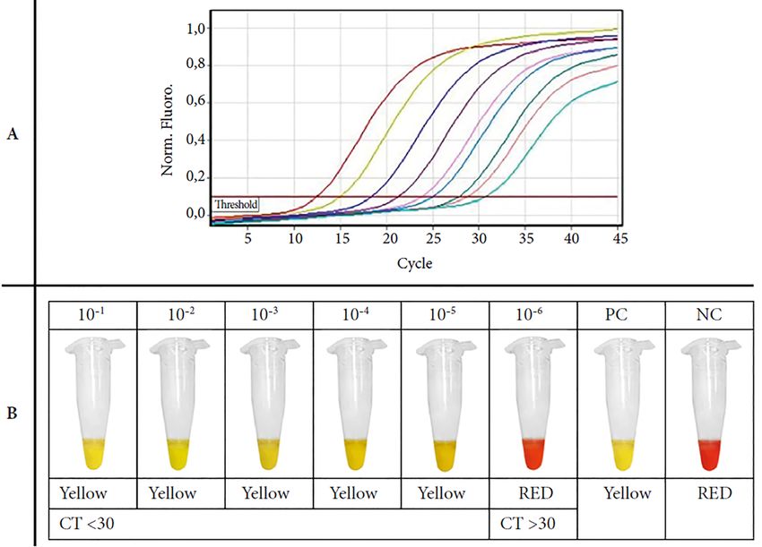
Rev Peru Med Exp Salud Publica. 2021;38(1):17-23.                                              RT-LAMP test: standardization and validation

    Molecular testing requires considerable financial and lo-
gistical investment compared to other diagnostic tools. The                 KEY MESSAGES
standard test suggested by the World Health Organization
(WHO) for the detection of SARS-CoV-2 is the real-time                      Motivation for the study: COVID-19 is a serious public health
                                                                            problem in Peru; it is important to develop new diagnostic
reverse-transcription polymerase chain reaction (RT-qPCR)                   methodologies for SARS-CoV-2.
test, which requires a molecular laboratory with expensive
                                                                            Main findings: The RT-LAMP test developed in-house
infrastructure, equipment and reagents, as well as specia-                  demonstrated adequate diagnostic performance and
lized personnel; resources that are not always available in                 concordance when compared to the RT-qPCR test, both
countries such as Peru (3,4).                                               in the laboratory and in the field. No cross-reactivity with
                                                                            other coronaviruses was identified in silico. In the field,
    In Peru, at the beginning of the pandemic, the RT-qP-
                                                                            a 5.5% reduction in sensitivity, and a 0.4% increase in
CR test could only be performed in a standardized manner                    specificity, was found in subjects who were between the
in Lima at the National Referral Laboratory for Respiratory                 first and second week of illness.
Viruses of the Instituto Nacional de Salud (INS). Progressi-                Implications: The in-house RT-LAMP test evaluated is an
vely, its processing was extended to regional laboratories in               effective alternative for molecular detection of SARS-CoV-2.
a decentralized manner. Currently, there are more than 50
laboratories nationwide, but the demand for these tests has
not been fully covered (5).
    The pressing need for other acute phase diagnostic alter-
                                                                         a) standardization, b) laboratory validation, and c) field va-
natives for SARS-CoV-2 infection has been evidenced by the
                                                                         lidation. The reference standard considered in the study was
development of tests based on the CRISPR/Cas system                  ,
                                                                   (6)
                                                                         RT-qPCR. The design of the standardization was descriptive
or the reverse transcription loop-mediated isothermal am-
                                                                         cross-sectional, and the design of the laboratory and field
plification method (RT-LAMP) (7). Other tests based on the
                                                                         validation was analytical cross-sectional.
loop-mediated isothermal amplification method (LAMP)
have been applied in Peru for diseases such as zika            , tu-
                                                             (8)
                                                                         Standardization
berculosis (9), malaria (10), and dengue (11); showing adequate
                                                                         This stage was carried out at the INS National Referral Labo-
performance. Its implementation is more feasible and viable
                                                                         ratory for Respiratory Viruses between June and July 2020.
since more affordable equipment is used, employing four
                                                                         To evaluate the performance of the RT-LAMP test accor-
to six primers, two/three forward and two/three reverse to
                                                                         ding to WHO standards, the SARS-CoV-2 strain isolated in
identify DNA targets for the amplification (12).
                                                                         VERO81 cells from a nasopharyngeal swab sample (NPS)
    The RT-LAMP test is presented as a fast and efficient al-
                                                                         with positive molecular diagnosis by RT-qPCR and NPS
ternative for the identification of suspicious cases, since the
                                                                         samples that were previously obtained during routine epi-
sample processing time in the laboratory is approximately
                                                                         demiological surveillance of COVID-19 discard were used.
50 minutes, compared to the four to eight hours required for
                                                                         Using the strain, we evaluated: a) detection limit (under se-
the RT-qPCR test (7).
                                                                         rial dilution in base 10), b) robustness to changes in primer
    This study aims to standardize, in the laboratory, an RT-
                                                                         concentration (original and half - 0.5P) and percentage re-
LAMP test developed in-house for the detection of SARS-
                                                                         duction of the final reaction volume (20% - 0.8V; 40% - 0.6V;
CoV-2; as well as to validate it in the field in patients suspicious
                                                                         50% - 0.5V and 60% - 0.4V), and c) reproducibility.
for COVID-19, obtaining diagnostic performance measures,
and using the results of the RT-qPCR test as reference.
                                                                         Laboratory validation
                                                                         Cross-reaction analysis was performed in silico by aligning
MATERIALS AND METHODS                                                    the RT-LAMP external primer sequences with known human
                                                                         coronavirus (HCoV) reference sequences (NC_005831.2,
Type of study                                                            HCoV-NL63; NC_002645.1, HCoV- 229E; NC_006213. 1,
A cross-sectional study was carried out to evaluate an RT-               HCoV-OC43 ATCC strain VR-759; NC_006577.2, HCoV-
LAMP test for the detection of SARS-CoV-2 in three stages:               HKU1; NC_004718.3, SARS-CoV-1; NC_019843.3, MERS-

8                                                                                                https://doi.org/10.17843/rpmesp.2021.381.7154
Rev Peru Med Exp Salud Publica. 2021;38(1):17-23. Escalante-Maldonado O et al.
CoV Middle East respiratory syndrome-related coronavirus; RT-qPCR reaction
FJ415324.1, HECoV 4408; NC_045512.2, Chinese SARS- Reactions were standardized in Rotor-Gene Multiplex RT-
CoV-2) (13). In addition, external primers were aligned with PCR Kit (Qiagen, Germany) using primers and probes for
194 Peruvian strains available from GISAID (https://www. SARS-CoV-2 detection (RdRP) and an internal control (hu-
gisaid.org/). All in vitro experiments were performed in tri- man GAPDH) (green channel, FAM: 470-510nm and oran-
plicate by the same operator under the same environmental ge, ROX: 585-610nm, respectively). Reactions were conside-
and equipment conditions .
(13)
red positive when cycle threshold (Ct) values < 37 (FAM)
To establish the diagnostic validity of the test in the la- and Ct < 40 (ROX) were obtained concomitantly. Primer se-
boratory, a sample size was calculated using the formula quences, probes and reaction conditions are available in the
for estimating the diagnostic performance by means of the supplementary material. More information on the in-house
Epidat program version 4.2, considering a sensitivity value RT-qPCR assay is available at: http://dx.doi.org/10.17504/
of 91.489% and a specificity of 99.531%, figures calculated protocols.io.bsm2nc8e.
according to that reported by Jiang et al, in their total sam-
ple (14). A significance level of 95%, absolute error of 5% and
RT-LAMP Reaction
a positivity probability of 39.5% were considered (based on
RT-LAMP reactions were performed according to Lamb et
the proportion of positive results usually obtained in diag-
al. , using WarmStart®️ Colorimetric LAMP 2X Master
(15)
nostic activities of the rapid field response teams by the INS
Mix DNA and RNA (Eiken Chemical Co., Ltd., Tokyo, Ja-
between July 6 and 8, 2020 in the jurisdiction of the Directo-
rate of Integrated Health Networks (DIRIS) Center). pan), which contains a pH indicator for colorimetric visu-
A loss rate of 20% was assumed in order to anticipate logis- alization. Robustness was tested from the standard primer
tical problems that could arise during sample handling and/or concentration and the final reaction volume. Forty-four µM
analysis. With these parameters, we established the need for of the FIP primers (16 µM), BIP (16 µM), F3 (2 µM), B3 (2
at least 379 samples. For this stage, samples of NPS were used, µM), LOOP F (4 µM), BUCLE B (4 µM), and 56 µM of water
which were previously obtained during routine epidemiologi- were used; in addition, 20 µL of the reagents MIX-LAMP
cal COVID-19 evaluations between the beginning of the pan- (12.5 µL), MIX-Primers (2.5 µL), RNA (5 µL), and 5 µL of
demic and July 2020; the vials were stored in INS laboratories. water were used. Reactions were carried out at 65 °C for 45
All samples evaluated were anonymized, and corresponded
min, and at 80 °C for 5 min.
to positive and negative subjects identified by RT-qPCR for
whom no additional information was available.
Field validation
The samples were selected in a non-probabilistic way by
After laboratory validation, the field evaluation was carried
convenience, the evaluation by RT-LAMP was blinded (the
out by selecting persons suspected of COVID-19 infection
evaluators were unaware of the previous result by RT-qP-
with up to 15 days of symptoms, who attended hospitals in
CR). The use of the samples was authorized by head reso-
Lima (Hospital Cayetano Heredia, Hospital Hipólito Una-
lution No. 00006918, health emergency decree No. 0064-
nue and Hospital Arzobispo Loayza), and persons who
2020-OGA/INS (April 7, 2020) and informative note No.
0055-2020 “Plan de Acción del Instituto Nacional de Salud were evaluated by home care teams (rapid response teams)
para Prevención, Diagnóstico y Control de COVID-19, en el between August and September 2020. People over 18 years
marco del Decreto SupremoNo. 008- 2020-SA”. of age, with mild symptoms, without previous diagnosis of
COVID-19 by molecular testing were included; pregnant
RNA extraction women, and serious or critical patients were excluded. The
RNA extraction from all samples was performed using the Ge- sample size for this stage was based on the same calculation
nElute™ total RNA purification kit (Sigma-Aldrich - Merck), made for the laboratory validation because they shared the
according to the manufacturer’s instructions (https://www. same objective (to identify diagnostic performance measu-
sigmaaldrich.com/technical-documents/protocols/biology/ res) although samples had different sources (stored and di-
viral-rna-purification.html), then frozen at -80 °C until fur- rectly obtained); for the selection of subjects in the field a
ther processing. non-probabilistic consecutive sampling was followed.
https://doi.org/10.17843/rpmesp.2021.381.7154 9Rev Peru Med Exp Salud Publica. 2021;38(1):17-23. RT-LAMP test: standardization and validation
The sex, age and clinical picture of each patient, and the samples because information on time of illness was not avai-
time of illness in days, from symptom onset were registered. lable; subjects who did not have information regarding time
Each participant underwent NPS, using the Yocon Biology of illness were excluded from the stratified evaluation. The
Technology Company sampling kit, which includes viral measures were calculated by means of point estimators and
transport media and flocked dacron swabs. Samples were 95% confidence intervals (95% CI); inferences were made
transported the same day to the INS using triple containers considering a significance level of 0.05.
with cold accumulators, at temperatures between 2 °C and 8
°C. All samples were analyzed following the procedure pre- Ethical considerations
viously described. The RT-qPCR and RT-LAMP results were Standardization and validation in the laboratory did not re-
obtained simultaneously from two different laboratories; the quire evaluation by the Institutional Research Ethics Commi-
evaluators of each laboratory were unaware of the results of ttee (CIEI), since the samples used were obtained in routine
the opposite test they were analyzing. activities established within the INS action plan, highlighting
that they were anonymized. Field validation was carried out
Statistical analysis using a research protocol approved by the INS CIEI, as shown
Data analysis was performed using the Stata statistical pac- in RD No. 283-2020-OGITT-INS. All study subjects included
kage version 16.1 (Stata Corporation, College Station, Texas, in this phase provided informed consent to participate and
USA). Summary measures of frequency and percentage their results were reported in less than 72 hours.
were used for the clinical and epidemiological characteris-
tics of the sample of subjects evaluated in the field validation. RESULTS
For both laboratory and field validation, the degree of con-
cordance between RT-qPCR and RT-LAMP test results was Performance evaluation of RT-LAMP compared
determined using Cohen’s Kappa index. Sensitivity, specifi- to RT-qPCR
city, positive and negative predictive value, accuracy value The detection limit of SARS-CoV-2 by RT-LAMP was 1000
and area under the curve for RT-LAMP were also calculated. copies/µL; which indicated that all cases with Ct values < 30
Stratified analysis of field results was performed according to were concordant between RT-qPCR and RT-LAMP (Figure
week of illness ; this was not feasible for the laboratory
(16,17)
1, Table 1). Regarding robustness evaluations, high-throu-
Figure 1. Standard curve of RT-qPCR reactions (panel A) and limit of detection by RT-LAMP (panel B) for
the detection of SARS-CoV-2.
10 https://doi.org/10.17843/rpmesp.2021.381.7154Rev Peru Med Exp Salud Publica. 2021;38(1):17-23. Escalante-Maldonado O et al.
Table 1. Comparison of the detection limit between RT-qPCR and RT-LAMP reactions for detecting the presence of SARS-CoV-2.
Concentration Ct Value Change of color
Serial Dilution
(Number of copies/µL) (RT-qPCR) (RT-LAMP)
10-1 107 13.6 Yes
10-2 106 16.7 Yes
10-3 105 20.4 Yes
10-4 104 25.0 Yes
10-5 103 29.2 Yes
10-6 102 35.1 No
10-7 101 - No
Ct: cycle threshold, RT-qPCR: Real-Time Reverse-Transcription Polymerase Chain Reaction, RT-LAMP: Reverse Transcription Loop-Mediated Isothermal Amplification.
ghput reactions were obtained with half the primer con- group was young adults (n=236, 61.6%). The most frequent
centrations (0.5P) and with 20 µL final volume (0.8V of the symptoms found were cough (n=268, 70.0%) and pharyn-
final standard reaction volume). geal pain (n=262, 68.4%); the mean time of illness was 7.1
(SD: 3.3) days; 56.1% were in the first week of illness and
Laboratory validation 43.6% in the second week, one subject was identified (0.3%)
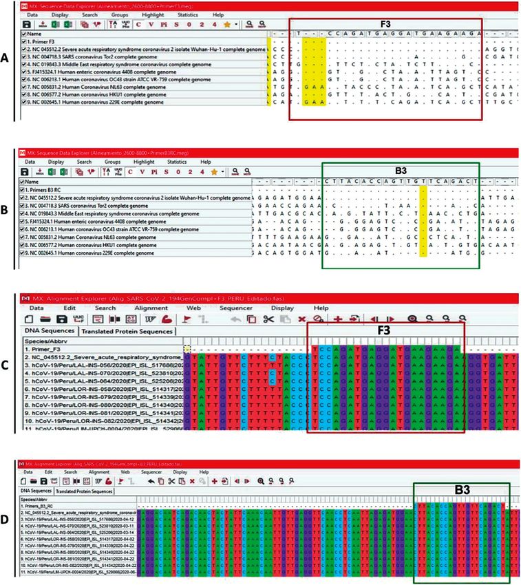
Cross-reaction analysis performed in silico, did not iden- who did not remember the date of onset of the symptoms,
tify consistent similarity with reference sequences of other and was excluded in the stratified evaluation by time of ill-
human coronaviruses (NL-63, HKU1, OC43, 229E, SARS- ness (Table 4).
CoV-1, MERS and HECoV, Figure 2). Furthermore, when Of the subjects evaluated, 37.3% (n=143) were positi-
these same primers were aligned with 194 Peruvian strains ve by RT-qPCR, while positivity by RT-LAMP was 33.7%
available in the GISAID initiative there was no exclusion of (n=129). The concordance of the results obtained between
conserved regions, which exhibit high similarity and specifi- both tests had a Kappa of 0.88 (95% CI: 0.84-0.94); when
city, this can be designated as absence of concomitant detec- stratified by week of symptoms it was found that during the
tion of other human coronaviruses other than SARS-CoV-2. first week this value was 0.92 (95% CI: 0.86-0.97), while for
For the evaluation of laboratory diagnostic performan- the second week there was a Kappa of 0.85 (95% CI: 0.76-
ce, 384 samples were included, of which 37.2% (n=143) had 0.93), in all scenarios the concordance was statistically sig-
previously positive results by RT-qPCR; when the results nificant (p < 0.001). In the overall sample, 20 (5.2%) cases
were obtained by RT-LAMP, a statistically significant con- were found with discordant results between both tests, three
cordance (p < 0.001) of 0.88 (95% CI: 0.83-0.93) was iden- FP with a FPR of 1.3%, and 17 FN with a FNR of 11.9%;
tified by Kappa test. Three false positives (FP) were found The FPR was 1.4% for the first week and 1.0% for the second
with a false positive rate (FPR) of 1.2%; in addition, there week, while the FNR for the first week of symptoms was
were 18 false negatives (FN) with a false negative rate (FNR) 7.9% and for the second week 16.4% (Table 2).
of 12.6% (Table 2). We found that the sensitivity of the overall sample was
The sensitivity obtained from the evaluated samples was 88.1% (95% CI: 81.6-92.9). For the first week of symptoms
87.4% (95% CI: 80.8-92.4), the specificity was 98.8% (95% CI: group sensitivity was 92.1% (95% CI: 83.6-97.0) and for the
96.4-99.7), the positive predictive value was 97.7% (95% CI: second week group it was 86.6% (95% CI: 72.5-91.5). As for
93.3-99.5), and negative 93.0% (95% CI: 89.1-95.8); the accura- specificity, the value for the overall sample was 98.8% (95%
cy value of the test was 94.5% (95% CI: 91.8-96.6), and the area CI: 96.4-99.7), for the first week 98.6 (95% CI: 94.9-99.8) and
under the ROC curve was 93.1% (95% CI: 90.3-95.9) (Table 3). for the second week 99.0% (95% CI: 94.6-100). The positive
predictive value (PPV) and negative predictive value (NPV)
Field validation for the overall sample were 97.7 (95% CI: 93.4- 99.5) and
For this stage of the study, 383 subjects were included ac- 93.3 (95% CI: 89.5-96.1), respectively; in the evaluation by
cording to the aspects foreseen in the research protocol, of week of symptoms the PPV in the first week was 97.2% (95%
which 51.7% (n=198) were women; the most frequent age CI: 90.3 - 99.7) and 98.2% (95% CI: 90.6 -100) for the second
https://doi.org/10.17843/rpmesp.2021.381.7154 11Rev Peru Med Exp Salud Publica. 2021;38(1):17-23. RT-LAMP test: standardization and validation Figure 2: Multiple sequence alignment between the F3 and B3 primers for RT-LAMP and the reference sequences of all known human coronaviruses, as well as with all available Peruvian SARS-CoV-2 strains in the GISAID initiative. Alignment was performed in ClustalW using MEGA. The sequence pri- mers (panel A - F3, panel B - B3) were aligned with all known human coronavirus reference sequences (NC_005831.2, HCoV-NL63; NC_002645.1, HCoV- 229E; NC_006213.1, HCoV-OC43 ATCC strain VR-759; NC_006577. 2, HCoV-HKU1; NC_004718.3, SARS-CoV-1; NC_019843.3, MERS; FJ415324.1, HE- CoV-4408 and NC_045512.2, SARS-CoV-2 Wuhan-Hu-1 isolate) and all 194 Peruvian SARS-CoV-2 strains (panel C - F3, panel D - B3). The yellow column in panels A and B, and the asterisks in panels C and D, represent conservative regions in the nsp3 gene fragment among all known human coronaviruses and all genome-wide Peruvian SARS-CoV-2 strains, respectively. week, the NPV was 95.8% (95% CI: 91.0-98.4) for the first the overall sample, while for those in the first week of symp- week and 90.0 (95% CI: 82.8-94.9) for the second week. The toms it was 95.3% (95% CI: 92.1-98.5), and 91.3% (95% CI: area under the ROC curve was 93.4% (95% CI: 90.7-96.2) in 86.7-95.9) for those in the second week. 12 https://doi.org/10.17843/rpmesp.2021.381.7154
Rev Peru Med Exp Salud Publica. 2021;38(1):17-23.                                                                                                     Escalante-Maldonado O et al.

Table 2. Concordance analysis between the results of laboratory and field evaluations obtained by RT-qPCR and RT-LAMP tests.

                                         RT-q CR                                                                                                  Results
                                                                                     Kappa
 RT-LAMP                                                         Total                                   p Value        Correct           Incorrect           FP            FN
                                Positive           Negative                         (95% CI)
                                                                                                                       (TP+TN)            (FP+FN)           (FPR)         (FNR)
 Laboratory evaluation
     Positive                      125                3           128          0.880 (0.831 – 0.930)
Rev Peru Med Exp Salud Publica. 2021;38(1):17-23. RT-LAMP test: standardization and validation
Table 3. Diagnostic performance measures of RT-LAMP in laboratory and field validation, considering RT-qPCR results as reference standard.
Laboratory validation (n=384)
Laboratory validation
(n=384) First week of symptoms Second week of symptoms
RT-LAMP General (n=383)
(n=215) (n=167)
% 95% CI % 95% CI % 95% CI % 95% CI
Sensitivity 87.4 80.8 – 92.4 88.1 81.6 – 92.9 92.1 83.6 – 97.0 86.6 72.5 – 91.5
Specificity 98.8 96.4 – 99.7 98.8 96.4 – 99.7 98.6 94.9 – 99.8 99.0 94.6 - 100
Positive predictive
977 93.3 – 99.5 97.7 93.4 – 99.5 97.2 90.3 – 99.7 98.2 90.6 - 100
value
Negative predictive
93.0 89.1 – 95.8 93.3 89.5 – 96.1 95.8 91.0 – 98.4 90.0 82.8 – 94.9
value
Accuracy 94.5 91.8 – 96.6 94.8 92.1 – 96.8 96.3 92.8 – 98.4 92.8 87.8 – 96.2
Area under the curve 93.1 90.3 – 95.9 93.4 90.7 – 96.2 95.3 92.1 – 98.5 91.3 86.7 – 95.9
95% CI: 95% confidence interval, RT-qPCR: Real-Time Reverse-Transcription Polymerase Chain Reaction, RT-LAMP: Reverse-Transcription Loop-Mediated Isothermal
Amplification.
during replication compared to the amount of RNA for the volume, technical errors can be made during small volume
RdRp gene, which would justify the lower sensitivity of the pipetting without compromising the results.
RT-LAMP assay. To overcome these difficulties, we designed RT-LAMP showed high sensitivity and specificity both
a new set of primers for other regions of the genome, espe- in the laboratory and in the field, obtaining results similar to
cially for RdRp. to properly compare the diagnostic perfor- those reported by Hu et al. (88.57% and 98.98%, respectively)
mance considering the same genomic region and the assays (21)
, and lower than those described by Jiang et al. (91.4% and
are in final validation phase according to the parameters pre- 99.5%, respectively) (14), as well as by Kitagawa et al. (100% and
sented in this work. 97.6%, respectively) (22). These differences could be associated
It was also demonstrated by in silico analysis that the set with the Ct values used to establish positivity by RT-qPCR;
of primers used for RT-LAMP was indeed specific for detec- moreover, only the positive samples that presented Ct values
ting the Peruvian SARS-CoV-2 strains and did not cross-react > 30 differed with those obtained by RT-LAMP in this study.
with other human coronaviruses in molecular testing. This is The concordance values obtained between both tests
considered as a limitation for this study because the analysis indicated the applicability of the RT-LAMP protocol as an
should be performed in vitro using clinical samples, which alternative to RT-qPCR. RT-LAMP could be implemented
was not possible because the INS does not have clinical sam- at the first level of healthcare, becoming useful to identify
ples positive for other human coronaviruses. Due to the need infected patients in the active transmission phase.
to quickly evaluate the performance of this diagnostic method We found that the RT-LAMP test had a PPV of 97.7%,
and eventually begin to transfer this technology to the point of similar to that reported by Jiang et al. (14), and much higher
care, the alternative of verifying the occurrence of cross-reac- than that mentioned by Hu et al. (PPV: 91.18%) (21); we must
tivity measured by in silico analysis was the most appropriate point out that the latter study evaluated 329 samples of
and scientifically feasible at the time. asymptomatic cases, unlike our study in which samples were
Perfect identity in the alignment region of primers F3 taken from symptomatic cases. Similarly, the RT-LAMP test
and B3 with all available Peruvian SARS-CoV-2 strains also had a NPV of 93.3%, which is lower than that reported by
indicated specific detection and possibly no false negative Jiang et al. (14), who found a NPV of 98.1%.
results due to the specificity of the primers. The degree of concordance in the identification of SARS-
The evaluation of the robustness of this protocol inclu- CoV-2 between RT-qPCR and RT-LAMP in the clinical as-
ded variables such as primer concentration and final reac- sessment was 94.8%, a result similar to that described in other
tion volume. This strategy considered the possibility that the studies such as that of Lu et al. (23) and Kitagawa et al. (22), where
reactions were performed by individuals who have no routi- it was over 90%. In our study we found 20 discordant results
ne contact with molecular biology techniques. Since the per- between RT-LAMP and RT-qPCR in the clinical assessment,
formance of the reactions was not compromised when using 17 FN and three FP; Jiang et al. , found five discordant
(14)
half the primer concentrations and 80% of the final reaction results, four FN and one FP. Kitagawa et al. (22) reported only
14 https://doi.org/10.17843/rpmesp.2021.381.7154Rev Peru Med Exp Salud Publica. 2021;38(1):17-23. Escalante-Maldonado O et al.
Table 4. Clinical and epidemiological characteristics of the subjects a higher performance in the first week of symptoms; these
evaluated in the field.
findings could be verified by the area under the curve, which
decreased from 95.3% in the first week to 91.3% by the se-
Characteristics n %
cond week of symptom onset.
Sex
It is worth mentioning that the analysis of quantitative
Male 185 48.3
data from RT-qPCR reactions of lower respiratory tract sam-
Female 198 51.7
ples is very reliable, especially in cases with small amounts of
Age group (years)
the virus (24); however, this analysis could not be performed
Young (≤ 29) 62 16.2
in our study; in which we only established a relation between
Young adult (30-59) 236 61.6
Ct values and the time of onset of the disease, showing that
Older adult (≥ 60) 85 22.2
samples collected from people in the first week of symptoms
Signs and Symptoms
presented lower Ct values; this could indirectly cause the vi-
Ageusia 19 5.0
ral load to be higher in these people.
Anosmia 37 9.7 The laboratory had the limitation of not having the cli-
Headache 214 55.9 nical and epidemiological data of the subjects from whom
Nasal congestion 127 33.2 the samples were taken, for this reason it was not possible
Diarrhea 80 20.9 to carry out a stratified analysis by time of illness. In the
Respiratory distress 90 23.5 field evaluation, the memory bias of the subjects included
Joint pain 27 7.0 in the study was recognized, due to the fact that the data
Sore throat 262 68.4 on the time of illness was obtained by self-reporting; only
Muscle Pain 113 29.5 one subject stated that he could not remember the date of
Chest Pain 67 17.5 onset of his symptoms, and was therefore excluded from the
Fever/chills 179 46.7 stratified analysis. Our results showed robust confidence in-
Irritability/confusion 2 0.5 tervals with amplitudes in accordance with the estimators
General malaise 232 60.6 used to determine the sample size, as well as the discordant
Nausea/vomiting 46 12.0 values obtained (FN and FP); studies in which sample sizes
Cough 268 70.0 are planned with lower absolute error could help to increase
Time of illness precision and thus reduce confidence intervals.
Not specified a 1 0.3 Finally, our data allows us to conclude that the RT-LAMP
First week 215 56.1 test developed in-house has been validated as a convenient
Second week 167 43.6 and acceptable alternative for the detection of SARS-CoV-2
RT-PCR result in symptomatic patients, these results being limited to clini-
Negative 240 62.7
cal pictures within the first two weeks of illness. This test is
proposed as an additional alternative to existing tests, which
Positive 143 37.3
helps to meet the diagnostic demand during the COVID-19
RT-LAMP result
pandemic. The rapid detection of cases would allow the es-
Negative 254 66.3
tablishment of effective control measures that would result
Positive 129 33.7
in the interruption of the chain of infection and a desirable
a Patient does not remember the onset of symptoms.
RT-qPCR: Reverse-Transcription Real-Time Polymerase Chain Reaction RT-LAMP: Reverse- reduction in incidence.
Transcription Loop-Mediated Isothermal Amplification
Acknowledgments: We thank the Pan American Health
two discordant cases, which were FP. Hu et al. (21) also iden- Organization (PAHO) for providing the reagents and establishing
tified four discordant samples (theoretically FP); however, the collaboration to perform the validations of the experiment. We
would like to acknowledge all the workers of the INS Microbiology
these were confirmed as positive for SARS-CoV-2 by a ge-
and Biomedicine Laboratory and all the people from other
netic sequencing test. institutions involved in the collection, handling and processing
When evaluating the performance of RT-LAMP by time of the samples, especially Jairo Méndez (PAHO), Rapid Response
of symptom onset, we found that sensitivity and NPV were Team (CDC/INS), Lely Solari, Faviola Valdivia, Helen Horna,
Gabriel de Lucio, Gabriel de Lucio, Lely Solari and Faviola Valdivia,
higher in the first week, and although PPV and specificity
as well as all the people from other institutions involved in the
showed an increase towards the second week, this increa- collection, handling and processing of the samples, especially Jairo
se was not significant. Furthermore, RT-qPCR has shown Méndez (PAHO), Helen Horna, Gabriel de Lucio, Yanina Zarate,
https://doi.org/10.17843/rpmesp.2021.381.7154 15Rev Peru Med Exp Salud Publica. 2021;38(1):17-23. RT-LAMP test: standardization and validation
Iris Pompa, Isidro Antipupa, Jhon Mayo, Carina Mantari, Kathia final version. All authors are responsible for the content of the article.
Tarqui, Romeo Pomari, Eduardo Juscamayta, Paquita García, Conflicts of interest: All authors except CG, EA, and JM are INS
Miryam Palomino, Pamela Rios, Priscila Lope, Johana Balbuena, staff. All authors declare that they have no conflicts of interest
Víctor Jiménez, Yolanda Angulo, Yuli Barrios, Paul Pachas, Noemi in relation to this publication. Cesar Cabezas is a member of the
Flores and Ana Zeppilli. editorial board of RPMESP. RDC is supported by the São Paulo
Research Foundation (FAPESP), grant no. 2019/01255-9, Brazil.
Authors’ contribution: All authors participated in the conception
and design of the study. LP, CG, and EA participated in data Funding: Fully funded by INS. The study was conducted within the
collection. GS participated in the statistical analysis of the data. All framework of regular INS activities.
authors participated in the interpretation of the data, drafting of the Supplementary material: Available in the electronic version of
manuscript, critical revision of the manuscript, and approval of the RPMESP.
REFERENCES
1. World Health Organization. Weekly epidemiological update - 5 January 13. GISAID - Initiative [Internet]. Múnich: GISAID; 2021 [cited on January
2021 [Internet]. Ginebra: WHO; 2021 [cited on January 25, 2021]. 6, 2021]. Available at: https://www.gisaid.org/.
Available at: https://www.who.int/publications/m/item/weekly-epide- 14. Jiang M, Pan W, Arasthfer A, Fang W, Ling L, Fang H, et al. Development
miological-update 5-january-2021. and Validation of a Rapid, Single-Step Reverse Transcriptase Loop-Media-
2. Del Brutto OH, Costa AF, Mera RM, Recalde BY, Bustos JA, García HH. ted Isothermal Amplification (RT-LAMP) System Potentially to Be Used
SARS-CoV-2-related mortality in a rural Latin American population. Int for Reliable and High-Throughput Screening of COVID-19. Front Cell
J Infect Dis. 2020;99:226–8. doi: 10.1016/j.ijid.2020.08.003. Infect Microbiol. 2020;10:331. doi: 10.3389/fcimb.2020.00331.
3. Wölfel R, Corman VM, Guggemos W, Seilmaier M, Zange S, Müller MA, 15. Lamb LE, Bartolone SN, Ward E, Chancellor MB. Rapid detection of novel
et al. Virological assessment of hospitalized patients with COVID-2019. coronavirus/Severe Acute Respiratory Syndrome Coronavirus 2 (SARS-
Nature. 2020;581(7809):465–9. doi: 10.1038/s41586-020-2196-x. CoV-2) by reverse transcription-loop-mediated isothermal amplification.
4. Organización Mundial de la Salud. Pruebas diagnósticas para el SARS- PloS One. 2020;15(6):e0234682. doi: 10.1371/journal.pone.0234682.
CoV-2: orientaciones provisionales, 11 de septiembre de 2020 [Internet]. 16. Carpenter CR, Mudd PA, West CP, Wilber E, Wilber ST. Diagnosing
Ginebra: OMS; 2021 [cited on January 25, 2021]. Available at: https://apps. COVID-19 in the Emergency Department: A Scoping Review of Clinical
who.int/iris/handle/10665/335830. Examinations, Laboratory Tests, Imaging Accuracy, and Biases. Acad
5. Instituto Nacional de Salud. Pruebas moleculares realizadas para el diag- Emerg Med Off J Soc Acad Emerg Med. 2020;27(8):653–70. doi: 10.1111/
nóstico de COVID-19 [Internet]. Lima: INS; 2021 [cited on January 25, acem.14048.
2021]. Available at: http://web.ins.gob.pe/es/indicador/pruebas-molecu- 17. Vidal-Anzardo M, Solis G, Solari L, Minaya G, Ayala-Quintanilla B, As-
lares-realizadas-para-el-diagnostico-de-covid-19.
tete-Cornejo J, et al. Evaluación en condiciones de campo de una prueba
6. Broughton JP, Deng X, Yu G, Fasching CL, Singh J, Streithorst J, et al. Rapid
serológica rápida para detección de anticuerpos IgM e IgG contra SARS-
Detection of 2019 Novel Coronavirus SARS-CoV-2 Using a CRISPR-based
CoV-2. Rev Peru Med Exp Salud Pública. 2020;37(2):203–9. doi: 10.17843/
DETECTR Lateral Flow Assay. MedRxiv Prepr Serv Health Sci. 2020. doi:
rpmesp.2020.372.5534.
10.1101/2020.03.06.20032334.
18. Kosack CS, Page A-L, Klatser PR. A guide to aid the selection of diag-
7. Huang WE, Lim B, Hsu C, Xiong D, Wu W, Yu Y, et al. RT‐LAMP
nostic tests. Bull World Health Organ. 2017;95(9):639–45. doi:10.2471/
for rapid diagnosis of coronavirus SARS‐CoV‐2. Microb Biotechnol.
BLT.16.187468
2020;13(4):950–61. doi: 10.1111/1751-7915.13586.
19. Corman VM, Landt O, Kaiser M, Molenkamp R, Meijer A, Chu DK,
8. Escalante-Maldonado O, Gavilán RG, García MP, Marcelo A, Pacheco
et al. Detection of 2019 novel coronavirus (2019-nCoV) by real-time
E, Cabezas C, et al. Desarrollo y validación del método de amplificación
RT-PCR. Eurosurveillance. 2020;25(3). doi: 10.2807/1560-7917.
isotérmica mediada en lazo para la detección del virus Zika. Rev Peru Med
ES.2020.25.3.2000045.
Exp Salud Publica. 2019;36(3):442–7. doi: 10.17843/rpmesp.2019.363.3941.
9. Gray CM, Katamba A, Narang P, Giraldo J, Zamudio C, Joloba M, et al. 20. Case JB, Bailey AL, Kim AS, Chen RE, Diamond MS. Growth, detection,
Feasibility and Operational Performance of Tuberculosis Detection by quantification, and inactivation of SARS-CoV-2. Virology. 2020;548:39–48.
Loop-Mediated Isothermal Amplification Platform in Decentralized Set- doi: 10.1016/j.virol.2020.05.015.
tings: Results from a Multicenter Study. J Clin Microbiol. 2016;54(8):1984– 21. Hu X, Deng Q, Li J, Chen J, Wang Z, Zhang X, et al. Development and
91. doi:10.1128/JCM.03036-15. Clinical Application of a Rapid and Sensitive Loop-Mediated Isothermal
10. Serra-Casas E, Manrique P, Ding XC, Carrasco-Escobar G, Alava F, Gave Amplification Test for SARS-CoV-2 Infection. mSphere. 2020;5(4):e00808-
A, et al. Loop-mediated isothermal DNA amplification for asympto- 20. doi: 10.1128/mSphere.00808-20.
matic malaria detection in challenging field settings: Technical perfor- 22. Kitagawa Y, Orihara Y, Kawamura R, Imai K, Sakai J, Tarumoto N, et al.
mance and pilot implementation in the Peruvian Amazon. PloS One. Evaluation of rapid diagnosis of novel coronavirus disease (COVID-19) us-
2017;12(10):e0185742. doi: 10.1371/journal.pone.0185742. ing loop-mediated isothermal amplification. J Clin Virol. 2020;129:104446.
11. Dauner AL, Mitra I, Gilliland T, Seales S, Pal S, Yang S-C, et al. Develop- doi: 10.1016/j.jcv.2020.104446.
ment of a pan-serotype reverse transcription loop-mediated isothermal 23. Lu R, Wu X, Wan Z, Li Y, Jin X, Zhang C. A Novel Reverse Transcription
amplification assay for the detection of dengue virus. Diagn Microbiol Loop-Mediated Isothermal Amplification Method for Rapid Detection of
Infect Dis. 2015;83(1):30–6. doi: 10.1016/j.diagmicrobio.2015.05.004. SARS-CoV-2. Int J Mol Sci. 2020;21(8):2826. doi: 10.3390/ ijms21082826.
12. Notomi T, Okayama H, Masubuchi H, Yonekawa T, Watanabe K, Amino 24. Yu F, Yan L, Wang N, Yang S, Wang L, Tang Y, et al. Quantitative Detection
N, et al. Loop-mediated isothermal amplification of DNA. Nucleic Acids and Viral Load Analysis of SARS-CoV-2 in Infected Patients. Clin Infect
Res. 2000;28(12):E63. doi: 10.1093/nar/28.12.e63. Dis Off Publ Infect Dis Soc Am. 2020; doi: 10.1093/cid/ciaa345.
16 https://doi.org/10.17843/rpmesp.2021.381.7154You can also read