Psoriasis Is Associated With Elevated Gut IL-1α and Intestinal Microbiome Alterations - Frontiers
←
→
Page content transcription
If your browser does not render page correctly, please read the page content below
ORIGINAL RESEARCH
                                                                                                                                       published: 01 October 2020
                                                                                                                                 doi: 10.3389/fimmu.2020.571319

                                           Psoriasis Is Associated With
                                           Elevated Gut IL-1α and Intestinal
                                           Microbiome Alterations
                                           Sergey Yegorov 1,2* † , Dmitriy Babenko 3,4† , Samat Kozhakhmetov 5 ,
                                           Lyudmila Akhmaltdinova 3 , Irina Kadyrova 3 , Ayaulym Nurgozhina 5 , Madiyar Nurgaziyev 5 ,
                                           Sara V. Good 6 , Gonzalo H. Hortelano 1 , Bakytgul Yermekbayeva 7 and
                                           Almagul Kushugulova 5
                                           1
                                            School of Sciences and Humanities, Nazarbayev University, Nur-Sultan, Kazakhstan, 2 Faculty of Education
                                           and Humanities, Suleyman Demirel University, Almaty, Kazakhstan, 3 Karaganda Medical University Research Centre,
                         Edited by:
                                           Karaganda, Kazakhstan, 4 Art Science LLP Innovative Center, Nur-Sultan, Kazakhstan, 5 Laboratory of Human Microbiome
              Juarez Antonio Simões
                                           and Longevity, National Laboratory Astana, Nazarbayev University, Nur-Sultan, Kazakhstan, 6 Department of Biology,
                         Quaresma,
                                           The University of Winnipeg, Winnipeg, MB, Canada, 7 “University Medical Center” Corporate Fund, Nur-Sultan, Kazakhstan
      Evandro Chagas Institute, Brazil
                      Reviewed by:
                             Yan He,       Background: Psoriasis is a chronic inflammatory condition that predominantly affects
   Southern Medical University, China
                                           the skin and is associated with extracutaneous disorders, such as inflammatory bowel
                         Qixiao Zhai,
           Jiangnan University, China      disease and arthritis. Changes in gut immunology and microbiota are important drivers
               Paulo Ricardo Criado,       of proinflammatory disorders and could play a role in the pathogenesis of psoriasis.
     Faculdade de Medicina do ABC,
                                Brazil
                                           Therefore, we explored whether psoriasis in a Central Asian cohort is associated with
                 *Correspondence:
                                           alterations in select immunological markers and/or microbiota of the gut.
                     Sergey Yegorov
          sergey.yegorov@nu.edu.kz;
                                           Methods: We undertook a case-control study of stool samples collected from
              yegorovsrg@gmail.com         outpatients, aged 30–45 years, of a dermatology clinic in Kazakhstan presenting with
     † These   authors have contributed    plaque, guttate, or palmoplantar psoriasis (n = 20), and age-sex matched subjects
                    equally to this work
                                           without psoriasis (n = 20). Stool supernatant was subjected to multiplex ELISA to
                   Specialty section:
                                           assess the concentration of 47 cytokines and immunoglobulins and to 16S rRNA gene
         This article was submitted to     sequencing to characterize microbial diversity in both psoriasis participants and controls.
               Microbial Immunology,
               a section of the journal    Results: The psoriasis group tended to have higher concentrations of most analytes in
              Frontiers in Immunology      stool (29/47 = 61.7%) and gut IL-1α was significantly elevated (4.19-fold, p = 0.007)
           Received: 10 June 2020          compared to controls. Levels of gut IL-1α in the psoriasis participants remained
      Accepted: 02 September 2020
        Published: 01 October 2020         significantly unaltered up to 3 months after the first sampling (p = 0.430). Psoriasis was
                            Citation:      associated with alterations in gut Firmicutes, including elevated Faecalibacterium and
               Yegorov S, Babenko D,       decreased Oscillibacter and Roseburia abundance, but no association was observed
  Kozhakhmetov S, Akhmaltdinova L,
            Kadyrova I, Nurgozhina A,
                                           between gut microbial diversity or Firmicutes/Bacteroidetes ratios and disease status.
              Nurgaziyev M, Good SV,
                                           Conclusions: Psoriasis may be associated with gut inflammation and dysbiosis.
 Hortelano GH, Yermekbayeva B and
   Kushugulova A (2020) Psoriasis Is       Studies are warranted to explore the use of gut microbiome-focused therapies in the
  Associated With Elevated Gut IL-1α       management of psoriasis in this under-studied population.
and Intestinal Microbiome Alterations.
         Front. Immunol. 11:571319.        Keywords: cytokines, Kazakhstan, Central Asia, mucosal immunity, gut microbiome, intestinal inflammation,
   doi: 10.3389/fimmu.2020.571319          psoriasis, skin disorder

Frontiers in Immunology | www.frontiersin.org                                   1                                        October 2020 | Volume 11 | Article 571319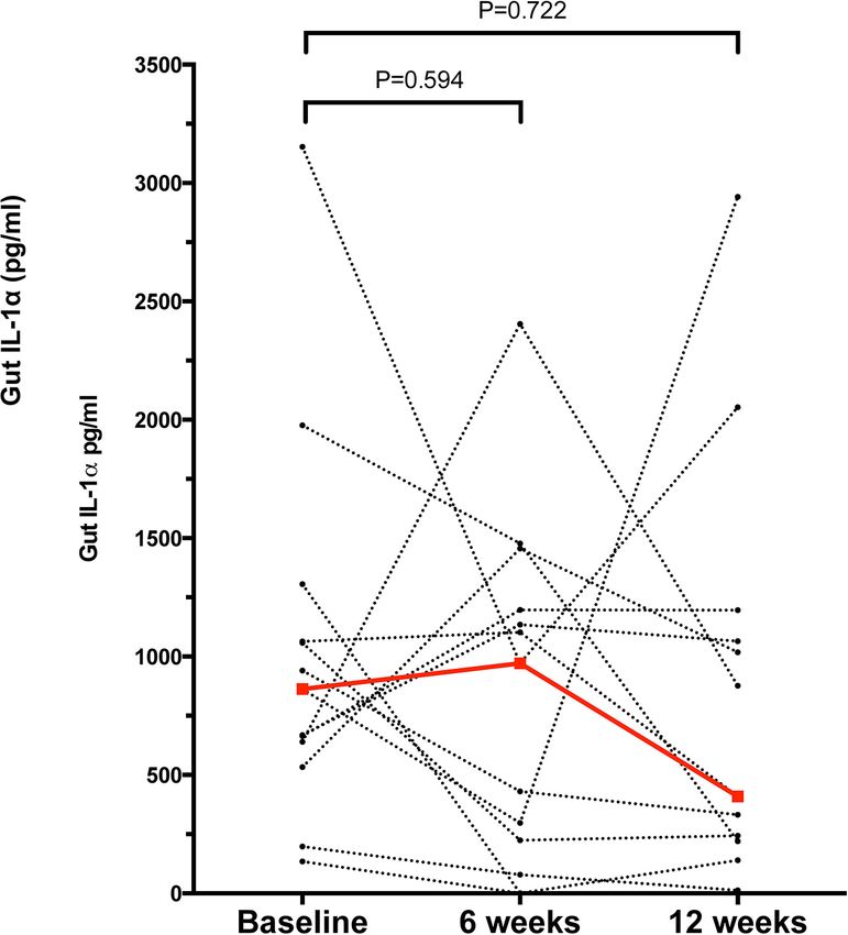
Yegorov et al.                                                                                           Psoriasis, Gut Immunology and Microbiome

INTRODUCTION                                                             the psoriasis patients at 6 and 12 weeks after the first visit. Fecal
                                                                         samples from all study participants were frozen within 2 h of
Psoriasis is a chronic autoimmune condition that predominantly           collection and kept frozen at −80◦ C until analysis.
affects the skin and manifests with variable severity (1). The              This study was designed as an exploratory case-control study
clinical classification of psoriasis is based on the pattern             to supply pilot data for future studies in the same population;
and extent of cutaneous involvement. For example, the most               therefore, no formal sample size calculations were performed
common phenotype, psoriasis vulgaris, or plaque-type psoriasis,          and the sample size was determined based on the available
is distinguished by the presence of well-defined areas of                study budget. In total, 40 individuals were included in the study
erythematous and indurated plaques and infiltration of the               (n = 20 with psoriasis, n = 20 controls). All experimental assays
epidermis and dermis by mononuclear cells (1). Frequently,               were performed by research personnel blinded to the psoriasis
psoriasis is associated with extracutaneous manifestations, such         status of participants. All research procedures were approved
as psoriatic arthritis (PsA) or cardiovascular disease, indicating       by the institutional review board of the UMC CF Academic
that systemic inflammatory processes likely underlie psoriatic           Council. Written consent to participate was collected from all
disease (1, 2).                                                          participants. We used the STROBE checklist when writing our
   The causes of psoriasis are incompletely understood; genetic          report (10).
predisposition plays a major role (1), but other factors such
as systemic inflammation and microbiota alterations have
also been implicated in disease pathogenesis. Furthermore,               Cytokine Measurements
the link between psoriasis and inflammatory bowel disease                Frozen stool samples were thawed, and approximately 4 mg of
(3) intriguingly points at the gut as an important contributor           each sample were dissolved in 200 µl of phosphate buffered
to psoriasis development. In support of this, recent studies             saline. The supernatant obtained by centrifugation at 16,000 g
have reported major alterations in both gut microbial                    for 15 min was then analyzed using the Milliplex Map
communities (4–7) and markers of immune response (6) in                  Human Magnetic Bead Panels for cytokines and chemokines
individuals with psoriasis.                                              (HCYTMAG-60K-PX41) and immunoglobulins (HGAMMAG-
   In earlier work, we characterized the gut microbiome of               301K-06) according to the manufacturer’s protocol on a Bio-Plex
adults with and without metabolic syndrome from Kazakhstan,              3D instrument (Bio-Rad). The ELISA assay details and analyte
a country in Central Asia (8). As part of our overarching                classification are given in Supplementary Table 1. To avoid the
objective to better understand the relationship between the              bias of inter-plate variation, paired samples were examined on the
mucosal and systemic correlates of chronic disease in this region        same ELISA plate.
(8, 9), here we expand on our earlier findings and focus on
the gut microenvironment in individuals with psoriasis. We
hypothesized that psoriasis in adult Central Asians is associated
                                                                         Microbiome Analysis
with gut inflammation and microbiome alterations. To test                DNA extraction and sequencing
this hypothesis, we assessed levels of select cytokines and              A subset of 21 stool samples (14 psoriasis and 7 controls)
immunoglobulins and performed a metagenomic analysis of                  from the first study visit was available for microbiome analysis.
stool samples obtained from dermatology clinic outpatients in the        Samples were thawed prior to DNA extraction using QIAamp
capital city of Kazakhstan.                                              DNA Mini Kit (Qiagen). The quality of extracted DNA was
                                                                         assessed using Qubit dsDNA HS Assay Kit (Thermo Fisher)
                                                                         on a Qubit 2.0 according to the manufacturer’s manual
MATERIALS AND METHODS                                                    (Invitrogen, Life Technologies). Next-generation sequencing
                                                                         libraries were prepared with NEXTflex 16S V1-V3 Amplicon-Seq
Study Setting and Participant                                            Kit (PerkinElmer), and library quality assessed using the Qubit
Recruitment                                                              2.0 system. Amplicons (96 samples per lane) were sequenced
Participants aged 30–45 years were recruited through an                  using the MiSeq platform (Illumina).
outpatient dermatology clinic at the Centre for Dermatology and
STD prophylaxis in the capital city of Kazakhstan, Nur-Sultan            Sequence analysis
(formerly Astana). The dermatological assessment of participants         The QC and raw sequence pre-processing were performed using
was done in accordance with the national clinical guidelines for         fastp (v. 0.20.0. April 2019) (11) with the following parameters:
psoriasis diagnosis and treatment of Kazakhstan.                         mean quality for 4 bp window size was 20, the adapter detection
   Study exclusion criteria were: use of antibiotics within              enabled, reads less than 120 bp were discarded, unmerged reads
3 months prior to the study, a diagnosis of psoriatic arthritis,         were included in the final FASTQ files. Total Sum Scaling (TSS)
presence of any other chronic condition of the skin or                   per-sample normalization was used to remove technical bias
gastrointestinal tract and presence of any severe comorbidity,           related to different sequencing depths among libraries and then
or pregnancy. Healthy controls were recruited from the local             scaled to units of reads per million per library. Taxonomic
communities through community-wide advertisement of the                  assignment was done using the naive Bayesian classifier method
study and matched by age, sex, and ethnicity. A fecal sample was         as implemented in the dada2 Bioconductor R package using an
collected at baseline from all participants, and subsequently from       RDP training set (v.16).

Frontiers in Immunology | www.frontiersin.org                        2                                   October 2020 | Volume 11 | Article 571319
Yegorov et al. Psoriasis, Gut Immunology and Microbiome
Statistical Analysis Comparison of Stool-Derived Cytokine
All statistical analyses and graphing were performed using Profiles
IBM SPSS V.23 (NY, United States) and GraphPad Prism Samples from a total of 40 participants were analyzed. Cytokine
V.6.0. (CA, United States), unless specified otherwise. and immunoglobulin data were obtained for 22 and 29 (out
Differences in demographic characteristics between groups of a total of 40) participants, respectively. Repeated efforts
were assessed using Independent-Samples Mann-Whitney to obtain ELISA data for outstanding measurements were
U and Chi-Square Tests. An ELISA analyte was considered unsuccessful, likely due to sample characteristics that were
“detectable” if the measured analyte concentration was equal psoriasis-independent. Overall, 26/41 cytokines and chemokines
to or above the assay’s lowest level of detection (LLOD), and 3/6 Igs were detectable (i.e., >0 pg/ml in >50% of the
and “undetectable” if the measured concentration was participants); the psoriasis group tended to have higher median
below the LLOD (Supplementary Table 1). Analytes were concentrations for most analytes (29/47 = 61.7%) compared to
grouped into (i) “detectable in >50% of the participants” the control group (Supplementary Table 2); no correlation was
and analyzed as continuous variables using Independent- observed between the stool analytes and the PASI scores. IL-1α
Samples Mann-Whitney U or (ii) “detectable in ≤50% of was the only cytokine significantly elevated in the psoriasis group
the participants” and analyzed as dichotomous variables. (4.19-fold, p = 0.007, Supplementary Table 2 and Figure 1A);
Differences among analyte concentrations across paired this difference was not associated with differences in socio-
study visits were assessed by Friedman’s Two-way ANOVA demographic or psoriasis-specific characteristics in this subset of
by Ranks Test (across all three visits) or by the Related participants (N = 23, Supplementary Table 3). Notably, IL-1α
Samples Wilcoxon Signed Rank Test (between two levels remained significantly unaltered 6 and 12 weeks after the
visits). The α and β microbial diversities were estimated initial sampling (Figure 2), similar to the rest of the analytes, with
using Shannon’s diversity index and UniFrac weighted the exception of IL-1β (p = 0.049) and IgG2 (p = 0.05), which
distance with Principal Coordinate Analysis (PCoA),
respectively. Group comparisons for β-diversity were done
via permutational multivariate ANOVA (PERMANOVA)
of dissimilarity using adonis2 function from the vegan R TABLE 1 | Socio-demographic and psoriasis-specific characteristics
package (12). of participants.
The linear discriminant analysis (LDA) effect size (LEfSe)
algorithm was applied to identify features significantly different Participant Psoriasis group (N = 20) Controls (N = 20) P value
characteristic
between the comparison groups with default settings (p ≤ 0.05
based on Kruskal-Wallis test and LDA score ≥2) (13). Median age (IQR) 34.5 (31.0–37.8) 33.0 (31.3–34.0) 0.205
Statistical analysis of microbiome data was performed using Men, n (%) 10 (50.0) 11 (55.0) 0.752
MicrobiomeAnalystR package (14). Pearson correlation analysis Mean BMI (range) 24.8 (21.4–28.7) 23.9 (18.6–32.7) 0.475
between the OTU relative abundance, PASI scores, and IL1α were Married, n (%) 13 (65.0) 19 (95.0) 0.018
performed on Log10-transformed data. Data with missing values Psoriasis type
were excluded from the analyses. Vulgaris 15 (75.0) – –
Guttate 3 (15.0) – –
Palmoplantar 2 (10.0) – –
Psoriasis present in 17 (85.0) – –
RESULTS a parent
Time since psoriasis first noted (years)
Participant Demographics and Clinical 10 8 (40.0) – –
psoriasis diagnostic data are given in Table 1. Age, sex, or BMI did Head hair damage 17 (85.0) – –
not significantly differ between groups. The median participant present
age was 33 years and significantly more individuals were not Nail damage 1 (5.0) – –
present
married in the psoriasis group (7/20) compared to the controls
Median PASI (IQR) 11.4 (6.7–16.4) – –
(1/20, p = 0.018). In the majority (85%) of the participants,
Psoriasis severity based on PASI
one or both parents had psoriasis. Psoriasis vulgaris was most
Mild [PASI < 7], n 5 (25.0) – –
prevalent (75%) while 25% of the participants had guttate and
(%)
palmoplantar psoriasis. Most participants (85%, 17/20) reported
Moderate 7 (35.0) – –
having had the condition for >5 years, indicating seasonality [7 ≤ PASI ≤ 12], n
in symptom presentation. In most participants psoriasis was (%)
associated with hair, but not nail, damage. The median psoriasis Severe 8 (40.0) – –
area and severity index (PASI) score was 11.4; mild, moderate, [PASI > 12], n (%)
and severe forms (15) were seen in 25, 35, and 40% of the BMI, body mass index; IQR, interquartile range; PASI, psoriasis area
participants, respectively (Table 1). and severity index.
Frontiers in Immunology | www.frontiersin.org 3 October 2020 | Volume 11 | Article 571319Yegorov et al. Psoriasis, Gut Immunology and Microbiome FIGURE 1 | Differences in the levels of analytes representative of major functional groups of immune response mediators in the gut of individuals with (“Ps”) and without psoriasis (controls, “C”). (A) IL-1α (Proinflammatory); (B) IL-4 (Th1/Th2); (C) Il-7 (Homeostatic); (D) IL-9 (Th9); (E) IL-10 (Anti-inflammatory); (F) IL-15 (Growth factor); (G) IFN-α2 (Interferons); (H) GM-CSF (Colony-stimulating factor); (I) MIP-1β (Chemokine). For all depicted analytes data were available for 23/40 (psoriasis+, N = 13, and psoriasis-, N = 10) participants. Statistical significance assessed by the Independent-Samples Mann-Whitney U test. Source data are provided as a Supplementary Material. Frontiers in Immunology | www.frontiersin.org 4 October 2020 | Volume 11 | Article 571319
Yegorov et al. Psoriasis, Gut Immunology and Microbiome
FIGURE 2 | Concentration of gut IL-1α in the psoriasis group at baseline, 6 and 12 weeks after the initial study visit. Each dot represents a participant, dashed lines
link samples paired across the study visits, n = 13 for paired comparisons. Red dots and line denote the median concentrations of IL-1α at each time point.
Statistical significance assessed by the Related Samples Wilcoxon Signed Rank Test. Source data are provided as a Supplementary Material.
fluctuated marginally over the period of 12 weeks after the first After correcting for multiple hypothesis testing, no OTUs
study visit (Supplementary Table 4). remained significantly different between groups (FDR = 0.05);
notwithstanding the overall differences in bacterial composition
Comparison of Gut Microbiome Profiles observed here are in agreement with those observed in other
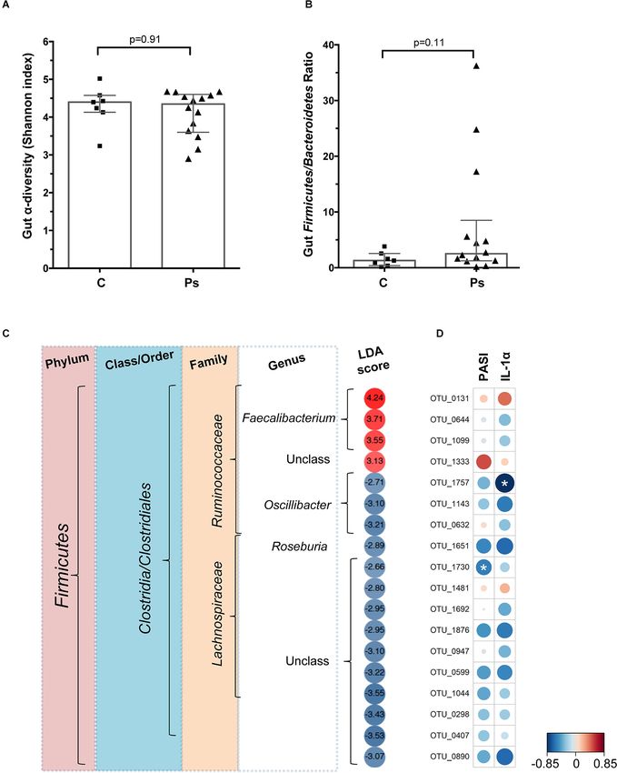
Gut microbiota diversity and Firmicutes/Bacteroides (F/B) ratio. studies (5, 6). Specifically, all 18 OTUs that were found to be
A total of 21 fecal samples, 14 psoriasis + and 7 controls, were differentially abundant were from the phylum Firmicutes and,
available for the microbiome analysis. In our earlier analysis except one unclassified OTU, were classified as Clostridia, Order
of the Kazakh gut metagenome, we found the Firmicutes to Clostridiales. The majority of these OTUs (14/18, 77.8%) were less
Bacteroidetes ratio to be significantly associated with metabolic abundant in psoriasis participants compared to controls. At the
syndrome. Therefore in the current analysis the F/B ratio family level, all 8 OTUs from the Lachnospiraceae were found to
comparison was a pre-specified endpoint. When compared to be less abundant in psoriasis patients, while 4 of the 7 OTUs from
controls, neither the microbial diversity indices nor the F/B ratios the family Ruminococcaceae were more abundant in psoriasis
of the psoriasis patients were significantly different (Figures 3A,B patients and 3 were less abundant (Figure 3D). Examining this at
and Supplementary Figure 1), although the median F/B ratio in the level of genus indicates that all OTUs from Faecalibacterium
the psoriasis group was double of that observed in the controls (n = 3 OTU) exhibited increased abundance, while those from the
(2.5 vs. 1.3), largely driven by an elevated F/B ratio in three Oscillibacter (n = 3 OTU) and Roseburia (n = 1 OTU) exhibited
psoriasis participants (Figure 3C). reduced abundance in the psoriasis group (Figure 3D). Lastly,
Bacterial taxa associated with psoriasis. The LefSe analysis the correlation analyses confirmed that there was a negative
(12) identified 18 operational taxonomic units (OTUs) that could correlation between OTU abundance and both disease severity
significantly discriminate among groups at p = 0.05 (Figure 3D). (PASI) and IL-1α levels for most OTUs; only two of these
Frontiers in Immunology | www.frontiersin.org 5 October 2020 | Volume 11 | Article 571319Yegorov et al. Psoriasis, Gut Immunology and Microbiome FIGURE 3 | (A) Microbial species α-diversity measured by Shannon index in the gut of the psoriasis individuals and controls. (B) Gut Firmicutes/Bacteroides ratio of psoriasis + individuals and controls. (C) Heatmap showing the LefSe-derived LDA scores for each of the OTU (n = 18) differentially abundant in the gut of psoriasis + participants. Data based on N = 21 samples. (D) Heatmap based on the Pearson coefficients of correlation between the psoriasis-associated microbial taxa, baseline IL-1α and PASI scores. Analysis based on N = 14 samples. Significant correlations marked by asterisks (*). Frontiers in Immunology | www.frontiersin.org 6 October 2020 | Volume 11 | Article 571319
Yegorov et al.                                                                                         Psoriasis, Gut Immunology and Microbiome

correlations reached statistical significance (Figure 3D). Three         known to strongly influence the human gut microbiome
taxa, OTU_0131 (from the genus Faecalibacterium), OTU_1333               composition (27).
(unclassified), and OTU_1481 (unclassified), exhibited positive              We found that psoriasis was associated with an elevated
correlations between OTU abundance and PASI and IL-                      abundance of members of the genus Faecalibacterium, which
1α (Figure 3D).                                                          is consistent with a metagenomic analysis from Spain (5). On
                                                                         the other hand, a study based on a PCR-aided identification
                                                                         of specific bacterial species in the Netherlands found that
DISCUSSION                                                               stool samples from psoriasis patients were depleted for
                                                                         Faecalibacterium prausnitzii (4). Notably, gut Faecalibacterium
Here, we examined associations between psoriasis, and gut                alterations have been associated with eczema and IBD (28, 29)
immunology and microbial parameters in an adult cohort based             and, taken together, these data highlight the dynamic role gut
in Kazakhstan. We found that psoriasis was associated with               Faecalibacterium spp. are potentially playing in the pathogenesis
elevated gut IL-1α and altered abundance of Firmicutes. Overall,         of both skin- and gut diseases.
these findings are consistent with the notion that psoriasis is              The causality of the relationship between psoriasis and
linked to gut dysbiosis and inflammation (5, 6, 16, 17).                 gut immunology and microbiota is unclear. Psoriasis is
   Few studies to date have directly assessed the immunological          associated with systemic inflammation (1), which could drive
changes occurring in the intestine of psoriasis patients. Scher          changes in the gut mucosa and thereby affect the gut
et al. reported elevation of soluble IgA and reduction of                microbiota. It has also been proposed that the proinflammatory
receptor activator of nuclear factor kappa-B ligand (RANKL),             environment of the gut, precipitated by dietary insults and
a critical factor controlling differentiation of intestinal lamina       genetic predisposition, could induce systemic inflammation and
propria cells, in PsA (6). In the current study, we saw                  thus cause skin-directed inflammation (30). Future mechanistic
trends toward elevation for >60% of the analytes from the                studies may clarify the exact mechanisms involved; meanwhile,
stools of psoriatic individuals, although only IL-1α, a cytokine         our finding of enhanced IL-1α, a key regulator of inflammatory
central to the regulation of inflammation (18), was significantly        processes, suggests that the gut may play a central role in
elevated compared to the controls and remained unchanged                 the induction of inflammatory responses in psoriasis (18).
in the psoriasis group for up to 3 months after the first                While current therapies for mild psoriasis target cutaneous
visit to the clinic. Elevated cytokines in stool were previously         manifestations and exhibit relatively low efficacies, the more
detected in the context of gut inflammation (19, 20), while              efficacious therapeutic modes for severe psoriasis based on
in a murine model of colitis IL-1α secreted by intestinal                the use of antibodies are expensive and can have severe
epithelium was the main driver of immune activation (21).                side effects (1). If the psoriatic disease is mediated by the
In psoriasis, IL-1α drives the formation of dermal clusters of           gut immune milieu and microbiota, psoriasis treatment could
T cells and antigen presenting cells and is involved in the              benefit from modulating the gut microenvironment using
development of dermal Th17 responses (22–24). Therefore, it              microecologic agents, such as probiotics, and fecal microbiota
is possible that increased levels of gut IL-1α in individuals            transplantation.
with psoriasis may contribute to increased inflammation via the              Our findings should be interpreted in the light of several
gut-skin axis (25) and may help explain the well-documented              limitations. First, due to technical and logistic limitations our
epidemiological link between psoriasis and inflammatory bowel            study had a small sample size and both the ELISA and
diseases (3).                                                            metagenomic analysis results were only available for subsets of
   To the best of our knowledge this study is the first to               the original cohort. While the relatively small sample size is an
characterize psoriasis in a population from Kazakhstan and to            important limitation of the study, this limitation was partially
assess its associations with gut immunology and microbiome.              overcome by repeated cytokine measures, which confirmed, in
Earlier, we performed a large-scale metagenomic analysis and             particular, the consistently high levels of IL-1α in the psoriasis
compared the Kazakh gut metagenome to its counterparts                   individuals compared to the controls. Notably, the significant
from other regions of the world (8). We found significant                fluctuation of gut IL-1β and IgG2 over the 3-month period may
differences between the microbiomes of Kazakhs and both                  be due to a natural tendency of these signaling molecules to
Europeans and East Asians. One distinguishing feature of the             change over time or due to assay-related variation. Given the
Kazakh gut metagenome is a remarkably dominant Prevotella-               relatively small sample size of our metagenomic analysis, our
rich enterotype, which is typically regarded as proinflammatory          analysis of psoriasis and gut microbiome interaction was likely
(26), and was associated with the metabolic syndrome in our              under-powered and should be revisited in future, larger, studies.
cohort (8). In our current analysis, psoriasis was not associated        Further, soluble analytes were measured in stool supernatant,
with changes in gut Prevotella abundance, but, consistent with           a highly heterogeneous sample, and further improvements of
studies from other cohorts, was associated with alterations in           the protocol may improve cytokine yields; work is underway
Firmicutes (5, 6). Although the direction of Firmicute change            in our laboratory in this direction. Lastly, this study used an
differs among studies, this suggests that psoriasis-associated gut       exploratory approach and therefore follow-up studies will be
microbiome signatures are generalizable to human populations             required to confirm these findings. It is noteworthy that despite
residing in different parts of the world, but may be modified            age and sex matching, there were significant differences in the
by differences in ethnic and environmental factors that are              marital status of psoriasis participants and controls. Psoriasis

Frontiers in Immunology | www.frontiersin.org                        7                                  October 2020 | Volume 11 | Article 571319
Yegorov et al. Psoriasis, Gut Immunology and Microbiome
is a socially stigmatizing condition that affects the individual’s AUTHOR CONTRIBUTIONS
ability to build relationships (1), underscoring the need for better
therapeutic and psychological instruments to help individuals SY, DB, SG, BY, and AK: conceptualization and data curation.
and their families affected by psoriasis. SY, DB, and SG: formal analysis and visualization. BY: funding
In summary, this study for the first time assessed psoriasis acquisition. SK, LA, IK, AN, BY, and AK: methodology. BY,
associations with gut immunology and microbiome in an under- AK, and GH: project administration, resources, and supervision.
studied population from Central Asia. Although limited by SY and DB: writing – original draft. All authors: writing –
a small sample size, the data presented here provide more review and editing.
evidence in support of the notion that gut immunology and
microbiota may play critical roles in the pathogenesis of
psoriasis, an autoimmune condition that is commonly thought FUNDING
to primarily affect the skin. Future studies are warranted to
explore the inclusion of gut microenvironment modulating This study was supported by the Science Committee of the
agents in the treatment and prophylaxis of psoriasis in Ministry of Education of the Republic of Kazakhstan Grants
this population. #AP05135585, #AP05134659, and #AP05135073. The funder had
no role in study design, data collection and analysis, decision to
publish, or preparation of the manuscript.
DATA AVAILABILITY STATEMENT
All processed ELISA and microbiome data generated and
analyzed during this study are included in the Supplementary ACKNOWLEDGMENTS
Material. The raw microbiome sequence data are accessible
through an online repository at: http://doi.org/10.5281/zenodo. We thank all the participants and research teams involved in
3782581. the study. This manuscript has been released as a pre-print at
medRxiv (31).
ETHICS STATEMENT
SUPPLEMENTARY MATERIAL
All study procedures were approved by the University
Medical Centre Ethics Committee. The patients/participants The Supplementary Material for this article can be found
provided their written informed consent to participate in online at: https://www.frontiersin.org/articles/10.3389/fimmu.
this study. 2020.571319/full#supplementary-material
REFERENCES 9. Yegorov S, Bromage S, Boldbaatar N, Ganmaa D. Effects of vitamin D
supplementation and seasonality on circulating cytokines in adolescents:
1. Greb JE, Goldminz AM, Elder JT, Lebwohl MG, Gladman DD, Wu JJ, et al. analysis of data from a feasibility trial in mongolia. Front Nutr. (2019) 6:166.
Psoriasis. Nat Rev Dis Primers. (2016) 2:16082. doi: 10.1038/nrdp.2016.82 doi: 10.3389/fnut.2019.00166
2. Eder L, Dey A, Joshi AA, Boehncke W-H, Mehta NN, Szentpetery S. 10. von Elm E, Altman DG, Egger M, Pocock SJ, Gøtzsche PC, Vandenbroucke JP,
Cardiovascular diseases in psoriasis and psoriatic arthritis. J Rheumatol Suppl. et al. The strengthening the reporting of observational studies in epidemiology
(2019) 95:20–7. doi: 10.3899/jrheum.190114 (STROBE) statement: guidelines for reporting observational studies. J Clin
3. Fu Y, Lee CH, Chi CC. Association of psoriasis with inflammatory bowel Epidemiol. (2008) 61:344–9. doi: 10.1016/j.jclinepi.2007.11.008
disease: a systematic review and meta-analysis. JAMA Dermatol. (2018) 11. Chen S, Zhou Y, Chen Y, Gu J. fastp: an ultra-fast all-in-one FASTQ
154:1417–23. doi: 10.1001/jamadermatol.2018.3631 preprocessor. Bioinformatics. (2018) 34:i884–90. doi: 10.1093/bioinformatics/
4. Eppinga H, Sperna Weiland CJ, Thio HB, van der Woude CJ, Nijsten TEC, bty560
Peppelenbosch MP, et al. Similar depletion of protective Faecalibacterium 12. Dixon P. VEGAN, a package of R functions for community ecology. J Vegetat
prausnitzii in psoriasis and inflammatory bowel disease, but not in hidradenitis Sci. (2003) 14:927–30. doi: 10.1111/j.1654-1103.2003.tb02228.x
suppurativa. J Crohns Colitis. (2016) 10:1067–75. doi: 10.1093/ecco-jcc/jjw070 13. Segata N, Izard J, Waldron L, Gevers D, Miropolsky L, Garrett WS, et al.
5. Codoner FM, Ramirez-Bosca A, Climent E, Carrión-Gutierrez M, Guerrero Metagenomic biomarker discovery and explanation. Genome Biol. (2011)
M, Pérez-Orquín JM, et al. Gut microbial composition in patients with 12:R60. doi: 10.1186/gb-2011-12-6-r60
psoriasis. Sci Rep. (2018) 8:3812. doi: 10.1038/s41598-018-22125-y 14. Chong J, Liu P, Zhou G, Xia J. Using MicrobiomeAnalyst for comprehensive
6. Scher JU, Ubeda C, Artacho A, Attur M, Isaac S, Reddy SM, et al. Decreased statistical, functional, and meta-analysis of microbiome data. Nat Protoc.
bacterial diversity characterizes the altered gut microbiota in patients with (2020) 15:799–821. doi: 10.1038/s41596-019-0264-1
psoriatic arthritis, resembling dysbiosis in inflammatory bowel disease. 15. Schmitt J, Wozel G. The psoriasis area and severity index is the adequate
Arthritis Rheumatol. (2015) 67:128–39. doi: 10.1002/art.38892 criterion to define severity in chronic plaque-type psoriasis. Dermatology.
7. Tan L, Zhao S, Zhu W, Wu L, Li J, Shen MX, et al. The Akkermansia (2005) 210:194–9. doi: 10.1159/000083509
muciniphila is a gut microbiota signature in psoriasis. Exp Dermatol. (2018) 16. Ely PH. Is psoriasis a bowel disease? Successful treatment with bile acids and
27:144–9. doi: 10.1111/exd.13463 bioflavonoids suggests it is. Clin Dermatol. (2018) 36:376–89. doi: 10.1016/j.
8. Kushugulova A, Forslund SK, Costea PI, Kozhakhmetov S, Khassenbekova clindermatol.2018.03.011
Z, Urazova M, et al. Metagenomic analysis of gut microbial communities 17. Visser MJE, Kell DB, Pretorius E. Bacterial dysbiosis and translocation in
from a Central Asian population. BMJ Open. (2018) 8:e021682. doi: 10.1136/ psoriasis vulgaris. Front Cell Infect Microbiol. (2019) 9:7. doi: 10.3389/fcimb.
bmjopen-2018-021682 2019.00007
Frontiers in Immunology | www.frontiersin.org 8 October 2020 | Volume 11 | Article 571319Yegorov et al. Psoriasis, Gut Immunology and Microbiome
18. Di Paolo NC, Shayakhmetov DM. Interleukin 1alpha and the 27. He Y, Wu W, Zheng HM, Li P, McDonald D, Sheng H-F, et al. Regional
inflammatory process. Nat Immunol. (2016) 17:906–13. doi: 10.1038/ni. variation limits applications of healthy gut microbiome reference ranges and
3503 disease models. Nat Med. (2018) 24:1532–5. doi: 10.1038/s41591-018-0164-x
19. Wedrychowicz A, Tomasik P, Zajac A, Fyderek K. Prognostic 28. Zheng H, Liang H, Wang Y, Miao M, Shi T, Yang F, et al. Altered gut microbiota
value of assessment of stool and serum IL-1beta, IL-1ra and IL- composition associated with eczema in infants. PLoS One. (2016) 11:e0166026.
6 concentrations in children with active and inactive ulcerative doi: 10.1371/journal.pone.0166026
colitis. Arch Med Sci. (2018) 14:107–14. doi: 10.5114/aoms.2017. 29. Hansen R, Russell RK, Reiff C, Louis P, McIntosh F, Berry SH, et al. Microbiota
68696 of de-novo pediatric IBD: increased Faecalibacterium prausnitzii and reduced
20. Enocksson A, Lundberg J, Weitzberg E, Norrby-Teglund A, Svenungsson B. bacterial diversity in Crohn’s but not in ulcerative colitis. Am J Gastroenterol.
Rectal nitric oxide gas and stool cytokine levels during the course of infectious (2012) 107:1913–22. doi: 10.1038/ajg.2012.335
gastroenteritis. Clin Diagnos Lab Immunol. (2004) 11:250–4. doi: 10.1128/cdli. 30. Stehlikova Z, Kostovcikova K, Kverka M, Rossmann P, Dvorak J, Novosadova
11.2.250-254.2004 I, et al. Crucial role of microbiota in experimental psoriasis revealed by a
21. Bersudsky M, Luski L, Fishman D, White RM, Ziv-Sokolovskaya N, Dotan gnotobiotic mouse model. Front Microbiol. (2019) 10:236. doi: 10.3389/fmicb.
S, et al. Non-redundant properties of IL-1alpha and IL-1beta during acute 2019.00236
colon inflammation in mice. Gut. (2014) 63:598–609. doi: 10.1136/gutjnl- 31. Yegorov S, Babenko D, Kozhakhmetov S, Akhmaltdinova L, Kadyrova I,
2012-303329 Nurgozhina A, et al. Psoriasis is associated with elevated gut IL-1α and
22. Natsuaki Y, Egawa G, Nakamizo S, Ono S, Hanakawa S, Okada T, et al. intestinal microbiome alterations: results of a cross-sectional study from
Perivascular leukocyte clusters are essential for efficient activation of effector Central Asia. medRxiv. (2020). . doi: 10.1101/2020.05.16.20103978
T cells in the skin. Nat Immunol. (2014) 15:1064–9. doi: 10.1038/ni.
2992 Conflict of Interest: DB was employed by Art Science LLP Innovative Center.
23. Kryczek I, Bruce AT, Gudjonsson JE, Johnston A, Aphale A, Vatan
L, et al. Induction of IL-17+ T cell trafficking and development by The remaining authors declare that the research was conducted in the absence of
IFN-gamma: mechanism and pathological relevance in psoriasis. any commercial or financial relationships that could be construed as a potential
J Immunol. (2008) 181:4733–41. doi: 10.4049/jimmunol.181.7. conflict of interest.
4733
24. Baliwag J, Barnes DH, Johnston A. Cytokines in psoriasis. Cytokine. (2015) Copyright © 2020 Yegorov, Babenko, Kozhakhmetov, Akhmaltdinova, Kadyrova,
73:342–50. doi: 10.1016/j.cyto.2014.12.014 Nurgozhina, Nurgaziyev, Good, Hortelano, Yermekbayeva and Kushugulova. This
25. O’Neill CA, Monteleone G, McLaughlin JT, Paus R. The gut-skin axis in is an open-access article distributed under the terms of the Creative Commons
health and disease: a paradigm with therapeutic implications. Bioessays. (2016) Attribution License (CC BY). The use, distribution or reproduction in other forums
38:1167–76. doi: 10.1002/bies.201600008 is permitted, provided the original author(s) and the copyright owner(s) are credited
26. Larsen JM. The immune response to Prevotella bacteria in chronic and that the original publication in this journal is cited, in accordance with accepted
inflammatory disease. Immunology. (2017) 151:363–74. doi: 10.1111/imm. academic practice. No use, distribution or reproduction is permitted which does not
12760 comply with these terms.
Frontiers in Immunology | www.frontiersin.org 9 October 2020 | Volume 11 | Article 571319You can also read


