Uremia Coupled with Mucosal Damage Predisposes Mice with Kidney Disease to Systemic Infection by Commensal Candida albicans
←
→
Page content transcription
If your browser does not render page correctly, please read the page content below
Uremia Coupled with Mucosal Damage Predisposes Mice with
Kidney Disease to Systemic Infection by Commensal Candida
albicans

                                                                                                Downloaded from http://www.immunohorizons.org/ by guest on January 25, 2021
Chetan V. Jawale, De-Dong Li, Kritika Ramani, Li Lin, Kelvin Li, Barbara Methe and Partha
Sarathi Biswas

ImmunoHorizons 2021, 5 (1) 16-24
doi: https://doi.org/10.4049/immunohorizons.2000114
http://www.immunohorizons.org/content/5/1/16
This information is current as of January 25, 2021.

     References    This article cites 40 articles, 9 of which you can access for free at:
                   http://www.immunohorizons.org/content/5/1/16.full#ref-list-1
   Email Alerts    Receive free email-alerts when new articles cite this article. Sign up at:
                   http://www.immunohorizons.org/alerts

ImmunoHorizons is an open access journal published by
The American Association of Immunologists, Inc.,
1451 Rockville Pike, Suite 650, Rockville, MD 20852
All rights reserved.
ISSN 2573-7732.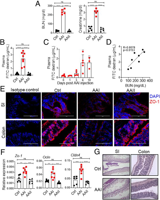
RESEARCH ARTICLE

                                                                                                                                        Infectious Disease

Uremia Coupled with Mucosal Damage Predisposes Mice with
Kidney Disease to Systemic Infection by Commensal
Candida albicans
Chetan V. Jawale,* De-Dong Li,* Kritika Ramani,* Li Lin,* Kelvin Li,† Barbara Methe,† and Partha Sarathi Biswas*

                                                                                                                                                                                Downloaded from http://www.immunohorizons.org/ by guest on January 25, 2021
*Division of Rheumatology and Clinical Immunology, Department of Medicine, University of Pittsburgh, Pittsburgh, PA 15261; and †Department of
Medicine, University of Pittsburgh, Pittsburgh, PA 15261

ABSTRACT
Infections are the second major cause of mortality in patients with kidney disease and accompanying uremia. Both vascular access
and non–access-related infections contribute equally to the infection-related deaths in patients with kidney disease. Dialysis is the
most common cause of systemic infection by Candida albicans in these patients. C. albicans also reside in the gastrointestinal tract as
a commensal fungus. However, the contribution of gut-derived C. albicans in non–access-related infections in kidney disease is
unknown. Using a mouse model of kidney disease, we demonstrate that uremic animals showed increased gut barrier permeability,
impaired mucosal defense, and dysbiosis. The disturbance in gut homeostasis is sufficient to drive the translocation of microbiota and
intestinal pathogen Citrobacter rodentium to extraintestinal sites but not C. albicans. Interestingly, a majority of uremic animals
showed fungal translocation only when the gut barrier integrity is disrupted. Our data demonstrate that uremia coupled with gut
mucosal damage may aid in the translocation of C. albicans and cause systemic infection in kidney disease. Because most of the
individuals with kidney disease suffer from some form of gut mucosal damage, these results have important implications in the risk
stratification and control of non–access-related opportunistic fungal infections in these patients. ImmunoHorizons, 2021, 5: 16–24.

INTRODUCTION                                                                            related infections over the past decade, prevention of non–access-
                                                                                        related infections is still a major problem. In most cases, the source
Kidney disease is a major public health problem in the 21st century                     of non–access-related infections in kidney disease remains un-
(1). Infection is the second leading cause of mortality (20%) in                        identified, which is a major constraint in controlling infections in
patients with kidney disease (2, 3). Although vascular access–                          these patients.
related infections are the primary cause of infection-related deaths                        Several predisposing factors make patients with kidney disease
in kidney disease, non–access-related infectious complications,                         susceptible to infection (6–8). These include dialysis access, the
although equally important, are often overlooked (4). A study                           presence of coexisting illnesses, vaccine hyporesponsiveness,
showed that infection-related hospitalizations are not due to                           immunosuppressive therapy, and uremia. Uremia is characterized
vascular access in 77% of identified cases; rather, the most common                      by the retention of ;900 metabolites in the blood in the absence of
reason was an infection of unknown source (5). Even though                              kidney function. A subset of these (;100) have a profound impact
advances in dialysis techniques have lowered the rate of access-                        on various biological systems and are termed uremic toxins (9).

Received for publication December 30, 2020. Accepted for publication December 30, 2020.
Address correspondence and reprint requests to: Dr. Partha Sarathi Biswas, University of Pittsburgh, 3500 Terrace Street, Pittsburgh, PA 15261. E-mail address:
psb13@pitt.edu
ORCIDs: 0000-0002-9474-390X (D.-D.L.); 0000-0003-3922-5664 (K.L.).
This work was supported by National Institutes of Health Grants AI142354, DK104680, and R21AI45242 to P.S.B.
Abbreviations used in this article: AAI, aristolochic acid I; AAII, aristolochic acid II; AAN, AAI nephropathy; BUN, blood urea nitrogen; DSS, dextran sulfate sodium; GI,
gastrointestinal; MLN, mesenteric lymph node; SI, small intestine; SILP, small intestinal lamina propria; Treg, regulatory T; UUO, unilateral ureteral obstruction; WT, wild-
type; YPD, yeast extract peptone dextrose.
This article is distributed under the terms of the CC BY-NC 4.0 Unported license.
Copyright © 2021 The Authors

16                                                                                                                      https://doi.org/10.4049/immunohorizons.2000114

ImmunoHorizons is published by The American Association of Immunologists, Inc.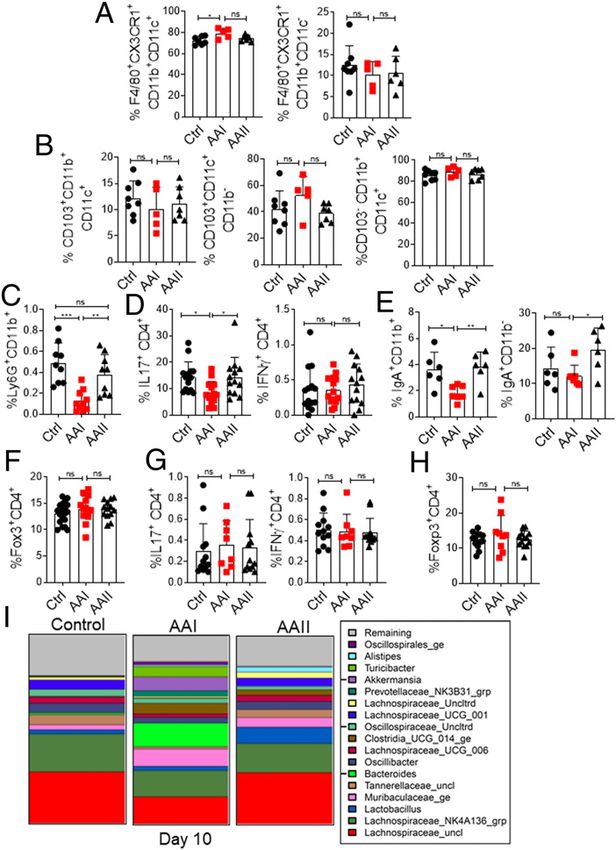
ImmunoHorizons                                                           GUT-DERIVED FUNGUS CAUSES SYSTEMIC INFECTION IN UREMIA               17

Uremia is implicated in immune dysfunction and is considered an                under specific pathogen-free conditions under the supervision
independent risk factor for infections independent of vascular                 of the Division of Laboratory Animal Resources. All animal
access (7, 10). Although the role of uremia in access-related                  experiments were conducted following National Institutes of
infections has been extensively studied (11, 12), nothing is known             Health guidelines under protocols approved by the University of
about the impact of uremia in non–access-related infections.                   Pittsburgh Institutional Animal Care and Use Committee (protocol
     The gastrointestinal (GI) tract harbors trillions of microorgan-          no. 20087922).
isms referred to as the gut microbiota, which play an important
role in digestion and host metabolism (13). The microbiota are also            Aristolochic acid I nephropathy
implicated in the development of metabolic, inflammatory, and                   To induce kidney dysfunction, mice were injected i.p. with
infectious diseases if gut homeostasis is altered (14, 15). The intestinal     10 mg/kg of aristolochic acid I (AAI) (Sigma). Mice in the chem-
epithelial cells maintain the symbiotic relationship between the               ical control group were injected i.p. with 10 mg/kg aristolochic
microbiota and the host by separating them. The mucosal barrier                acid II (AAII) (Sigma), and control animals received an i.p.
function is regulated by continuous interaction between the micro-             injection of equal volume of PBS. For the pharmacological
biota and host immune cells. Such interactions are also critical for           inhibition study, mice were treated as follows: 1) a single i.p.
maintaining the balance between symbionts and pathobionts and                  injection of AAI at 10 mg/kg, 2) the probenecid + AAI group mice

                                                                                                                                                        Downloaded from http://www.immunohorizons.org/ by guest on January 25, 2021
prevent dysbiosis. Hence, altered gut homeostasis aids in the leakage          were i.p. injected with probenecid at 150 mg/kg bodyweight 30
of microorganisms into the circulation, leading to systemic infections         min before the administration of AAI, and 3) the probenecid-
and inflammatory diseases.                                                      only group mice were i.p. injected with probenecid at 150 mg/kg.
     Accumulating evidence indicates that cross-talk between host
and microbiota is pathophysiologically relevant in kidney disease.             Dextran sulfate sodium–mediated gut epithelial injury
Uremia impacts both the composition and metabolism of the gut                  Mice received 2.5% dextran sulfate sodium (DSS) (36,000–
microbiota (16). Additionally, 80% of all uremic patients show GI              50,000 m.w.) (MP Biomedicals) in their drinking water for 5 d.
manifestations, in which the gut epithelial lining is damaged (17,             Control animals received autoclaved water for the entire period.
18). In contrast, many uremic toxins that originated from microbial            Mice were monitored daily for body weight.
metabolism cause damage to renal and vascular cells (16, 19). The
past decade has seen a major effort in understanding the role of gut-           C. albicans intestinal colonization
derived uremic toxins in kidney and vascular disease. However, not             Mice were orally gavaged with 2 3 108 CFU of C. albicans (strain
much emphasis has been placed on investigating whether gut                     SC5314). For establishment of C. albicans intestinal colonization,
microorganisms can cause non–access-related opportunistic in-                  mice were supplemented with ampicillin (1 mg/ml) in drinking
fections in uremia.                                                            water 2 d prior to oral C. albicans gavage. Thereafter, mice were
     Candida albicans causes a severe nosocomial systemic in-                  maintained on ampicillin-supplemented drinking water through-
fection known as disseminated candidiasis (20). C. albicans                    out the experiment. C. albicans colonization in the GI tract was
accounts for 79% of all systemic fungal infections in patients with            enumerated by culturing fecal contents on yeast extract peptone
kidney disease (21). Invasive medical procedures are the major                 dextrose (YPD) agar supplemented with 0.010 mg/ml vancomycin
sources of disseminated candidiasis in these patients (21). C. albicans        and 0.100 mg/ml gentamicin. Liver and spleen tissues were
is also a commensal fungus in the GI tract of humans (22). In mouse            homogenized and plated on YPD agar for the determination of
models, the fungus generally resides in the GI tract and does not              systemic dissemination of C. albicans.
translocate unless the animals show neutropenia and compromised
gut barrier integrity (23–25). However, it is unclear whether                  Citrobacter rodentium infection
commensal C. albicans can cause systemic infection in uremia.                  Mice were infected by oral gavage with 2 3 109 CFUs of
     In this report, using a well-validated mouse model of kidney              C. rodentium (strain DBS100). Mice were weighed daily and
disease and associated uremia, we demonstrate that disruption                  monitored for signs of illness or distress. Bacterial counts were
of the gut mucosal layer in uremia may aid in the translocation                determined in freshly collected fecal pellets or aseptically collected
of C. albicans and cause systemic infection. Collectively, these               liver and spleens by homogenization in PBS, followed by plating of
results highlight the role of commensal C. albicans in causing                 serial dilutions of the homogenate on MacConkey agar plates.
non–vascular access-related opportunistic infection in a subset of
kidney disease patients, in which uremia is coupled with gut                   Unilateral ureteral obstruction
mucosal damage.                                                                A unilateral ureteral obstruction (UUO) kidney disease model was
                                                                               induced in 8-wk-old WT male mice by left ureteral ligation.

MATERIALS AND METHODS                                                          Measurement of serum blood urea nitrogen and
                                                                               creatinine levels
Mice                                                                           Serum blood urea nitrogen (BUN) was measured using BUN
C57BL/6NTac (wild-type [WT]) male mice were purchased from                     Enzymatic Kit (Bioo Scientific), and creatinine levels were measured
Taconic Biosciences (Germantown, NY). All mice were housed                     with QuantiChrom Creatinine Assay Kit (BioAssay Systems).

https://doi.org/10.4049/immunohorizons.2000114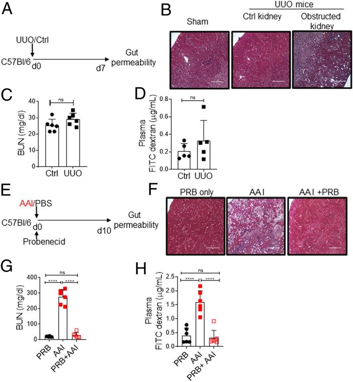
18    GUT-DERIVED FUNGUS CAUSES SYSTEMIC INFECTION IN UREMIA                                                                 ImmunoHorizons

FITC–dextran assay for intestinal permeability                           rRNA followed by sequencing on an Illumina MiSeq. Sequences
Food was withheld from mice for 4 h prior to FITC–dextran                from the Illumina MiSeq were deconvolved and then processed
gavage. FITC–dextran (4 kDa; Sigma) was resuspended in PBS at a          through the Center for Medicine and the Microbiome in-house
concentration of 100 mg/ml and orally gavaged to each mouse at           sequence quality control pipeline, which includes dust low
a dose of 40 mg/100 g body weight. Four hours later, mice were           complexity filtering, quality value trimming, trimming of primers
euthanized, and blood was collected immediately by retro-orbital         used for 16S rRNA gene amplification, and minimum read length
bleeding in tubes containing anticoagulant. Plasma was isolated          filtering. Forward and reverse reads were merged into contigs
from blood samples. One hundred microliters of plasma was added          and then processed through the Center for Medicine and the
to a black 96-well microplate in duplicate. The concentration of         Microbiome’s Mothur-based 16S clustering and sequence anno-
FITC in plasma was determined by spectrophotometry fluorom-               tation pipeline. Sequence taxonomic classifications was per-
etry with an excitation of 485 nm (20 nm bandwidth) and an               formed with the Ribosomal Database Project and Naive Bayesian
emission wavelength of 528 nm (20 nm bandwidth) using standard           classifier with the Silva reference database. Microbiota profiles
serially diluted FITC–dextran. Plasma from mice gavaged with PBS         were statistically quantified and analyzed using three distinct
was used to determine background.                                        methods at the genus taxonomic level: distance-based methods
                                                                         (intersample difference) used the Manhattan distance metric,

                                                                                                                                                   Downloaded from http://www.immunohorizons.org/ by guest on January 25, 2021
Analysis of bacterial or C. albicans translocation                       distribution-based methods (e.g., diversity) used the Tail statistic
Mice were euthanized, and aseptically collected liver and spleen         and Shannon index, and abundance-based methods used the
samples were homogenized in PBS. Tissue homogenates were                 additive-log ratio transformation. Sample time points (baseline
plated on brain heart infusion agar and YPD agar plates analysis of      and 10 d post–AAI injection) from the same subject were paired,
bacterial and fungal translocation, respectively.                        and linear models were fit with the paired differences as the
                                                                         response (paired analyses).
Preparation of tissue cell suspensions and flow
cytometric analysis                                                      Histology
Spleen and mesenteric lymph nodes (MLN) were harvested from              The terminal ileum and colon tissues was fixed with 10% buffered
mice and subjected to mechanical dissociation to prepare single          formaldehyde and embedded in paraffin. Slices 4-mm thick were
cell suspensions, followed by RBC lysis by ammonium–chloride–            made, stained with H&E, and then observed by a pathologist
potassium lysing buffer (Thermo Fisher Scientific). Small intestinal       blinded to this study design with light microscopy.
lamina propria (SILP) leukocytes were isolated. Briefly, Peyer’s
patches were carefully excised from a 10-cm piece of a terminal part     Immunofluorescence staining
of small intestine (SI). Intestinal tissues were opened longitudinally   Sections from frozen SI and colon were fixed in 4% para-
and washed with HBSS to remove luminal contents. Tissues were            formaldehyde and stained with ZO-1 primary Ab (Invitrogen).
incubated with 20 ml of HBSS containing 5 mM EDTA for 20 min at          The secondary Ab used was goat anti-rabbit Cy3. Sections were
37°C in a shaking incubator to remove epithelial cells. Tissues were     subsequently stained with DAPI nuclear stain. Images were
cut into small pieces and incubated in 10 ml of RPMI 1640                acquired with EVOS FL Auto microscope (Life Technologies).
containing 0.3 mg/ml collagenase D and 0.1 mg/ml DNase I for
20 min at 37°C in a shaking water bath. Then, 10% FBS was added to       Statistical analysis
stop the activity of digesting enzymes, and the tissue suspension        All data are expressed as mean 6 SD. Statistical analyses were
was passed through a 70-microns cell strainer. Cell suspension was       performed using the ANOVA, Mann–Whitney, or unpaired Student
washed twice with RPMI 1640 containing 10% FBS and passed                t test through GraphPad Prism 7 program: *p , 0.05, **p , 0.01,
through a 40-mm cell strainer for removal of tissue debris.              ***p , 0.001, and ****p , 0.0001. All experiments were performed
                                                                         a minimum of twice in independently performed replicates.
RNA extraction and real-time PCR
RNA was extracted from intestinal tissues using RNeasy kits
(QIAGEN, Valencia, CA). cDNA was synthesized by Super-                   RESULTS
Script III First Strand Kits (Thermo Fisher Scientific,
Waltham, MA). Quantitative real-time PCR was performed                   Uremic mice show increased gut permeability
with the PerfeCTa SYBR Green FastMix (Quanta BioSciences,                Using a mouse model of AAI nephropathy (AAN) (26), we tested
Beverly, MA) and analyzed on an ABI 7300 real-time in-                   whether uremia negatively impacts gut homeostasis and causes
strument. Primers were from obtained QuantiTect Primer                   disseminated candidiasis. In this study, mice injected with AAI
Assays (QIAGEN). The expression of each gene was normal-                 show kidney dysfunction and uremia (Fig. 1A). Control animals
ized to that of GAPDH.                                                   received PBS. AAII was used as a nonnephrotoxic control. We first
                                                                         measured the impact of uremia on intestinal barrier function in
Gut microbiome sequencing and analysis                                   mice with AAN. Uremic mice showed a 10-fold increase in the gut
Fecal DNA was isolated using QIAamp stool DNA extraction kit.            barrier permeability than controls (Fig. 1B). The increase in barrier
Microbial community analysis used PCR amplification of the of 16S         permeability was highest around 6–10 d post–AAI injection, the

                                                                                                  https://doi.org/10.4049/immunohorizons.2000114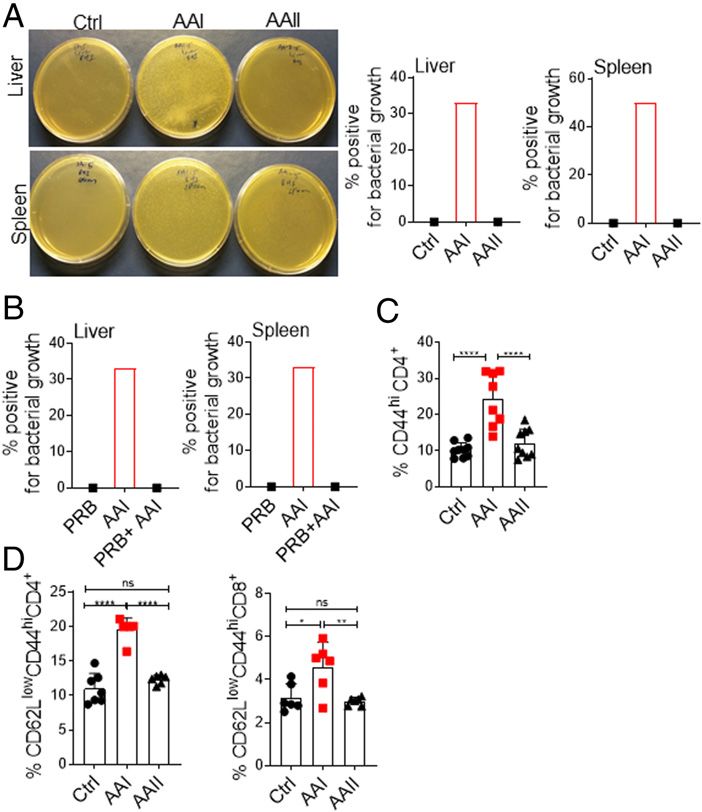
ImmunoHorizons                                                           GUT-DERIVED FUNGUS CAUSES SYSTEMIC INFECTION IN UREMIA                19

                                                                               To test this issue, we adapted an UUO model of renal fibrosis, in
                                                                               which one of the ureters is ligated below the kidney, whereas the
                                                                               contralateral ureter is left intact, allowing for normal function by
                                                                               the nonligated kidney (28). Consequently, UUO causes unilateral
                                                                               kidney damage but not uremia (Fig. 2A–C). Mice with UUO
                                                                               showed no changes in the gut barrier permeability in comparison
                                                                               with non-UUO control (Fig. 2D), indicating that the increase in gut
                                                                               permeability is due to uremia and not kidney damage per se.
                                                                               Moreover, to determine whether increased permeability is due to a
                                                                               direct effect of AAI on the intestinal epithelial cells, AAI-injected
                                                                               mice were treated with probenecid, an organic anion transporter
                                                                               inhibitor that prevents kidney dysfunction and uremia without
                                                                               neutralizing AAI (29) (Fig. 2E–G). Uremic mice treated with

                                                                                                                                                         Downloaded from http://www.immunohorizons.org/ by guest on January 25, 2021
FIGURE 1. Increased gut barrier permeability in AAI-injected mice.
C57BL/6 (WT) mice (n = 6–8) were either injected with AAI, PBS
(control), or AAII. (A) Serum BUN and creatinine levels (n = 5) were
measured at day 10 post–AAI injection. At (B) day 10 (n = 6–8) and (C)
indicated time points post–AAI injection (n = 3–6), mice were gavaged
with FITC–dextran and assessed for FITC–dextran concentration in the
plasma. (D) Correlation between gut barrier permeability and BUN level
(n = 8). (E) SI and colon sections were stained for ZO-1 expression. (F)
Transcript expression of tight junction protein genes were evaluated by
quantitative real-time PCR (n = 6–7). (G) H&E staining of SI and colon of
uremic and control mice. Images from one of three mice/group for (E
and G). Original magnification 3200. Data pooled from at least two
independent experiments for (A–D and F) and expressed as mean 6 SD             FIGURE 2. Uremia drives increased gut permeability.
(A, B, C, and F). Statistical analyses by Pearson correlation (D) and one-     (A) Schematic diagram of the experimental design. WT mice (n = 5–6)
way ANOVA (A–C and F). **p , 0.01, ***p , 0.001, ****p , 0.0001.               were subjected to UUO. At day 7 postsurgery, UUO and non-UUO
                                                                               control mice were gavaged with FITC–dextran and assessed for barrier
                                                                               permeability. (B) Kidney histopathology following Masson trichome
time point at which BUN level peaked in the uremic group, as                   staining, (C) serum BUN level, and (D) plasma FITC–dextran concen-
shown before (26) (Fig. 1C, 1D). Consequently, uremic animals                  tration were measured at day 7 postsurgery. (E) Schematic diagram of
showed a reduced expression of ZO-1 tight junction protein in the              the experimental design. Groups of uremic mice (n = 6–7) were either
epithelial lining of the SI and colon (Fig. 1E). There was an increase         treated with probenecid (AAI+PRB) or left untreated (AAI). Control mice
in the expression of a few tight junction protein genes (ZO1,                  received probenecid only (PRB only). Mice were evaluated for (F) kidney
occludin, and claudin 4), signifying the onset of repair responses in          fibrosis, (G) serum BUN level, and (H) gut barrier permeability. Images
dysfunctional gut epithelium, as previously described (27) (Fig. 1F).          from one of three mice/group for (B and F). Original magnification
The loss of barrier function in uremia was not due to mucosal                  3200. The data are pooled from at least two independent experiments
damage of the SI and colon (Fig. 1G).                                          for (C, D, G, and H) and expressed as mean 6 SD (C, D, G, and H).
    We next determined whether kidney damage is responsible for                Statistical analyses by Student t test (C and D) and one-way ANOVA (G
the alteration in gut barrier permeability independent of uremia.              and H). ns, statistically not significant. ****p , 0.0001.

https://doi.org/10.4049/immunohorizons.2000114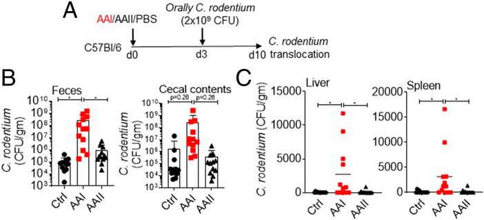
20     GUT-DERIVED FUNGUS CAUSES SYSTEMIC INFECTION IN UREMIA                                                                    ImmunoHorizons

                                                                             Uremia causes alterations in the mucosal defense and
                                                                             composition of gut microbiota
                                                                             The innate and adaptive immune cells have a distinct yet
                                                                             complementary role in maintaining gut homeostasis and mucosal
                                                                             immune defense (30). In the SILP, the percentages of macrophage
                                                                             and dendritic cell subsets were comparable between the uremic
                                                                             and control mice (Fig. 3A, 3B). Strikingly, we observed a reduction
                                                                             in the percentage of neutrophils in the SILP of uremic mice rather
                                                                             than in controls (Fig. 3C). Uremic mice also demonstrated a
                                                                             reduction in the percentage of Th17 but not Th1 cells (Fig. 3D).
                                                                             Markedly, the percentage of IgA+CD11b+ plasmablasts in the SILP
                                                                             of uremic mice was reduced compared to the percentage in
                                                                             control animals (Fig. 3E). There was a trend toward diminished
                                                                             IgA+CD11b2 plasmablasts in uremia, although the difference be-
                                                                             tween the groups did not achieve statistical significance. How-

                                                                                                                                                       Downloaded from http://www.immunohorizons.org/ by guest on January 25, 2021
                                                                             ever, the frequency of regulatory T (Treg) cells was similar
                                                                             between the groups (Fig. 3F). We observed no difference in the
                                                                             frequencies of Th17, Th1, and Treg cells in the MLN between
                                                                             uremic and nonuremic groups (Fig. 3G, 3H). These results
                                                                             indicate that uremia negatively impacts the number of neutro-
                                                                             phils, Th17, and IgA-producing plasmablasts in the SILP.
                                                                                 Next, we assessed the relative change in the diversity and
                                                                             composition of gut microbiota in uremic and control animals
                                                                             between days 0 (baseline) and 10 post–AAI injection. There was no
                                                                             significant difference in the diversity of microbiota between
                                                                             uremic and control groups at day 10 post–AAI injection (Fig. 3I).
                                                                             Both control and AAII-injected animals demonstrated altered
                                                                             microbiota composition between days 0 and 10. Interestingly,
                                                                             uremic mice showed significantly greater change (p = 0.0094) in
                                                                             the overall microbiota composition than control animals at day 10
FIGURE 3. Compromised gut mucosal immunity and dysbiosis in                  postinjection. Whereas the control group showed a reduction
uremia.                                                                      in the abundance of Tannerellaceae and Lactobacillus, uremic
At day 10 post–AAI injection, SILP (n = 5–19) were evaluated for the fre-    animals exhibited an increase in the Tannerellaceae. Collectively,
quency of (A) macrophages (liveCD45+CD11b+ F4/80+CX3CR1+CD11c+;              these results show a modest impact of uremia on the composition
liveCD45+CD11b+F4/80+CX3CR1+CD11c2), (B) dendritic cells (liveCD45+          of gut microbiota in uremic mice.
CD11b+CD103+CD11c+, liveCD45+CD11b2CD103+CD11c+; liveCD45+
CD11b+ CD1032CD11c+), (C) neutrophils (liveCD45+CD11b+Ly6G+), (D)            Uremic mice exhibit translocation of gut microbiota and
               +     +      +                    +     +      +
Th17 (liveCD45 CD4 IL-17 ) and Th1 (liveCD45 CD4 IFN-g ), (E) IgA-           C. rodentium
producing plasmablasts (liveCD45+CD11b+IgA+; liveCD45+CD11b2IgA+),           We assessed whether the uremia-driven disturbance in gut
and (F) Treg cells (liveCD4+Foxp3+) by FACS at day 10 post–AAI injection.    hemostasis can drive the translocation of gut microbiota. Although
(G) Percentages of Th17 and Th1 cells in the MLN (n = 8–12) were de-         we were unable to detect any bacterial colonies in the control or
termined by intracellular cytokine staining following in vitro stimulation   AAII mice, 30–50% of uremic animals showed translocation of
with PMA/ionomycin. (H) Frequency of Treg cells was determined in the        microbiota in the liver and spleen at day 10 post–AAI injection (Fig.
MLN by FACS. (I) At day 10 post–AAI injection, fecal pellets from uremic     4A). Moreover, probenecid-treated uremic mice did not show any
and control (n = 5) mice were subjected to targeted 16S rRNA se-             bacterial colony in the liver and spleen (Fig. 4B). The translocation
quencing. Data pooled from at least two independent experiments for          of microbiota caused the activation of T cells in the MLN and
(A–H) and expressed as mean 6 SD (A–H). Statistical analyses by one-way      spleen of uremic mice, as shown before (Fig. 4C, 4D) (12).
ANOVA (A–H) and pair-wise using Wilcoxon rank sum test (I). *p , 0.05,           We next evaluated the ability of harmful bacteria C. rodentium
**p , 0.01, ***p , 0.001.                                                    to cause systemic infection in uremic animals (Fig. 5A). C.
                                                                             rodentium is a murine intestinal pathogen that is closely related
                                                                             to human pathogens, including enteropathogenic Escherichia coli
probenecid showed reduced FITC–dextran in the plasma, in-                    and enterohemorrhagic E. coli (31). Following oral infection, we
dicating that AAI does not exert any direct effect on the barrier             observed more C. rodentium in the fecal pellet and cecal contents
permeability (Fig. 2H). These data suggest that uremia drives                of uremic mice (Fig. 5B). Few control mice exhibited very low
increased barrier permeability in mice with kidney disease (12).             colony counts of C. rodentium in the liver and spleen, as shown

                                                                                                      https://doi.org/10.4049/immunohorizons.2000114
ImmunoHorizons GUT-DERIVED FUNGUS CAUSES SYSTEMIC INFECTION IN UREMIA 21
treated uremic and control mice with oral antibiotic to aid in robust
colonization (34) (Fig. 6D, 6E). However, uremic animals showed
no fungal dissemination in the organs (Fig. 6F). Thus, unlike C.
rodentium, C. albicans cannot translocate from the gut in uremia.
Most kidney disease patients suffer from some form of GI
manifestations, in which the gut mucosa is damaged (17). These
include perforation of intestine, intestinal ulcer, uremic colitis,
ischemic colitis, diverticular disease, and intestinal hemorrhage.
Hence, we tested whether C. albicans can cause systemic infection
when the gut mucosa is disrupted in uremia. Accordingly, uremic
and control mice were fed with antibiotic to favor colonization
followed by oral gavage with 2.5% DSS to damage the epithelial
lining (23) (Fig. 6G). AAI-injected mice treated with DSS showed
reduced survival (30%) in comparison with AAI only, control only,
and control + DSS animals (100%) (Fig. 6H). Control animals with
Downloaded from http://www.immunohorizons.org/ by guest on January 25, 2021
DSS showed no fungal dissemination in the liver, as shown before
(23) (Fig. 6I). There was no fungal translocation in uremic mice
not receiving DSS. Interestingly, 65% of the uremic animals fed
with DSS showed fungal translocation in the liver. These data
indicate that C. albicans can translocate and cause systemic
infection only when the mucosal barrier is damaged in the
majority of uremic animals. Thus, it is reasonable to speculate that
uremic individuals with GI manifestations may have a higher risk
of systemic infection by gut-derived C. albicans.
FIGURE 4. Uremic mice exhibit translocation of gut microbiota.
(A) Mice (n = 6) were subjected to AAN and evaluated for the trans- DISCUSSION
location of gut microbiota in the liver and spleen at day 10 post–AAI
injection. Images from one of six mice/group. (B) Uremic mice (n = 6) Prevention of infection is one of the few avenues to reduce
were either treated with probenecid (AAI+PRB) or left untreated (AAI) hospitalizations, control costs, and improve quality of life for
and assessed for microbiota translocation in the liver and spleen. patients with kidney disease. In the past decade, technological
Uremic and control groups (n = 6–9) were evaluated for the activation advances in the dialysis procedure have lowered the incidence of
of T cells in the (C) MLN (liveCD4+CD44hi) and (D) spleen (live- access-related infections. However, it has not been successful in
CD4+CD62LloCD44hi and liveCD8+CD62LloCD44hi) by FACS. Pooled preventing infection-related hospitalizations in these patients (2,
data from at least two experiments for (A–D) and expressed as mean 6 SD 3). This is partly due to an alarming rise in the prevalence of
(C and D). Statistical analyses by one-way ANOVA (C and D). *p , 0.05, previously unappreciated non–access-related infections. Currently,
**p , 0.01, ****p , 0.0001.
before (32) (Fig. 5C). In contrast, 60–70% of the uremic animals
showed higher C. rodentium burden in the liver and spleen. Our
data suggest that gut microbiota and C. rodentium can translocate
from the gut and cause systemic infection in uremia.
Uremia and mucosal damage are required for the
translocation of C. albicans
The role of gut-derived C. albicans in non–access-related infections
in kidney disease is unknown. Based on our data (Figs. 4, 5), we
hypothesize that C. albicans could translocate and cause disseminated FIGURE 5. Uremic mice exhibit translocation of C. rodentium.
candidiasis in uremia. C. albicans is not a commensal fungus in (A) Schematic diagram of the experimental design. AAI, control, and AAII-
mice. Hence, colonization of the fungus requires alteration in gut injected mice (n = 12) were gavaged with C. rodentium at day 3 post–AAI
homeostasis (33). Following oral gavage, uremic mice showed a injection. At day 7, C. rodentium burden in the (B) fecal pellet and cecal
modest increase in fungal colonization in the gut at day 7 postinjection content and (C) liver and spleen were measured. Pooled data from at
(Fig. 6A, 6B). We were unable to detect any fungal colony in the liver least two experiments for (B and C) and expressed as mean 6 SD (B and
and spleen of uremic animals at this time point (Fig. 6C). We also C). Statistical analyses by one-way ANOVA (B and C). *p , 0.05.
https://doi.org/10.4049/immunohorizons.200011422 GUT-DERIVED FUNGUS CAUSES SYSTEMIC INFECTION IN UREMIA ImmunoHorizons
Downloaded from http://www.immunohorizons.org/ by guest on January 25, 2021
FIGURE 6. Uremic mice show fungal translocation following DSS treatment.
(A) Schematic representation of the experimental design. Mice (n = 10–15) were gavaged with C. albicans at day 2 post–AAI injection. At day 8,
C. albicans and microbiota burden in the (B) fecal pellet and (C) liver and spleen were evaluated. (D) Schematic diagram of the experimental plan.
Mice (n = 6–13) were fed with ampicillin in the drinking water throughout the experiment. At day 10 post–oral fungal infection, mice were either injected
with AAI or PBS. Fungal and bacterial CFU in the (E) fecal pellet and (F) liver and spleen were determined. (G) Schematic representation of the experimental
plan. Oral antibiotic–treated animals were fed with 2.5% DSS in water 3 d after AAI injection. (H) Survival (n = 4–8) was evaluated for 9 d post–AAI
injection. (I) Mice were evaluated for the translocation of C. albicans in the liver. Pooled data from at least two experiments for (B, C, E, F, H, and I) and
expressed as mean 6 SD (B and E). Statistical analyses by one-way ANOVA (B), Mann–Whitney t test (E), and log-rank test (H). *p , 0.05, **p , 0.01.
our knowledge about the risk and source of non–access-related systemic infection when uremia is coupled with mucosal damage.
infections in patients with kidney disease is surprisingly rudimen- These results identify gut-derived commensal C. albicans as a source
tary. Using a clinically relevant mouse model of kidney disease and of non–access-related systemic infection in patients with kidney
associated uremia, we show that gut commensal C. albicans causes disease and showing GI damage.
https://doi.org/10.4049/immunohorizons.2000114ImmunoHorizons GUT-DERIVED FUNGUS CAUSES SYSTEMIC INFECTION IN UREMIA 23
Our data show that the loss of barrier function in uremic mice is a higher risk of systemic infection by commensal C. albicans. To
due to an increase in the gut barrier permeability and mucosal date, no studies have looked at the prevalence of disseminated
damage. The level of uremia correlates with the loss of barrier candidiasis in uremic patients with GI manifestations. Addition-
function, indicating that uremia is directly implicated in the ally, these findings have compelling implications in the risk
increased gut permeability. Although our study provided evidence stratification and clinical management of infection control and
of leaky gut in 100% of the uremic mice, only 30–50% of the prevention in patients with kidney disease.
animals consistently showed translocation of microbiota. These
data imply that factors other than an increase in gut permeability
DISCLOSURES
play a major role in bacterial translocation in uremia. Moreover,
when microbiota and C. rodentium showed translocation,
The authors have no financial conflicts of interest.
C. albicans failed to do so. The fungal yeast (2–10 mm) and hyphae
(.10 mm) are bigger in size than bacteria (0.5–5 mm), which may
act as an impediment for the fungal yeast and hyphal form to pass ACKNOWLEDGMENTS
through the tight junctions of GI epithelial cells. Additionally,
control or uremic mice do not show any fungal translocation We thank Drs. Sarah Gaffen, Tim Hand, and Mandy McGeachy for
Downloaded from http://www.immunohorizons.org/ by guest on January 25, 2021
without mucosal damage. These data argue against the fact that suggestions.
C. albicans can translocate by causing damage to GI epithelial cells
as proposed by others (35).
REFERENCES
Dysbiosis observed in this study may be due to iatrogenic
causes or uremia per se. Loss of kidney function leads to diffusion 1. Nugent, R. A., S. F. Fathima, A. B. Feigl, and D. Chyung. 2011. The
of urea in the GI tract. Subsequent hydrolysis of urea by urease burden of chronic kidney disease on developing nations: a 21st cen-
expressed by some gut microbes results in the formation of large tury challenge in global health. Nephron Clin. Pract. 118: c269–c277.
quantities of ammonia, which could affect the growth of 2. Sarnak, M. J., and B. L. Jaber. 2000. Mortality caused by sepsis in
patients with end-stage renal disease compared with the general
commensal bacteria (16, 19). We observed a modest change in the
population. Kidney Int. 58: 1758–1764.
gut microbiota in our mouse model of AAN. This is in contrast to 3. Dalrymple, L. S., K. L. Johansen, G. M. Chertow, S. C. Cheng,
chronic kidney disease patients, in which a markedly altered B. Grimes, E. B. Gold, and G. A. Kaysen. 2010. Infection-related
change in terms of quantity and quality of microbiota is evident (16, hospitalizations in older patients with ESRD. Am. J. Kidney Dis. 56:
19). There may be several reasons for this discrepancy. First, AAN is 522–530.
an acute kidney injury model, in which the gut bacteria are exposed 4. Collins, A. J., R. N. Foley, C. Herzog, B. M. Chavers, D. Gilbertson,
A. Ishani, B. L. Kasiske, J. Liu, L.-W. Mau, M. McBean, et al. 2010.
to uremic toxins for a relatively short period of time (i.e., 7–10 d).
Excerpts from the US renal data system 2009 annual data report. Am.
Second, mice and human microbiota differ considerably, and their J. Kidney Dis. 55: S1–S420, A6–A7.
susceptibility to uremia may reflect the difference in alterations in 5. Allon, M., T. A. Depner, M. Radeva, J. Bailey, S. Beddhu, D. Butterly,
gut bacterial content (36). Finally, uremic toxins differ between D. W. Coyne, J. J. Gassman, A. M. Kaufman, G. A. Kaysen, et al;
mice and humans, making it difficult to compare their impact on the HEMO Study Group. 2003. Impact of dialysis dose and membrane on
infection-related hospitalization and death: results of the HEMO
gut microbiota (37).
Study. J. Am. Soc. Nephrol. 14: 1863–1870.
We observed a decrease in the percentages of innate and 6. Pastan, S., J. M. Soucie, and W. M. McClellan. 2002. Vascular access
adaptive immune cells in the SILP of uremic mice. Uremia- and increased risk of death among hemodialysis patients. Kidney Int.
induced neutrophil apoptosis may account for the reduced 62: 620–626.
number of neutrophils in the uremic gut (38). This observation 7. Vaziri, N. D., M. V. Pahl, A. Crum, and K. Norris. 2012. Effect of
is in line with a previous report showing that immunosuppressive uremia on structure and function of immune system. J. Ren. Nutr. 22:
149–156.
treatment–induced neutropenia is required for the fungus to
8. Syed-Ahmed, M., and M. Narayanan. 2019. Immune dysfunction and
translocate and cause systemic infection in nonuremic animals risk of infection in chronic kidney disease. Adv. Chronic Kidney Dis.
(23). Additionally, IgA-producing plasmablast deficiency in 26: 8–15.
uremic mice can be simultaneously mediated by increased B cell 9. Dobre, M., T. W. Meyer, and T. H. Hostetter. 2013. Searching for
apoptosis and reduced expression of BAFF-R, as demonstrated uremic toxins. Clin. J. Am. Soc. Nephrol. 8: 322–327.
before (39). The role of IgA in C. albicans colonization and 10. Cohen, G., and W. H. Hörl. 2012. Immune dysfunction in uremia—an
update. Toxins (Basel) 4: 962–990.
translocation is poorly understood. Interestingly, we did not see
11. Andersen, K., M. S. Kesper, J. A. Marschner, L. Konrad, M. Ryu,
any change in Treg cells in the gut, a hallmark of chronic kidney S. Kumar Vr, O. P. Kulkarni, S. R. Mulay, S. Romoli, J. Demleitner,
disease patients (40). et al. 2017. Intestinal dysbiosis, barrier dysfunction, and bacterial
The GI symptoms are reported in up to 80% of kidney disease translocation account for CKD-related systemic inflammation. J. Am.
patients (17). Some of these symptoms, including intestinal Soc. Nephrol. 28: 76–83.
12. Anders, H. J., K. Andersen, and B. Stecher. 2013. The intestinal
necrosis, spontaneous colonic perforation, uremic colitis, gastric
microbiota, a leaky gut, and abnormal immunity in kidney disease.
ulcer, GI hemorrhage, and acute diverticulitis, result in the damage Kidney Int. 83: 1010–1016.
of mucosa and development of sepsis. Thus, it is reasonable to 13. Amato, K. R., R. Martinez-Mota, N. Righini, M. Raguet-Schofield, F. P.
speculate that uremic individuals with GI manifestations may have Corcione, E. Marini, G. Humphrey, G. Gogul, J. Gaffney, E. Lovelace,
https://doi.org/10.4049/immunohorizons.200011424 GUT-DERIVED FUNGUS CAUSES SYSTEMIC INFECTION IN UREMIA ImmunoHorizons
et al. 2016. Phylogenetic and ecological factors impact the gut 28. Ramani, K., R. J. Tan, D. Zhou, B. M. Coleman, C. V. Jawale, Y. Liu, and
microbiota of two neotropical primate species. Oecologia 180: 717–733. P. S. Biswas. 2018. IL-17 receptor signaling negatively regulates the
14. Rooks, M. G., and W. S. Garrett. 2016. Gut microbiota, metabolites and development of tubulointerstitial fibrosis in the kidney. Mediators
host immunity. Nat. Rev. Immunol. 16: 341–352. Inflamm. 2018: 5103672.
15. Tilg, H., N. Zmora, T. E. Adolph, and E. Elinav. 2020. The intestinal 29. Baudoux, T. E., A. A. Pozdzik, V. M. Arlt, E. G. De Prez, M. H.
microbiota fuelling metabolic inflammation. Nat. Rev. Immunol. 20: Antoine, N. Quellard, J. M. Goujon, and J. L. Nortier. 2012. Pro-
40–54. benecid prevents acute tubular necrosis in a mouse model of aristo-
16. Meijers, B., P. Evenepoel, and H. J. Anders. 2019. Intestinal micro- lochic acid nephropathy. Kidney Int. 82: 1105–1113.
biome and fitness in kidney disease. Nat. Rev. Nephrol. 15: 531–545. 30. Belkaid, Y., and T. W. Hand. 2014. Role of the microbiota in immunity
17. Ala-Kaila, K., and A. Pasternack. 1989. Gastrointestinal complications and inflammation. Cell 157: 121–141.
in chronic renal failure. Dig. Dis. 7: 230–242. 31. Bouladoux, N., O. J. Harrison, and Y. Belkaid. 2017. The mouse model
18. Carrera-Jiménez, D., P. Miranda-Alatriste, X. Atilano-Carsi, R. Correa- of infection with Citrobacter rodentium. Curr. Protoc. Immunol. 119:
Rotter, and Á. Espinosa-Cuevas. 2018. Relationship between nutritional 19.15.1–19.15.25.
status and gastrointestinal symptoms in geriatric patients with end-stage 32. Vallance, B. A., W. Deng, K. Jacobson, and B. B. Finlay. 2003. Host
renal disease on dialysis. Nutrients 10: 425. susceptibility to the attaching and effacing bacterial pathogen Cit-
robacter rodentium. Infect. Immun. 71: 3443–3453.
19. Ramezani, A., and D. S. Raj. 2014. The gut microbiome, kidney disease,
33. Koh, A. Y. 2013. Murine models of Candida gastrointestinal coloni-
and targeted interventions. J. Am. Soc. Nephrol. 25: 657–670.
zation and dissemination. Eukaryot. Cell 12: 1416–1422.
Downloaded from http://www.immunohorizons.org/ by guest on January 25, 2021
20. Lionakis, M. S., I. D. Iliev, and T. M. Hohl. 2017. Immunity against
34. Shao, T. Y., W. X. G. Ang, T. T. Jiang, F. S. Huang, H. Andersen, J. M.
fungi. JCI Insight 2: e93156.
Kinder, G. Pham, A. R. Burg, B. Ruff, T. Gonzalez, et al. 2019. Com-
21. Gandhi, B. V., M. M. Bahadur, H. Dodeja, V. Aggrwal, A. Thamba, and
mensal Candida albicans positively calibrates systemic Th17 immu-
M. Mali. 2005. Systemic fungal infections in renal diseases. J. Post-
nological responses. Cell Host Microbe 25: 404–417.e6.
grad. Med. 51(Suppl. 1): S30–S36.
35. Allert, S., T. M. Förster, C.-M. Svensson, J. P. Richardson, T. Pawlik,
22. Limon, J. J., J. H. Skalski, and D. M. Underhill. 2017. Commensal B. Hebecker, S. Rudolphi, M. Juraschitz, M. Schaller, M. Blagojevic,
fungi in health and disease. Cell Host Microbe 22: 156–165. et al. 2018. Candida albicans-induced epithelial damage mediates
23. Koh, A. Y., J. R. Köhler, K. T. Coggshall, N. Van Rooijen, and G. B. translocation through intestinal barriers. mBio 9: e00915-18.
Pier. 2008. Mucosal damage and neutropenia are required for Candida 36. Hugenholtz, F., and W. M. de Vos. 2018. Mouse models for human
albicans dissemination. PLoS Pathog. 4: e35. intestinal microbiota research: a critical evaluation. Cell. Mol. Life Sci.
24. Kobayashi-Sakamoto, M., R. Tamai, E. Isogai, and Y. Kiyoura. 2018. 75: 149–160.
Gastrointestinal colonisation and systemic spread of Candida albicans 37. Itoh, Y., A. Ezawa, K. Kikuchi, Y. Tsuruta, and T. Niwa. 2013. Cor-
in mice treated with antibiotics and prednisolone. Microb. Pathog. 117: relation between serum levels of protein-bound uremic toxins in
191–199. hemodialysis patients measured by LC/MS/MS. Mass Spectrom.
25. Fan, D., L. A. Coughlin, M. M. Neubauer, J. Kim, M. S. Kim, X. Zhan, (Tokyo) 2: S0017.
T. R. Simms-Waldrip, Y. Xie, L. V. Hooper, and A. Y. Koh. 2015. 38. Majewska, E., Z. Baj, Z. Sulowska, J. Rysz, and M. Luciak. 2003.
Activation of HIF-1a and LL-37 by commensal bacteria inhibits Effects of uraemia and haemodialysis on neutrophil apoptosis and
Candida albicans colonization. Nat. Med. 21: 808–814. expression of apoptosis-related proteins. Nephrol. Dial. Transplant. 18:
26. Jawale, C. V., K. Ramani, D.-D. Li, B. M. Coleman, R. S. Oberoi, 2582–2588.
S. Kupul, L. Lin, J. V. Desai, G. M. Delgoffe, M. S. Lionakis, et al. 2020. 39. Pahl, M. V., S. Gollapudi, L. Sepassi, P. Gollapudi, R. Elahimehr, and
Restoring glucose uptake rescues neutrophil dysfunction and protects N. D. Vaziri. 2010. Effect of end-stage renal disease on B-lymphocyte
against systemic fungal infection in mouse models of kidney disease. subpopulations, IL-7, BAFF and BAFF receptor expression. Nephrol.
Sci. Transl. Med. 12: eaay5691. Dial. Transplant. 25: 205–212.
27. Kigerl, K. A., J. C. Hall, L. Wang, X. Mo, Z. Yu, and P. G. Popovich. 40. Meier, P. 2009. FOXP3+ regulatory T-cells in chronic kidney disease:
2016. Gut dysbiosis impairs recovery after spinal cord injury. J. Exp. molecular pathways and clinical implications. Adv. Exp. Med. Biol.
Med. 213: 2603–2620. 665: 163–170.
https://doi.org/10.4049/immunohorizons.2000114You can also read