Sacrococcygeal Teratoma : A Tumor at the Center of Embryogenesis - KoreaScience
←
→
Page content transcription
If your browser does not render page correctly, please read the page content below
Review Article
J Korean Neurosurg Soc 64 (3) : 406-413, 2021
https://doi.org/10.3340/jkns.2021.0015                                                                                        pISSN 2005-3711 eISSN 1598-7876

Sacrococcygeal Teratoma : A Tumor at the Center of
Embryogenesis
Ji Hoon Phi
Division of Pediatric Neurosurgery, Seoul National University Children’s Hospital, Seoul National University College of Medicine, Seoul, Korea

Sacrococcygeal teratoma (SCT) is an extragonadal germ cell tumor (GCT) that develops in the fetal and neonatal periods. SCT is a
type I GCT in which only teratoma and yolk sac tumors arise from extragonadal sites. SCT is the most common type I GCT and is
believed to originate through epigenetic reprogramming of early primordial germ cells migrating from the yolk sac to the gonadal
ridges. Fetal SCT diagnosed in utero presents many obstetrical problems. For high-risk fetuses, fetal interventions (devascularization
and debulking) are under development. Most patients with SCT are operated on after birth. Complete surgical resection is the
key for tumor control, and the anatomical location of the tumor determines the surgical approaches. Incomplete resection and
malignant histology are risk factors for recurrence. Approximately 10–15% of patients have a tumor recurrence, which is frequently
of malignant histology. Long-term surveillance with monitoring of serum alpha fetoprotein and magnetic resonance imaging is
required. Survivors of SCT may suffer anorectal, urological, and sexual sequelae later in their life, and comprehensive evaluation and
care are required.

Key Words : Teratoma, Sacrococcygeal · Germ cell tumor · Fetus · Surgery.

INTRODUCTION                                                                            tumor (YST) is possible. Patients with SCT are also commonly
                                                                                        plagued with many gastrointestinal, urological, and neurolog-
   Sacrococcygeal teratoma (SCT) is a rare neoplasm of early                            ical abnormalities. Despite the huge impact of this tumor on
infancy. SCT is a congenital tumor usually diagnosed in utero                           the quality of life of affected children, many of clinically im-
or during the neonatal period. Histologically, the majority of                          portant questions on SCT are currently difficult to answer.
SCTs are mature teratomas, one of the subtypes of germ cell                             The major difficulties of clinicians dealing with SCT are the
tumors (GCTs). Despite its histological similarity, SCT is dif-                         extremely low incidence of the disease and the lack of clear,
ferent from GCTs developing in the gonads and other extrago-                            unified guidelines for its management. Regarding the patho-
nadal sites after puberty. SCT is a benign neoplasm, and sur-                           genesis, the occurrence of SCT in such an unusual location at
gery provides a cure for the disease. However, recurrence and                           such a young age has been an enigma for a long time. Discov-
malignant transformation into teratocarcinoma or yolk sac                               eries in developmental biology and molecular genetics have

• Received : January 20, 2021 • Revised : February 11, 2021 • Accepted : March 10, 2021
•A ddress for reprints : Ji Hoon Phi
  Division of Pediatric Neurosurgery, Seoul National University Children’s Hospital, Seoul National University College of Medicine, 101 Daehak-ro, Jongno-gu, Seoul 03080,
  Korea
  Tel : +82-2-2072-3639, Fax : +82-2-744-8459, E-mail : phijh@snu.ac.kr, ORCID : https://orcid.org/0000-0002-9603-5843

T his is an Open Access article distributed under the terms of the Creative Commons Attribution Non-Commercial License (http://creativecommons.org/licenses/by-nc/4.0)
which permits unrestricted non-commercial use, distribution, and reproduction in any medium, provided the original work is properly cited.

406       Copyright © 2021 The Korean Neurosurgical Society
Sacrococcygeal Teratoma | Phi JH

revealed the origin and pathogenesis of SCT to a great degree.        in the incidence of SCT. However, a nationwide study from
This review will focus on the current knowledge of the clinical       Finland showed a higher birth prevalence of SCT (1 in 10000),
aspects and biology of SCT from developmental perspectives.           supporting some genetic contribution to the etiology of
                                                                      SCT31).

EPIDEMIOLOGY
                                                                      PATHOGENESIS
   Approximately 90% of human GCTs develop in the gonads,
especially in the testis, but a few cases develop in extragonadal        The pathogenesis of SCT should be understood in the broad
sites such as the brain, neck, mediastinum, and retroperitone-        context of GCT development. All GCTs have been thought to
um. However, in neonates and young infants, the sacrococcy-           be derived from primitive germ cells (germ cell hypothesis)42).
geal area is the most common site for GCT development. SCT            The hypothesis explains the peculiar anatomical distribution
develops from the sacrum and coccyx, protruding outwards              of extragonadal GCTs along the midline of the body28). Pri-
and growing into the pelvic cavity. It is a rare disease with an      mordial germ cells (PGCs) migrate from the yolk sac to their
incidence of 1 in 20000–40000 live births4). There is a clear fe-     final destinations in the gonadal ridges by passing the hindgut
male predominance with a 3 : 1 to 4 : 1 female-to-male ratio35,39),   where SCT can arise22). However, research on embryos and
which is in contrast to the well-recognized male preponderance        embryonic stem (ES) cells revealed that teratomas could be
of adolescent GCTs (both gonadal and extragonadal). Testicu-          derived from ES cells, making ES cells candidate cells for de-
lar GCTs (TGCTs) have one of the highest familial risks among         veloping into SCT6). Stem cells residing in extraembryonic tis-
cancers : having a brother with TGCTs increases the risk              sues can also induce teratoma formation, adding other candi-
by 8–10 times and its presence in dizygotic or monozygotic            date cells to the possible origins of SCT41). The sacrococcygeal
twins elevates the risk by 37- or 76.5-fold16). Interestingly, most   region also corresponds to the primitive streak and Hensen’s
patients with SCT are sporadic, and no definite familial ten-         node in early embryonic development. If there are some resid-
dency exists, although a family history of twin gestation is ob-      ual totipotent stem cells from Hensen’s node, they can develop
served in 10–15% of patients4).                                       later into teratoma or YST, the two most common pathologies
   In a thorough examination, associated congenital anoma-            encountered in SCT13,14).
lies are found in 15–30% of patients with SCT17). The most               Genomic sequencing of gonadal and extragonadal GCTs re-
common anomalies are in the urogenital system, such as hy-            vealed that the majority of GCTs lack driver mutations for
dronephrosis, which can be considered as a consequence of             cancer initiation and progression. TGCTs are bona fide malig-
compression by the tumor mass. Although the proportion of             nancies, but they are among the human cancers with the least
congenital anomalies is higher than that of the normal popu-          mutational burden (0.3–0.5 mutation/Mb)19,40). TGCTs are di-
lation (3–4%), causative associations are rare except for the         vided into seminomas (germinomas in extragonadal sites)
Currarino triad. The Currarino triad is a very rare syndrome          and nonseminomas, including embryonal carcinoma (EC),
and consists of sacral bony defects, anorectal anomalies, and a       YST, choriocarcinoma, and teratoma. Mutations in KIT and
presacral mass15). Approximately 40% of the presacral masses          KIT signaling (KRAS, NRAS) are found in 20–60% of semi-
in the Currarino triad are benign teratomas15). The Currarino         nomas, but fewer mutations are observed in nonsemino-
triad is an autosomal dominant syndrome linked to muta-               mas19,40). TP53 mutations, a strong common driver of human
tions in the HLXB9 gene21).                                           cancers, are virtually not found in TGCTs. These findings in-
   GCTs are characterized by dramatic geographical differenc-         dicate that GCT is not a mutation-driven tumor but a devel-
es in incidence according to the tumor location. The incidence        opment-driven neoplasm.
of TGCTs in Europe is approximately 10 times that in East                Oosterhuis and Looijenga27) proposed a unifying classifica-
Asia8). Conversely, the incidence of intracranial GCT is five         tion scheme for all GCTs of the human body. They envisaged
times higher in East Asia than in western countries32). Cur-          GCTs as tumors derived through genomic reprogramming
rently, there is no evidence of such a geographical difference of     from their developmental cells of origin27). In their scheme,

                                                                                        J Korean Neurosurg Soc 64 (3) : 406-413   407
J Korean Neurosurg Soc 64 | May 2021 there are five types of GCTs28). Type I GCTs are infantile GCTs pletely erased in type II GCTs which develop from late PGCs arising exclusively from extragonadal sites, with the sacrococ- through reprogramming28). cygeal region as the most frequent location. Type I GCTs are Therefore, the germ cell hypothesis is correct because all characterized by a female preponderance and consist of tera- GCTs (type I to type V) develop from germ cells of different tomas and YSTs. Rarely, somatic malignancy develops from developmental statuses. Genetic polymorphisms, gains of ge- mature teratoma through transformation48). Type I GCTs are netic mutations, and epigenetic (or microenvironmental) believed to be derived from early PGCs during their migra- changes affect the fate of germ cells and induce reprogram- tion. Migrating PGCs depends on KIT- KITLG (KIT-ligand) ming to ES-like cells that lead to GTCs. signaling for their survival. If early PGCs are reprogrammed to attain pluripotency by an abnormal microenvironment and to become ES cells, they can survive without the KIT pathway CLINICAL FEATURES and form teratomas. A genome-wide association study (GWAS) revealed that a variant of the anti-apoptosis gene Approximately 25–50% of SCTs are diagnosed in utero be- BAK1 is associated with type I GCTs, suggesting that evading fore birth (i.e., true congenital neoplasms) during routine ul- apoptotic death after the loss of KIT signaling is important for trasonographic surveillance (Fig. 1)10,24). In utero diagnosis of type I GCT pathogenesis33). SCT warrants vigilant fetal monitoring because of the higher Type II GCTs are gonadal and extragonadal GCTs in ado- risks of polyhydroamniosis, fetal hydrops, or placentomegaly, lescents and young adults28). If late PGCs in gonads and in ex- which may lead to premature delivery or fetal demise. The di- tragonadal sites (ectopically located PGCs) are reprogrammed agnosis of SCT prior to a gestational age (GA) of 30 weeks is into totipotent ES cells, they develop into seminoma (germi- especially hazardous4). Fetal dystocia and rupture of tumors noma/dysgerminoma) as a default pathway, and further neo- during labor are also concerns of obstetricians. Since SCT is a plastic transformation leads to EC27). Through totipotent dif- hypervascular tumor, tumor rupture and massive hemorrhage ferentiation of EC cells, choriocarcinoma, YST, teratoma, and are possible during labor. Cesarean section may reduce the mixed GCT can subsequently develop. Type III GCTs are risks accompanying labor and delivery. However, babies with spermatocytic tumors (former spermatocytic seminomas) a small SCT (
Sacrococcygeal Teratoma | Phi JH

                  A                                                                B

                  C                                   D

Fig. 1. Sacrococcygeal teratoma of a female newborn. A : In utero ultrasonography taken at gestational age of 35+3 week reveals a multi-cystic mass
protruding outward from the sacrococcygeal area; (B) coronal and (C) sagittal T1-weighted magnetic resonance images show a huge multi-cystic
heterogeneous mass located in both intrapelvic and extracorporeal spaces; (D) a gross photograph of the tumor resected at 6th postnatal day.

retention. Abdominopelvic magnetic resonance imaging                        serum AFP are normally elevated due to hepatic production3).
(MRI) is indicated for all patients to assess the extent of the                SCT has been classified according to their anatomical loca-
tumor. Associated malformations such as hip dislocation, spi-               tion. The Altman classification system divides SCT into four
nal dysraphism, and cardiac anomalies should be identified17).              types (Fig. 2)1). Type I : tumors predominantly external (45%);
If lower limb weakness or hypotonia is observed, intradural                 type II : tumors presenting externally but with a significant
spinal extension of the tumor or associated spinal dysraphism               intrapelvic portion (35%); type III : tumors predominantly in-
are considered.                                                             trapelvic (10%); type IV : presacral tumors without an exter-
   Neonates with a large SCT are at risk of developing con-                 nal component (10%). Large type II–IV SCT can exert mass
sumptive coagulopathy, thrombocytopenia, disseminated in-                   effects on intrapelvic organs and present with severe problems
travascular coagulation, and profuse hemorrhage26). There-                  such as constipation, fecal incontinence, neurogenic bladder,
fore, platelet count and coagulation profile should be checked.             and urinary incontinence30). Large type III SCT often requires
Serum alpha fetoprotein (AFP) and human β-chorionic go-                     extensive abdomino-sacral resection and carries high risks of
nadotropin (β-hCG) levels aid is the diagnosis of GCT and its               a poor functional outcome24).
subtypes before surgery. Especially, YST can produce high lev-
els of AFP. However, interpretation of the serum AFP value
needs caution because in neonates and infants, the levels of

                                                                                                J Korean Neurosurg Soc 64 (3) : 406-413        409
J Korean Neurosurg Soc 64 | May 2021
A B
C D
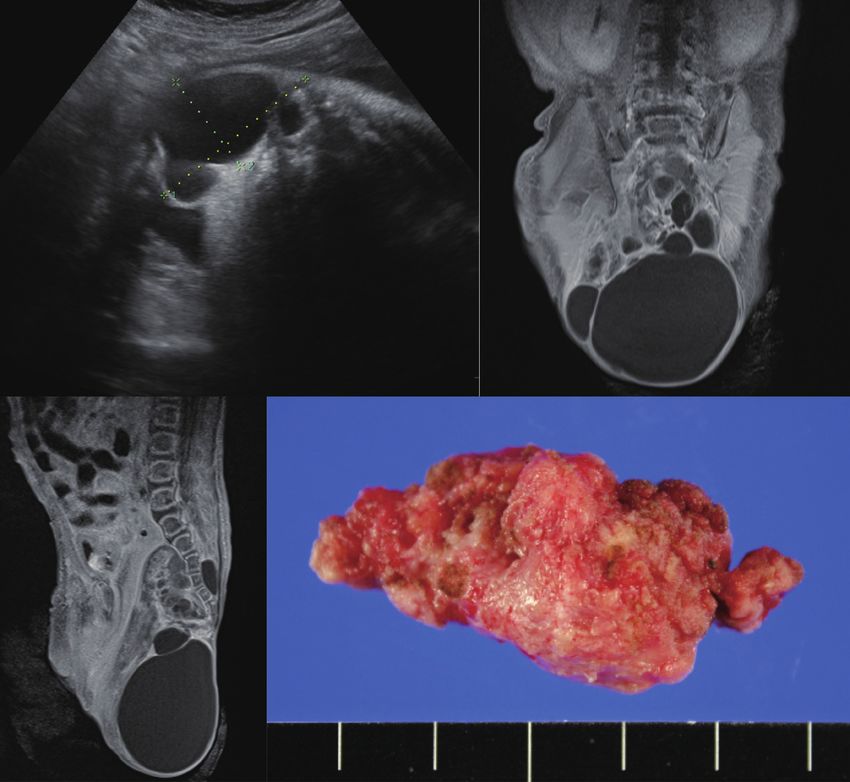
Fig. 2. Altman classification of sacrococcygeal teratoma according to the anatomical configuration. A : Type I : predominantly external with small
internal components. B : Type II : dumbbell-shaped tumor with similar internal and external components. C : Type III : predominantly internal and
smaller external components. D : Type IV : exclusively internal (intrapelvic) tumor.
TREATMENT 15%23,29,45). Incomplete resection and immature/malignant his-
tology are the most important risk factors for recurrence23,45).
Complete surgical resection is the standard for SCT. De- Preoperatively, older age at diagnosis (>2 months) and predom-
layed treatment may lead to tumor rupture and hemorrhage. inantly solid components within the mass are suggestive of ma-
SCT has a tendency toward malignant degeneration, which lignant histology and a poor prognosis4,9,47). To prevent incom-
makes early surgery mandatory. In a study, 91% of patients di- plete resection, removing the coccyx bone and preventing
agnosed at birth had mature/immature teratomas, whereas tumor tissue spillage during surgery have been empha-
27% of infants diagnosed later had YST35). The majority of pa- sized4,45,47). Although immature teratoma histology is a risk fac-
tients (Altman type I and II) can be operated on via the poste- tor for recurrence, postoperative chemotherapy is not recom-
rior (sacral) approach only. Large type III tumors with both mended because it is ineffective in preventing recurrence23).
external and internal components may require combined Recurrent SCT tends to have a malignant histology. One study
transabdominal and posterior approaches4). Purely intrapelvic showed that in eight patients with recurrent SCT, initial mature
tumors (type IV) may be approached via laparoscopy4). teratoma recurred as immature teratoma (one patient) and YST
The surgical outcome and prognosis of SCT are favorable. (four patients)45). The majority of recurrences are diagnosed
The recurrence rate after surgery has been estimated as 10– 6–36 months after the initial surgery45,46). Serum AFP is useful
410 https://doi.org/10.3340/jkns.2021.0015Sacrococcygeal Teratoma | Phi JH
in monitoring for recurrences. In a systemic review, serum AFP to fetal demise. Open in utero tumor debulking is a challeng-
levels were elevated in 75% of patients with recurrent SCT43). ing, but viable option for such cases. Researchers in the Chil-
Therefore, careful monitoring with digital rectal exam and se- dren’s Hospital of Philadelphia reported the early outcome of
rum AFP check at 3-month intervals for 3–4 years is generally four patients who received open fetal surgery for SCT12). All
recommended4,29). MRI or computed tomography should be patients experienced preterm birth after surgery, but three of
performed if suspicious clinical finding or elevated AFP level is the four patients have survived without the disease except for
noted4). The German study protocols included routine imaging one patient who died early. Interventional techniques to ablate
surveillance at 3-month intervals for the first year and at feeding arteries to SCT are also under investigation to reverse
6-month intervals in the second year38). However, long-term fetal cardiac failure. Researchers in Toronto tried fetoscopic
surveillance is warranted because patients with a very late re- laser ablation, radiofrequency ablation, and intravascular coil-
currence after 5–15 years have been reported29). ing in five fetuses with SCT44). Although they used minimally
Patients with recurrent SCT can be salvaged by surgery and invasive techniques, only two patients survived after the pro-
chemotherapy46). Malignant recurrence requires intensive cedures. Experience from Brazil and France showed similar
management, and multimodality treatment can establish sec- results of percutaneous interventions for fetal SCT : two out of
ond and third remissions in many patients38). There is no stan- five patients survived37). Therefore, fetal treatments of SCT,
dard chemotherapeutic regimen for malignant recurrent SCT, open or interventional, are still investigational. However, con-
but it has been emphasized that platinum compound should sidering the poor prognosis of fetuses developing cardiac fail-
be included to achieve stable remission38). JEB (carboplatin, et- ure and hydrops in utero, these early intervention techniques
oposide, bleomycin) and ICE (ifosfamide, carboplatin and et- deserve to be scrutinized and developed.
oposide) regimens were applied to recurrent malignant YST in Interventions can be applied to postnatal patients with large
the UK study23). The 5 year overall survival of recurrent, malig- SCTs to reduce the chance of spontaneous hemorrhage, stabi-
nant SCT (excluding teratoma recurrence) was 42%38). Func- lize the cardiopulmonary status, and decrease surgical bleed-
tional outcome, however, is a concern for survivors. A study re- ing. In the 1990s, staged operations, i.e., devascularization
ported anorectal sequelae in 29% and urological dysfunction in surgery followed by tumor resection were tried for patients
33% of survivors30). Chronic constipation and fecal inconti- with huge SCTs36). In 2006, Cowles et al. first reported suc-
nence are common anorectal problems, and neurogenic blad- cessful embolization of feeding vessels in a newborn of GA 36
der, vesicoureteral reflux, and urinary incontinence constitute weeks with a large hypervascular SCT, followed by similar at-
common urologic sequelae. A questionnaire survey from the tempts by others5,18). Preoperative embolization seems to be a
Netherland indicated that long-term survivors of SCT com- promising and effective treatment, but the technique needs
plained of impaired bowel function (46%), urinary inconti- great expertise and skill. Currently, interventional approaches
nence (31%) and unacceptable scarring (40%)7). Extensive pelvic are not routinely practiced, and only selective centers with
surgery in early life also tends to lead to sexual dysfunction in such resources can utilize them successfully for SCT9).
adulthood17). Therefore, long-term surveillance and evaluation
of functional status are important for the survivor programs
for SCT. CONFLICTS OF INTEREST
In addition to classic surgical resection for SCT, a number
of innovative interventions have been developed to rescue No potential conflict of interest relevant to this article was
complicated fetuses with a large SCT or to decrease surgical reported.
bleeding in neonates. Fetal SCT, especially when diagnosed
before GA 30 weeks, has a far worse prognosis than neonatal
SCT. If the tumor has a large cystic portion, cyst aspiration in INFORMED CONSENT
utero can prevent preterm labor or reduce fetal dystocia dur-
ing delivery12). Rapid growth of hypervascular, solid SCT This type of study does not require informed consent.
causes high output cardiac failure and fetal hydrops, leading
J Korean Neurosurg Soc 64 (3) : 406-413 411J Korean Neurosurg Soc 64 | May 2021
AUTHOR CONTRIBUTIONS 1122-1126, 2007
8. Ferlay J, Soerjomataram I, Dikshit R, Eser S, Mathers C, Rebelo M, et al. :
Cancer incidence and mortality worldwide: sources, methods and major
Conceptualization : JHP
patterns in GLOBOCAN 2012. Int J Cancer 136 : E359-E386, 2015
Data curation : JHP
9. Fumino S, Tajiri T, Usui N, Tamura M, Sago H, Ono S, et al. : Japanese
Formal analysis : JHP clinical practice guidelines for sacrococcygeal teratoma, 2017. Pediatr
Funding acquisition : JHP Int 61 : 672-678, 2019
Methodology : JHP 10. Gabra HO, Jesudason EC, McDowell HP, Pizer BL, Losty PD : Sacrococcy-
Project administration : JHP geal teratoma--a 25-year experience in a UK regional center. J Pediatr
Surg 41 : 1513-1516, 2006
Visualization : JHP
11. Gross SJ, Benzie RJ, Sermer M, Skidmore MB, Wilson SR : Sacrococ-
Writing - original draft : JHP
cygeal teratoma: prenatal diagnosis and management. Am J Obstet
Writing - review & editing : JHP Gynecol 156 : 393-396, 1987
12. Hedrick HL, Flake AW, Crombleholme TM, Howell LJ, Johnson MP,
Wilson RD, et al. : Sacrococcygeal teratoma: prenatal assessment, fetal
ORCID intervention, and outcome. J Pediatr Surg 39 : 430-438, 2004
13. Izant RJ Jr, Filston HC : Sacrococcygeal teratomas. Analysis of forty-three
Ji Hoon Phi https://orcid.org/0000-0002-9603-5843 cases. Am J Surg 130 : 617-621, 1975
14. Keslar PJ, Buck JL, Suarez ES : Germ cell tumors of the sacrococcygeal
region: radiologic-pathologic correlation. Radiographics 14 : 607-620;
●
Acknowledgements quiz 621-622, 1994
15. Köchling J, Pistor G, Märzhäuser Brands S, Nasir R, Lanksch WR : The
This study was supported by a grant from National Research Currarino syndrome--hereditary transmitted syndrome of anorectal,
Foundation (NRF) of Korea (No. 2018R1D1A1A02086005; to sacral and presacral anomalies. Case report and review of the literature.
Phi JH) and a grant from Seoul National University Hospital Eur J Pediatr Surg 6 : 114-119, 1996
(No. 04-20183140; to Phi JH). 16. Kratz CP, Mai PL, Greene MH : Familial testicular germ cell tumours.
Best Pract Res Clin Endocrinol Metab 24 : 503-513, 2010
17. Kremer MEB, Althof JF, Derikx JPM, van Baren R, Heij HA, Wijnen
MHWA, et al. : The incidence of associated abnormalities in patients
References with sacrococcygeal teratoma. J Pediatr Surg 53 : 1918-1922, 2018
18. Lahdes-Vasama TT, Korhonen PH, Seppänen JM, Tammela OK, Iber T :
1. Altman RP, Randolph JG, Lilly JR : Sacrococcygeal teratoma: American Preoperative embolization of giant sacrococcygeal teratoma in a prema-
academy of pediatrics surgical section survey-1973. J Pediatr Surg 9 : ture newborn. J Pediatr Surg 46 : e5-e8, 2011
389-398, 1974 19. Litchfield K, Summersgill B, Yost S, Sultana R, Labreche K, Dudakia D,
2. Anteby EY, Yagel S : Route of delivery of fetuses with structural anoma- et al. : Whole-exome sequencing reveals the mutational spectrum of
lies. Eur J Obstet Gynecol Reprod Biol 106 : 5-9, 2003 testicular germ cell tumours. Nat Commun 6 : 5973, 2015
3. Bader D, Riskin A, Vafsi O, Tamir A, Peskin B, Israel N, et al. : Alpha- 20. Lobo J, Gillis AJM, Jerónimo C, Henrique R, Looijenga LHJ : Human germ
fetoprotein in the early neonatal period--a large study and review of the cell tumors are developmental cancers: impact of epigenetics on patho-
literature. Clin Chim Acta 349 : 15-23, 2004 biology and clinic. Int J Mol Sci 20 : 258, 2019
4. Barksdale EM Jr, Obokhare I : Teratomas in infants and children. Curr 21. Lynch SA, Wang Y, Strachan T, Burn J, Lindsay S : Autosomal dominant
Opin Pediatr 21 : 344-349, 2009 sacral agenesis: Currarino syndrome. J Med Genet 37 : 561-566, 2000
5. Cowles RA, Stolar CJ, Kandel JJ, Weintraub JL, Susman J, Spigland NA : 22. Mamsen LS, Brøchner CB, Byskov AG, Møllgard K : The migration and
Preoperative angiography with embolization and radiofrequency abla- loss of human primordial germ stem cells from the hind gut epithelium
tion as novel adjuncts to safe surgical resection of a large, vascular towards the gonadal ridge. Int J Dev Biol 56 : 771-778, 2012
sacrococcygeal teratoma. Pediatr Surg Int 22 : 554-556, 2006 23. Mann JR, Gray ES, Thornton C, Raafat F, Robinson K, Collins GS, et al. :
6. Damjanov I, Solter D, Belicza M, Skreb N : Teratomas obtained through Mature and immature extracranial teratomas in children: the UK Chil-
extrauterine growth of seven-day mouse embryos. J Natl Cancer Inst dren’s Cancer Study Group Experience. J Clin Oncol 26 : 3590-3597,
46 : 471-475, 1971 2008
7. Derikx JP, De Backer A, van de Schoot L, Aronson DC, de Langen ZJ, van 24. Masahata K, Ichikawa C, Makino K, Abe T, Kim K, Yamamichi T, et al. :
den Hoonaard TL, et al. : Long-term functional sequelae of sacrococcygeal Long-term functional outcome of sacrococcygeal teratoma after resec-
teratoma: a national study in the Netherlands. J Pediatr Surg 42 : tion in neonates and infants: a single-center experience. Pediatr Surg
412 https://doi.org/10.3340/jkns.2021.0015Sacrococcygeal Teratoma | Phi JH
Int 36 : 1327-1332, 2020 ous fetal therapies for large sacrococcygeal teratomas: cohort study and
25. Monk D, Mackay DJG, Eggermann T, Maher ER, Riccio A : Genomic literature review. Ultrasound Obstet Gynecol 47 : 712-719, 2016
imprinting disorders: lessons on how genome, epigenome and environ- 38. Schneider DT, Wessalowski R, Calaminus G, Pape H, Bamberg M, Engert
ment interact. Nat Rev Genet 20 : 235-248, 2019 J, et al. : Treatment of recurrent malignant sacrococcygeal germ cell tu-
26. Murphy JJ, Blair GK, Fraser GC : Coagulopathy associated with large mors: analysis of 22 patients registered in the german protocols MAKEI
sacrococcygeal teratomas. J Pediatr Surg 27 : 1308-1310, 1992 83/86, 89, and 96. J Clin Oncol 19 : 1951-1960, 2001
27. Oosterhuis JW, Looijenga LHJ : Human germ cell tumours from a devel- 39. Schropp KP, Lobe TE, Rao B, Mutabagani K, Kay GA, Gilchrist BF, et al. :
opmental perspective. Nat Rev Cancer 19 : 522-537, 2019 Sacrococcygeal teratoma: the experience of four decades. J Pediatr
28. Oosterhuis JW, Stoop H, Honecker F, Looijenga LH : Why human ex- Surg 27 : 1075-1078; discussion 1078-1079, 1992
tragonadal germ cell tumours occur in the midline of the body: old con- 40. Shen H, Shih J, Hollern DP, Wang L, Bowlby R, Tickoo SK, et al. : Inte-
cepts, new perspectives. Int J Androl 30 : 256-263; discussion 263- grated molecular characterization of testicular germ cell tumors. Cell
264, 2007 Rep 23 : 3392-3406, 2018
29. Padilla BE, Vu L, Lee H, MacKenzie T, Bratton B, O’Day M, et al. : Sa- 41. Sobis H, Vandeputte M : Yolk sac-derived rat teratomas are not of germ
crococcygeal teratoma: late recurrence warrants long-term surveillance. cell origin. Dev Biol 51 : 320-323, 1976
Pediatr Surg Int 33 : 1189-1194, 2017 42. Teilum G, Albrechtsen R, Norgaard-Pedersen B : The histogenetic-
30. Partridge EA, Canning D, Long C, Peranteau WH, Hedrick HL, Adzick embryologic basis for reappearance of alpha-fetoprotein in endodermal
NS, et al. : Urologic and anorectal complications of sacrococcygeal tera- sinus tumors (yolk sac tumors) and teratomas. Acta Pathol Microbiol
tomas: prenatal and postnatal predictors. J Pediatr Surg 49 : 139-142; Scand A 83 : 80-86, 1975
discussion 142-143, 2014 43. van Heurn LJ, Knipscheer MM, Derikx JPM, van Heurn LWE : Diagnostic
31. Pauniaho SL, Heikinheimo O, Vettenranta K, Salonen J, Stefanovic V, Rit- accuracy of serum alpha-fetoprotein levels in diagnosing recurrent sa-
vanen A, et al. : High prevalence of sacrococcygeal teratoma in Finland crococcygeal teratoma: a systematic review. J Pediatr Surg 55 : 1732-
- a nationwide population-based study. Acta Paediatr 102 : e251- 1739, 2020
e256, 2013 44. Van Mieghem T, Al-Ibrahim A, Deprest J, Lewi L, Langer JC, Baud D, et
32. Phi JH, Wang KC, Kim SK : Intracranial germ cell tumor in the molecular al. : Minimally invasive therapy for fetal sacrococcygeal teratoma: case
era. J Korean Neurosurg Soc 61 : 333-342, 2018 series and systematic review of the literature. Ultrasound Obstet Gy-
33. Poynter JN, Hooten AJ, Frazier AL, Ross JA : Associations between vari- necol 43 : 611-619, 2014
ants in KITLG, SPRY4, BAK1, and DMRT1 and pediatric germ cell tumors. 45. Yao W, Li K, Zheng S, Dong K, Xiao X : Analysis of recurrence risks for
Genes Chromosomes Cancer 51 : 266-271, 2012 sacrococcygeal teratoma in children. J Pediatr Surg 49 : 1839-1842,
34. Rattan KN, Singh J : Neonatal sacrococcygeal teratoma: our 20-year 2014
experience from a tertiary care centre in North India. Trop Doct : 46. Yoneda A, Usui N, Taguchi T, Kitano Y, Sago H, Kanamori Y, et al. : Im-
49475520973616, 2020 pact of the histological type on the prognosis of patients with prenatally
35. Rescorla FJ, Sawin RS, Coran AG, Dillon PW, Azizkhan RG : Long-term diagnosed sacrococcygeal teratomas: the results of a nationwide Japa-
outcome for infants and children with sacrococcygeal teratoma: a report nese survey. Pediatr Surg Int 29 : 1119-1125, 2013
from the Childrens Cancer Group. J Pediatr Surg 33 : 171-176, 1998 47. Yoon HM, Byeon SJ, Hwang JY, Kim JR, Jung AY, Lee JS, et al. : Sacro-
36. Robertson FM, Crombleholme TM, Frantz ID 3rd, Shephard BA, Bianchi coccygeal teratomas in newborns: a comprehensive review for the radi-
DW, D’Alton ME : Devascularization and staged resection of giant sacro- ologists. Acta Radiol 59 : 236-246, 2018
coccygeal teratoma in the premature infant. J Pediatr Surg 30 : 309- 48. Yoshida M, Tanaka M, Gomi K, Ohama Y, Kigasawa H, Iwanaka T, et
311, 1995 al. : Malignant steroidogenic tumor arising from sacrococcygeal mature
37. Sananes N, Javadian P, Schwach Werneck Britto I, Meyer N, Koch A, teratoma. Hum Pathol 42 : 1568-1572, 2011
Gaudineau A, et al. : Technical aspects and effectiveness of percutane-
J Korean Neurosurg Soc 64 (3) : 406-413 413You can also read