Hemangioma rubi no couro cabeludo* Cherry hemangioma in the scalp
←
→
Page content transcription
If your browser does not render page correctly, please read the page content below
Pereira                                                                                                                                                                                83
                                                                                                            Artigo de Revisão / Review Article

Hemangioma rubi no couro cabeludo*
Cherry hemangioma in the scalp*
José Marcos Pereira1
                  Resumo: O hemangioma rubi (HR) é dermatose de origem vascular extremamente freqüente,
                  acometendo mais de 75% da população acima de 70 anos de idade. Em geral compõe-se de
                  lesões múltiplas,localizadas predominantemente no alto do tronco e braços. Clinicamente é
                  caracterizado por lesões que variam desde máculas puntiformes até lesões papulosas com
                  cinco milímetros de diâmetro. As mais novas são vermelhas, em tons vivos, e as mais antigas
                  podem ser azuladas. O HR é de etiologia desconhecida. Histologicamente chama atenção uma
                  neoformação de vasos capilares, que se tornam dilatados e com fenestrações em suas paredes.
                  A membrana basal está muito espessada e existe abundante estroma de colágeno entre os
                  vasos. O presente trabalho demonstra a alta incidência do HR no couro cabeludo Em amostra
                  de 171 pacientes, sendo 85 homens e 86 mulheres, o autor observou que 123 deles (72%) tin-
                  ham HR no couro cabeludo, localização em que o HR nunca foi descrito na literatura.
                  Palavras-chave: couro cabeludo; hemangioma.

                  Summary: Cherry hemangioma (CH) is an extremely frequent dermatosis with vascular
                  origin involving more than 75% of the population over 70 years of age. Normally they are
                  multiplex spots and focus predominantly on the upper trunk and arms. Clinically they are
                  characterized by pinpoint maculae and papules with up to 5 millimeters in diameter. The
                  most recent lesions can be a strong red color while the older ones are bluish. The etiology of
                  CH is still unknown. From the histologic standpoint, the neoformation of the capillary tube
                  draws attention: they are very dilated and with fenestration along the wall. The basement
                  membranes are thickened and there is abundant collagen stroma between the veins. This
                  work demonstrates the high frequency of CH in the scalp. In a sample of 171 patients (85
                  men and 86 women) the author noted that 123 (72%) had CH in the scalp. However, accor-
                  ding to the literature, CH in the scalp has never been described.
                  Key-words: scalp; hemangioma.

INTRODUÇÃO                                                                                        INTRODUCTION
       O hemangioma rubi (HR) também é conhecido na                                                      Cherry hemangioma (CH) [known in the Portuguese
língua portuguesa como hemangioma senil, angioma senil,                                           language as hemangioma senil, angioma senil, angioma
angioma rubi, mancha de Morgan ou Mancha de Campbell                                              rubi, mancha de Morgan or Mancha de Campbell de
de Morgan. Na língua inglesa, como cherry angioma, seni-                                          Morgan] is also denominated cherry angioma, senile
le hemangiomas, cherry ou ruby spots, Campbell de                                                 hemangioma, cherry or ruby spots, Campbell de Morgan's
Morgan's spots, Morgan's spots, senile angioma, petechial                                         spots, Morgan's spots, senile angioma, petechial angioma
angiomata1 ou capillary angiomas.                                                                 or capillary angioma.
       A doença foi descrita pela primeira vez em 1872 por                                               The disease was described for the first time in 1872
Campbell de Morgan, cirurgião do Middlesex Hospital                                               by Campbell de Morgan, surgeon at Middlesex Hospital
(1842 a 1875), na Inglaterra, em cujo livro On the origin of                                      (1842 to 1875), in England. In his textbook "On the origin
Cancer, ele associava o HR à presença de neoplasias, em                                           of Cancer", he associated CH to the presence of neoplasias
particular câncer de estômago.2                                                                   and in particular to cancer of the stomach.2
       Dermatose de origem vascular extremamente                                                         It is an extremely common dermatosis of vascular

Recebido em 17.08.2001. / Received in August, 17 th of 2001.
Aprovado pelo Conselho Consultivo e aceito para publicação em 10.06.2002. / Approved by the Consultive Council and accepted for publication in June, 10 th of 2002.
* Trabalho realizado na clinica privada do autor. / Work done at private clinic of the author.
1
    Ex-professor instrutor de dermatologia da Faculdade de Ciências Médicas da Santa Casa de São Paulo. / Ex-professor of Dermatology at the College of Medical Sciences, Santa Casa
    Charitable Hospital, São Paulo.

©2004 by Anais Brasileiros de Dermatologia

An bras Dermatol, Rio de Janeiro, 79(1):83-89, jan./fev. 2004.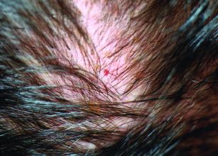
84        Pereira

comum, são raras as pessoas que não têm pelo menos um            origin and it is rare for individuals not to present at least
HR. A lesão pode ser única ou múltipla, às vezes chegando        one CH. The lesion can be single or multiple, sometimes
a centenas espalhadas pelo corpo, principalmente no alto         reaching hundreds dispersed throughout the body, but
do tronco e nos braços.3                                         mainly in the upper trunk and arms.3
        Clinicamente a lesão inicial pode lembrar uma peté-              Clinically, the initial lesion can be similar to
quia, sendo apenas uma lesão maculosa, plana, vermelha e         petechiae, being just a macular, flat, red and punctiform
puntiforme. Com a evolução tornam-se pápulas vermelho-           lesion. As they develop, they become brilliant-red papules
brilhantes de um a cinco milímetros (Figura 1). As mais          with one to five millimeters in diameter (Figure 1). With
antigas podem assumir coloração azul-escura. São lesões          time they assume a dark-blue coloration. The lesions are
assintomáticas, benignas4 e não compressíveis à vitropres-       asymptomatic, benign4 and not compressible.5 The lesions
são.5 Sangram quando traumatizadas, podendo formar sobre         bleed on suffering trauma and can form a black clot on the
a superfície um coágulo negro, o que pode simular um             surface, mimicking a malignant melanoma. They grow
melanoma maligno. Não involuem e crescem lentamente.             slowly without involution.
        A real incidência dos HRs é desconhecida, pois pou-              The real incidence of CH is unknown, because few
cos trabalhos abordam o assunto. Keller6 em uma série de         works have covered the subject. Keller 6 in a series of 696
696 voluntários e 583 pacientes de uma clínica na                volunteers and 583 patients from a clinic in Germany found
Alemanha encontrou HR em 34,5% das pessoas com 30                CH in 34.5% of those under 30 years of age, and in 40.15%
anos de idade e em 40,15% daquelas com 31 anos; Murison          of those over 31 years age; Murison et al.2 observed 1,300
e colaboradores2 observaram 1300 pacientes de um hospi-          patients at a hospital in Glasgow (UK) and found that 5%
tal em Glasgow e constataram que 5% dos adolescentes e           of the adolescents and 75% of the patients above 70 years
75% dos pacientes acima de 70 anos tinham HR. Não havia          had CH. There was no sex bias and CH increased in num-
diferença de manifestação nos dois sexos, e o HR aumenta-        ber and size with age, however the growth index decreased
va em número e tamanho com a idade, porém seu índice de          with time. Surprisingly, in a population of soldiers in the
crescimento diminuía com o tempo. Curiosamente, numa             air force there was much less CH than in the patients of
população de soldados da força aérea havia muito menos           hospitals. The authors also observed that CH seems to be
HR do que nos pacientes de hospitais. Os autores também          more frequent in patients with neoplasia than in those
observaram que o HR parece ser mais freqüente em pacien-         without neoplasia.
tes com neoplasias do que naqueles sem neoplasia.                        Although no study has yet clarified the etiopatho-
        Embora não exista qualquer trabalho que explique a       genesis of CH, several factors have been related to its
etiopatogenia do HR, vários fatores foram relacionados a         onset, as described below.
seu aparecimento, os quais serão descritos a seguir.                     In diabetics the lesions are more numerous and have
        Em diabéticos as lesões são mais numerosas e mais        a greater volume;7 furthermore, according to Jaimovich,8
volumosas;7 e, segundo Jaimovich,8 elas aumentam sob             they expand under high temperatures.
temperaturas altas.                                                      Epidemic outbreaks of CH have been described.
        Têm sido descritos surtos                                                         These being patients that within a
epidêmicos de HR. Trata-se de                                                             period of days refer to the emer-
pacientes que no prazo de poucos                                                          gence of several lesions. Seville et
dias referem o aparecimento de                                                            al.9 in 1968 observed that in the
várias lesões. Seville e colaborado-                                                      Lancaster Moor Hospital ( UK)
res9 em 1968 observaram que na                                                            some 1,000 patients were attended
Inglaterra, no Lancaster Moor                                                             with onset of several CH lesions in a
Hospital, foram atendidos cerca de                                                        few days. The patients presented
1000 pacientes com aparecimento                                                           clinical and histopathological signs
de várias lesões de HR em poucos                                                          of CH, but with no systemic altera-
dias. Eram pacientes com clínica e                                                        tion or symptom. Some patients
histopatologia de HR e não com                                                            referred to the emergence of the
qualquer alteração ou sintoma sis-                                                        lesions after sun bathing. One
têmico. Alguns pacientes referiam                                                         patient followed-up for two months
o aparecimento das lesões após                                                            showed that the lesions appeared
tomada de sol. Um paciente obser-                                                         during the hottest days. Honish et

                      Figura 1: Múltiplos                                               Figure 1: Multiple
                           hemangiomas                                                  cherry hemangiomas
                        rubis no tronco .                                               in the trunk

An bras Dermatol, Rio de Janeiro, 79(1):83-89, jan./fev. 2004.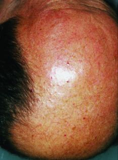
Pereira                                                                                                                      85

vado por dois meses mostrou que as lesões apareciam nos          al.10 reported an outbreak of CH among patients and work-
dias de maior calor. Honish e colaboradores10 relataram um       ers of a rest clinic, in 1988 in Edmonton (USA). A total of
surto de HR entre pacientes e dirigentes de uma clínica de       147 cases were observed in 302 people over a 10-day peri-
repouso, em 1988 em Edmonton. Foram observados 147               od. The patients' age varied from 33 to 100 years, and the
casos entre 302 pessoas no intervalo de 10 dias. A idade dos     number of lesions per patient from two to 78, with a mean
pacientes variava de 33 a 100 anos, e o número de lesões         of 30. The anatomicopathological exam was compatible
por paciente, de duas a 78, com a média de 30. O exame           with CH. The same authors described that in 1985, in
anatomopatológico foi compatível com HR. Os mesmos               Edmonton, three rest clinics and a hospital had an outbreak
autores descrevem que, em 1985, em Edmonton, em três             of CH and in 1987, a similar outbreak occurred at another
clínicas de repouso e um hospital houve um surto de HR, e        rest clinic. Although this suggests the possibility of a conta-
que, em 1987, surto igual ocorreu em outra clínica de            gious agent, exhaustive studies attempting to prove this
repouso. Embora isso possa sugerir um agente infectocon-         were fruitless.
tagioso, exaustivos estudos foram infrutíferos para compro-               CH has also been associated with exposure to chemi-
vá-lo.                                                           cal products. Cohen et al. 11 described two cases of CH after
        O HR também foi associado à exposição de produtos        exposure to derivatives of bromine. Raymond et al.1 2 have
químicos. Cohen e colaboradores11 descreveram dois casos         reported that four months after seven people were exposed
de HR após exposição a derivados do bromo. Raymond e             to steam of the solvent 2-butoxyethanol, six developed typi-
colaboradores12 relatam que, quatro meses depois de sete         cal CH lesions in the arms, trunk and thighs. Firooz et al.13
pessoas terem sido expostas ao vapor de 2-butoxietanol,          observed 250 people that came into contact with mustard
que é um solvente, seis desenvolveram nos braços, tronco e       gas and after 18 months, approximately 10% presented
coxas lesões típicas de HR. Firooz e colaboradores 13 obser-     onset of CH.
varam 250 pessoas que entraram em contato com gás mos-                    A greater frequency of CH was also observed after
tarda, das quais, após 18 meses, cerca de 10% tiveram apa-       liver transplant; 14 in graft-versus-host disease; 15 after
recimento de HR.                                                 cyclosporin therapy;1 6 and following argon laser therapy
        Maior freqüência do HR também foi observada após         for dermatosis.17
transplante de fígado; 14 na doença enxerto-versus-hospedei-              There is considerable controversy regarding the
ro;15 na terapia com ciclosporina;16 e após terapia de derma-    etiopathogenesis of CH. According to Jaimovich,8 it is a
tose com laser de argônio.17                                     non-tumoral self-limiting hyperplasia that is not associ-
        A etiopatogenia do HR é desconhecida. Segundo            ated to neoangiogenesis with abnormally increased
Jaimovich,8 trata-se de hiperplasia não tumoral autolimi-        endothelial proliferation, and the angiogenic growth
tante que não se associa a alguma neoangiogênese com             factors of which, such as TNF-α, FGF-β and VEGF, do
proliferação endotelial anormalmente aumentada, e cujos          not appear to be related to its onset. Hagiwara et al., 18
fatores de crescimento angiogênicos, como alfa-TNF ,             based on the principal that mastocytes are related to the
beta-FGF e VEGF, não parecem estar relacionados a seu            angiogenesis, counted their number in CH. In normal
aparecimento. Hagiwara e colaboradores,18 partindo do            tissue, the mean number of mastocytes was
princípio de que mastócitos estão relacionados à angiogê-        6.85±4.9/mm2 ; while in the presence of CH this was
nese, contaram seu número no HR. Em tecidos normais o            85.3±45.6/mm 2 . Tamm et al. 1 9 in immunohistochemical
número médio de mastócitos era 6,85±4,9/mm2 ; na pre-            studies demonstrated that the perivascular hyaline tis-
sença de HR, 85,3±45,6/mm2 . Tamm e colaboradores 19             sue observed in CH is composed of collagen IV and VI.
mediante estudos imuno-histoquímicos mostraram que o             The authors established the hypothesis that collagen
tecido hialinizado perivascular observado no HR é com-           type VI serves as a platform in the tissues with a high
posto de colágeno IV e VI. Os autores estabeleceram a            concentration of collagenolytic enzymes and that the
hipótese de que o colágeno tipo VI serve como uma plata-         increase of collagen type VI in CH is related to its for-
forma nos tecidos com alta concentração de enzimas cola-         mation. Eichhorn et al.2 0 observed that most of the blood
genolíticas e que o aumento no tipo VI de colágeno no HR         vessels in CH are fenestrated and have a positive reac-
está relacionado a sua formação. Eichhorn e colaborado-          tion to carbonic anhydrase, which is an enzyme. The
res20 observaram que a maioria dos vasos sangüíneos nos          authors cogitate the possibility that this enzyme is rela-
HR é fenestrada e com reação positiva à anidrase carbôni-        ted to the maintenance of the fenestration. Tuder et al., 21
ca, que é uma enzima. Os autores cogitam a possibilidade         in immunohistochemical studies using Ki67 markers
de que essa enzima esteja relacionada à manutenção das           specific for G2 cells and phase S of the mitoses, con-
fenestrações. Tuder e colaboradores, 21 por estudos imuno-       cluded that CH is not a true neoplasia, but a composite
histoquímicos com marcadores Ki67 específicos para               of mature veins similar to dermal venulae.
células G2 e fase S das mitoses, concluíram que o HR não                  The histopathology is very characteristic. In the ini-
é uma verdadeira neoplasia, mas um composto de vasos             tial phase CH has the appearance of a capillary heman-
maturos relembrando vênulas dérmicas.                            gioma 22 or angioblastoma6 - formed by numerous narrow

An bras Dermatol, Rio de Janeiro, 79(1):83-89, jan./fev. 2004.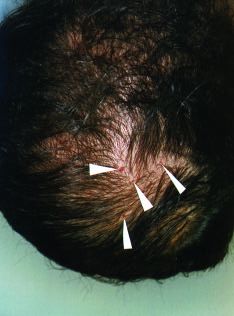
86        Pereira

        A histopatologia é bastante característica. Na fase inicial   capillary neoformations and prominent endothelial cells
os HR têm a aparência de hemangioma capilar22 ou angioblas-           arranged in a lobular form and located exactly between the
toma6 - formados por numerosos capilares neoformados com              dermis and the epidermis. With time the capillaries become
luzes estreitas e proeminentes células endoteliais arranjadas em      voluminous and are characteristic of the tubular or spheri-
um modo lobular, localizados exatamente entre a derme e a epi-        cal dilations of the capillary loops in the papillary der-
derme. Com o tempo os capilares ficam volumosos, e são                mis. 23,24,25 Each dilated vein is connected with one or more
características as dilatações tubulares ou esféricas das alças        neighboring loops by tortuous vascular channels. The veins
capilares da papila dérmica.23,24,25 Cada vaso dilatado é conecta-    in the horizontal plexus are not involved. 23,24,25 There is little
do com a ou as alças vizinhas por canais vasculares tortuosos.        vascular space, and the intercapillary stroma presents
Os vasos no plexo horizontal não são afetados.23,24,25 Há poucos      edema and homogenization of the collagen. The walls of the
espaços vasculares, e o estroma intercapilar mostra edema e           capillaries are sometimes hyalinized. Cavernous spaces
homogeneização do colágeno. As paredes dos capilares são às           can also be observed. Important findings are the fenestra-
vezes hialinizadas. Espaços cavernosos também podem ser               ted endothelium of the capillaries and the considerably
observados. Achados importantes são o endotélio fenestrado            thickened basement membrane. 26
dos capilares e a membrana basal bastante espessada.26                           Electron microscopy reveals that CH is located
        A microscopia eletrônica mostra que os HR estão loca-         immediately under the epidermis and is very different
lizados imediatamente sob a epiderme e são muito diferentes           from adjacent tissues. The walls of all the veins are
dos tecidos adjacentes. As paredes de todos os vasos são for-         formed by a single layer of endothelial cells, that fre-
madas por apenas uma camada de células endoteliais, que com           quently present the so-called microtubular bodies in the
freqüência apresentam no citoplasma os chamados corpos                cytoplasm.2 7 Within the veins one finds blood and fibrin,
microtubulares.27 No interior dos vasos encontram-se sangue e         surrounded by agglomerates of fine collagen fibers. The
fibrina, circundados por aglomerados de fibras colágenas finas.       most characteristic images of CH under electron
As imagens mais características do HR à microscopia eletrôni-         microscopy are fenestration in the endothelium, that can
ca são fenestrações no endotélio, que podem ser intercelulares        be intercellular or transcellular, 27 as well as a very thick-
ou transcelulares,27 bem como a da membrana basal muito               ened and multilaminated basal membrane. 19,21,27
espessada e multilaminada.19,21,27 Além disso é possível obser-       Furthermore, it is possible to observe villous projections
var projeções vilosas para dentro da luz dos vasos.28                 into the lumen of the veins.2 8
        Quanto ao diagnóstico diferencial são desejáveis                         Several observations should be made regarding the
algumas observações. Quando o HR é circundado por halo                differential diagnosis. When CH is surrounded by purpuric
purpúrico, deve-se pensar em amiloidose.29 Na síndrome de             halo, one should consider amyloidosis.29 In POEMS syn-
Poems (polineuropatia, organomegalia, M-proteínas e alte-             drome (polyneuropathy, organomegaly and skin changes),
rações cutâneas skin), os hemangiomas lembram muito o                 the hemangiomas are very similar to CH30. An important
HR30. Diagnóstico diferencial importante é com a histioci-            differential diagnosis to consider is the histiocytosis X,
tose X, cujas lesões iniciais são idênticas às do HR31. Como          whose initial lesions are identical to those of CH31. As
já referido, o HR sangra quando traumatizado, e a formação            already mentioned, CH bleeds with trauma, such that the
de um coágulo negro pode simular melanoma maligno.                    formation of a black clot can simulate malignant melanoma.
        A maioria dos pacientes não se incomoda com o HR,                        Most of the patients are not inconvenienced by CH,
porém alguns desejam removê-lo por questão de estética ou             however some want to remove it for aesthetic purposes or
mesmo por causa de pequenos sangramentos. Várias técni-               even because of minor bleeding. Several techniques can be
cas podem ser usadas, entre elas: curetagem,32 laser33,34 e ele-      used, including curettage,32 laser33,34 and electrosurgery. 35
trocirurgia.35                                                                   Although the clinical and histopathological
        Embora o HR tenha sido muito bem caracterizado                aspects of CH have been very well characterized since it
clínica e histopatologicamente desde sua primeira aparição            first appeared in the medical literature, its presence in
em publicação, sua presença no couro cabeludo nunca foi               the scalp had not been described previously. The objec-
descrita na literatura médica. O presente trabalho tem o              tive of the present work was to clarify the frequency of
objetivo de mostrar a freqüência do HR no couro cabeludo.             CH in the scalp.

CASUÍSTICA                                                            PATIENTS
       No período de setembro a dezembro de 2000, foram                       From September to December 2000, 171 patients
observados no Centro Dermatológico de Guarulhos, no Estado            were observed at the Dermatological Center of
de São Paulo, 171 pacientes. Foram selecionados os primeiros          Guarulhos, in the State of São Paulo. The first 85 men and
85 homens e as primeiras 86 mulheres, para que houvesse uni-          the first 86 women were selected, so that there was uni-
formidade quanto à variável sexo. Cada paciente foi examina-          formity in terms of gender. Each patient was examined
do sentado em uma cadeira, em ambiente iluminado por seis             while seated in a chair and illuminated by six dichroic
lâmpadas dicróicas, de modo que o examinador tivesse fácil            lamps, such that the examiner had easy access to all areas

An bras Dermatol, Rio de Janeiro, 79(1):83-89, jan./fev. 2004.
Pereira 87
Figura 2: Figure 2:
Hemangioma Cherry hemangioma
rubi no couro cabeludo in the scalp
acesso a todas as regiões do of the scalp. The clinical
couro cabeludo. O exame clínico exam was made with the
foi feito a olho nu e com uso de naked eye and the use of a
lupa biocular da Nikon, denomi- 20X biocular magnifying
nada Naturescope, cujo poten- glass (Naturescope, Nikon).
cial de aumento é de 20 vezes. The patients' age
A idade dos pacientes ranged from 18 to 75 years
variou de 18 a 75 anos, tendo (mean, 40 years). They were
como média 40 anos. Todos all white, since CH is difficult
eram brancos, uma vez que o to characterize in black skin.
HR é de difícil caracterização na pele negra. Nenhum dos None of the patients examined sought medical attendance
pacientes examinados veio à consulta em função do HR. due to the CH.
RESULTADOS RESULTS
Foi constatado que 123(72%) dos 171 pacientes tinham It was observed that 123(72%) of the 171 patients had
HR no couro cabeludo, sendo 62 homens e 61 mulheres. Não CH in the scalp, of which 62 were men and 61 women. CH was
apresentavam o HR 48 pacientes(28%), 23 homens e 25 not present in 48 (28%) patients (23 men and 25 women). The
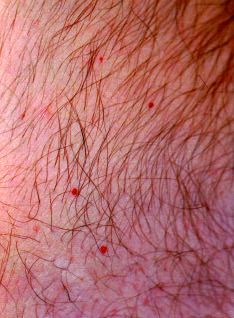
mulheres. O tamanho das lesões variava de puntiforme até size of the lesions varied from punctiform to five millimeters
cinco milímetros, e seu número por paciente, de uma a 10, com in diameter, and their number per patient ranged from one to
a média de cinco (Figuras 2 e 3). Havia maior quantidade de 10, with a mean of five (Figures 2 and 3). There was a higher
HR, em geral acima de cinco lesões por paciente, na faixa etá- number of CH, in general over five lesions per patient, in the
ria entre 30 e 40 anos e naqueles com alopecia androgenética age group between 30 and 40 years and those with andro-
acima do grau V, segundo classificação de Hamilton/Norwood. genic alopecia above level V, according to the classification of
Todos os pacientes com HR no couro cabeludo o apresentavam Hamilton/Norwood. All the patients with CH in the scalp also
também no tronco, e 23 (19%) tinham a face acometida. presented it in the trunk and 23 (19%) had facial involvement.
As lesões de HR não apresentavam relação qualquer The CH lesions did not present any relationship to
com doenças próprias do couro cabeludo, tais como derma- diseases of the scalp, such as seborrheic dermatitis, psori-
tite seborréica, psoríase e pseudopelada. asis and pseudopelade.
DISCUSSÃO DISCUSSION
Embora seja dermatose Although an extremely fre -
extremamente freqüente, o HR é quent dermatosis, there is little
pouco referido em livros clássicos reference to CH in the classic text-
de dermatologia, e, em. livros books of dermatology, while in
específicos de tricologia, a doença books specifically about tricholo-
não é citada. Apesar de ampla- gy, the disease is not mentioned at
mente usada, a expressão heman- all. Although in widespread use,
gioma ou angioma senil não é ade- the expression hemangioma or
quada para essa dermatose, uma senile angioma is not appropriate
vez que ela é encontrada em 5% for this dermatosis, since it is
dos adolescentes 2 e em 40,15% found in five percent of adoles-
das pessoas examinadas com 31 cents2 and in 40.15% of the sub-
anos de idade,6 ou seja, uma jects examined aged up to 31
população bastante jovem. Tem years, 6 or that is, a very young
sido descrita em incidência de até population. Incidences have been
Figura 3: Múltiplos Figure 3: Multiple
hemangiomas rubis cherry hemangiomas
no couro cabeludo in the scalp
An bras Dermatol, Rio de Janeiro, 79(1):83-89, jan./fev. 2004.88 Pereira
75% em pessoas acima de 70 anos de idade.2 Esses valo- described of up to 75% in people over 70 years of age.2
res, contudo, podem ser muitos maiores, principalmente These values, however, could be much larger, mainly in
no tronco e braços de indivíduos de pele clara e idade em the trunk and arms of those with clear skin and aged
torno dos 30 anos, independente do sexo. O exame aten- around 30 years, irrespective of their gender. Careful
to, em ambiente bem iluminado e com o uso de um der- examination in a well illuminated local and with the use
matoscópio, pode identificar minúsculas lesões puntifor- of a dermatoscope, could identify miniscule punctiform
mes de HR. As lesões maiores, já bem mais formadas e lesions of CH. The largest, already well formed and char-
características, são encontradas em idades um pouco acteristic lesions are found in individuals with a more
mais avançadas. advanced age.
O HR é de etiologia desconhecida, porém chamam The etiology of CH is unknown, however epidem-
atenção surtos epidêmicos9,10 e seu desencadeamento após ic outbreaks call attention9,10 as well as its appearance
exposição a produtos químicos.11,12,13 Não existe justificativa after exposure to chemical products. 11,12,13 There is no
plausível para sua grande incidência no alto do tronco e nos plausible justification for its greater incidence in the
braços. Em suas observações o autor tem notado grande inci- upper trunk and arms. In his observations the author
dência de HR no couro cabeludo, onde nenhum trabalho ou has noticed a great incidence of CH in the scalp, even
livro de toda a literatura pesquisada cita sua presença. Keller6 though no textbook or work in all of the literature
em série de 1279 pessoas, encontrou alta ocorrência de HR researched mentions its presence in this region. Keller6
em indivíduos com mais de 30 anos de idade. Desenhou in a series of 1279 individuals, found a high occurrence
então um boneco e assinalou a localização de todos os HRs of CH in individuals over 30 years of age. A model was
encontrados - das centenas de pontos assinalados, apenas drawn, on which the location was marked of all the CH
dois foram colocados no couro cabeludo, embora textual- found - of the hundreds of points marked, only two were
mente não tenha havido qualquer referência ao fato. O pre- in the scalp, although no reference was made in the text
sente trabalho tem a finalidade de mostrar que o HR é muito to this fact. The present work has the purpose of show-
freqüente no couro cabeludo. De 171 pacientes examinados, ing that CH is very frequent in the scalp. Of 171 patients
123, ou seja, 72%, o apresentavam nessa localização, inci- examined, 123 (72%) presented it in this location, an
dência alta o bastante para justificar esta publicação. As incidence high enough to justify this publication. The
lesões observadas eram assintomáticas, acometiam igual- lesions observed were asymptomatic, with no sex bias
mente ambos os sexos, mais freqüentes e maiores em pacien- and were more frequent and larger in patients over thir-
tes acima da terceira década de vida e naqueles com alopecia ty years of age and in those with advanced androgenet-
androgenética avançada, ou seja, além do grau V de ic alopecia, in other words, beyond degree V of
Hamilton/Norwood (Figura 4). Esse fato talvez seja explica- Hamilton/Norwood (Figure 4). This fact may be
do pela ação solar sobre o couro cabeludo, conforme descri- explained by the solar action on the scalp, as described,
to, quando do relato de aumento da incidência de HR após in the report of an increased incidence of CH after solar
exposição solar. 9 Nenhum paciente veio à consulta em função exposure.9 No patient sought consultation due to CH in
do HR no couro cabeludo; porém, the scalp; however, once
quando foram informados sobre sua informed of its presence many
presença muitos quiseram tirá-lo por wanted to have it removed for
questão de estética ou porque as lesões aesthetic reasons or because
sangram com facilidade. the lesions bleed with ease.
CONCLUSÃO CONCLUSION
O HR é a dermatose de origem CH is the most frequent
vascular mais freqüente no ser huma- dermatosis of vascular origin in
no. Sua incidência no couro cabeludo the human being. Its incidence in
é bastante alta, acometendo igualmen- the scalp is very high, involving
te homens e mulheres, mais freqüente men and women equally and it is
no adulto de 30 a 40 anos de idade. A more frequent in adults from 30 to
alopecia androgenética avançada 40 years of age. Advanced andro-
parece estar relacionada a maior inci- genetic alopecia seems to be rela-
Figura 4: Múltiplos Figure 4 - Multiple
hemangiomas cherry hemangiomas
rubis em área calva. in bald area.
An bras Dermatol, Rio de Janeiro, 79(1):83-89, jan./fev. 2004.Pereira 89
dência do HR. Embora a maioria das pessoas não tome ted to a higher incidence of CH. Although most of the people
conhecimento de sua presença no couro cabeludo, muitos are not aware of its presence in the scalp, many patients,
pacientes, ao serem notificados, manifestam interesse em once notified manifest interest in having it removed for aes-
tirá-lo, ou pelo aspecto estético, ou porque sangram com thetic reasons, or because they can easily bleed following
facilidade aos mínimos traumatismos. q minimal traumatism. q
REFERÊNCIAS / REFERENCES
1. Brannen M, Nixon RK, Doucette JW. Petechial angiomata. Arch Dermatol Res 1992; 284:275-282.
Arch Dermatol 1961; 83:386-390. 20. Eichhorn M, Jungkunz W, Worl J, Marsch WC. Carbonic
2. Murison AR, Sutherland JW, Williamson AM. De Morgan anhydrase is abundant in fenestrated capillaries of cherry heman-
spots. Brit M J 1947; 1:634-636. gioma. Acta Derm Venereol(Stockh) 1994; 74:51-53.
3. Odom RB, James WD, Beger TG. Dermal and subcutaneous 21. Tuder RM, Young R, Karasek M, Bensch K. Adult cutaneous
tumors. In; Andrews. Disease of the skin. Saunders Company, hemangiomas are composed of nonreplicating endotelial cell. J
USA. 2000 pp: 733-799. Investg Dermatol 1987; 89:594-597.
4. Johnson WC. Tumores Vasculares. In: Bondi EE, Jegsothy BV, 22. Lever WF, Lever GS - Tumors of vascular tissue. In: Lever
Lazarus GS. Dermatologia, Diagnóstico e tratamento. Ed. Artes WF, Lever GS. Histopathology of the skin. JB Lippincott USA
Médicas, Brasil; 1993; pp.214-226. 1975 pp: 591- 617.
5. Reed RJ, O'Quinn SE. Vascular neoplasms. In: Fitzpatrick TB, 23. Braverman IM. Cutaneous microvasculature. In. Freedberg
Arndt KA, Clark WH, Eisen AZ, Van Scott EJ, Vaughan JH. IM, Eisen AZ, Wolf K, Austen KF, Goldsmith LA, Katz SI,
Dermatology in general medicine.McCraw-Hill Inc. USA, 1971; Fitzpatrick TB. Dermatology in general medicine. McGraw-Hill
pp: 533-556. USA, fifth edition, USA 1999; pp: 299-305.
6. Keller VR. Zur klinik und histologie der senilen angiome. 24. Braverman MS, Braverman IM. Three-dimensional recon-
Dermatologica 1957; 114:345-359. structions of objects from serial sections using a microcomputer
7. Shah K, Shah AC, Shah PC. Campbell de Morgan's spots in dia- graphics system. J Invest Dermatol 1986; 86:290-294.
betes mellitus. Brit J Dermat 1966; 78:493-494. 25. Braverman IM, Keh-Yen A. Ultrastructure and three-dimen-
8. Jaimovich L. Por qué se multiplican los "pontos rubí" con la sional reconstruction of serveral macular and papular telangiec-
edad ? Act Terap Dermatol 1999; 22:233-240. tases. J Invest Dermatol 1983;81:489-497.
9. Seville RH, Rao PS, Hutchinson DN, Birchal G. Outbreak of 26. Calonje E, Wilson-Jones E. Vascular Tumors. In; Lever's
Campbell de Morgan Spots. Brit Med J 1970; 1:408-409. Histopathology of the skin. Lippincott-Raven USA. 1997. pp:
10. Honish A, Grimsrud K, Miedzinski L, Gold E, Cherry RR. 889-953.
Outbreak of Campbell de Morgan spots in a nursing home Alberta. 27. Stehbens WE, Ludatscher RM. Fine structure of senile
Can Dis Wkly Rep 1988; 14:211-212. angiomas of human skin. Angiology 1968; 19:581-592.
11. Cohen AD, Cagnano E, Vardy DA. Cherry angiomas associat- 28. Sala E, Crosti C, Menni S, Piccino R. Cherry hemangioma: na
ed with exposure to bromides. Dermatology 2001; 202:52-53. SEM study. J Cutan Path 1984; 11:531-533.
12. Raymond LW, Williford LS, Burke WA. Eruptive cherry 29. Schmidt CP. Purpuric halos around hemangiomas in systemic
angiomas and irritant symptoms after one acute exposure to the amyloidosis. Cutis 1991; 48:141-143.
glycol ether solvente 2-butoxyethanol. J Occup Environ Med 30. Kanitakis J, Roger H, Soubrier M, Dubost JJ, Chouvet B,
1998; 12:1059-1064. Souteyrand P. Cutaneous angiomas in POEMS syndrome.Arch
13. Firooz A, Komeili A, Dowlati Y. Eruptive melanocytic and Dermatol 1988; 124:695-698.
cherry angiomas secondary to exposure to sulfur mustard gas. J 31. Messenger GG, Kamei R, Honig PJ. Histiocytosis X resem-
Am Acad Dermatol 1999; 40:646-647. bling cherry angiomas. Ped Dermatol 1985; 3:75-78.
14. Chu P, Le Boit PE. An eruptive vascular proliferation resem- 32. Aversa AJ, Miller III OF. Cryo-curettage of cherry angiomas.
bling acquired tufted angioma in the recipient of a liver transplant. J Dermatol Surg Oncol 1983; 9:930-931.
J Am Acad Dermatol 1992; 26:322-325. 33. Landthaler M, Haina D, Waidelich W, Braun-Falco O. A three-
15. Garnis S, Billick RC, Srolovitz H. Eruptive vascular tumors year experience with the Argon LASER in dermatotherapy. J
associated with chronic graft-versus-host disease. J Am Acad Dermatol Surg Oncol 1984; 10:456-461.
Dermatol 1984; 10:918-921. 34. Aghassi D, Anderson RR, Gonzalez S. Time-sequence histo-
16. De Felipe I, Redondo P. Eruptive angiomas after treatment logic imaging of laser-treated cherry angiomas with in vivo con-
with cyclosporine in a patient with psoriasis. Arch Dermatol 1998; cofocal microscopy. J Am Acad Dermatol 2000; 43:37-41.
134:1487-1488. 35. Spiller FS, Spiller RF. Cryoanesthesia and electrosurgical
17. Wollina U, Zielinski M, Knopf B, Hipler C. Eruptives kapil- treatment of benign skin tumors. Cutis 1985; 35:551-552.
läres hämangiom nach argon-laser-therapie eines naevus flam-
meus. Hautarzt 1989; 40:212-214.
18. Hagiwara K, Khaskhely NM, Uezato H, Nonaka S. Mast cell ENDEREÇO PARA CORRESPONDÊNCIA : / MAILING ADDRESS:
"densities" in vascular proliferations: A preliminary study of pyo- José Marcos Pereira
genic granuloma, portwine stain, cavernous hemangioma, cherry Rua Sílvio Rodini, 611 apto 101
angioma, Kaposi's sarcoma, and malignant hemangioendothe-
São Paulo SP 02241-000
lioma. J Dermatol 1999; 26:577-586.
19. Tamm E, Jungkunz W, Marsch WC, Lutjen-Drecoll E.
Tel.: (11) 6452-8727
Increase in types IV and VI collagen in cherry haemangiomas. E-mail: jmp@terra.com.br
An bras Dermatol, Rio de Janeiro, 79(1):83-89, jan./fev. 2004.You can also read