Herpes simplex virus 1 KOS 63 strain is virulent and causes titer dependent corneal nerve damage and keratitis - Nature
←
→
Page content transcription
If your browser does not render page correctly, please read the page content below
www.nature.com/scientificreports
OPEN Herpes simplex virus‑1 KOS‑63
strain is virulent and causes
titer‑dependent corneal nerve
damage and keratitis
Hamid‑Reza Moein1,2, Victor G. Sendra1,2, Arsia Jamali1,2, Ahmad Kheirkhah2,
Deshea L. Harris1,2 & Pedram Hamrah1,2,3*
To investigate the acute clinical, immunological, and corneal nerve changes following corneal HSV-1
KOS-63 strain inoculation. Corneas of C57BL/6 mice were inoculated with either low dose (Ld) or
high dose (Hd) HSV-1 KOS-63 or culture medium. Clinical evaluation was conducted up to 7 days post
inoculation (dpi). Viral titers were assessed by standard plaque assay. Excised corneas were stained
for CD45 and beta-III tubulin. Corneal flow cytometry was performed to assess changes in leukocyte
subpopulations. Corneal sensation was measured using a Cochet-Bonnet esthesiometer. Naïve,
sham-infected (post scarification), and McKrae-infected C57BL/6 corneas served as two negative and
positive controls, respectively. Compared to Ld infected mice, Hd HSV-1 KOS-63 demonstrated higher
incidence of corneal opacity (1.5 ×) and neovascularization (2.6 × ; p < 0.05). At 7 dpi Hd infected mice
showed more severe corneal opacity (2.23 vs. 0.87; p = 0.0003), neovascularization (6.00 vs. 0.75;
p < 0.0001), and blepharitis (3.11 vs. 2.06; p = 0.001) compared to the Ld group. At 3 dpi epitheliopathy
was significantly larger in the Hd group (23.59% vs. 3.44%; p = 0.001). Similarly, corneal opacity was
significantly higher in Hd McKrae-infected corneas as compared with Ld McKrae-infected corneas at
3 and 5 dpi. No significant corneal opacity, neovascularization, blepharitis, and epitheliopathy were
observed in naïve or sham-infected mice. Higher viral titers were detected in corneas (1 and 3 dpi)
and trigeminal ganglia (TG) (3 and 5 dpi) in Hd versus Ld KOS-63 groups (p < 0.05). Leukocyte density
showed a gradual increase over time from 1 to 7 dpi in both KOS-63 and McKrae-infected corneas.
Corneal flow cytometric analysis (3 dpi) demonstrated a higher percentage of Gr-1 + (71.6 vs. 26.3)
and CD11b + (90.6 vs. 41.1) cells in Hd versus Ld KOS-63 groups. Corneal nerve density significantly
decreased in both Hd KOS-63 and Hd McKrae infected corneas in comparison with naïve and sham-
infected corneas. At 3 dpi corneal nerve density was lower in the Hd versus Ld KOS-63 groups (16.79
vs. 57.41 mm/mm2; p = 0.004). Corneal sensation decreased accordingly at 5 and 7 dpi in both Ld and
Hd KOS-63-infected mice. Corneal inoculation with HSV-1 KOS-63 strain shows acute keratitis and
nerve degeneration in a dose-dependent fashion, demonstrating virulence of this strain.
Herpes simplex virus type 1 (HSV-1) is a double-stranded enveloped DNA virus with a relatively large genome
(152 Kb)1,2. HSV keratitis is the leading cause of infectious blindness in the developed w
orld1, with approximately
40,000 cases of blindness or severe visual impairment occurring annually w orldwide1. Its prevalence is estimated
with 500,000 cases in the United States alone, and the annual worldwide incidence is estimated at 1.5 m illion1.
During dendritic epithelial keratitis, the virus replicates in the corneal epithelium, and enters sensory nerve
endings, subsequently traveling to the trigeminal ganglia (TG), where it becomes latent and can lead to recurrent
keratitis upon reactivation1–5. Detection of HSV-1 virus in the infected epithelium by innate immune system
results in the production of a variety of cytokines and chemokines, and subsequent infiltration of leukocytes and
pro-angiogenic factors that result in opacification, neovascularization, and corneal s carring5. Animal models of
1
Department of Ophthalmology, Center for Translational Ocular Immunology, Tufts Medical Center, Tufts
University School of Medicine, Boston, MA 02111, USA. 2Department of Ophthalmology, Schepens Eye Research
Institute/Massachusetts Eye and Ear, Harvard Medical School, Boston, MA, USA. 3Cornea Service, New England
Eye Center, Tufts Medical Center, Tufts University School of Medicine, Boston, MA, USA. *email: Phamrah@
tuftsmedicalcenter.org
Scientific Reports | (2021) 11:4267 | https://doi.org/10.1038/s41598-021-83412-9 1
Vol.:(0123456789)
www.nature.com/scientificreports/
corneal HSV-1 keratitis can mimic clinical and pathological aspects of human corneal HSV keratitis and have
resulted in increased knowledge on the pathophysiology of this d isease6–12.
Both human and animal studies have shown increased leukocyte i nfiltration6,8,10,11,13,14, corneal nerve degen-
eration, and decreased sensitivity following HSV k eratitis7,14–16. However, animal and virus strains, as well as the
viral inoculation dose have been shown to affect the severity of HSV keratitis in animal models3,11,17,18. To date,
several HSV-1 strains have been utilized in animal studies. McKrae, RE, and KOS are among the most commonly
used strains in animal models of ocular i nfection6,16,17,19–23. McKrae and RE strains have been clinically isolated
from corneas of patients with HSV keratitis21–23, while the HSV-1 KOS-63 strain was derived in 1963 from a
labial lesion of the virologist, Kendall Owen S mith24.
Given that the HSV-1 KOS-63 strain does not cause encephalitis and is thus not lethal following footpad
inoculation, it has been considered to be avirulent9,21,25. The HSV-1 KOS-63 strain has been shown to express less
Pro–Ala–Thr repeats in the ICP34.5 protein, important in HSV neuro-virulence, than the neurovirulent McK-
rae and RE HSV-1 strains, which can cause encephalitis and lead to the animal’s death3,26. Most of the previous
studies referred to KOS-63 as KOS strain and although it has been used in ocular HSV-1 infection, controversy
remains regarding the ocular virulence of this strain9,27–32. While some groups have shown that the HSV-1 KOS
can result in stromal keratitis9,16,27,28,33,34, other groups have demonstrated that inoculation with the KOS strain
does not result in acute or recurrent k eratitis29,30 or stromal k eratitis31,32. In addition, previous murine studies,
in which the KOS strain was used9,16,27,28,33,34, did not provide detailed information on the TG infection, clinical
(especially epithelial involvement) and immunological responses following HSV keratitis in the acute phase
(up to 7 dpi) in C57BL/6 mice. Many transgenic mice are bred on C57BL/6 background, which has been shown
to be the most resistant hosts to the HSV-1 virus17. Interestingly, many fluorescent recombinant HSV-1 strains
are derived from the KOS parent strain. Thus, with increased use of fluorescent recombinant viral stains and
transgenic mice, confirmation regarding the ocular virulence of the KOS strain in C57BL/6 mice is critical. The
current study, therefore, aimed to assess the virulence of the HSV-1 KOS-63 strain in C57BL/6 mice in an acute
HSV keratitis model in a titer-dependent fashion. Herein, we demonstrate that corneal inoculation of C57BL/6
mice with the HSV-1 KOS-63 strain results in corneal disease and corneal nerve damage in a titer-dependant
fashion and that the virus can be detected in their TGs.
Materials and methods
Animals. Six-to-twleve-week-old male and female C57BL/6 mice (Charles River Laboratories, Inc, Wilm-
ington, MA) were used in this study. All the procedures were approved by Institutional Animal Care and Use
Committee of Tufts Medical Center and the Schepens Eye Research Institute. Our study was compliant with
Association for Research in Vision and Ophthalmology statement for the use of animals in ophthalmic and
visual research.
Virus preparation. HSV-1 KOS-63 strain (kindly provided by Dr. Paul R. Kinchington, University of Pitts-
burgh, Pittsburgh, PA; GenBank accession number: JQ780693) and McKrae strain (Kindly provided by Dr.
Homayon Ghiasi, Cedars-Sinai Medical Center, Los Angeles, CA; GenBank accession number: MN136524.1)
were used in this study. Vero cells (African green monkey kidney cells, kindly provided by Dr. Judy Lieberman,
Boston Children’s Hospital, Boston, MA) were cultured in RPMI 1640 medium with L-glutamine (Corning/
Mediatech, Inc. Manassas, VA) on 6-well plates. After a monolayer was formed, the cells were infected with the
virus and incubated for 48 h at 37 °C. Virus-infected Vero cells were incubated with gentleMACS dissociator
(Miltenyi Biotec GmbH, Bergisch Gladbach, Germany) to extract the virus from the infected cells. High-speed
centrifugation (40,000 G for 30 min at 4 °C) was applied to pellet the virus. The virus pellet was re-suspended
in RPMI 1640 as above. The prepared virus was titrated using a standard plaque assay10 and aliquots of the virus
were kept at − 80 °C for further use.
Corneal HSV‑1 inoculation and study groups. Mice were anesthetized with intraperitoneal injections
of ketamine (100 mg/kg) and xylazine (10 mg/kg) mixture. A drop of 0.5% proparacaine hydrochloride (Akorn,
Lake Forest, IL) was applied on the right eye to obtain local anesthesia. Right corneas were then scarified in
a 5 × 5 grid pattern with a 30-gauge needle. Five microliter (µl) of RPMI containing 2 × 10⁴ (Low dose, Ld) or
2 × 10⁶ (High dose, Hd) plaque forming unit (PFU) of HSV-1 were then applied on the scarified corneas. Naïve
and sham-infected mice were used as negative controls throughout the study. Sham-infected mice were scarified
similar to infected mice and corneas were inoculated with 5 µl of RPMI. HSV-1 McKrae infected mice served
as positive controls in this study. One dose of buprenorphine (0.1 mg/kg) was injected subcutaneously right
after the corneal scarification for pain control. The studies were carried out in compliance with the ARRIVE
guidelines35.
Clinical grading. Mice were anesthetized at days 1, 3, 5, and 7 post-inoculation (dpi) with a ketamine/xyla-
zine mixture and evaluated for blepharitis, corneal opacity, neovascularization, and epitheliopathy. Blepharitis
was scored as 0, no puffiness of eyelids; 1, noticeably puffy lids; 2, puffy lids plus moderate crusting; 3, eyelids
50% swollen shut with severe crusting; and 4, eyelids totally shut9. Corneal opacity was scored using slit-lamp
bio-microscopy, using a modified s cale27: 0, no opacity; 1, epithelial involvement of < 50%; 1.5, epithelial involve-
ment of > 50% of the cornea; 2, stromal involvement (haze) of < 50% of the cornea and iris visible; 2.5, stromal
involvement of > 50% of the cornea and iris visible; 3, stromal involvement of < 50%, iris partially invisible; 3.5,
stromal involvement of > 50%, iris partially invisible; and 4, totally opaque cornea with invisible iris. Corneal
neovascularization was graded from 0 to 12, based on the number of clock hours (with every 30° considered as
one clock hour) that new vessels, extending from the limbus toward the cornea, o ccupied36. To assess corneal
Scientific Reports | (2021) 11:4267 | https://doi.org/10.1038/s41598-021-83412-9 2
Vol:.(1234567890)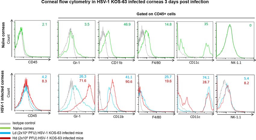
www.nature.com/scientificreports/
epitheliopathy we used a previously described m ethod37. In brief, a 1.0 mg fluorescein sodium ophthalmic strip
(FUL-GLO, Akorn, Lake Forest, IL) was diluted in 1 ml 1 × PBS (Life Technologies, Carlsbad, CA) and 5 µl of
0.1% fluorescein solution was applied on the ocular surface. After 15 s, the cornea was washed with PBS 2–3
times and using a cotton tip applicator, the excess fluid gently removed after each wash to prevent pooling of flu-
orescein. Photos were obtained by a Canon digital camera (PowerShot A2200), mounted on top of the slit-lamp
binocular (SL-1E, Topcon, Oakland, NJ), using white light (prior to fluorescein staining) and cobalt blue light.
Images were then analyzed with ImageJ software38 (public domain Java image processor, NIH, Bethesda, MD;
https://imagej.nih.gov/ij/) to calculate the percentage of epithelial defect. For this purpose, the area of epitheliop-
athy, defined as the distinct fluorescein stained areas (including dendrites), was selected using polygon selection
tool and measured first. Then the epitheliopathy area was divided by the area of the whole cornea, measured by
selection of the whole cornea using oval selection tool. Clinical grading was performed by 3 masked observers.
Viral titration of corneas and trigeminal ganglia. We pooled and homogenized 2 corneas or 2 ipsi-
lateral TGs from 2 infected mice in 300 µl of RPMI using gentle MACS dissociator for each experiment. A
standard plaque assay10 was performed to titer the virus. In brief, serial dilutions of the homogenized superna-
tants were added in duplicate to the monolayer of Vero cells in 6 well plates. Plates were incubated at 37 °C for
1 h. Subsequently, solution of 0.5% methylcellulose (Sigma-Aldrich, St. Louis, MO) and 5% fetal bovine serum
(FBS; Gemini BioProducts, West Sacramento, CA) in Dulbecco’s Modified Eagle’s Medium (DMEM, Corning/
Mediatech, Inc., Manassas, VA) were added to each well and plates were incubated again at 37 °C. After 48–72 h,
well contents were vacuumed and 1 ml of 1% crystal violet (Sigma-Aldrich) was added to each well. After 30 min
plates were washed with PBS and number of white spots was counted to calculate the virus titer as PFU/ml
(detection limit of 100 PFU/ml).
Corneal whole‑mount immunofluorescent staining. Harvested corneas were fixed in chilled acetone
for 15 min at − 20 °C and washed 3 times in PBS for 10 min each. Corneas were blocked in 3% bovine serum
albumin containing 1% anti-FcR monoclonal antibody (CD16/CD32; BioXCell, West Lebanon, NH) for 60 min
at room temperature. Corneas were then stained for PE-conjugated rat anti-mouse CD45 (30-F11) (Biolegend,
San Diego, CA) and NL637-conjungated mouse anti-βIII-Tubulin (R&D, Minneapolis, MN, USA) overnight at
4 °C on the shaker. Corneas were washed 3 times in PBS for 10 min each, flattened after 3 radial incisions, and
mounted on a slide using 4′, 6-diamidino-2-phenylindole (DAPI) mounting media (Vectashield, Vector Labo-
ratories, Inc. Burlingame, CA, USA).
Confocal microscopy and image analysis. The whole-mounted corneas were imaged using a confo-
cal Leica TCS SP5 (Leica Microsystems, Wetzlar, Germany). Central and peripheral areas of each cornea were
assessed separately, with the central area defined as the area within 0.6 mm of the corneal center, and the periph-
ery defined as being within a 0.9–1.5-mm radial distance from the center. Three different non-overlapping fields
from the peripheral cornea and one central field were imaged for each cornea. At least three different corneas
were stained and imaged per group. Stacked images were analysed off-line with IMARIS software version 8.0
(Bitplane AG, Zurich, Switzerland) to calculate the number of CD45 (pan-leukocyte marker) positive cells. The
number of cells in peripheral corneal fields was then averaged. Corneal nerves were traced using NeuronJ39,40
(http://www.imagescience.org/meijering/software/neuronj/), a free semi-automatic software. Nerve density was
reported as mm/mm2.
Flow cytometry. At 3 dpi, infected corneas were excised and digested in DNAse I (2 mg/ml) and colla-
genase D (2 mg/ml) (Roche Diagnostics GmbH, Manheim, Germany) in RPMI for 30 min at 37 °C. Single-cell
suspensions were filtered through a 70 µm cell strainer (VWR International, LLC, Radnor, PA) and washed with
PBS to stop the enzyme reaction. The remaining tissues were further digested at 37 °C for additional 30 min.
Remaining single cells were filtered and washed again and added to the previously digested cells. Similarly, both
corneas of wild-type C57BL/6 mice were excised and single cells were obtained. Corneal single cell suspensions
were blocked in 1% anti-FcR monoclonal antibody in PBS for 15 min at 4 °C to minimize non-specific staining
and then further incubated with the desired conjugated antibodies for 1 h at 4 °C. Conjugated antibodies used
for immunohistochemistry staining included: Pacific Blue rat anti-mouse CD45 (30-F11), phycoerythrin (PE)-
conjugated mouse anti-mouse NK-1.1 (PK136), PE-conjugated rat anti-mouse Gr-1 (RB6-8C5), Alexa fluor-
647-conjugated rat anti-mouse F4/80 (BM8), and Alexa fluor-647-conjugated rat anti-mouse/human CD11b
(M1/70) all from Biolegend. Fluorescein isothiocyanate (FITC)-conjugated rat anti-mouse CD11c (HL3) was
purchased from BD Pharmingen (San Diego, CA). Isotype controls included Pacific Blue rat IgG2b, κ (RTK4530),
Alexa fluor-647-conjugated rat IgG2a, κ (RTK2758), PE-conjugated rat IgG2b, κ (RTK4530), FITC Armenian
Hamster IgG (HTK888), and Alexa fluor-647-conjugated rat IgG2b, κ (RTK4530) all from Biolegend. Stained
corneal single cell suspensions were washed with PBS, centrifuged (1300 RPM for 10 min) and re-suspended in
4% paraformaldehyde. The stained cells were analyzed using a BD LSR II flow cytometer (BD Biosciences, San
Jose, CA). FlowJo software (FlowJo, LLC, Ashland, OR) was used for additional cell analysis. Doublet cells were
excluded by eliminating disproportions between forward scatter-width (FSC-W) versus forward scatter-height
(FSC-H). Gates were plotted based on appropriate isotype control staining for each antibody.
Corneal sensation. Corneal nerve sensitivity was measured by a Cochet-Bonnet esthesiometer (Luneau
Technology, Pont-de-l’Arche, France). In brief, the mono-filament was applied to the central cornea of un-
anesthetized mice, avoiding contact with whiskers and eyelashes. Maximum mono-filament length is 6 cm; the
Scientific Reports | (2021) 11:4267 | https://doi.org/10.1038/s41598-021-83412-9 3
Vol.:(0123456789)www.nature.com/scientificreports/
micro-filament length required to measure a blink reflex was recorded. The procedure was repeated three times
to ensure reproducibility and average length was used for analysis.
Statistical analysis. Statistical analysis was performed with Prism version 6.01 (GraphPad Software, Inc.,
La Jolla, CA). We used chi square test to compare incidence percentage of corneal opacity, blepharitis, and
neovascularisation between two different groups of infected mice. Student’s t-test was used to compare means
between the two groups and one-way ANOVA, followed by Tukey’s test used to compare the means among three
or more groups. Results were shown as mean ± standard error of the mean (SEM) and p values of < 0.05 were
considered as statistically significant.
Results
Titer‑dependent incidence and clinical severity in mice infected with Ld (2 × 10⁴ PFU) versus
Hd (2 × 10⁶ PFU) HSV‑1 KOS‑63. Ten to 17 mice at each time point were evaluated for clinical signs of
the disease in either 2 × 10⁴ PFU (Ld) or 2 × 10⁶ PFU (Hd) KOS-63-infected mice. At the end of the follow-up
period (7 dpi), the incidence of blepharitis did not show a significant difference between the two doses of the
virus (93.7% and 94.4% in Ld and Hd KOS-63-infected mice, respectively, p = 0.93). However, the incidence of
mice, which developed corneal opacity was significantly higher in the Hd KOS-63-infected mice (88.2%) as
compared to the Ld KOS-63-infected mice (56.2%), p = 0.03. The incidence of corneal neovascularisation was
also significantly higher in the Hd KOS-63-infected mice (81.8%), as compared to the Ld KOS-63-infected mice
(31.2%), p = 0.009.
Representative white light and cobalt blue filtered images for comparison of corneal opacity and epitheli-
opathy in the Ld and Hd HSV-1 KOS-63 infected corneas, as well as naïve and sham-infected mice at different
time points are illustrated in Fig. 1. Blepharitis was not present at 1 dpi in any of the groups; however, at 3 dpi
blepharitis scores were 0.18 ± 0.10 and 0.62 ± 0.12 in the Ld- and Hd-infected mice, respectively (p = 0.68). At
7 dpi, blepharitis scores increased significantly to 2.06 ± 0.26 and 3.11 ± 0.25 in the Ld- and Hd-infected mice,
respectively (p = 0.001). Blepharitis scores showed an increasing trend from 1 to 7 dpi (ANOVA: p < 0.0001)
(Fig. 2A).
Corneal opacity scores continuously increased in both groups from 1 to 7 dpi (ANOVA: p < 0.0001). Corneal
opacity was significantly higher in the Hd KOS-63-infected mice as compared to Ld KOS-63-infected mice at 7
dpi (2.23 ± 0.27 vs. 0.87 ± 0.25, respectively; p = 0.0003) (Fig. 2B). Further, corneal neovascularization increased
significantly from 1 to 7 dpi in both groups, and was more prominent in the Hd compared to the Ld KOS-
63-infected at all time points (ANOVA: p < 0.0001). Similar to HSV-1 KOS-63-infected mice, corneal opacity was
higher in Hd as compared to Ld HSV-1 McKrae-infected mice (Supplementary Fig. 1). There was no significant
difference among Hd KOS-63 vs. Hd McKrae infected corneas at 1, 3, and 5 dpi (p > 0.99). At 5 dpi, corneal opac-
ity was significantly higher in Hd McKrae corneas as compared to Ld KOS-63 corneas (0.83 ± 0.16 vs. 2.0 ± 0.0;
p = 0.029) (Supplementary Fig. 1).
At 7 dpi, the time point with highest neovascularization, 6.00 ± 1.39 and 0.75 ± 0.35 clock hours of the Hd
and Ld KOS-63-infected corneas demonstrated presence of vessels, respectively (p < 0.0001) (Fig. 2C). Moreover,
epithelial defects were larger in the Hd as compared to the Ld KOS-63-infected mice at all time points, but were
only significant at 3 dpi (23.59 ± 6.67% vs. 3.44 ± 0.96%, respectively; p = 0.001) (Fig. 2D). No opacity, neovascu-
larization, and blepharitis were observed in naïve and sham-infected mice (1, 3, and 5 days post-scarification;
Fig. 2 and Supplementary Fig. 2). Blepharitis scores were significantly higher starting at 5 dpi in the Hd KOS-
63-infected mice, as compared to naïve and sham-infected mice (Fig. 2A). Opacity scores were significantly
higher only in the Hd KOS-63-infected mice, than in naïve and sham-infected mice (Fig. 2B). Neovasculariza-
tion was significantly increased only in Hd KOS-63 infected mice between 3 and 7 dpi, as compared to controls
(Fig. 2C). Very minor epithelial changes were present in sham-infected corneas (only at 1-day post-scarification)
(0.34 ± 0.27% vs. 0.0% in naïve mice; p = 0.99) (Fig. 2D and Supplementary Fig. 2).
HSV‑1 KOS‑63 strain is capable of infecting corneas and ipsilateral trigeminal ganglia. In
infected corneas, the maximum viral load was detected at 1 dpi but it was undetectable at 7 dpi in both Hd and
Ld KOS-63-infected groups. However, the corneal viral titer was significantly higher in the Hd compared with
the Ld KOS-63-infected mice at 1 dpi (5.32 ± 0.20 vs. 4.47 ± 0.06 Log PFU, respectively; p = 0.004) and 3 dpi
(4.22 ± 0.07 vs. 3.30 ± 0.12 Log PFU, respectively; p = 0.001) (Fig. 3A). Interestingly, corneal viral titers in Hd
McKrae-infected mice were 4.82 ± 0.02 Log PFU at 7 dpi, as compared to undetected levels in Ld and Hd KOS-
63-infected mice (p < 0.0001; Supplementary Fig. 2A). While at 1 dpi, viral titers in TG of infected mice were not
detectable, viral titers in TGs significantly increased at 3 and 5 dpi in the Hd KOS-63-infected group (6.27 ± 0.06
and 5.68 ± 0.07 Log PFU, respectively) compared with Ld KOS-63-infected group (5.29 ± 0.14 and 4.76 ± 0.18 Log
PFU) (both p < 0.001). However, at 7 dpi viral titers reached similar levels in the Hd and Ld KOS-63-infected
mice (3.37 ± 1.58 and 3.30 ± 1.51 Log PFU, respectively) (Fig. 3B). TG viral titers in Ld and Hd KOS-63-infected
mice were significantly higher than in McKrae-infected mice at 3 dpi (3.07 ± 0.08 Log PFU; p < 0.0001 for both),
but similar at 7 dpi (Supplementary Fig. 2B). Naïve and sham-infected controls were negative (Fig. 3).
Immune cell infiltration in acute herpes simplex keratitis induced by HSV‑1 KOS‑63 strain. The
number of bone marrow (BM)-derived immune cells ( CD45+ cells), identified by immunohistochemistry stain-
ing in the central and peripheral corneas of naïve, sham control, and HSV-1 KOS-63-infected mice at 1, 3,
and 7 dpi have been demonstrated in Fig. 4. At 3 and 7 dpi, C D45+ cell density increased in the central cor-
2
nea of both Ld (686.5 ± 8.50 and 735.9 ± 88.89 cells/mm , respectively) and Hd (738.8 ± 68.85 and 790.0 ± 116.8
cells/mm2, respectively) KOS-63-infected mice compared to naïve (170.7 ± 16.04 cells/mm2) and sham-infected
Scientific Reports | (2021) 11:4267 | https://doi.org/10.1038/s41598-021-83412-9 4
Vol:.(1234567890)www.nature.com/scientificreports/
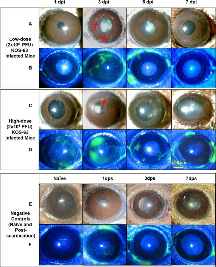
Figure 1. Representative slit-lamp photos following corneal inoculation with low dose (Ld, 2 × 10⁴ PFU; A,
B), high dose (Hd, 2 × 10⁶ PFU; C, D) HSV-1 KOS-63 strain, and negative controls (naïve and sham-infected;
E, F). Representative white light images from Ld (A) and Hd (C) inoculated corneas at 1, 3, 5, and 7 days
post infection (dpi) demonstrate the corneal opacity at these time points. Hd HSV-1 infected mice developed
more severe corneal opacity as compared to Ld infected mice. Red arrows are showing the opacity at 3 dpi.
Representative cobalt blue filtered light images from Ld (B) and Hd (D) inoculated corneas demonstrate corneal
epithelial defect following staining the corneas with 0.1% fluorescein at 1, 3, 5, and 7 dpi. In both Ld and Hd
KOS-63 infected mice corneal epithelial defects peaked at 3 dpi. Representative white light (E) and blue filtered
(F) corneal images in naïve and 1,3, and 7 days post scarification (dps).
(143.1 ± 16.15 cells/mm2) mice (all p < 0.05). However, there was no significant difference in the CD45+ cell den-
sity in the central cornea between Ld and Hd KOS-63-infected groups at any time point (all p > 0.05; Fig. 4A).
Scientific Reports | (2021) 11:4267 | https://doi.org/10.1038/s41598-021-83412-9 5
Vol.:(0123456789)www.nature.com/scientificreports/
Figure 2. Comparison of clinical scores in naïve, sham-infected (1-day post scarification (dps)), and following
corneal infection with Ld and Hd HSV-1 KOS-63 strain at 1, 3, 5, and 7 days post infection (dpi). (A)
Blepharitis scores. At 1 dpi no blepharitis was observed but it increased gradually in both Hd and Ld infected
mice. Blepharitis score was significantly higher in the Hd infected mice compared to Ld infected mice at 7 dpi
(**p = 0.001). No blepharitis was observed in naïve and sham infected mice (1–7 dps), 1 dps is presented as
representative. (B) Corneal opacity scores are demonstrated in both Ld and Hd infected mice. Opacity scores
were higher in the Hd infected mice as compared to Ld infected mice and was significant at 5 dpi (*p = 0.04)
and 7 dpi (**p = 0.003). No corneal opacity was observed in naïve and sham infected mice (1–7 dps), 1 dps is
presented as representative. (C) Corneal neovascularization scores according to the clock hours area of vessel
coverage. Neovascularization was more significant in the Hd infected mice compared to Ld infected mice at
7 dpi (ǂp < 0.0001). No corneal neovascularization was observed in naïve and sham-infected mice (1–7 dps), 1
dps is presented as representative. (D) Percentage of corneal epithelial defect was higher in the Hd infected mice
compared to Ld infected mice in all time points and was significant at 3 dpi (**p = 0.001). No corneal opacity
was observed in naïve mice. Minor epithelial defect remained after corneal scarification only at 1 dps, which
was not statistically significant compared to naïve mice. Bars are showing mean ± SEM. ANOVA: p < 0.0001. ¥,
demonstrates the statistical significance (p < 0.05) between the marked column and naïve and sham-infected. p
values are calculated by one-way ANOVA followed by Tukey’s multiple comparison test.
In both Ld and Hd KOS-63-infected groups, the number of C D45+ cells in the peripheral cornea increased
2
significantly at 1 dpi (639.4 ± 121 and 1092 ± 61.66 cells/mm , respectively), as compared to naïve (273.2 ± 21.92
cells/mm2; p = 0.03 and p < 0.0001, respectively) and sham-infected mice (334.4 ± 34.16 cells/mm2; p = 0.02 and
p < 0.0001, respectively), and remained elevated until 7 dpi. The density of CD45+ cells significantly increased
in the Hd-infected group compared to the Ld-infected group at 1 dpi (p = 0.01) (Fig. 4B). Additionally, we
compared CD45+ cell density in central and peripheral cornea between HSV-1 KOS-63 and HSV-1 McKrae-
infected mice. C D45+ cell density was significantly increased in the central corneas of McKrae-infected mice
(2088 ± 180.4 cells/mm2) at 7 dpi, as compared to Ld and Hd KOS-63-infected mice (p < 0.0001 for both; Sup-
plementary Fig. 3A). CD45+ cell density in the corneal periphery was significantly lower at 1 dpi (291.0 ± 32.85
cells/mm2) and 3 dpi (361.8 ± 37.71 cells/mm2) in McKrae-infected corneas as compared to similar timepoints
in Hd KOS-63-infected corneas (p < 0.001 and p = 0.045, respectively; Supplementary Fig. 3B). However, at 7
dpi CD45+ cell density in peripheral corneas was significantly higher in McKrae-infected mice (3925.0 ± 230.1
cells/mm2), as compared to both Ld and Hd KOS-63-infected mice (p < 0.0001 for both; Supplementary Fig. 3B).
To further analyze the subpopulation of infiltrated cells in corneas of HSV-1 KOS-63 strain infected mice,
we used flow cytometry. The percentage of CD45 + cells in the cornea increased to 4.2% and 8.3% in Ld and
Hd groups, respectively, compared to naïve corneas (2.1%). We then gated on CD45+ cells and compared the
percentage of other immune cell subpopulations between naïve and infected mice (Fig. 5). While Gr-1+ cells
Scientific Reports | (2021) 11:4267 | https://doi.org/10.1038/s41598-021-83412-9 6
Vol:.(1234567890)www.nature.com/scientificreports/
Figure 3. The viral titer levels in corneas and trigeminal ganglia (TG) of Ld and Hd HSV-1 KOS-63 infected
mice at 1, 3, 5, and 7 days dpi. Corneal and TG of naïve and sham-infected mice served as controls. (A) Mean
viral titer in corneas; Hd infected mice had higher viral titer at 1dpi (*p = 0.004) and 3 dpi (**p = 0.001). (B)
Mean viral titer in the TGs; It demonstrated higher titers in the Hd-infected mice compared to Ld-infected
mice at 3 dpi (**p = 0.0008) and 5 dpi (ǂp < 0.0001). At each time point 6–10 TGs or corneas, from 6 to 10 mice
and from 3 to 4 independent experiments, were homogenized and tittered in pairs (2 corneas or 2 TGs were
homogenized in a same tube for titration). Bars are showing mean ± SEM. ANOVA: p < 0.0001. ¥, demonstrates
the statistical significance (p < 0.05) between the marked column and naïve and sham-infected. p values are
calculated by one-way ANOVA followed by Tukey’s multiple comparison test.
Figure 4. Number of CD45 + inflammatory cells in the central (A) and peripheral (B) cornea in naïve, sham-
infected, Ld and Hd KOS-63-infected mice. Whole mount confocal images of corneas, which were stained with
PE-conjugated rat anti-mouse CD45 (30-F11) antibody, were analyzed with IMARIS software to calculate the
number of CD45 + cells in the cornea. At each post infection timepoint 3–9 corneas were stained. In addition,
6 and 9 naïve and sham-infected (1 day post scarification) corneas were stained, respectively. Bars are showing
mean ± SEM. ANOVA: p < 0.0001; *p = 0.01, calculated by one-way ANOVA followed by Tukey’s multiple
comparison test. ¥, demonstrates the statistical significance (p < 0.05) between the marked column and the
sham-infected controls.
constituted 3.5% of leukocytes ( CD45+ cells) in naïve corneas, their percentage increased to 26.3% and 71.6% in
the Ld and Hd infected mice, respectively. 41.1% and 90.6% of C D45+ cells expressed C
D11b+ in the Ld and Hd
infected groups, respectively. In addition, F4/80+ cells constituted 25.7%, and 19.6% of CD45 + cells in Ld- and
Hd-infected mice, respectively (Fig. 5). Furthermore, 74.1% and 28.7% of corneal CD45+ cells expressed CD11c+
in Ld- and Hd-infected mice, respectively. Moreover, natural killer cells ( CD45+ / NK-1.1+) were not detected in
Scientific Reports | (2021) 11:4267 | https://doi.org/10.1038/s41598-021-83412-9 7
Vol.:(0123456789)www.nature.com/scientificreports/
Figure 5. Flow cytometry histograms of Ld and Hd HSV-1 KOS-63-infected corneas at 3 dpi (lower row)
compared with naïve corneas (upper row). Histograms are representative from 2 independent experiments. In
one experiment corneas of 6 Hd- and 6 Ld-infected C57BL/6 mice were pooled and digested separately. In the
other experiment 27 corneas of naïve C57BL/6 mice harvested and digested together in a same fashion. Single
cell suspensions were stained for different cell markers. Gating was performed using isotype control antibodies.
CD45+ population was selected in each group initially and percentage of C D45+ cells to the total number of cells
is demonstrated in the histograms. Percentage of Gr-1+ cells, CD11b+ cells, and F4/80+ were demonstrated after
gating on CD45+ cell population. In the lower row blue line demonstrates the Ld and red line demonstrates the
Hd HSV-1 KOS-63-infected mice. Gray line represents isotype control in all histograms.
D45+ cells,
naïve corneas, while in the Ld and Hd infected corneas these cells constituted 5.4% and 8.2% of the C
+
respectively (Figure not shown). In summary, at 3 dpi the percentage of C D45 cells was 2 times higher in the
Hd-infected corneas as compared to Ld-infected. Moreover, gating on C D45+ cells, Gr-1+, CD11b+, and NK cell
percentage were higher in the Hd- versus Ld-infected corneas. Interestingly, C D11c+ cells constituted majority
of CD45+ cells in the Ld-infected corneas at 3 dpi and had higher percentage compared to Hd-infected (Fig. 5).
Different immune cell subpopulation at 3 dpi in McKrae-infected corneas are demonstrated in Supplementary
Fig. 3C. The majority of corneal immune cells in McKrae-infected corneas were Gr-1+ cells (48.2%).
HSV‑1 KOS‑63 strain results in nerve damage after corneal inoculation. Figure 6A demonstrates
representative confocal microscope micrographs of βIII-tubulin stained central and peripheral corneal nerves
in naïve, sham-infected, and Hd or Ld HSV-1 KOS-63-infected mice. Total nerve density did not significantly
decrease in sham-infected mice compared to naïve mice in either the central (94.72 ± 4.07 vs. 119.46 ± 0.58 mm/
mm2; p = 0.23) or peripheral corneas (79.12 ± 7.70 vs. 105.28 ± 8.61 mm/mm2; p = 0.39). At 1 dpi, there was no
significant loss of nerves in the central cornea of Ld KOS-63-infected mice, as compred to sham-infetced mice.
However, the central corneal nerve density significantly decreased at 3 and 7 dpi for the Ld and for all time-
points for the Hd KOS-63-infected mice, as compared to sham-infected (p < 0.05) (Fig. 6B). As early as 3 dpi,
the central corneal nerve density was significantly lower in the Hd compared with Ld KOS-63-infected mice
(16.79 ± 3.17 vs. 57.41 ± 14.64 mm/mm2, respectively; p = 0.004) (Fig. 6B). Peripheral corneal nerves further
decreased significantly at 7 dpi in the Ld KOS-63-infected mice (30.87 ± 3.61 mm/mm2) compared to sham-
infected (p = 0.005). Morevoer, in the Hd KOS-63-infected corneas, peripheral corneal nerves decreased signifi-
cantly at 3 dpi (33.48 ± 12.99 mm/mm2) and 7 dpi (12.79 ± 8.55 mm/mm2) compared to sham-infected (p = 0.01
and p < 0.0001, respectively) (Fig. 6C). Interestingly, there was no significant difference in peripheral corneal
nerve density between the Ld and Hd KOS-63-infected groups (Fig. 6C). However, we did observe a linear
decrease in the central and peripheral corneal nerve densities during the 7 days follow-up period in both Ld and
Hd KOS-63-infected mice (ANOVA: p < 0.0001). Similar to Hd KOS-63-infected corneas, central corneal nerve
density was significantly reduced at 1 dpi in Hd McKrae-infected corneas (62.20 ± 4.32 mm/mm2), as compared
to naïve and sham-infected controls. (p = 0.0003 and p = 0.02, respectively (Supplementary Fig. 4).
Additionally, corneal esthesiometry confirmed significantly decreased corneal nerve sensitivity, starting from
5 dpi in both Ld and Hd HSV-1 KOS-63-infected corneas. At 5 dpi corneal nerve sensitivity was significantly
lower in the Hd-infected corneas, as compared to Ld-infected corneas (filament length of 0.77 ± 0.37 cm vs.
4.23 ± 0.26 cm; p < 0.0001). Similarly, at 7 dpi, corneal nerve sensitivity was significatly lower in the Hd-infected
corneas as compared to Ld-infected corneas (filament length of 0.0 ± 0.0 cm vs. 1.73 ± 0.61 cm; p = 0.007) (Fig. 7).
Scientific Reports | (2021) 11:4267 | https://doi.org/10.1038/s41598-021-83412-9 8
Vol:.(1234567890)www.nature.com/scientificreports/
Figure 6. Corneal nerve density in the center and periphery of naïve and sham-infected corneas as well as Ld
and Hd HSV-1 KOS-63-infected corneas. (A) Sample confocal micrographs of whole mount corneas stained
with NL637 conjugated beta III tubulin antibody. There is a non-significant corneal nerve loss in the sham
control group compared with naïve corneas. Sample central and peripheral corneal nerve tracing in naïve
and sham-infected mice is demonstrated using NeuronJ software. Diminished corneal nerves in the infected
corneas were observed at 1, 3, and 7 dpi especially in the central cornea of Hd infected mice. (B) Corneal nerve
density (mm/mm2) was calculated with NeuronJ software after tracing the nerves in whole mount confocal
micrographs. (C) Corneal nerve density (mm/mm2) in the peripheral cornea was calculated the same as
central cornea. Each group is demonstrating the average of at least 3 whole mount stained corneas. Naïve and
sham-infected central and peripheral corneal nerves were not significantly different although there was a trend
towards more corneal nerve loss in the sham-infected group compared to the naïve mice corneas. ANOVA:
p < 0.0001; *p = 0.01; **p = 0.004 was calculated by one-way ANOVA followed by Tukey’s multiple comparison
test. ¥, demonstrates the statistical significance (p < 0.05) between the marked column and the sham-infected
controls.
Discussion
Herein, we demonstrate that the HSV-1 KOS-63 strain can cause a titer-dependant keratitis in C57BL/6 mice
when applied topically after corneal scarification. Although the HSV-1 KOS cannot infect the central nervous
system and cause encephalitis when applied peripherally, through corneal i noculation9,21,25, we show that it can
infect the corneal nerves as part of the peripheral nervous system, and subsequently migrate to the TGs. This
is further demonstrated by corneal nerve degeneration observed in our studies. While development of stromal
keratitis following inoculation with KOS strain in C57BL/6 mice has been recently reported, the presence of
the virus in the TGs or corneal leukocyte and nerve alterations have not been previously a ssessed41. However,
in contrast, Nguyen et al.32 did not observe stromal keratitis following ocular inoculation of HSV-1 KOS on
scarified C57BL/6 mice.
We show that Hd KOS-63 strain not only increases the frequency of corneal opacity and neovasculariza-
tion, but also results in increased severity of ocular manifestations as compared to Ld of virus. Similarly, using
McKrae strain as a positive control we observed a titer-dependent keratitis. Consistent with our study, higher
doses of HSV-1 strain RE demonstrated more severe corneal neovascularization in BALB/c mice at 15 dpi42.
Moreover, Zheng et al.43 demonstrated that 2.5 × 10⁷ PFU of HSV-1 McKrae caused significantly higher corneal
opacity scores than a 100 times lower dose. However, albeit higher dose of HSV-1 KOS-63 (5 × 10⁴ PFU vs.
2.5 × 10⁴ PFU) increased the incidence of herpetic stromal keratitis in BALB/c mice, it did not cause more severe
keratitis27. Corneal infection using other HSV-1 strains on mice (CJ394, OD4, 994, 401, 394)9,18 or rabbits (RE
Scientific Reports | (2021) 11:4267 | https://doi.org/10.1038/s41598-021-83412-9 9
Vol.:(0123456789)www.nature.com/scientificreports/
Figure 7. Corneal esthesiometry results. Corneal sensitivity was measured using Cochet-Bonnet esthesiometer
in the central corneas. Maximum filament length is 6 cm and indicates full sensitivity. Corneal nerve sensitivity
was significantly decreased after 5 dpi in both low dose (Ld) and high dose (Hd) HSV-1 KOS-63-infected
corneas as compared to naïve corneas. Bars are showing mean ± SEM. p values are calculated by one-way
ANOVA followed by Tukey’s multiple comparison test. ***p < 0.0001, **p = 0.007. ¥, demonstrates the statistical
significance (p < 0.05) between the marked column and naïve cornea.
and McKrae)22 did not demonstrate any titer-dependant severity of the disease. Corneal epithelial defects at 3 dpi
were at its highest for both Hd and Ld HSV-1 KOS-63-infected corneas; this is in accordance with a significant
rise of inflammatory cells ( CD45+ cells) in the central cornea at 3 dpi, as compared to naïve corneas. Significantly
larger epithelial defects were observed in the Hd HSV-1 KOS-63 infected corneas as compared to the Ld group.
Higher corneal viral titer in the Hd KOS-63 infected group may, in part, explain the increased severity of clinical
signs observed in this group, as compared to Ld KOS-63 infected group. Similarly, mice that developed herpetic
stromal keratitis (HSK), using RE strain, showed higher viral titer in their tear films (although not significant)
compared to those without HSK44.
We demonstrate increased number of infiltrating leukocyte (CD45+ cells) in corneas of both KOS-63
infected groups. We have previously shown an increased influx of C D45+ cells in the cornea after HSV-1 McK-
10,45
rae infection . Here we show that although KOS-63-infected corneas have higher number of C D45+ cells at 3
and 5 dpi compared to McKrae infected mice, their density is lower at 7 dpi, suggesting an earlier, but less severe
inflammatory response with KOS-63 infection. Interestingly, analysis of corneal leukocyte subpopulations is dif-
ferent between Hd and Ld KOS-63-infected mice. C D11b+ cells are suggested to be involved in causing blepharitis
8
in HSV-1 infected m D11b+ cells in the Hd KOS-63-infected mice might
ice . Therefore, a higher percentage of C
explain the more severe blepharitis in the Hd as compared to the Ld KOS-63-infected group. Neutrophils are a
source of vascular endothelial growth factor (VEGF)-A and have a crucial role in inducing angiogenesis follow-
ing HSV-1 ocular infections46. Interestingly, we show a larger infiltration of CD45+ Gr-1+ cells into the cornea
of the Hd KOS-63-infected mice (71.6%) as compared to the Ld-infected group (26.3%), which can explain the
higher neovascularization score in the former group. NK cells and macrophages play a role in the control and
clearance of the virus from the c ornea6. Percentages of both cells increase at 3 dpi as compared to naïve mice.
CD11c+ DCs are believed to control the virus replication in the cornea indirectly by increasing migration of NK
cells to the center of c ornea47 and by systemic dissemination of the virus to the TGs, thus decreasing the virus
titer in the cornea10. Interestingly, we have recently demonstrated that depletion of C D11c+ cells resulted in more
10
severe acute HSV k eratitis . The current study shows that Hd-infected mice have a lower frequency of C D11c+
cells among C D45+ immune cells (28.7%) as compared to Ld-infected mice (74.1%) at 3 dpi, which can partly
explain the higher viral titer in the cornea and more sever keratitis in the Hd-infected mice. It is plausible that in
the presence of a lower viral load, DCs can increase and protect the cornea from severe damage, while a higher
viral load could prevent increased DC density and therefore cause more severe clinical symptoms.
Our data shows that both central and peripheral subbasal corneal nerve density decreases gradually over the
first 7 days after inoculation, with more severe loss in the central cornea and in Hd KOS-63-infected mice. This
has been in concordance with significant decreased corneal sensation at 5 and 7 dpi. Corneal nerve damage fol-
lowing HSV infection has been well demonstrated in clinical and preclinical studies7,10,15,16,48,49. Both KOS and
McKrae HSV-1 strains have previously shown to result in nerve degeneration in BALB/c and C57BL/6 mice,
respectively7,10,16,45. We show that as soon as 1 dpi, Hd HSV-1 KOS-63 and Hd McKrae reduce the central corneal
nerve density by 42.34% and 34.10%, as compared to sham-infected C57BL/6 mice, respectively. Previously, our
group has shown similar results with McKrae infected m ice10,45. Further, Chucair-Elliott et al., using the same
mouse model, demonstrated a significant decrease in corneal nerves at 6, 8, and 14 dpi with 103 PFU/cornea
HSV-1 McKrae infection, as compared to uninfected c orneas7. Moreover, Yun et al. demonstrated dramatic
Scientific Reports | (2021) 11:4267 | https://doi.org/10.1038/s41598-021-83412-9 10
Vol:.(1234567890)www.nature.com/scientificreports/
reduction of corneal nerve fibers in BALB/c mice at 28 dpi with the HSV-1 KOS s train16, albeit without quantifi-
cation of nerve density. Furthermore, quantification methods for nerve density have been qualitative in the latter
two studies and are thus not comparable to the quantitative method used herein. The exact cause of denervation
has not been elucidated yet, although direct nerve damage by the virus and viral-induced inflammation could
be leading c auses10,16. Based on the current study, higher corneal nerve loss (at 3 dpi) in the Hd-infected group
compared to the Ld group can be explained either by a higher corneal viral titer and/or by a higher number of
PMNs and NK cells in the Hd-infected mice compared to the Ld-infected mice. On the other hand, more severe
nerve loss in the Hd-infected mice as compared to Ld-infected mice may explain more severe neovascularization
and immune infiltration into the cornea and not necessarily direct effect of the virus. We have shown an inverse
correlation between corneal nerve density and immune cell infiltration in the cornea in clinical and pre-clinical
studies50. Tepelus et al. also found similar inverse correlation in patients with dry eye disease51. Interestingly, Yun
et al.16 concluded that corneal nerve damage can be maintained even without significant corneal inflammation
after HSV-1 infection. These studies assessed nerve loss from different perspectives and still the cause of nerve
damage in HSV keratitis remains controversial.
It has been previously reported that the HSV-1 KOS is less virulent than strain 17 and McKrae and has
mutated US9 and US8A genes as compared with strain 1752,53. Different variants of KOS have been used in
laboratories up to date, including KOS-63, KOS-79, and KOS1.1 (KOS (M))21,54. In addition, different mutant
derivatives of HSV-1 KOS have been used to study the virulence, latency, and reactivation of HSV-155–57. Bowen
et al.54 demonstrated that despite more than 99.2% similarity among different passages of a same virus (HSV-1
KOS-63), there are small genomic differences. Specifically, the HSV-1 KOS-63 strain that was used for the first
time by Dix et al.21 is different in 3 genes (proteins), gB (UL27), VP1/2 (UL36), and RR (UL39), as compared
to the HSV-1 KOS-63 from Kinchington group (which is used in this study)54. UL36 and UL39 proteins were
shown previously to influence virulence and replication of HSV-1 virus, respectively58. Thus, different sub-strains,
mutants, and maybe different passages of HSV-1 KOS used by different laboratories may provide additional
explanation for variable clinical scores and the controversy in the reported literature.
Corneal wounding may result in activation of resident leukocytes and infiltration of leukocytes to the c ornea59.
Therefore, in order to eliminate the possibility that scarification creates a confounding factor, we used sham-
infected mice as an additional control group, in which scarified corneas were inoculated with PBS. We demon-
strate that superficial corneal scarification, which is much less extensive than corneal wounds described by Stepp
et al.59 do not lead to any significant corneal disease, immune cell infiltration, or corneal nerve damage. Similarly,
BenMohamed et al.12 and Yuan et al.60 did not show any significant eye disease and corneal neovascularization
post scarification, respectively. Prior studies have shown that HSV-1 McKrae can infect the murine corneas
without scarification, although this has not been shown with the KOS s train12,29, thus requiring scarification to
study the Ld and Hd KOS-63 strain effects. However, in this study in order to exclude any confounding factor we
performed scarification prior to corneal inoculation with HSV-1 KOS-63 or HSV-1 McKrae stains. Given that
the primary objective of the study was to compare the Hd and Ld HSV-1 KOS-63 corneal infection, these results
were compared to the more virulent McKrae strain that was used as a positive control. We used a modified cor-
neal opacity scoring system, with a more detailed scale than mild, moderate and severe opacity, which has been
widely used previously. Furthermore, we used clock hours to report clinical neovascularization. Although this
method is sensitive to differentiate subtle changes at different time points post infection but it cannot distinguish
the extent of neovascularization. Thus, we chose this scoring system, as our goal was to assess virulence at the
acute phase of the disease and to tease out minor changes. Moreover, since we followed infected mice for only
7 days to assess the acute phase of HSV keratitis, we cannot extend our dose-dependent findings to the chronic
phase of infection. In addition, the plaque assay that we used for virus titration has the detection limit of 10 PFU.
In conclusion, our results are indicative of ocular virulence of HSV-1 KOS-63 in a dose-dependent fashion
when applied on scarified corneas of C57BL/6 mice. Moreover, we showed that HSV-1 KOS-63 dose can affect
incidence, viral titers in the cornea and TG, immune cell infiltration to the cornea, and finally corneal nerve
damage at different time points pi. Therefore, the choice of a suitable viral strain and viral dose, based on the
aims of the study, is of great importance. However, whether the viral load or different immune cell infiltrates
(or both) are responsible for more sever clinical manifestations and corneal nerve damage in the Hd versus Ld
infected mice is not clear.
Received: 26 April 2020; Accepted: 27 January 2021
References
1. Farooq, A. V. & Shukla, D. Herpes simplex epithelial and stromal keratitis: an epidemiologic update. Surv. Ophthalmol. 57, 448–462
(2012).
2. Rowe, A. M. et al. Herpes keratitis. Prog. Retin. Eye Res. 32, 88–101 (2013).
3. Kaye, S. & Choudhary, A. Herpes simplex keratitis. Prog. Retin. Eye Res. 25, 355–380 (2006).
4. Koganti, R., Yadavalli, T. & Shukla, D. Current and emerging therapies for ocular herpes simplex virus type-1 infections. Micro-
organisms 7, 429 (2019).
5. Koujah, L., Suryawanshi, R. K. & Shukla, D. Pathological processes activated by herpes simplex virus-1 (HSV-1) infection in the
cornea. Cell Mol. Life Sci. 76, 405–419 (2019).
6. Biswas, P. S. & Rouse, B. T. Early events in HSV keratitis–setting the stage for a blinding disease. Microbes Infect. 7, 799–810 (2005).
7. Chucair-Elliott, A. J., Zheng, M. & Carr, D. J. Degeneration and regeneration of corneal nerves in response to HSV-1 infection.
Invest. Ophthalmol. Vis. Sci. 56, 1097–1107 (2015).
8. Ghiasi, H. et al. Corneal macrophage infiltrates following ocular herpes simplex virus type 1 challenge vary in BALB/c mice vac-
cinated with different vaccines. Vaccine 19, 1266–1273 (2000).
Scientific Reports | (2021) 11:4267 | https://doi.org/10.1038/s41598-021-83412-9 11
Vol.:(0123456789)www.nature.com/scientificreports/
9. Grau, D. R., Visalli, R. J. & Brandt, C. R. Herpes simplex virus stromal keratitis is not titer-dependent and does not correlate with
neurovirulence. Invest. Ophthalmol. Vis. Sci. 30, 2474–2480 (1989).
10. Hu, K., Harris, D. L., Yamaguchi, T., von Andrian, U. H. & Hamrah, P. A dual role for corneal dendritic cells in herpes simplex
keratitis: local suppression of corneal damage and promotion of systemic viral dissemination. PLoS ONE 10, e0137123 (2015).
11. Hendricks, R. L. & Tumpey, T. M. Contribution of virus and immune factors to herpes simplex virus type I-induced corneal
pathology. Invest. Ophthalmol. Vis. Sci. 31, 1929–1939 (1990).
12. BenMohamed, L. et al. Prior corneal scarification and injection of immune serum are not required before ocular HSV-1 Infection
for UV-B-induced virus reactivation and recurrent herpetic corneal disease in latently infected mice. Curr. Eye Res. 41, 747–756
(2016).
13. Hamrah, P., Pavan-Langston, D. & Dana, R. Herpes simplex keratitis and dendritic cells at the crossroads: lessons from the past
and a view into the future. Int. Ophthalmol. Clin. 49, 53–62 (2009).
14. Rosenberg, M. E., Tervo, T. M., Muller, L. J., Moilanen, J. A. & Vesaluoma, M. H. In vivo confocal microscopy after herpes keratitis.
Cornea 21, 265–269 (2002).
15. Hamrah, P. et al. Corneal sensation and subbasal nerve alterations in patients with herpes simplex keratitis: an in vivo confocal
microscopy study. Ophthalmology 117, 1930–1936 (2010).
16. Yun, H., Rowe, A. M., Lathrop, K. L., Harvey, S. A. & Hendricks, R. L. Reversible nerve damage and corneal pathology in murine
herpes simplex stromal keratitis. J. Virol.. 88, 7870–7880 (2014).
17. Metcalf, J. F. & Michaelis, B. A. Herpetic keratitis in inbred mice. Invest. Ophthalmol. Vis. Sci. 25, 1222–1225 (1984).
18. Kintner, R. L. & Brandt, C. R. The effect of viral inoculum level and host age on disease incidence, disease severity, and mortality
in a murine model of ocular HSV-1 infection. Curr. Eye Res. 14, 145–152 (1995).
19. Su, Y. H., Oakes, J. E. & Lausch, R. N. Ocular avirulence of a herpes simplex virus type 1 strain is associated with heightened
sensitivity to alpha/beta interferon. J. Virol. 64, 2187–2192 (1990).
20. Gimenez, F., Suryawanshi, A. & Rouse, B. T. Pathogenesis of herpes stromal keratitis–a focus on corneal neovascularization. Prog.
Retin. Eye Res. 33, 1–9 (2013).
21. Dix, R. D., McKendall, R. R. & Baringer, J. R. Comparative neurovirulence of herpes simplex virus type 1 strains after peripheral
or intracerebral inoculation of BALB/c mice. Infect. Immun. 40, 103–112 (1983).
22. Wander, A. H., Centifanto, Y. M. & Kaufman, H. E. Strain specificity of clinical isolates of herpes simplex virus. Arch. Ophthalmol.
98, 1458–1461 (1980).
23. Kollias, C. M., Huneke, R. B., Wigdahl, B. & Jennings, S. R. Animal models of herpes simplex virus immunity and pathogenesis.
J. Neurovirol. 21, 8–23 (2015).
24. Smith, K. O. Relationship between the envelope and the infectivity of herpes simplex virus. Proc. Soc. Exp. Biol. Med. 115, 814–816
(1964).
25. Thompson, R. L., Cook, M. L., Devi-Rao, G. B., Wagner, E. K. & Stevens, J. G. Functional and molecular analyses of the avirulent
wild-type herpes simplex virus type 1 strain KOS. J. Virol. 58, 203–211 (1986).
26. Mao, H. & Rosenthal, K. S. Strain-dependent structural variants of herpes simplex virus type 1 ICP345 determine viral plaque
size, efficiency of glycoprotein processing, and viral release and neuroinvasive disease potential. J. Virol. 77, 3409–3417 (2003).
27. Opremcak, E. M. et al. Immunogenetic influence of Igh-1 phenotype on experimental herpes simplex virus type-1 corneal infec-
tion. Invest. Ophthalmol. Vis. Sci. 29, 749–754 (1988).
28. Brandt, C. R. et al. Evaluation of a theta-defensin in a Murine model of herpes simplex virus type 1 keratitis. Invest. Ophthalmol.
Vis. Sci. 48, 5118–5124 (2007).
29. Mott, K. R., Wechsler, S. L. & Ghiasi, H. Ocular infection of mice with an avirulent recombinant HSV-1 expressing IL-4 and an
attenuated HSV-1 strain generates virulent recombinants in vivo. Mol. Vis. 16, 2153–2162 (2010).
30. Stroop, W. G. & Banks, M. C. Herpes simplex virus type 1 strain KOS-63 does not cause acute or recurrent ocular disease and does
not reactivate ganglionic latency in vivo. Acta Neuropathol. 87, 14–22 (1994).
31. Wander, A. H., Bubel, H. C. & McDowell, S. G. The pathogenesis of herpetic ocular disease in the guinea pig. Arch Virol. 95,
197–209 (1987).
32. Nguyen, Q. D. et al. Effect of fas and fas ligand deficiency in resistance of C57BL/6 mice to HSV-1 keratitis and chorioretinitis.
Invest. Ophthalmol. Vis. Sci. 42, 2505–2509 (2001).
33. Tamesis, R. R., Messmer, E. M., Rice, B. A., Dutt, J. E. & Foster, C. S. The role of natural killer cells in the development of herpes
simplex virus type 1 induced stromal keratitis in mice. Eye (Lond.) 8(Pt 3), 298–306 (1994).
34. Heiligenhaus, A., Bauer, D., Zheng, M., Mrzyk, S. & Steuhl, K. P. CD4+ T-cell type 1 and type 2 cytokines in the HSV-1 infected
cornea. Graefes Arch. Clin. Exp. Ophthalmol. 237, 399–406 (1999).
35. Percie du Sert, N. et al. The ARRIVE guidelines 2.0: Updated guidelines for reporting animal research. PLoS Biol. 18, e3000410
(2020).
36. Kenyon, B. M. et al. A model of angiogenesis in the mouse cornea. Invest. Ophthalmol. Vis. Sci. 37, 1625–1632 (1996).
37. Tighe, S. et al. Topical cryopreserved amniotic membrane and umbilical cord eye drops promote re-epithelialization in a murine
corneal abrasion model. Invest. Ophthalmol. Vis. Sci. 58, 1586–1593 (2017).
38. Schneider, C. A., Rasband, W. S. & Eliceiri, K. W. NIH image to ImageJ: 25 years of image analysis. Nat. Methods 9, 671–675 (2012).
39. Meijering, E. et al. Design and validation of a tool for neurite tracing and analysis in fluorescence microscopy images. Cytom. A
58, 167–176 (2004).
40. Cottrell, P. et al. Neuron J is a rapid and reliable open source tool for evaluating corneal nerve density in herpes simplex keratitis.
Invest. Ophthalmol. Vis. Sci. 55, 7312–7320 (2014).
41. Bhattacharjee, P. S. et al. Effective treatment of ocular HSK with a human apolipoprotein E mimetic peptide in a mouse eye model.
Invest. Ophthalmol. Vis. Sci. 49, 4263–4268 (2008).
42. Biswas, P. S., Banerjee, K., Kim, B., Smith, J. & Rouse, B. T. A novel flow cytometry based assay for quantification of corneal angio-
genesis in the mouse model of herpetic stromal keratitis. Exp. Eye Res. 80, 73–81 (2005).
43. Zheng, X. et al. Increased severity of HSV-1 keratitis and mortality in mice lacking the 2–5A-dependent RNase L gene. Invest.
Ophthalmol. Vis. Sci. 42, 120–126 (2001).
44. Divito, S. J. & Hendricks, R. L. Activated inflammatory infiltrate in HSV-1-infected corneas without herpes stromal keratitis. Invest.
Ophthalmol. Vis. Sci. 49, 1488–1495 (2008).
45. Jamali, A. et al. Characterization of resident corneal plasmacytoid dendritic cells and their pivotal role in herpes simplex keratitis.
Cell Rep. 32, 108099 (2020).
46. Suryawanshi, A. et al. Ocular neovascularization caused by herpes simplex virus type 1 infection results from breakdown of bind-
ing between vascular endothelial growth factor A and its soluble receptor. J. Immunol. 186, 3653–3665 (2011).
47. Frank, G. M., Buela, K. A., Maker, D. M., Harvey, S. A. & Hendricks, R. L. Early responding dendritic cells direct the local NK
response to control herpes simplex virus 1 infection within the cornea. J. Immunol. 188, 1350–1359 (2012).
48. Villani, E. et al. In vivo confocal microscopy of the ocular surface: from bench to bedside. Curr. Eye Res. 39, 213–231 (2014).
49. Shaheen, B. S., Bakir, M. & Jain, S. Corneal nerves in health and disease. Surv. Ophthalmol. 59, 263–285 (2014).
50. Hamrah, P., Seyed-Razavi, Y. & Yamaguchi, T. Translational immunoimaging and neuroimaging demonstrate corneal neuroim-
mune crosstalk. Cornea 35(Suppl 1), S20–S24 (2016).
Scientific Reports | (2021) 11:4267 | https://doi.org/10.1038/s41598-021-83412-9 12
Vol:.(1234567890)www.nature.com/scientificreports/
51. Tepelus, T. C. et al. Correlation between corneal innervation and inflammation evaluated with confocal microscopy and symp-
tomatology in patients with dry eye syndromes: a preliminary study. Graefes Arch. Clin. Exp. Ophthalmol. 255, 1771–1778 (2017).
52. Macdonald, S. J., Mostafa, H. H., Morrison, L. A. & Davido, D. J. Genome sequence of herpes simplex virus 1 strain KOS. J. Virol.
86, 6371–6372 (2012).
53. Negatsch, A., Mettenleiter, T. C. & Fuchs, W. Herpes simplex virus type 1 strain KOS carries a defective US9 and a mutated US8A
gene. J. General Virol. 92, 167–172 (2011).
54. Bowen, C. D. et al. Viral forensic genomics reveals the relatedness of classic herpes simplex virus strains KOS, KOS63, and KOS79.
Virology 492, 179–186 (2016).
55. Soliman, T. M., Sandri-Goldin, R. M. & Silverstein, S. J. Shuttling of the herpes simplex virus type 1 regulatory protein ICP27
between the nucleus and cytoplasm mediates the expression of late proteins. J. Virol. 71, 9188–9197 (1997).
56. Cai, W. et al. The herpes simplex virus type 1 regulatory protein ICP0 enhances virus replication during acute infection and reac-
tivation from latency. J. Virol. 67, 7501–7512 (1993).
57. Sedarati, F., Izumi, K. M., Wagner, E. K. & Stevens, J. G. Herpes simplex virus type 1 latency-associated transcription plays no role
in establishment or maintenance of a latent infection in murine sensory neurons. J. Virol. 63, 4455–4458 (1989).
58. Brandt, C. R. Virulence genes in herpes simplex virus type 1 corneal infection. Curr. Eye Res. 29, 103–117 (2004).
59. Stepp, M. A. et al. Wounding the cornea to learn how it heals. Exp. Eye Res. 121, 178–193 (2014).
60. Yuan, X. & Wilhelmus, K. R. Corneal neovascularization during experimental fungal keratitis. Mol. Vis. 15, 1988–1996 (2009).
Acknowledgements
We thank Dr. Paul R. Kinchington for providing HSV-1 KOS-63 virus, Dr. Homayon Ghiasi for the HSV-1
McKrae virus, and Dr. Judy Lieberman for providing Vero cells. This work was partly presented as a poster at
International society of eye research, San Francisco, CA; July 2014.
Author contributions
H.M. and P.H. wrote the main manuscript text, H.M., A.J., and V.S. prepared the figures and conducted the
experiments and image analysis. A.K. helped with clinical scoring and preparing figures. All authors reviewed
the manuscript.
Funding
NIH K08-EYE020575 (PH), NIH R01-EY022695 (PH).
Competing interests
The authors declare no competing interests.
Additional information
Supplementary Information The online version contains supplementary material available at https://doi.
org/10.1038/s41598-021-83412-9.
Correspondence and requests for materials should be addressed to P.H.
Reprints and permissions information is available at www.nature.com/reprints.
Publisher’s note Springer Nature remains neutral with regard to jurisdictional claims in published maps and
institutional affiliations.
Open Access This article is licensed under a Creative Commons Attribution 4.0 International
License, which permits use, sharing, adaptation, distribution and reproduction in any medium or
format, as long as you give appropriate credit to the original author(s) and the source, provide a link to the
Creative Commons licence, and indicate if changes were made. The images or other third party material in this
article are included in the article’s Creative Commons licence, unless indicated otherwise in a credit line to the
material. If material is not included in the article’s Creative Commons licence and your intended use is not
permitted by statutory regulation or exceeds the permitted use, you will need to obtain permission directly from
the copyright holder. To view a copy of this licence, visit http://creativecommons.org/licenses/by/4.0/.
© The Author(s) 2021
Scientific Reports | (2021) 11:4267 | https://doi.org/10.1038/s41598-021-83412-9 13
Vol.:(0123456789)You can also read