Advances in biomaterials for adipose tissue reconstruction in plastic surgery
←
→
Page content transcription
If your browser does not render page correctly, please read the page content below
Nanotechnology Reviews 2020; 9: 385–395
Review Article
Zhiyu Peng#, Pei Tang#, Li Zhao*, Lina Wu, Xiujuan Xu, Haoyuan Lei, Min Zhou,
Changchun Zhou, and Zhengyong Li*
Advances in biomaterials for adipose tissue
reconstruction in plastic surgery
https://doi.org/10.1515/ntrev-2020-0028 various biomaterials preparation technology and transfor-
received January 16, 2020; accepted January 24, 2020 mation research also provide the basis for clinical transfor-
Abstract: Adipose tissue reconstruction is an important mation of fat tissue reconstruction.
technique for soft tissue defects caused by facial plastic Keywords: adipose tissue reconstruction, biomaterial,
surgery and trauma. Adipose tissue reconstruction can be plastic surgery
repaired by fat transplantation and biomaterial filling, but
there are some problems in fat transplantation, such as
second operation and limited resources. The application of
advanced artificial biomaterials is a promising strategy. In
this paper, injectable biomaterials and three-dimensional 1 Introduction
(3D) tissue-engineered scaffold materials for adipose tissue
reconstruction in plastic surgery are reviewed. Injectable Adipose tissue reconstruction is an plastic surgery aimed at
biomaterials include natural biomaterials and artificial achieving volume recovery and adipose tissue regeneration
biomaterials, which generally have problems such as high [1,2]. The surgery is not only used for routine cosmetic
absorptivity of fillers, repeated injection, and rejection. In treatment, such as wrinkle removal, but also widely used for
recent years, the technology of new 3D tissue-engineering congenital defects, trauma, or reconstruction of surgically
scaffold materials with adipose-derived stem cells (ADSCs) removed tissues, such as breast collapse after breast tumor
and porous scaffold as the core has made good progress in surgery [3] and soft tissue deficiency after maxillofacial
fat reconstruction, which is expected to solve the current surgery [4]. Adipose tissue reconstruction usually uses the
problem of clinical adipose tissue reconstruction, and transfer of autologous adipose tissue or the construction of
biomaterials suitable for filling requirements. Adipose tissue
metastasis has the advantages of low immune rejection and
# These authors contribute equally to this paper. light inflammatory response, which has been introduced
into daily clinical treatment [5]. However, autologous fat
transplantation is accompanied by inevitable fat absorption
* Corresponding author: Li Zhao, Department of Health Policy and and donor region complications [6]. So, researchers are
Management, West China School of Public Health, West China
turning to biomaterials. Biomaterials are inanimate mate-
Fourth Hospital, Sichuan University, 610041, Chengdu, China,
e-mail: lizydd@sina.com
rials used for medical purposes to contact with living tissues
* Corresponding author: Zhengyong Li, Department of Burn and [7]. Common implantable biomaterials include hyaluronic
Plastic Surgery, West China School of Medicine, West China acid (HA), collagen, and polymethyl methacrylate (PMMA).
Hospital, Sichuan University, 610041, Chengdu, China, Compared with autologous fat transplantation, biomaterials
e-mail: zhaoli@scu.edu.cn have the advantages of easy access, no damage to the donor
Zhiyu Peng: Department of Burn and Plastic Surgery, West China
area and modification according to requirements, but also
School of Medicine, West China Hospital, Sichuan University,
610041, Chengdu, China; Department of Thoracic Surgery, West have the disadvantages of foreign body reaction and
China School of Medicine, West China Hospital, Sichuan University, inflammation, deformation, and repeated injection due to
610041, Chengdu, China absorption of filler materials [7–10]. Although fat transplan-
Pei Tang, Min Zhou: Department of Burn and Plastic Surgery, West tation and biomaterials have been widely used in clinical
China School of Medicine, West China Hospital, Sichuan University,
practice, problems such as multiple operations because of
610041, Chengdu, China
Lina Wu, Xiujuan Xu, Haoyuan Lei, Changchun Zhou: National
material absorption and rejection reactions are not com-
Engineering Research Center for Biomaterials, Sichuan University, pletely solved. The idea of three-dimensional (3D) biological
610064, Chengdu, China scaffolds is to construct extracellular matrix (ECM) scaffolds
Open Access. © 2020 Zhiyu Peng et al., published by De Gruyter. This work is licensed under the Creative Commons Attribution 4.0 Public
License.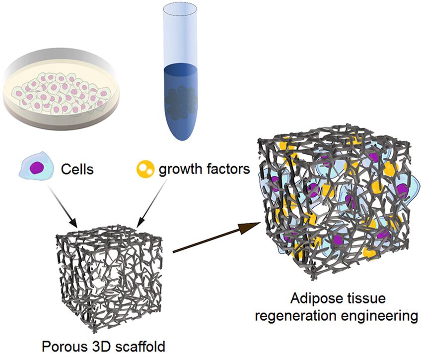
386 Zhiyu Peng et al.
and combine them with adipose-derived stem cells (ADSCs) various bovine collagen products have been on the market for
and other cells, so as to provide an environment for the correcting scars, crow’s feet, and periorbital wrinkles [5,17].
differentiation and growth of adipocytes and ultimately However, it has been reported that bovine collagen has a high
achieve the reconstruction of adipose tissue [11]. Now many incidence of rejection [18]. The second is pig collagen, which is
studies of the combination of injectable biomaterials and extracted from the tendons, dermis, and bones of pigs.
biomaterial stents with fat transplantation show great Recently, materials using pig collagen have been promoted
potential in preclinical studies. This paper reviews the as intraepithelial fillers. Compared with bovine collagen, pig
recent research advances in biomaterials for surgery of collagen has higher wrinkle resistance and no serious adverse
adipose tissue reconstruction. reactions [19]. The third is human collagen material extracted
from the dermis of the human body. It can be extracted from
adipose tissue after liposuction or produced by recombining
yeast or bacteria of human collagen [20,21]. Human collagen
2 Injectable biomaterials for material has been used in clinical practice, delaying the
degradation of collagen lysine residues by cross-linking them
adipose tissue reconstruction in with glutaraldehyde, mainly for deep scars and wrinkles [20].
plastic surgery The human collagen material has shown good clinical effect,
but the price is expensive, and the long-term effect is yet to be
The materials used for adipose tissue reconstruction are verified by clinical studies (Table 1).
generally injectable hydrogel materials because they can be
simply injected subcutaneously, greatly avoiding pain and
complications in patients. In addition, the injected bioma- 2.1.2 HA
terials can fill the complex lacuna in the patient’s surgical
area, avoiding the formation of dead space [12]. Therefore, HA is a naturally occurring glycosaminoglycan that is usually
injectable biomaterials are widely used in adipose tissue extracted from bacteria or the cockscomb of roosters. HA has
reconstruction. Injectable in situ gels can be prepared by shown low immunogenicity in preclinical studies, and
various physical and chemical methods. Physical methods several HA materials have been approved for clinical use
include temperature, pH, electric and magnetic fields, [22,23]. However, HA degrades quickly and requires repeated
enzymatic method, optical method, and hydrophobic injections and lacks the ability to induce adipose cells, so it is
interaction, etc., while chemical methods include various usually used as a temporary filler to induce tissue repair [24].
crosslinking methods, such as Schiff crosslinking method, Several studies have focused on reducing the degradation
Michael crosslinking method, and enzyme-catalyzed cross- rate of HA to ensure its volume persistence and improve its
linking method [5,7,13]. The preparation of various inject- biological performance. At present, there are two main
able biomaterials is complicated, but the materials can be methods to degrade the degradation rate of HA, one is the
divided into natural materials and synthetic materials. The double-crosslinking method [24] and the other is the
basic research and clinical application progress of various combination of HA with biopolymers such as gelatin,
injectable biomaterials are introduced from natural bio- chitosan, cellulose, or synthetic polymers such as polyethy-
materials and synthetic biomaterials, respectively. lene glycol (PEG) [25–27]. Compared with pure HA, the
combination of HA and gelatin shows good biocompatibility
and lower degradation rate. Although the anaphylaxis caused
by HA is weak, there are still pain, bruising, and temporary
2.1 Natural biological materials edema in the local area which have been reported [28].
2.1.1 Collagen
2.1.3 Chitosan
Collagen is the main component of connective tissues,
ligaments, and tendons, and collagen in the natural ECM is Chitosan is a natural linear polysaccharide obtained by
an important biomaterial [14,15]. Currently, there are three partial deacetylation of chitin. In tissue engineering
main sources of collagen biomaterials. The first is bovine applications, its water-soluble analog N-SCS is often used
collagen extracted from bovine tendons, dermis, and bones as a tissue filler [29]. Tan et al. [30] developed a
[16]. Bovine collagen can form a firm structure through biodegradable hydrogel construct composed of succinyl
collagen fibers after injection to help correct facial defects. Now chitosan (SCS), PEG, and insulin, in which insulin acts asTable 1: Advanced biomaterials for adipose tissue regeneration and their applications
Name Molecular structure/formula Resource Advantages Disadvantages Application
Natural Collagen [5,14–21] None Bovine collagen, pig Superior biocompatibility Rejection reaction; Facial soft tissue filler for crow’s
biomaterials collagen and human possible disease feet, periorbital wrinkles, and
collagen transfection deep scars
Bacteria or the
Hyaluronic Mild swelling; rapid Temporary filler to induce tissue
cockscomb of Minimal rejection reaction
acid [22–28] degradation repair
roosters
Nontoxic; slowly Rejection reaction; limited
Partial deacetylation
Chitosan [29,30,68] biodegradable; promotion of cell Soft tissue filler; wound healing
of chitin
biocompatible proliferation
Partial hydrolysis of A good vehicle for adipose Poor mechanical Biological scaffold for adipose
Gelatin [31,32,69]
collagen tissue engineering properties tissue engineering
Decellularized None Adipose tissue Potential benefits for Rapid degradation; Biological scaffold for adipose
adipose repairing of damaged repeated injection tissue engineering; wound
tissue [33–35] tissues healing cartilage and bone
regeneration, nerve injury
repairing
Volume enhancer and profiler for
Synthetic Artificially Rejection reaction; nasolabial line, radial upper lip
PMMA [36,37] Long-lasting
biomaterials synthesized nonbiodegradable line, interbrow line and
corner line
Superior biocompatibility;
Artificially Soft tissue reconstruction;
PLLA [38–41] provoke collagen Rapid biodegradation
synthesized biological stimulant
deposition
Long-lasting;
Calcium
Advances in biomaterials for adipose tissue reconstruction in plastic surgery
Artificially biocompatibility;
hydroxyapatite Rejection reaction Nasolabial folds
synthesized biodegradability; low
[42,43]
toxicity
387388 Zhiyu Peng et al.
an adipogenic factor. When the hydrogel was cultured with such as macrophage phagocytosis and inflammation.
cells for 6 h, the adhesion of cells to SCS/PEG hydrogel was Artefill [37] is a new generation of developed PMMA
significantly greater than that to pure SCS hydrogel (the microspheres, with a size of 30–50 µm, which is reported
relative DNA content increased by about 1.5 times). that it has a less risk of phagocytosis and can reduce the
formation of granuloma. At present, it has been reported
2.1.4 Gelatin as a volume enhancer and profiler for nasolabial line,
radial upper lip line, interbrow line, and corner line.
Gelatin is a water-soluble biocompatible protein, which is
the product of partial hydrolysis of collagen. It retains 2.2.2 Poly-L-lactic acid
biological interaction signals including Arg-Gly-Asp
(RGD) sequence, so it is often used in various tissue Originally synthesized from α-hydroxy acids, poly-L-lactic
engineering applications. Tuin et al. [31] evaluated the acid (PLLA) is a biodegradable thermoplastic polymer that
potential of an injectable hydrogel composed of recombi- has been widely used in orthopedics, neurosurgery, and
nant gelatin enriched by HA and RGD in rats. They found suture materials of craniofacial surgery, absorbable plates,
that after 4 weeks of transplantation, the introduction of and screws [38]. The main use of PLLA in soft tissue
recombinant gelatin induced soft tissue regeneration, reconstruction is to enlarge and fill the deep dermis or
characterized by significant vascularization and infiltration subcutaneous layer and repair facial fat atrophy [39]. PLLA
of fibroblasts and macrophages. Using transglutaminase to expands at the moment of injection to fill and repair the
hydrolyze and cross-link gelatin hydrogel and encapsulating lacuna and degrades after a period of time. PLLA can
ADSCs, Zeno Alarake et al. [32] obtained good results in vitro. stimulate collagen production and vascularization in the
whole process, which ultimately causes dermal fiber prolif-
eration, acting as a biological stimulant [40]. Therefore, PLLA
2.1.5 Decellularized adipose tissue is also approved as a treatment for nasolabial fold, facial
wrinkles, line, and contour enhancement defects [40,41].
Because decellularized adipose tissue (DAT) has the
potential to enhance the regeneration and repair of
damaged tissues, some studies have shown that ADSCs 2.2.3 Calcium hydroxyapatite
combined with DAT is an effective biofiller [33–35]. Zhang
et al. [33] studied the potential of heparinized DAT loaded Calcium hydroxyapatite (CH) and its by-products natu-
with basic fibroblast growth factor (bFGF) in mice. These rally occurring in human bone and tooth enamel are
injectable scaffolds induced significant adipogenesis and composed of calcium and phosphate ions and have
neovascularization within 6–12 weeks, whereas the non- strong biocompatibility [42]. CH can be prepared as
crosslinked DAT (excluding bFGF) formed adipogenesis to a microspheres and suspended in carboxymethyl cellulose
lesser extent and was almost completely absorbed after 12 gel, which has been approved for the correction of facial
weeks. Tan et al. [35] also observed that after subcutaneous wrinkles and the filling of soft tissues [43]. The CH
injection of acellular pig fat tissue, fat generation was microspheres in the gel can promote the formation of
significantly enhanced after ADSC inoculation. Therefore, new tissues through collagen deposition. The gel usually
the application of DAT in combination with ADSCs is degrades within 2–3 months and its by-products,
considered to be highly synergistic, as these systems calcium and phosphate ions, can be removed by
demonstrate long-term lipogenesis and angiogenesis in vivo. phagocytosis of macrophages. Therefore, some studies
believe that the treatment effect of CH on nasolabial
folds is better than that of pure collagen materials [43].
2.2 Synthetic biomaterials
2.2.1 PMMA 3 Tissue engineering scaffold
PMMA is a synthetic and nondegradable biocompatible
materials for adipose tissue
polymer, which is often used as an intradermal filler in reconstruction in plastic surgery
tissue repair. The PMMA microspheres developed originally
are called Arteplast [36], because of their small molecular The application of 3D tissue-engineered scaffold materials
diameter (less than 20 µm), causing immune responses for adipose tissue repair is an important method in the fieldAdvances in biomaterials for adipose tissue reconstruction in plastic surgery 389
of adipose tissue reconstruction. The principle is to methods include solvent casting method and particle
encapsulate adipose cells, ADSCs, or other cells and growth leaching method, freeze-drying method, and freeze-gel
factors through tissue engineering, and then inoculate them methods, which are simple and economical. For example,
on 3D scaffolds simulating ECM [11]. Good tissue engi- Phull et al. [44] used the solvent casting method and the
neering scaffolds in orthopedic adipose tissue reconstruc- particle leaching method to produce macroporous struc-
tion should meet the following requirements: (1) good tures for soft tissue engineering. They used alginate
biocompatibility and nontoxicity; (2) the scaffold shall have microspheres as pore-foaming agents, and a mechanically
interpore connections with an aperture greater than 100 µm stable scaffold material with large voids was prepared by
to provide effective oxygen and nutrients and to ensure gelatin for the surrounding matrix. The results showed that
good cell proliferation and migration; (3) it can be designed the gelatin matrix with ADSCs could support the growth
and shaped flexibly and it should be convenient to and differentiation of cells along the fat line.
construct 3D scaffold quickly [7,11]. The progress of adipose Additive manufacturing forming technology includes
reconstruction scaffold material depends on the progress of two kinds of technology methods of conventional 3D printing,
preparation technology. The biggest advantage of 3D which can fabricate post complex cells, growth factors, and
scaffold is that it can provide a good biomimetic environ- direct 3D bio-fabrication. Additive manufacturing forming
ment for cells and cytokines, etc. At present, the preclinical technology has the advantage that can make custom-made
research on the combination of cells and biological scaffold scaffold for patients according to their characteristics, and
material shows a good application prospect. The manufac- scaffold materials, seed cells, and growth factors can be
turing technology of 3D adipose tissue engineering scaffold prepared accurately; it is widely used in the manufacture of
materials, the cells and growth factors used in adipose tissue artificial organs, such as bone tissue, skin, trachea, and so on
reconstruction, and the application of 3D scaffold materials [45–47]. In conventional 3D printing manufacturing technol-
in adipose tissue reconstruction are introduced (Figure 1). ogies, fused deposition modelling (FDM) technology based
on extrusion molding can be used to fabricate 3D porous
scaffolds of adipose tissue layer by layer. The main
disadvantage of this method is that cells cannot be printed
with materials and cells must be inoculated after printing.
Chhaya et al. [48] added PLLA scaffold with an aperture more
than 1 mm to human umbilical vein endothelial cells, and
then subcutaneously implanted the scaffold into nude mice
without thymus. Angiogenesis and adipose tissue formation
were observed in all mammary scaffolds. Bio-fabrication is
the application of “bio-ink” containing cell components to
produce 3D scaffolds. Its advantage is that scaffolds and cells
are prepared as “bio-ink” in advance, and then, they are
simultaneously printed [49]. Current research on bio-fabrica-
tion focuses on how to solve the survival rate of cells and how
do cells stay alive and differentiate into tissue after printing
[50]. Gruene et al. [51] made 3D biological scaffolds by using
laser-assisted methods in bio-fabrication, and the results
Figure 1: Cells, growth factors, and porous scaffolds constitute the showed that bio-fabrication had no effect on the differentia-
adipose tissue regeneration engineering. tion potential and proliferation ability of stem cells.
3.1 Preparation technology of 3D tissue
engineering scaffold for adipose tissue 3.2 Cells and cytokines of tissue engineering
reconstruction for adipose tissue reconstruction
At present, the methods applied to the construction of 3D 3.2.1 ADSCs
biological scaffolds can be divided into two types:
traditional construction and additive manufacturing Adipose tissue contains ADSCs. ADSCs are similar to bone
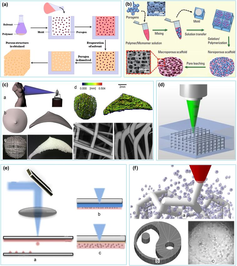
forming technology (Figure 2). Traditional construction marrow stem cells in their ability to differentiate and390 Zhiyu Peng et al. Figure 2: Figure 2: Examples of preparation methods of tissue engineering scaffolds. (a) Solvent casting [70]. (b) Particulate leaching method [71]. (c) Scaffold fabricated by FDM method: (a) laser scanning; (b) CAD model fabricating; (c) porous patient-specific scaffolds generating; (d) MicroCT scans show the filament and volume of the scaffolds; (e) SEM image bars and struts of the scaffold [46]. (d) Scaffold fabricated by laser fabrication approach [64]. (e) Scaffolds fabricated by the LAB method. (f) Two-photon polymerization (2PP) method: (a) schematic illustration showing 2PP structures (gray) fabricated by a focused laser beam (red) within the suspension of cells (blue); (b) CAD model used for laser cell encapsulation; (c) an optical online image of a 2PP-produced 400 μm wide Yin–Yang hydrogel structure with cells inside [73].
Advances in biomaterials for adipose tissue reconstruction in plastic surgery 391
regenerate, and they can supplement lost volume at repair As a signaling protein, FGF has many functions such as
sites by proliferating and differentiating into adipocytes [52]. In development, metabolism, and adipokine. Adipokines are
addition, they also secrete various growth factors, such as involved in the remodeling, lipogenesis, and angiogenesis of
vascular endothelial growth factor (VEGF), hepatocyte growth adipose tissue by autocrine/paracrine. Ogushi et al. [59]
factor, fibroblast growth factor-2 (FGF-2), insulin-like growth cross-linked ADSCs with FGF enzymes in vivo and in vitro
factor 1, etc., playing an important role in adipose tissue and then loaded them into injectable carboxymethyl
regeneration and angiogenesis [53]. It has been reported that cellulose with phenolic hydroxyl for adipose tissue engi-
ADSCs mixed with HA filler can differentiate into fibroblasts, neering. The results showed that the survival rate of
smooth muscle cells, preadipocytes, progenitor cells, adipocyte was 92.8%, and the adipocyte proliferation was
endothelial cells, and stem cells after injection [54]. good. In addition, after subcutaneous injection of Lewis rats
Several studies have shown that ADSCs promote the for 10 weeks, new vascularized adipose tissue and lipogen-
regeneration of adipose tissue. Yao et al. [55] encapsulated esis were observed at the injection site. Therefore, FGF can be
ADSCs of human in alginate microspheres and alginate/ used as a component of ADSCs and fat transfer to promote
gelatin microspheres to simulate the natural fat lobule in adipose tissue generation and angiogenesis.
adipose tissue. It was found that alginate/gelatin micro- VEGF was first identified as an important role in
spheres containing ADSCs showed higher cell proliferation angiogenesis in 1983 [60]. In addition, it has been found to
and adipogenic differentiation than that of alginate micro- play a role in wound healing, fatty tissue expansion, tumor
spheres. Turner et al. [56] prepared DAT as a substrate to growth, and age-related macular degeneration. Kim et al. [61]
induce ADSCs. The results showed that when ADSCs were studied the effects of ADSCs and VEGF on myogenic
inoculated on DAT microcarriers containing adipogenic differentiation in injectable heat-sensitive PEG–poly-e-capro-
medium, the adipogenic differentiation level was higher, lactone (PCL) hydrogels. The results showed that the
while the gelatin microcarriers showed no substantial combination of ADSCs and VEGF in the hydrogel showed
adipogenesis. In addition to promoting adipose tissue obvious proliferation, differentiating into muscle tissue and
regeneration, ADSCs are related to promoting angiogenesis vascularization enhancement after subcutaneous injection
and collagen expression and have good biocompatibility into the neck of mice for 4 weeks. Gorkun et al. [62] studied
and tolerance. Butler et al. [57] subcutaneously injected the effect of VEGF on endothelial cell differentiation after
human microvascular endothelial cells (HMEC) and ADSCs ADSCs or umbilical cord pluripotent MMSCs were cultured
coated with collagen hydrogel in mice, finding that on unmodified and polyglycolated fibrin hydrogel. The
combined transplantation with ADSCs had effects of anti- results showed that poly(ethylene glycol) hydrogel had better
apoptosis and can promote angiogenesis on HMEC. multibranch formation than pure fibrin gels, and the
differentiated ADSCs had stronger angiogenesis capacity
than the differentiated umbilical MMSCs. Therefore, it can
3.2.2 Growth factors of tissue engineering for adipose be believed that VEGF can play an important role in
tissue reconstruction angiogenesis of ADSCs and fat transfer.
Platelet-rich plasma (PRP) is a concentrated solution of a
large number of platelets in a small volume of plasma. It has
been reported that PRP contains a large number of cytokines 3.3 Application of 3D scaffolds for adipose
that promote cell proliferation, differentiation, and angio- tissue reconstruction
genesis. Liao et al. studied the effect of PRP on proliferation
and adipogenic differentiation of ADSCs in vitro and found The application of 3D scaffold materials for adipose tissue
that PRP significantly increased the proliferation of ADSCs. reconstruction has made good progress, and some fruitful
Recently, Li et al. [58] studied the effects of PRP and results of 3D porous scaffolds have been obtained in vivo.
conditioned medium on ADSCs in nude mice after sub- For example, (D,L)-lactide, GelMod, alginate, and DAT
cutaneous injection of PRP combined with ADSCs. The bioinks and PCL have been used in scaffold printing for
results showed that the residual fat volume of PRP and ADSC adipose tissue engineering [7]. Chang et al. [63] used
group was significantly higher than that of other groups after gelatin/HA cold gel scaffolds in nude mice, and the results
90 days. Therefore, the combination of PRP with ADSCs and showed that this type of scaffolds is expected to provide a
fat transfer can significantly increase the proliferation and stable structure and chemical environment, enabling
adipose differentiation of ADSCs and improve the survival cells to attach and proliferate, and supporting the
rate of grafts after fat transplantation. biological functions and fat generation of ADSCs.392 Zhiyu Peng et al.
Ovsianikov et al. [64] prepared a methylacrylamide- ADSCs are pluripotent mesenchymal stem cells with the
modified gelatin (GelMod) scaffold with an aperture of function of supplementing tissue defects through prolif-
250 mm by bio-fabrication for adipose tissue engineering eration and maturation, so they are widely used in fat
applications. The results showed that the crosslinking regeneration of various cell. The combination of ADSCs,
degree had no effect on the enzymatic degradation ability growth factors, and biological scaffolds has shown a
of the gel-based structure, and the scaffold successfully broad prospect in studies related to adipose tissue
supported the adhesion, proliferation, and adipogenic regeneration, but most of these studies are still in the
differentiation of ASC. preclinical stage. More clinical studies are needed to
However, the study of biological scaffolds still has verify its safety and efficacy in the future.
many limitations. Compared with injectable materials,
the placement of 3D biological scaffolds requires surgical Conflict of interest: The authors declare no conflict of
incision, which is more complicated, and clinical interest regarding the publication of this paper.
verification is also required for the location and fixation
of implants. The study of scaffold’s microenvironments Acknowledgments: This work was supported by the
on regulation of cell fate is still a difficult issue [65]. National Key Research and Development Program of
Another difficulty that can be predicted is the fabrication China (No. 2018YFC1106800), the National Natural
of a tri-microbial scaffold, which uses materials that are Science Foundation of China (Nos. 31971251 and
same as injectable biomaterials. Its progress depends on 81871574), the Sichuan Province Science & Technology
advances in the fabrication of biomaterials. Therefore, it Department Projects (2016CZYD0004, 2017SZ0001,
can be expected that the production time of biological 2018GZ0142, and 2019YFH0079), the Research
scaffolds is longer and more difficult than that of in situ Foundation for Young Teachers of Sichuan University
injectable materials [66]. The studies in vivo reported so (2018SCUH0017), and the “111” Project (No. B16033).
far indicate that only very small amounts of material
have been used in mouse or rat models. How these
results will be applied to humans in future studies? And
whether the hydrogels can achieve the same effect by References
increasing their volume? These problems are unclear.
Hillel et al. [67] reported differences in inflammatory [1] Lequeux C, Rodriguez J, Boucher F, Rouyer O, Damour O,
responses between mice and humans. Implants made of Mojallal A, et al. In vitro and in vivo biocompatibility,
polyethylene glycol and HA caused a greater inflamma- bioavailability and tolerance of an injectable vehicle for
adipose-derived stem/stromal cells for plastic surgery indica-
tory response in human body Therefore, more clinical
tions. Plast Reconstr Aesthet Surg. 2015;68(11):1491–7. doi:
studies are needed to verify the effectiveness and safety 10.1016/j.bjps.2015.07.022. PubMed PMID: 26282247. Epub
of biological scaffold materials. 2015/08/05.
[2] Auclair E, Blondeel P, Del Vecchio DA. Composite breast
augmentation: soft-tissue planning using implants and fat.
Plast Reconstr Surg. 2013;132(3):558–68. doi: 10.1097/
PRS.0b013e31829ad2fa. PubMed PMID: 23985632.
4 Summary and expectation [3] Malvezzi M, Carioli G, Bertuccio P, Rosso T, Boffetta P, Levi F, et al.
European cancer mortality predictions for the year 2016 with focus
Injectable biomaterials are widely used in soft tissue on leukaemias. Ann Oncol. 2016;27(4):725–31. doi: 10.1093/
reconstruction. Natural biomaterial has good biocompat- annonc/mdw022. PubMed PMID: 26812903. Epub 2016/01/26.
ibility, and it is a kind of natural degradable nonperma- [4] Cohen SR, Hewett S, Ross L, Delaunay F, Goodacre A,
Ramos C, et al. Regenerative cells for facial surgery:
nent soft tissue filling material. However, natural
biofilling and biocontouring. Aesthet Surg J. 2017;
biomaterials tend to have a higher absorption rate and 37(Suppl 3):S16–32. doi: 10.1093/asj/sjx078. PubMed
are more difficult to produce long-lasting filling. Synthetic PMID: 29025218.
biomaterials have low biocompatibility, but some me- [5] Cho K-H, Uthaman S, Park I-K, Cho C-S. Injectable biomater-
chanical property, chemical property, and degradation ials in plastic and reconstructive surgery: a review of the
property can be customized according to requirements. At current status. Tissue Eng Regen Med. 2018;15(5):559–74.
doi: 10.1007/s13770-018-0158-2. PubMed PMID: 30603579.
present, many kinds of injectable scaffolds with good
[6] Coleman SR, Saboeiro AP. Fat grafting to the breast revisited:
biological and mechanical properties have shown good safety and efficacy. Plast Reconstr Surg. 2007;119(3):775–87.
effects in preclinical studies by combining relevant cells doi: 10.1097/01.prs.0000252001.59162.c9. PubMed PMID:
and cytokines with natural and synthetic biomaterials. 17312477.Advances in biomaterials for adipose tissue reconstruction in plastic surgery 393
[7] Van Nieuwenhove I, Tytgat L, Ryx M, Blondeel P, Stillaert F, [20] Kim BS, Choi JS, Kim JD, Yoon HI, Choi YC, Cho YW. Human
Thienpont H, et al. Soft tissue fillers for adipose tissue collagen isolated from adipose tissue. Biotechnol Prog.
regeneration: from hydrogel development toward clinical 2012;28(4):973–80. doi: 10.1002/btpr.1555. PubMed PMID:
applications. Acta Biomater. 2017;63:37–49. doi: 10.1016/ 22549937. Epub 2012/06/08.
j.actbio.2017.09.026. PubMed PMID: 28941654, Epub 2017/ [21] Báez J, Olsen D, Polarek JW. Recombinant microbial systems for
09/20. the production of human collagen and gelatin. Appl Microbiol
[8] Radhakrishnan J, Krishnan UM, Sethuraman S. Hydrogel Biotechnol. 2005;69(3):245–52. doi: 10.1007/s00253-005-
based injectable scaffolds for cardiac tissue regeneration. 0180-x. PubMed PMID: 16240115. Epub 2005/11/15.
Biotechnol Adv. 2014;32(2):449–61. doi: 10.1016/j.biote- [22] Collins MN, Birkinshaw C. Hyaluronic acid based scaffolds for
chadv.2013.12.010. PubMed PMID: 24406815. Epub 2014/ tissue engineering – a review. Carbohydr Polym.
01/07. 2013;92(2):1262–79. doi: 10.1016/j.carbpol.2012.10.028.
[9] Spector M, Lim TC. Injectable biomaterials: a perspective on PubMed PMID: 23399155. Epub 2012/10/17.
the next wave of injectable therapeutics. Biomed Mater. [23] Bogdan Allemann I, Baumann L. Hyaluronic acid gel
2016;11(1):014110. doi: 10.1088/1748-6041/11/1/014110. (Juvéderm) preparations in the treatment of facial wrinkles
PubMed PMID: 26836246. and folds. Clin Interv Aging. 2008;3(4):629–34. doi: 10.2147/
[10] Alam M, Gladstone H, Kramer EM, Murphy JP, Nouri K, cia.s3118. PubMed PMID: 19281055.
Neuhaus IM, et al. ASDS guidelines of care: injectable fillers. [24] Tan H, Li H, Rubin JP, Marra KG. Controlled gelation and
Dermatol Surg. 2008;34(Suppl 1):S115–S48. doi: 10.1111/ degradation rates of injectable hyaluronic acid-based hydro-
j.1524-4725.2008.34253.x. PubMed PMID: 18547175. gels through a double crosslinking strategy. J Tissue Eng
[11] Van Hoorick J, Declercq H, De Muynck A, Houben A, Van Regen Med. 2011;5(10):790–7. doi: 10.1002/term.378.
Hoorebeke L, Cornelissen R, et al. Indirect additive manu- PubMed PMID: 22002922. Epub 2011/01/10.
facturing as an elegant tool for the production of self- [25] Korurer E, Kenar H, Doger E, Karaoz E. Production of a
supporting low density gelatin scaffolds. J Mater Sci Mater composite hyaluronic acid/gelatin blood plasma gel for
Med. 2015;26(10):247. doi: 10.1007/s10856-015-5566-4. hydrogel-based adipose tissue engineering applications.
PubMed PMID: 26411443. Epub 2015/09/28. J Biomed Mater Res A. 2014;102(7):2220–9. doi: 10.1002/
[12] Ercan H, Durkut S, Koc-Demir A, Elçin AE, Elçin YM. Clinical jbm.a.34901. PubMed PMID: 23913820. Epub 2013/08/10.
applications of injectable biomaterials. Adv Exp Med Biol. [26] Domingues RMA, Silva M, Gershovich P, Betta S, Babo P,
2018;1077:163–82. doi: 10.1007/978-981-13-0947-2_10. Caridade SG, et al. Development of injectable hyaluronic acid/
PubMed PMID: 30357689. cellulose nanocrystals bionanocomposite hydrogels for tissue
[13] Sheikholeslam M, Wright MEE, Jeschke MG, Amini-Nik S. engineering applications. Bioconjug Chem.
Biomaterials for skin substitutes. Adv Healthc Mater. 2015;26(8):1571–81. doi: 10.1021/acs.bioconjchem.5b00209.
2018;7(5):1700897(1–20). doi: 10.1002/adhm.201700897. PubMed PMID: 26106949. Epub 2015/07/20.
PubMed PMID: 29271580. Epub 2017/12/22. [27] Fan M, Ma Y, Zhang Z, Mao J, Tan H, Hu X. Biodegradable
[14] Jin G-Z, Kim H-W. Effects of type I collagen concentration in hyaluronic acid hydrogels to control release of dexametha-
hydrogel on the growth and phenotypic expression of rat sone through aqueous Diels-Alder chemistry for adipose
chondrocytes. Tissue Eng Regen Med. 2017;14(4):383–91. doi: tissue engineering. Mater Sci Eng C Mater Biol Appl.
10.1007/s13770-017-0060-3. PubMed PMID: 30603494. 2015;56:311–7. doi: 10.1016/j.msec.2015.04.004. PubMed
[15] Luca A, Butnaru M, Maier SS, Knieling L, Bredetean O, PMID: 26249595. Epub 2015/04/15.
Verestiuc L, et al. Atelocollagen-based hydrogels crosslinked [28] Fallacara A, Manfredini S, Durini E, Vertuani S. Hyaluronic acid
with oxidised polysaccharides as cell encapsulation matrix for fillers in soft tissue regeneration. Facial Plast Surg.
engineered bioactive stromal tissue. Tissue Eng Regen Med. 2017;33(1):87-96. doi: 10.1055/s-0036-1597685. PubMed
2017;14(5):539–56. doi: 10.1007/s13770-017-0063-0. PubMed PMID: 28226376. Epub 2017/02/22.
PMID: 30603508. [29] Aiping Z, Tian C, Lanhua Y, Hao W, Ping L. Synthesis and
[16] Felician FF, Xia C, Qi W, Xu H. Collagen from marine biological characterization of N-succinyl-chitosan and its self-assembly
sources and medical applications. Chem Biodivers. of nanosphere. Carbohydr Polym. 2006;66:274–9. doi:
2018;15(5):e1700557. doi: 10.1002/cbdv.201700557. PubMed 10.1016/j.carbpol.2006.03.014
PMID: 29521032. Epub 2018/05/21. [30] Tan H, Luan H, Hu Y, Hu X. Covalently crosslinked chitosan-
[17] Rostan E. Collagen fillers. Facial Plast Surg Clin North Am. poly(ethylene glycol) hybrid hydrogels to deliver insulin for
2007;15(1):55–61. doi: 10.1016/j.fsc.2006.11.001. PubMed adipose-derived stem cells encapsulation. Macromol Res.
PMID: 17317556. 2012;21:392–9. doi: 10.1007/s13233-013-1023-8
[18] Kastellorizios M, Tipnis N, Burgess DJ. Foreign body reaction [31] Tuin A, Zandstra J, Kluijtmans S, Bouwstra J, Harmsen M,
to subcutaneous implants. Adv Exp Med Biol. Luyn M. Hyaluronic acid-recombinant gelatin gels as a
2015;865:93–108. doi: 10.1007/978-3-319-18603-0_6. scaffold for soft tissue regeneration. Eur Cell Mater.
PubMed PMID: 26306445. 2012;24:220–330. doi: 10.22203/eCM.v024a23
[19] Lee JH, Choi YS, Kim SM, Kim YJ, Rhie JW, Jun YJ. Efficacy and [32] Zeno Alarake N, Frohberg P, Groth T, Pietzsch M. Mechanical
safety of porcine collagen filler for nasolabial fold correction properties and biocompatibility of in situ enzymatically cross-
in Asians: a prospective multicenter, 12 months follow-up linked gelatin hydrogels. Int J Artif Organs.
study. J Korean Med Sci. 2014;29(Suppl 3):S217–21. doi: 2017;40(4):159–68. doi: 10.5301/ijao.5000553
10.3346/jkms.2014.29.S3.S217. PubMed PMID: 25473212. [33] Zhang S, Lu Q, Cao T, Toh WS. Adipose tissue and
Epub 2014/11/21. extracellular matrix development by injectable decellularized394 Zhiyu Peng et al.
adipose matrix loaded with basic fibroblast growth factor. [48] Chhaya MP, Melchels FPW, Holzapfel BM, Baldwin JG,
Plast Reconstr Surg. 2016;137(4):1171–80. doi: 10.1097/ Hutmacher DW. Sustained regeneration of high-volume
PRS.0000000000002019 adipose tissue for breast reconstruction using computer aided
[34] Brown C, Yan J, Han TT, Marecak D, Amsden B, Flynn L. Effect design and biomanufacturing. Biomaterials. 2015;52:551–60.
of decellularized adipose tissue particle size and cell density doi: 10.1016/j.biomaterials.2015.01.025. PubMed PMID:
on adipose-derived stem cell proliferation and adipogenic 25818460. Epub 2015/03/18.
differentiation in composite methacrylated chondroitin [49] Moroni L, Boland T, Burdick JA, De Maria C, Derby B,
sulphate hydrogels. Biomed Mater. 2015;10(4):045010. Forgacs G, et al. Biofabrication: a guide to technology and
doi: 10.1088/1748-6041/10/4/045010 terminology. Trends Biotechnol. 2018;36(4):384–402. doi:
[35] Tan Q-W, Zhang Y, Luo J-C, Zhang D, Xiong B-J, Yang J-Q, et al. 10.1016/j.tibtech.2017.10.015. PubMed PMID: 29137814. Epub
Hydrogel derived from decellularized porcine adipose tissue as 2017/11/11.
a promising biomaterial for soft tissue augmentation. J Biomed [50] Wang X, Liu C. 3D Bioprinting of adipose-derived stem cells
Mater Res A. 2017;105(6):1756–64. doi: 10.1002/jbm.a.36025 for organ manufacturing. Adv Exp Med Biol. 2018;1078:3–14.
[36] Lemperle G, Ott H, Charrier U, Hecker J, Lemperle M. PMMA doi: 10.1007/978-981-13-0950-2_1. PubMed PMID: 30357615.
microspheres for intradermal implantation: part I, animal [51] Gruene M, Pflaum M, Deiwick A, Koch L, Schlie S, Unger C,
research. Ann Plast Surg. 1991;26(1):57–63. doi: 10.1097/ et al. Adipogenic differentiation of laser-printed 3D tissue
00000637-199101000-00009 grafts consisting of human adipose-derived stem cells.
[37] Greco T, Antunes M, Yellin S. Injectable fillers for volume Biofabrication. 2011;3(1):015005. doi: 10.1088/1758-5082/3/
replacement in the aging face. Facial Plast Surg. 1/015005. PubMed PMID: 21358040. Epub 2011/03/01.
2012;28(1):8–20. doi: 10.1055/s-0032-1305786 [52] Jin HJ, Bae YK, Kim M, Kwon S-J, Jeon HB, Choi SJ, et al.
[38] Attenello NH, Maas CS. Injectable fillers: review of material Comparative analysis of human mesenchymal stem cells from
and properties. Facial Plast Surg. 2015;31(1):29–34. doi: bone marrow, adipose tissue, and umbilical cord blood as
10.1055/s-0035-1544924, PubMed PMID: 25763894, Epub sources of cell therapy. Int J Mol Sci. 2013;14(9):17986–8001.
2015/03/12. doi: 10.3390/ijms140917986. PubMed PMID: 24005862.
[39] Garson S, Delay E, Sinna R, Cornette de Saint Cyr B, Taha F. [53] Combellack EJ, Jessop ZM, Naderi N, Griffin M, Dobbs T,
The third dimension of the face aging, improvement of its Ibrahim A, et al. Adipose regeneration and implications for
understanding. Ann Chir Plast Esthet. 2017;62(5):387–98. breast reconstruction: update and the future. Gland Surg.
doi: 10.1016/j.anplas.2017.09.001. PubMed PMID: 28943216. 2016;5(2):227–41. doi: 10.3978/j.issn.2227-684X.2016.01.01.
Epub 2017/09/22. PubMed PMID: 27047789.
[40] Bartus C, William Hanke C, Daro-Kaftan E. A decade of [54] Yoshimura K, Sato K, Aoi N, Kurita M, Hirohi T, Harii K. Cell-
experience with injectable poly-L-lactic acid: a focus on assisted lipotransfer for cosmetic breast augmentation: sup-
safety. Dermatol Surg. 2013;39(5):698–705. doi: 10.1111/ portive use of adipose-derived stem/stromal cells. Aesthetic
dsu.12128. PubMed PMID: 23379657. Epub 2013/02/04. Plast Surg. 2008;32(1):48–57. doi: 10.1007/s00266-007-9019-
[41] Kontis TC. Contemporary review of injectable facial fillers. 4. PubMed PMID: 17763894. Epub 2007/09/01.
JAMA Facial Plast Surg. 2013;15(1):58–64. doi: 10.1001/ [55] Yao R, Zhang R, Luan J, Lin F. Alginate and alginate/gelatin
jamafacial.2013.337. PubMed PMID: 23183718. microspheres for human adipose-derived stem cell encapsu-
[42] Hobar PC, Pantaloni M, Byrd HS. Porous hydroxyapatite lation and differentiation. Biofabrication. 2012;4(2):025007.
granules for alloplastic enhancement of the facial region. Clin doi: 10.1088/1758-5082/4/2/025007. PubMed PMID:
Plast Surg. 2000;27(4):557–69. doi: PubMed PMID: 11039889. 22556122. Epub 2012/05/04. .
[43] Luebberding S, Alexiades-Armenakas M. Facial volume aug- [56] Turner AEB, Yu C, Bianco J, Watkins JF, Flynn LE. The
mentation in 2014: overview of different filler options. J Drugs performance of decellularized adipose tissue microcarriers as
Dermatol. 2013;12(12):1339–44. doi: PubMed PMID: 24301234. an inductive substrate for human adipose-derived stem cells.
[44] Phull MK, Eydmann T, Roxburgh J, Sharpe JR, Lawrence- Biomaterials. 2012;33(18):4490–9. doi: 10.1016/j.biomater-
Watt DJ, Phillips G, et al. Novel macro-microporous gelatin ials.2012.03.026. PubMed PMID: 22456084. Epub 2012/03/26.
scaffold fabricated by particulate leaching for soft tissue [57] Butler MJ, Sefton MV. Cotransplantation of adipose-derived
reconstruction with adipose-derived stem cells. J Mater Sci mesenchymal stromal cells and endothelial cells in a modular
Mater Med. 2013;24(2):461–7. doi: 10.1007/s10856-012- construct drives vascularization in SCID/bg mice. Tissue Eng
4806-0. PubMed PMID: 23143193. Epub 2012/11/10. Part A. 2012;18(15–16):1628–41. doi: 10.1089/
[45] Song P, Hu C, Pei X, Sun J, Sun H, Wu L, et al. Dual modulation ten.TEA.2011.0467. PubMed PMID: 22655687. Epub 2012/
on crystallinity and macro/micro structures of 3D printed 07/09.
porous titanium implants to enhance the stability and [58] Li K, Li F, Li J, Wang H, Zheng X, Long J, et al. Increased
osseointegration. J Mater Chem B. 2019;7(17):2865–77. survival of human free fat grafts with varying densities of
[46] Zhang B, Sun H, Wu L, Ma L, Xing F, Kong Q, et al. 3D printing human adipose-derived stem cells and platelet-rich plasma.
of calcium phosphate bioceramic with tailored biodegradation J Tissue Eng Regen Med. 2017;11(1):209–19. doi: 10.1002/
rate for skull bone tissue reconstruction. Bio-Design Manuf. term.1903. PubMed PMID: 24978937. Epub 2014/06/30.
2019;2:161–71. [59] Ogushi Y, Sakai S, Kawakami K. Adipose tissue engineering
[47] Zhang B, Pei X, Song P, Sun H, Li H, et al. Porous bioceramics using adipose-derived stem cells enclosed within an inject-
produced by inkjet 3D printing: effect of printing ink able carboxymethylcellulose-based hydrogel. J Tissue Eng
formulation on the ceramic macro and micro porous archi- Regen Med. 2013;7(11):884–92. doi: 10.1002/term.1480.
tectures control. Compos B Eng. 2018;155:112–21. PubMed PMID: 22489051. Epub 2012/04/04.Advances in biomaterials for adipose tissue reconstruction in plastic surgery 395
[60] Senger DR, Galli SJ, Dvorak AM, Perruzzi CA, Harvey VS, Dvorak HF. [67] Hillel AT, Unterman S, Nahas Z, Reid B, Coburn JM, Axelman J,
Tumor cells secrete a vascular permeability factor that promotes et al. Photoactivated composite biomaterial for soft tissue
accumulation of ascites fluid. Science. 1983;219(4587):983–5. doi: restoration in rodents and in humans. Sci Transl Med.
10.1126/science.6823562. PubMed PMID: 6823562. 2011;3(93):93ra67. doi: 10.1126/scitranslmed.3002331.
[61] Kim MH, Hong HN, Hong JP, Park CJ, Kwon SW, Kim SH, et al. The PubMed PMID: 21795587.
effect of VEGF on the myogenic differentiation of adipose tissue [68] Patrulea V, Ostafe V, Borchard G, Jordan O. Chitosan as a
derived stem cells within thermosensitive hydrogel matrices. starting material for wound healing applications. Eur J Pharm
Biomaterials. 2010;31(6):1213–8. doi: 10.1016/j.biomater- Biopharm. 2015;97(Pt B):417–26. doi: 10.1016/
ials.2009.10.057. PubMed PMID: 19914711. Epub 2009/11/14. j.ejpb.2015.08.004. PubMed PMID: 26614560.
[62] Gorkun AA, Shpichka AI, Zurina IM, Koroleva AV, Kosheleva NV, [69] Echave MC, Saenz del Burgo L, Pedraz JL, Orive G. Gelatin as
Nikishin DA, et al. Angiogenic potential of spheroids from biomaterial for tissue engineering. Curr Pharm Des.
umbilical cord and adipose-derived multipotent mesenchymal 2017;23(24):3567–84. doi: 10.2174/
stromal cells within fibrin gel. Biomed Mater. 2018;13(4):044108. 0929867324666170511123101. PubMed PMID: 28494717.
doi: 10.1088/1748-605X/aac22d. PubMed PMID: 29722292. [70] Bartis D, Pongracz J. Three dimensional tissue cultures and
[63] Chang K-H, Liao H-T, Chen J-P. Preparation and characteriza- tissue engineering. 1st ed. University of Pécs Press, University
tion of gelatin/hyaluronic acid cryogels for adipose tissue of Pécs; 2011, doi: 10.13140/2.1.2793.5047
engineering: in vitro and in vivo studies. Acta Biomater. [71] Bencherif SA, Braschler TM, Renaud P. Advances in the design
2013;9(11):9012–26. doi: 10.1016/j.actbio.2013.06.046. of macroporous polymer scaffolds for potential applications in
PubMed PMID: 23851171. Epub 2013/07/10. dentistry. J Periodontal Implant Sci. 2013;43(6):251–61. doi:
[64] Ovsianikov A, Deiwick A, Van Vlierberghe S, Pflaum M, 10.5051/jpis.2013.43.6.251. PubMed PMID: 24455437. Epub
Wilhelmi M, Dubruel P, et al. Laser fabrication of 3D gelatin 2013/12/31.
scaffolds for the generation of bioartificial tissues. Materials [72] Ferris CJ, Gilmore KG, Wallace GG, In het Panhuis M.
(Basel). 2011;4(1):288–99. doi: 10.3390/ma4010288. Biofabrication: an overview of the approaches used for
PubMed PMID: 28879989. printing of living cells. Appl Microbiol Biotechnol.
[65] Xing F, Li L, Zhou C, Long C, Wu L, Lei H, et al. Regulation and 2013;97(10):4243–58. doi: 10.1007/s00253-013-4853-6.
directing stem cell fate by tissue engineering functional PubMed PMID: 23525900. Epub 2013/03/24.
microenvironments: scaffold physical and chemical cues. [73] Ovsianikov A, Mühleder S, Torgersen J, Li Z, Qin X-H, Van
Stem Cells Int. 2019;2019:2180925. Vlierberghe S, et al. Laser photofabrication of cell-
[66] Murphy SV, Atala A. 3D bioprinting of tissues and organs. Nat containing hydrogel constructs. Langmuir.
Biotechnol. 2014;32(8):773–85. doi: 10.1038/nbt.2958. 2014;30(13):3787–94. doi: 10.1021/la402346z. PubMed
PubMed PMID: 25093879. PMID: 24033187. Epub 2013/10/01.You can also read