Progranulin: A proangiogenic factor in visceral adipose tissue in tumoral and non tumoral visceral pathology
←
→
Page content transcription
If your browser does not render page correctly, please read the page content below
EXPERIMENTAL AND THERAPEUTIC MEDICINE 22: 1337, 2021

              Progranulin: A proangiogenic factor in visceral adipose
               tissue in tumoral and non‑tumoral visceral pathology
IOANA BINIȘOR1*, ILEANA MONICA BANIȚĂ1, DRAGOȘ ALEXANDRU2*, MIHAELA CEZARINA MEHEDINȚI3*,
            SANDA JURJA4*, ANA‑MARINA ANDREI5 and CĂTĂLINA GABRIELA PISOSCHI5

            Departments of 1Histology and 2Medical Informatics and Biostatistics, University of Medicine and Pharmacy
            of Craiova, 200349 Craiova; 3Department of Histology, ‘Dunărea de Jos’ University of Galați, 80010 Galați;
               4
                 Department of Ophthalmology, ‘Ovidius’ University of Constanta, 900470 Constanta; 5Department of
                     Biochemistry, University of Medicine and Pharmacy of Craiova, 200349 Craiova, Romania

                                        Received July 23, 2021; Accepted August 23, 2021

                                                  DOI: 10.3892/etm.2021.10772

Abstract. The connection between central obesity and the           remaining 50% of adipose tissue consists of a complex
development and metastasis of various visceral tumors is           population of immature adipocytes, proinflammatory cells
largely accepted and one of the main causes seems to be            (macrophages and lymphocytes) and fully differentiated cells
the local synthesis of proangiogenic molecules. Progranulin        such as endothelial and nerves cells. Two major adipose tissue
(PRG), recently identified as an adipokine, is a novel             varieties are described, with different cell phenotype and func‑
pleiotropic growth factor acting on the proliferation and          tions: white adipose tissue (WAT) and brown adipose tissue
development of fast‑growing epithelial cells, cancer cells, and    (BAT), the latter being well developed in newborns and small
also a proangiogenic factor whose expression is induced in         mammals, with its major function being thermoregulation as
activated endothelial cells. One of the molecules that seems to    well as playing an anti‑obese role (2‑5).
trigger the angiogenic activity of PRG is vascular endothelial         As a result of the heterogeneity of their structure and
growth factor (VEGF). Two groups of human subjects were            function, adipose tissues are now considered to form a
considered and adipose tissue was processed for an immu‑           complex organ provided with metabolic, endocrine and
nohistochemical and morphometric study after surgery for           immunologic functions (6‑8). The normal metabolic functions
abdominal tumoral or non‑tumoral pathology. The presence           of WAT and their disruption involve a mediated crosstalk
of PRG in adipose pads of the omentum was analyzed and its         between adipocytes and cells from the stromo‑vascular
association with VEGF, CD34 and collagen IV in tumoral and         fraction (SVF). This crosstalk is made possible by the active
non‑tumoral visceral pathology was examined. The results           secretion of adipokines or adipocytokines from the adipose
showed that PRG but not VEGF expression was upregulated in         tissue with metabolic and pro/anti‑inflammatory effects (9).
adipose tissue in tumoral visceral pathology. In conclusion, the   As a consequence, there is a direct link between obesity,
involvement of the proangiogenic activity of PRG and VEGF          cardiovascular diseases and in the last 10 years cancer
in adipose tissue under tumor conditions may be dependent on       development (1,10,11).
the visceral tumor type.                                               The development of metabolic syndrome and cancer are
                                                                   not dependent on the total amount of WAT in an organism,
Introduction                                                       but rather on the distribution of the fat depots (7,12). Visceral
                                                                   adipose tissue accumulation leading to central obesity is
Adipose tissue which is widespread in humans is comprised          accepted as a source of inflammatory molecules as well as
of 50% of completely developed adipocytes (1) while the            ‘oncogenic fat’ (11), whereas subcutaneous adipose tissue is
                                                                   usually considered to be protective (13). The list of central
                                                                   obesity‑related cancers is in active extension, most of
                                                                   which are hosted in the visceral area (gastric, endometrial,
                                                                   ovarian, colorectal, prostatic, renal, liver, gallbladder or
Correspondence to: Professor Ileana Monica Baniță, Department
                                                                   esophageal) (11,14,15). Two main mechanisms connect
of Histology, University of Medicine and Pharmacy of Craiova,
2‑4 Petru Rares Street, 200349 Craiova, Romania                    visceral fat to visceral cancer: i) the dysregulation of
E‑mail: monica.banita@umfcv.ro                                     metabolism (through the increase in insulin secretion and
                                                                   insulin growth factor synthesis in WAT) (16) and ii) mild
*
    Contributed equally                                            chronic inflammation (17,18).
                                                                       At present, two important findings connect undoubt‑
Key words: omental adipose tissue, visceral tumors, progranulin,   edly visceral fat to visceral cancer: the most common site
VEGF, collagen IV, CD34                                            for ovarian cancer metastasis is the epiploon, and primary
                                                                   human omental adipocytes induce in vitro cell proliferation
                                                                   and invasion for the majority of visceral tumors (19,20).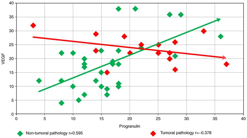
2                      BINIȘOR et al: PROGRANULIN IN TUMORAL AND NON-TUMORAL VISCERAL PATHOLOGY

Even if cancer‑associated adipocytes are demonstrated to               included into two groups: Group I included subjects with
supply energy for tumor development, the role of visceral              non‑tumoral pathology (NTP) (n=30): 15 patients with inflam‑
adipose tissue from obese or lean subjects in priming tumor            matory pathology (gastric ulcer, angiocolitis, peritonitis,
development is still under debate.                                     pancreatitis) and 15 patients with obstructive mechanical
     The adipose tissue is one of the most plastic organs in adults    pathology (hernia, eventration) and all 15 subjects of the NTP
gifted with the ability of continuous remodeling, expanding            group were obese; Group II included subjects with tumoral
or retracting according to the energetic balance. Being a              pathology (TP): 15 patients suffering from visceral cancer
compulsory condition for the expansion of any tissue in order to       (stomach, liver, ovary, gut) from which 3 were obese.
provide the substances requested for its survival, angiogenesis            Obese patients were those with abdominal obesity
may be considered in a mechanistic approach, a common feature          (Ø>94 cm in men and >80 cm in women) and a body mass
of adipose and tumor tissue. WAT secretome includes various            index (BMI) >30, according to the criteria of the International
pro‑angiogenic hormones, cytokines, and growth factors                 Federation of Diabetes (44). BMI was calculated as weight
secreted by the stromo‑vascular cells, namely endothelial cells,       divided by the height squared (kg/m2).
and also by adipocytes and preadipocytes (21‑23). Visceral                 Informed consent was obtained from all the participants
WAT is more vascularized (24). It expresses more adipokines            included in the study, which was approved by the Ethics
with angiogenic activity, endothelial cells exhibit more potent        Committee of the University of Medicine and Pharmacy of
angiogenic molecules and newly emerging adipocytes in                  Craiova (no. 85/2019).
obese conditions are highly angiogenic (25). These are added               For each subject a fragment of omental adipose tissue was
to the substances elaborated by the proinflammatory and                obtained and rinsed in saline solution.
mesenchymal cells resident in the stromovascular fraction of
the adipose depots, mainly growth factors (26‑28). Adipokines          Histological staining. Immediately after sampling, the tissue
lead to the establishment of a proangiogenic milieu.                   fragments were fixed in 10% buffered formalin for 24‑48 h at
     However, it is not clear whether all the substances secreted by   room temperature and then processed for paraffin embedding.
the adipose tissue may have a paracrine or an endocrine effect on      Sections of 3‑4 µm were mounted on a glass slide and routinely
visceral tumor initiation and progression, but the proangiogenic       stained with hematoxylin and eosin (H&E), and trichrome
factors may be truly incriminated, as angiogenesis is a major          Masson according to the producer's data sheet (Bio‑Optica).
common event required both for tumor and adipose development.
     Progranulin (PRG), also known as granulin (GRN)‑epithelin         Immunohistochemistry. Serial sections of 3‑4 µm were
precursor (29), proepithelin, (30) or acrogranin (31), is a novel      dewaxed and rehydrated. Antigen retrieval was performed
pleiotropic growth factor acting on the proliferation and              after microwave incubation of sections in citrate buffer at
development of fast‑growing epithelial cells, cancer cells (32).       pH 6.0, for 20 min. Endogenous peroxidase was blocked
It is also a proangiogenic factor whose expression is induced          after incubation with 3% hydrogen peroxide in methanol
in activated endothelial cells (33) or in growing placenta (34).       solution. After blocking, non‑specific binding with 3%
PRG, recently identified as an adipokine (7), is a 68‑ to 86‑kDa       skimmed milk in PBS at pH 7.4‑7.6 for 30 min, the sections
secreted glycoprotein which can be fragmented by some matrix           were incubated overnight, at 4º C, with one of the primary
metalloproteinases (MMPs) into small homologous subunits,              antibodies mentioned in Table I. Sections were then rinsed
granulins/epithelins (35,36), with proinflammatory activity.           in PBS and processed for amplification of the immune
Previous findings emphasize the association between PRG                signal using a detection labeling polymer [Histofine ®
and type 2 diabetes as well as non‑fatty liver disease, and also,      Simple Stain™ MAX PO (MULTI) 414151 Nichirei] as
its function in promoting vasodilatation (37). Some studies            mentioned in Table I. The immunostaining was detected
demonstrated the connection between PRG, vascular endothelial          with 3,3'‑diaminobenzidine (Vector Laboratories), and
growth factor (VEGF) expression and the augmentation of                finally the nuclear counterstaining was performed with
vascular density in breast cancer (38,39). VEGF is the most potent     Mayer's hematoxylin (Bio‑Optica). Skimmed milk, the
angiogenic factor for tumor development and invasion (40,41) and       chemicals used for buffers, solvents and hydrogen peroxide
it is secreted by adipocytes as a proangiogenic factor stimulating     were purchased from Merck.
the migration and proliferation of endothelial cells (23,42,43).           For each antibody tested, a negative control was performed
     The aim of the present study was to examine the possible          in which the primary antibody was replaced by 10 mM PBS,
relationship between PRG and VEGF in adipose visceral                  pH 7.4‑7.6.
tissue under tumorigenic or inflammatory conditions. An                    The sections were examined and images were captured
immunohistochemical study was conducted to quantify the                with a Nikon Eclipse 55i microscope equipped with a 5 Mp
expression of the aforementioned markers. In order to assess the       color‑cooled CCD (charge‑coupled device) camera, under
degree of maturation and functional differentiation of the vascular    the Image ProPlus 7 AMS image analysis software (Media
network in visceral (omental) fat related to PRG and VEGF              Cybernetics Inc.).
proangiogenic activity, an immunohistochemical identification              Each immunostaining section for the antibodies mentioned
of vessels with CD34, and collagen IV (Col IV) was performed.          above was examined by two different researchers according to
                                                                       the following: immunohistochemical (IHC) reactions (brown
Materials and methods                                                  deposits in labeled structures) were graded as absent (negative
                                                                       signal) or present (moderate or strong intensity of the signal).
Tissue samples. Human adipose tissues were obtained after              Images were finally processed using the Microsoft Office
abdominal surgery for visceral pathology. The subjects were            Picture Manager.
EXPERIMENTAL AND THERAPEUTIC MEDICINE 22: 1337, 2021 3
Table I. Antibodies and immunohistochemical techniques used for the morphological study.
Antigen Amplification
Antibody Dilution Source retrieval method
Monoclonal anti‑human 1:20 Thermo Fisher Citrate buffer HISTOFINE Simple
VEGF Scientific, Inc. pH 6.0 Stain MAX PO
JH121 (MULTI) 414151
Nichirei
Monoclonal anti‑human 1:30 Dako Citrate buffer HISTOFINE Simple
collagen IV M0785 pH 6.0 Stain MAX PO
(MULTI) 414151
Nichirei
Monoclonal anti‑human 1:20 Thermo Fisher Citrate buffer HISTOFINE Simple
progranulin Scientific, Inc. pH 6.0 Stain MAX PO
2D4‑2F1 (MULTI) 414151
Nichirei
Monoclonal anti‑human 1:50 Dako Citrate buffer HISTOFINE Simple
CD34 M7080 pH 6.0 Stain MAX PO
(MULTI) 414151
Nichirei
Morphometry. After general examination of the sections, for the two groups of patients was analyzed. The small number of
each subject and each histological or IHC‑stained slide with obese subjects in the TP group prevented us from performing
the antibodies mentioned above, three different microscopic a statistically significant correlation between lean and obese
fields on the same section were chosen randomly in the area subjects for all the markers.
of interest captured with a x20 objective lens and examined
by two different researchers. A count of the vessels marked Results
by tagging manually on the field was performed by two
researchers and the averages were used as primary values in Hematoxylin and eosin or trichrome staining. Histological
the statistical analysis. staining with hematoxylin and eosin or trichrome showed
the characteristic distribution of adipose cells in the
Statistical analysis. Statistical analysis was performed adipose lobules. In the samples from the obese subjects a
using Microsoft Excel (Microsoft Corp.), together with the pronounced heterogeneity of adipose cell dimension was
XLSTAT add‑on for MS Excel (Addinsoft SARL) and IBM observed (Fig. 1A).
SPSS Statistics 20.0 (IBM Corp.) for processing the data. Data The Student's t‑test did not reveal any significant difference
were recorded using Microsoft Excel files, then the data were between patients with mechanical or inflammatory pathology
statistically analyzed to determine the relationship between and those with tumor pathology, in terms of the number of
histological parameters of the patients. A descriptive analysis capillaries identified by trichrome or hematoxylin and eosin
of the study group (percentages of cases for categorical date, staining (P=0.0787) (Table II).
mean and standard deviation for numerical data) and complex Each lobule contained some middle vessels (arterioles or
statistical tests (Chi square and Fisher's exact test, Student's venules) and many very small capillaries (new capillaries)
t‑test) were performed using the abovementioned statistical around the adipose cells (Fig. 1A). All these vessels were
software. consistently marked with CD34 in inflammatory and in
To test the normality of the data for the parameters involved tumoral pathology (Fig. 1B and K) and inconsistently with
in this study (number of blood vessels counted using different PRG (Fig. 1C) and Col IV (Fig. 1E and F). In both groups,
histological markers) the Anderson‑Darling test was used. As proinflammatory cells were noted inside the vessel lumen,
the numerical variables investigated had a normal distribution perivascular and in the SVF between the adipose cells. In
of data, globally or inside each studied group, parametric visceral adipose tissue from the TP group, these stromal cells
statistical tests such as the Student's t‑test were employed were largely positive for PRG and CD34 (Fig. 1H, I and L).
and the results are summarized as the mean value ± standard The result of the Student's t‑test revealed a significant
deviation. P4 BINIȘOR et al: PROGRANULIN IN TUMORAL AND NON-TUMORAL VISCERAL PATHOLOGY
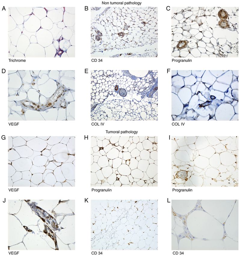
Figure 1. Microscopic aspects of the adipose tissue sections from patients with non‑tumoral pathology (NTP) group and tumoral pathology (TP) group.
Microscopic aspects of the adipose tissue sections from patients with non‑tumoral pathology (NTP) group (A‑F): (A) Trichrome. Adipose tissue from epiploon,
inflammatory cells were present in vessels and in stromo‑vascular fraction (SVF), intermingled with the adipocytes. Magnification, x400. (B) CD34. General
view of an adipose lobule: double positive concentric rings are noted around the vessels. Magnification, x100. (C) PRG. Positive cells for PRG in vessels and
in SVF. Magnification, x20. (D) VEGF. Endothelial positivity for VEGF in SVF cells and in endothelial cells. Magnification, x40. (E) Col IV. General aspect
of an adipose pad, vessels on the periphery are positive for Col IV. Magnification, x45. (F) Col IV. Few capillaries inside of the adipose lobule immunolabeled
with Col IV. Magnification, x400. (G‑L) Microscopic aspects of the adipose tissue sections from patients with tumoral pathology (TP group): (G) VEGF was
positive in endothelial cells and in mature adipocytes. Magnification, x400. (H) Adipose cells and capillaries with intense reaction for PRG. Magnification,
x200. (I) PRG intense positive reaction in vessels. Magnification, x400. (J) VEGF. Positive reaction in vessels walls. Magnification, x400. (K) CD34. Numerous
vessels positive for CD34. Magnification, x100. (L) CD34. New forming vessels and stromal cells positive for CD34. Magnification, x400. SVF, stromo‑vascular
fraction; PRG, progranulin; VEGF, vascular endothelial growth factor; Col IV, collagen IV.
not for inflammatory and mechanical pathologies, nor for peripheral area and PRG was constantly present in endothelial
tumoral pathologies. cells of the very small neoformation capillaries (Fig. 1H and I).
In adipose tissue samples collected from patients of the TP
Number of capillaries. CD34 and PRG immune reaction group, the adipose cells also showed a positive reaction for
showed a stronger positivity deep in the lobule than in the PRG (Fig. 1H and I). In the medium vessels, CD34‑positiveEXPERIMENTAL AND THERAPEUTIC MEDICINE 22: 1337, 2021 5
Table II. Number of capillaries identified with usual histo‑ Table IV. Number of capillaries labeled with CD34.
logical staining.
Pathology vs. Non‑tumoral Tumoral
Pathology vs. Non‑tumoral Tumoral CD34 pathology pathology
Trichrome/H&E pathology pathology
No. of cases 30 15
No. of cases 30 15 Mean 21.63 31.93
Mean 28.23 32.47 SD 9.39 7.60
SD 8.16 5.64 C.V. (%) 43.42% 23.80%
C.V. (%) 28.89% 17.38% P‑value 0.0006 HS
P‑value 0.0787 NS (Student's t‑test)
(Student's t‑test)
SD, standard deviation; C.V., coefficient of variation; HS, highly
SD, standard deviation; H&E, hematoxylin and eosin; C.V., coef‑ significant.
ficient of variation; NS, not significant.
(Table VI). Any significant difference was noted between the
Table III. Number of capillaries labeled with progranulin
number of blood vessels identified in normal‑weight and obese
(PRG).
patients neither for inflammatory and mechanical pathologies
Pathology vs. Non‑tumoral Tumoral nor for tumoral pathologies.
PRG pathology pathology Finally, the relationship between PRG and VEGF for the
two groups of patients, NTP with inflammatory and mechan‑
No. of cases 30 15 ical pathology, and TP with tumoral pathology, was examined.
Mean 16.17 22.53 For the first group, the Pearson correlation coefficient was
r= 0.595, showing a statistically significant direct correlation,
SD 7.00 8.53
with a significance level of 95% for 30 samples (P6 BINIȘOR et al: PROGRANULIN IN TUMORAL AND NON-TUMORAL VISCERAL PATHOLOGY
Table V. Comparison between the number of vessels labeled for VEGF between lean and obese patients.
Non‑tumoral pathology Tumoral pathology
Pathology ‑‑‑‑‑‑‑‑‑‑‑‑‑‑‑‑‑‑‑‑‑‑‑‑‑‑‑‑‑‑‑‑‑‑‑‑‑‑‑‑‑‑‑‑‑‑‑‑‑‑‑‑‑‑‑‑‑‑‑‑‑‑‑‑‑‑‑‑‑‑‑‑‑‑‑ ‑‑‑‑‑‑‑‑‑‑‑‑‑‑‑‑‑‑‑‑‑‑‑‑‑‑‑‑‑‑‑‑‑‑‑‑‑‑‑‑‑‑‑‑‑‑‑‑‑‑‑‑‑‑‑‑‑‑‑‑‑‑‑‑‑‑‑‑‑‑‑‑‑‑‑
Weight Normal weight Obese Normal weight Obese
No of cases 15 15 12 3
Mean 20.27 16.07 25.00 17.67
SD 11.07 7.91 4.22 3.79
C.V. (%) 54.64% 49.26% 16.88% 21.43%
P‑value 0.242 (NS) 0.017 (S)
(Student's t‑test)
SD, standard deviation; CV, coefficient of variation; NS, not significant; S, significant.
Table VI. Number of capillaries labeled for collagen IV. mechanical and inflammatory pathology (NTP) and those with
tumor pathology (TP) in terms of the number of capillaries
Pathology vs. Non‑tumoral Tumoral identified, similar to PRG, was not evident. However, VEGF
Collagen IV pathology pathology expression showed a significant difference between lean and
obese subjects in the TP group. This aspect may be explained
No. of cases 30 15
by the canonical model of vascular development whereby the
Mean 22.83 17.80 oncogenic vascularization and the vascular network in adipose
SD 7.55 4.36 pads are both induced by the rapid growth of the tissue and
C.V. (%) 33.08% 24.51% the onset of hypoxic conditions, with an increase in VEGF
P‑value 0.0217 S expression (49).
(Student's t‑test) As observed in the IHC reactions for PRG and VEGF,
not only the capillaries demonstrate an immune positivity for
SD, standard deviation; CV, coefficient of variation; NS, not the two molecules, but also the adipocytes and cells from
significant; S, significant.
the stromal tissue. Similar results were reported by others
showing that both SVF and mature adipocytes express
VEGF (24,50).
In obese subjects, when the blood supply becomes deficient,
cells from the SVF contribute to PRG expression in adipose as in all fast‑growing tissues, the whole human adult adipose
tissue (45). Correlating these results with the serum level of tissue can induce angiogenesis from its own cells, adipocytes
PRG authors of that study suggested that PRG contributes or stromal cells, or can recruit distant endothelial cells (24). In
to the crosstalk between macrophages and adipocytes in the human and animal models, it was demonstrated that VEGF is
adipose tissue and could be one of the molecules involved in produced by adult adipocytes and stromovascular cells (2,51).
the increase of the macrophage number in the visceral adipose Corroborating our results with findings by others (24,52), an
tissue of obese subjects. interesting hypothesis to understand the adaptation of the
PRG is less expressed in quiescent vessels and more vascular network density to the adipose pads volume was that
intensely in tissue with active angiogenesis: in normal tissues the synthesis of adipogenic factors is related to the variation of
(developing placenta or wound healing) (33,34) or tumoral weight rather than to the absolute weight. Our results demon‑
diseases (46‑48). The results of the present study demonstrated strated that weight may influence the expression of VEGF in
that the vessels marked with PRG were more numerous in the tumoral pathology but not in inflammatory or mechanical
adipose pads of the tumoral milieu than in the inflammatory diseases, and that weight does not seem to have the same
ones, and the positivity observed from the IHC reaction was influence for PRG expression.
not restricted to vessels, but was expressed also in adipocytes The present study was carried out on a small number of
and migrated cells. Thus, PRG overexpression in the vessel cases, and must therefore be considered a pilot study. Research
walls and equally in adipocytes and stromal cells may be on a larger group of patients should be conducted to perform a
induced by proangiogenic molecules synthetized by the comparison with significant results between VEGF and PRG
neighboring tumor. in lean and obese subjects.
The angiogenic action of PRG seems to be performed at When and how locally secreted PRG and VEGF could
least under certain circumstances (such as in breast cancer influence individually or by molecular crosstalk the initiation
and breast cancer cell lines) (39) under the influence of the and the development of visceral tumors is a controversial
main proangiogenic factor, VEGF. In order to assess the subject. It is often emphasized that the tumor microenviron‑
collaboration between the two molecules, we performed an ment provides angiogenetic factors released by the tumor cells
IHC study for the expression of VEGF in blood vessels of or by the stromal cells surrounding the tumors (20,53). VEGF,
the same lots. A significant difference between patients with leptin and recently described PRG are similarly expressedEXPERIMENTAL AND THERAPEUTIC MEDICINE 22: 1337, 2021 7
Figure 2. Correlation between PRG and VEGF expression. PRG, progranulin; VEGF, vascular endothelial growth factor.
in adipose tissue and the tumoral vascular network develop‑ cells and probably by the pericytes occurs (60,61). Col IV is the
ment (25,50,54‑56). Overexpression of PRG was reported in main constituent of the lamina densa of the basal membrane,
renal, breast or ovarian cancers (57‑59), organs embedded secreted by the endothelial cells and pericytes in the final step
in an active metabolic adipose tissue. Tangkeangsirisin et al of the sequential angiogenic process (61) and required for
demonstrated that PRG could promote tumor angiogenesis and blood vessel maturation and homeostasis (62).
metastasis in human breast cancer by stimulating the synthesis In order to assess the incidence of mature capillaries in the
of VEGF and MMP9 (39). two lots of subjects we performed an IHC study using the Col IV
In a mouse developmental model, Toh et al demonstrated antibody. The results showed a significant difference between
that the expression of VEGF and its receptors is compulsory for patients with mechanical and inflammatory pathology (NTP
early vasculogenesis in embryo, but PRG was expressed later, group and those with TP), in terms of the number of capillaries
its synthesis being instead necessary for vessel growth and identified. In the TP group, few vessels were labeled inside the
stability and may represent a novel angiogenic pathway (48). adipose lobules, and those located superficially and the small new
The results reported by Tangkeangsirisin and Serrero for forming vessels were inconstantly positive. Considering Col IV
breast tumors support the hypothesis that the increased VEGF as a marker for mature functional vascular wall, we may affirm
expression was determined rather than an indirect effect of that in the adipose tissue of the TP group, PRG stimulates the
stromal cells found in the tumor environment by the PRG formation of newly less mature and functional vessels, lacking
direct stimulation of VEGF expression (39). a basal membrane. Considering this observation, we supposed
The statistical correlations between the two angiogenetic that a proportion of the remaining hidden capillaries may be
factors analyzed in our experiment showed interesting newly formed. We used CD34, a major endothelial cell marker
differences. As shown in Fig. 2, for the first group (NTP), present on hematopoietic stem cells and on progenitor cells (63),
the Pearson correlation coefficient showed a statistically in order to label these presumed capillaries. In terms of the
significant direct correlation (r= 0.595), while for the second number of capillaries identified, a highly significant difference
smaller group (TP), the Pearson coefficient showed an (P8 BINIȘOR et al: PROGRANULIN IN TUMORAL AND NON-TUMORAL VISCERAL PATHOLOGY
Considering these results together with the significant Authors' information
number of vessels marked by PRG in the TP group, we were
able to sustain that in the PRG omental adipose pads had a This study is part of the Ph.D. thesis of Ioana Binisor, from the
significant contribution to induce angiogenesis in a tumoral University of Medicine and Pharmacy of Craiova, Romania.
ambience, and that this capacity is complementary with VEGF
action in NTP. References
In summary, PRG is an adipokine able to act as an
effective proangiogenic factor in visceral adipose tissue, with 1. Divella R, De Luca R, Abbate I, Naglieri E and Daniele A:
Obesity and cancer: The role of adipose tissue and
the formation of new capillaries in visceral tumors. However, adipo-cytokines-induced chronic inflammation. J Cancer 7:
whether the angiogenic response to PRG synthesis is influenced 2346‑2359, 2016.
or not by inflammation and obesity remains to be determined. 2. Marlatt KL and Ravussin E: Brown adipose tissue: An update on
recent findings. Curr Obes Rep 6: 389‑396, 2017.
VEGF is more efficient in stimulating the formation of new 3. Yoneshiro T, Aita S, Matsushita M, Kayahara T, Kameya T,
vessels in inflammatory conditions depending on weight. The Kawai Y, Iwanaga T and Saito M: Recruited brown adipose tissue as
an antiobesity agent in humans. J Clin Invest 123: 3404‑3408, 2013.
collaboration of the proangiogenic activity of PRG and VEGF 4. A nd rei A M, Berb e ca r u‑Iova n A, D i n‑A nghel F R I,
in the adipose tissue under tumor conditions may be dependent Stănciulescu CE, Berbecaru‑Iovan S, Baniţă IM and Pisoschi CG:
on the visceral tumor type. Chapter 16. Interplay between hypoxia, inflammation and
adipocyte remodeling in the metabolic syndrome. In: Hypoxia
and Human Diseases. Zheng J (ed). IntechOpen, pp303‑329, 2017.
Acknowledgements 5. Andrei AM, Berbecaru‑Iovan A, Din‑Anghel FRI, Binişor ID,
Marinescu RM, Goga LD, Baniță IM and Pisoschi CG: New
insights into brown adipose tissue as a pharmacological target in
We are thankful for the technical support provided by Cristina obesity. Farmacia 68: 1‑7, 2020.
Stănciucă from the Histology Department, University of 6. Fusaru AM, Stănciulescu CE, Surlin V, Taisescu C, Bold A,
Pop OT, Baniţă IM, Crăiţoiu S and Pisoschi CG: Role of innate
Medicine and Pharmacy of Craiova, Romania. immune receptors TLR2 and TLR4 as mediators of the inflam‑
matory reaction in human visceral adipose tissue. Rom J Morphol
Embryol 53 (3 Suppl): 693‑701, 2012.
Funding 7. Francisco V, Pino J, Gonzalez‑Gay MA, Mera A, Lago F, Gómez R,
Mobasheri A and Gualillo O: Adipokines and inflammation: Is it a
No funding was received. question of weight? Br J Pharmacol 175: 1569‑1579, 2018.
8. Schäffler A and Schölmerich J: Innate immunity and adipose
tissue biology. Trends Immunol 31: 228‑235, 2010.
Availability of data and materials 9. Hotamisligil GS: Inflammation and metabolic disorders.
Nature 444: 860‑867, 2006.
10. Cozzo AJ, Fuller AM and Makowski L: Contribution of adipose
The datasets used and/or analyzed during the current study are tissue to development of cancer. Compr Physiol 8: 237‑282, 2017.
available from the corresponding author on reasonable request. 11. Venniyoor A: The most important questions in cancer research
and clinical oncology‑question 2‑5. Obesity‑related cancers:
More questions than answers. Chin J Cancer 36: 18, 2017.
Authors' contributions 12. Mendonça F and Soares R: Obesity and cancer phenotype:
Is angiogenesis a missed link? Life Sci 139: 16‑23, 2015.
13. Booth AD, Magnuson AM, Fouts J, Wei Y, Wang D,
IB, MCM, DA and SJ made equal contributions to the Pagliassotti MJ and Foster MT: Subcutaneous adipose tissue
conception and editing of this manuscript. IB, IMB, MCM, accumulation protects systemic glucose tolerance and muscle
metabolism. Adipocyte 7: 261‑272, 2018.
SJ and CGP were involved in literature research and wrote 14. Arnold M, Pandeya N, Byrnes G, Renehan PAG, Stevens GA,
the manuscript. Data curation was performed by IB, IMB Ezzati PM, Ferlay J, Miranda JJ, Romieu I, Dikshit R, et al:
and CGP. DA supported the statistical analysis and reviewed Global burden of cancer attributable to high body‑mass index in
2012: A population‑based study. Lancet Oncol 16: 36‑46, 2015.
the results. IB, IMB, SJ and CGP conceived, planned and 15. Lauby‑Secretan BP, Scoccianti C, Loomis D, Grosse Y, Bianchini F
followed the execution of the experiments. AMA, MCM and Straif K; International Agency for Research on Cancer
Handbook Working Group: Body fatness and cancer‑viewpoint of
and IMB performed morphometry. All authors contributed the IARC working group. N Engl J Med 375: 794‑798, 2016.
to manuscript revision, read and approved the final version. 16. Pollak M: The insulin and insulin‑like growth factor receptor
AMA, MCM, IB, IMB, SJ and CGP are all responsible for the family in neoplasia: An update. Nat Rev Cancer 12: 159‑169, 2012.
17. Colotta F, Allavena P, Sica A, Garlanda C and Mantovani A:
authenticity of the data obtained. Cancer‑related inflammation, the seventh hallmark of cancer:
Links to genetic instability. Carcinogenesis 30: 1073‑1081, 2009.
18. Khandekar MJ, Cohen P and Spiegelman BM: Molecular
Ethics approval and consent to participate mechanisms of cancer development in obesity. Nat Rev
Cancer 11: 886‑895, 2011.
The study was approved by The Ethics Committee of The 19. Niema n K M, Ken ny H A, Pen icka CV, Lada nyi A,
Buell‑Gutbrod R, Zillhardt MR, Romero IL, Carey MS,
University of Medicine and Pharmacy of Craiova, Romania Mills GB, Hotamisligil GS, et al: Adipocytes promote ovarian
(approval no. 85/2019). All patients included in the study cancer metastasis and provide energy for rapid tumor growth.
Nat Med 17: 1498‑1503, 2011.
provided informed consent for data publication. 20. Nieman KM, Romero IL, Van Houten B and Lengyel E: Adipose
tissue and adipocytes support tumorigenesis and metastasis.
Patient consent for publication Biochim Biophys Acta 1831: 1533‑1541, 2013.
21. Rehman J, Traktuev D, Li J, Merfeld‑Clauss S, Temm‑Grove CJ,
Bovenkerk JE, Pell CL, Johnstone BH, Considine RV and
Not applicable. March KL: Secretion of angiogenic and antiapoptotic factors by
human adipose stromal cells. Circulation 109: 1292‑1298, 2004.
22. Halberg N, Wernstedt‑Asterholm I and Scherer PE: The adipocyte
Competing interests as an endocrine cell. Endocrinol Metab Clin North Am 37:
753‑768, 2008.
23. Christiaens V and Lijnen HR: Angiogenesis and development of
The authors declare that they have no competing interests. adipose tissue. Mol Cell Endocrinol 318: 2‑9, 2010.EXPERIMENTAL AND THERAPEUTIC MEDICINE 22: 1337, 2021 9
24. Ledoux S, Queguiner I, Msika S, Calderari S, Rufat P, Gasc JM, 49. Corvera S and Gealekman O: Adipose tissue angiogenesis:
Corvol P and Larger E: Angiogenesis associated with visceral Impact on obesity and type‑2 diabetes. Biochim Biophys
and subcutaneous adipose tissue in severe human obesity. Acta 1842: 463‑472, 2014.
Diabetes 57: 3247‑3257, 2008. 50. Zhang QX, Magovern CJ, Mack CA, Budenbender KT, Ko W
25. Sun K, Kusminski CM and Scherer PE: Adipose tissue and Rosengart TK: Vascular endothelial growth factor is
remodeling and obesity. J Clin Invest 121: 2094‑2101, 2011. the major angiogenic factor in omentum: Mechanism of the
26. Cao Y: Angiogenesis and vascular functions in modulation omentum‑mediated angiogenesis. J Surg Res 67: 147‑154, 1997.
of obesity, adipose metabolism, and insulin sensitivity. Cell 51. Fusaru AM, Pisoschi CG, Bold A, Taisescu C, Stănescu R,
Metab 18: 478‑489, 2013. Hîncu M, Crăiţoiu S and Baniţă IM: Hypoxia induced VEGF
27. Lijnen HR: Angiogenesis and obesity. Cardiovasc Res 78: synthesis in visceral adipose depots of obese diabetic patients.
286‑293, 2008. Rom J Morphol Embryol 53: 903‑909, 2012.
28. Hammarstedt A, Gogg S, Hedjazifar S, Nerstedt A and Smith U: 52. Voros G, Maquoi E, Demeulemeester D, Clerx N, Collen D and
Impaired adipogenesis and dysfunctional adipose tissue in Lijnen HR: Modulation of angiogenesis during adipose tissue
human hypertrophic obesity. Physiol Rev 98: 1911‑1941, 2018. development in murine models of obesity. Endocrinology 146:
29. Zanocco‑Marani T, Bateman A, Romano G, Valentinis B, He ZH 4545‑4554, 2005.
and Baserga R: Biological activities and signaling pathways of 53. Folkman J: Role of angiogenesis in tumor growth and metastasis.
the granulin/epithelin precursor. Cancer Res 59: 5331‑5340, 1999. Semin Oncol 29 (Suppl 16): S15‑S18, 2002.
30. Shoyab M, McDonald VL, Byles C, Todaro GJ and Plowman GD: 54. Hicklin DJ and Ellis LM: Role of the vascular endothelial
Epithelins 1 and 2: Isolation and characterization of two growth factor pathway in tumor growth and angiogenesis. J Clin
cysteine‑rich growth‑modulating proteins. Proc Natl Acad Sci Oncol 23: 1011‑1027, 2005.
USA 87: 7912‑7916, 1990. 55. Papetti M and Herman IM: Mechanisms of normal and
31. Anakwe OO and Gerton GL: Acrosome biogenesis begins tumor‑derived angiogenesis. Am J Physiol Cell Physiol 282:
during meiosis: Evidence from the synthesis and distribution C947‑C970, 2002.
of an acrosomal glycoprotein, acrogranin, during guinea pig 56. Gonzalez‑Perez RR, Lanier V and Newman G: Leptin's
spermatogenesis. Biol Reprod 42: 317‑328, 1990. pro‑angiogenic signature in breast cancer. Cancers (Basel) 5:
32. Jian J, Konopka J and Liu C: Insights into the role of progranulin 1140‑1162, 2013.
in immunity, infection, and inflammation. J Leukoc Biol 93: 57. Lu R and Serrero G: Inhibition of PC cell‑derived growth factor
199‑208, 2013. (PCDGF, epithelin/granulin precursor) expression by antisense
33. He Z, Ong CH, Halper J and Bateman A: Progranulin is a PCDGF cDNA transfection inhibits tumorigenicity of the human
mediator of the wound response. Nat Med 9: 225‑229, 2003. breast carcinoma cell line MDA‑MB‑468. Proc Natl Acad Sci
34. Desmarais J, Cao M, Bateman A and Murphy B: Spatiotemporal USA 97: 3993‑3998, 2000.
expression pattern of progranulin in embryo implantation 58. Donald CD, Laddu A, Chandham P, Lim SD, Cohen C, Amin M,
and placenta formation suggests a role in cell proliferation, Gerton GL, Marshall FF and Petros JA: Expression of progran‑
remodeling, and angiogenesis. Reproduction 136: 247‑257, 2008. ulin and the epithelin/granulin precursor acrogranin correlates
35. Jian J, Li G, Hettinghouse A and Liu C: Progranulin: A key with neoplastic state in renal epithelium. Anticancer Res 21:
player in autoimmune diseases. Cytokine 101: 48‑55, 2018. 3739‑3742, 2001.
36. Wei J, Hettinghouse A and Liu C: The role of progranulin in 59. Jones MB, Michener CM, Blanchette JO, Kuznetsov VA,
arthritis. Ann NY Acad Sci 1383: 5‑20, 2016. Raffeld M, Serrero G, Emmert‑Buck MR, Petricoin EF,
37. Korolczuk A and Bełtowski J: Progranulin, a new adipokine at Krizman DB, Liotta LA and Kohn EC: The granulin‑epithelin
the crossroads of metabolic syndrome, diabetes, dyslipidemia precursor/PC‑cell‑derived growth factor is a growth factor for
and hypertension. Curr Pharm Des 23: 1533‑1539, 2017. epithelial ovarian cancer. Clin Cancer Res 9: 44‑51, 2003.
38. Li LQ, Min LS, Jiang Q, Ping JL, Li J and Dai LC: Progranulin 60. Sand JMB, Genovese F, Gudmann NS and Karsdal MA:
expression in breast cancer with different intrinsic subtypes. Chapter 4‑type IV collagen. In: Biochemistry of Collagens,
Pathol Res Pract 208: 210‑216, 2012. Laminins and Elastin. Karsdal MA (ed). 2nd edition. Academic
39. Tangkeangsirisin W and Serrero G: PC cell‑derived growth factor Press, pp37‑49, 2019.
(PCDGF/GP88, progranulin) stimulates migration, invasiveness 61. Maragoudakis ME: The role of basement membrane in
and VEGF expression in breast cancer cells. Carcinogenesis 25: angiogenesis. In: Vascular Endothelium. Catravas JD, Gillis CN
1587‑1592, 2004. and Ryan US (eds). Springer, Boston, MA, pp111‑120, 1989.
40. Ferrara N: Vascular endothelial growth factor: Basic science and 62. Pöschl E, Schlötzer‑Schrehardt U, Brachvogel B, Saito K,
clinical progress. Endocr Rev 25: 581‑611, 2004. Ninomiya Y and Mayer U: Collagen IV is essential for basement
41. Tammela T, Enholm B, Alitalo K and Paavonen K: The biology membrane stability but dispensable for initiation of its assembly
of vascular endothelial growth factors. Cardiovasc Res 65: during early development. Development 131: 1619‑1628, 2004.
550‑563, 2005. 63. Goncharov NV, Nadeev AD, Jenkins RO and Avdonin PV:
42. Ikeda K: Adipose tissue angiogenesis: An emerging therapeutic Markers and biomarkers of endothelium: When something is
target for obesity and methabolic disease. Austin J Clin rotten in the state. Oxid Med Cell Longev 2017: 9759735, 2017.
Cardiolog 1: 1005, 2014. 64. Daquinag AC, Zhang Y and Kolonin MG: Vascular targeting of
43. Lemoine AY, Ledoux S and Larger E: Adipose tissue angiogenesis adipose tissue as an anti‑obesity approach. Trends Pharmacol
in obesity. Thromb Haemost 110: 661‑668, 2013. Sci 32: 300‑307, 2011.
44. Alberti KG, Eckel RH, Grundy SM, Zimmet PZ, Cleeman JI, 65. Lin G, Garcia M, Ning H, Banie L, Guo YL, Lue TF and Lin CS:
Donato KA, Fruchart JC, James WP, Loria CM, Smith SC Jr, et al: Defining stem and progenitor cells within adipose tissue. Stem
Harmonizing the metabolic syndrome. A joint interim statement Cells Dev 17: 1053‑1063, 2008.
of the international diabetes federation task force on epidemiology 66. Sidney LE, Branch MJ, Dunphy SE, Dua HS and Hopkinson A:
and prevention; national heart, lung, and blood institute; Concise review: Evidence for CD34 as a common marker for
American heart association; world heart federation; international diverse progenitors. Stem Cells 32: 1380‑1389, 2014.
atherosclerosis society; and international association for the 67. Zannettino ACW, Paton S, Arthur A, Khor F, Itescu S, Gimble JM
study of obesity. Circulation 120: 1640‑1645, 2009. and Gronthos S: Multipotential human adipose‑derived stromal
45. Youn BS, Bang SI, Klöting N, Park JW, Lee N, Oh JE, Pi KB, stem cells exhibit a perivascular phenotype in vitro and in vivo.
Lee TH, Ruschke K, Fasshauer M, et al: Serum progranulin J Cell Physiol 214: 413‑421, 2008.
concentrations may be associated with macrophage infiltration 68. Hu Y, Zhang Z, Torsney E, Afzal AR, Davison F, Metzler B and
into omental adipose tissue. Diabetes 58: 627‑636, 2009. Xu Q: Abundant progenitor cells in the adventitia contribute
46. Davidson B, Alejandro E, Flørenes VA, Goderstad JM, Risberg B, to atherosclerosis of vein grafts in ApoE‑deficient mice. J Clin
Kristensen GB, Trope CG and Kohn EC: Granulin‑epithelin Invest 113: 1258‑1265, 2004.
precursor is a novel prognostic marker in epithelial ovarian
carcinoma. Cancer 100: 2139‑2147, 2004.
47. Gonzalez EM, Mongiat M, Slater SJ, Baffa R and Iozzo RV: This work is licensed under a Creative Commons
A novel interaction between perlecan protein core and Attribution-NonCommercial-NoDerivatives 4.0
progranulin: Potential effects on tumor growth. J Biol Chem 278: International (CC BY-NC-ND 4.0) License.
38113‑38116, 2003.
48. Toh H, Cao M, Daniels E and Bateman A: Expression of the
growth factor progranulin in endothelial cells influences growth
and development of blood vessels: A novel mouse model. PLoS
One 8: e64989, 2013.You can also read


