Human pluripotent stem cell-derived alveolar organoids for modeling pulmonary fibrosis and drug testing
←
→
Page content transcription
If your browser does not render page correctly, please read the page content below
Kim et al. Cell Death Discovery (2021)7:48
                                    https://doi.org/10.1038/s41420-021-00439-7                                                                                                           Cell Death Discovery

                                     ARTICLE                                                                                                                                                    Open Access

                                    Human pluripotent stem cell-derived alveolar
                                    organoids for modeling pulmonary fibrosis and
                                    drug testing
                                    Jung-Hyun Kim1, Geun Ho An2,3, Ji-Young Kim1, Roya Rasaei1, Woo Jin Kim1, Xiong Jin2, Dong-Hun Woo2,
                                    Choongseong Han2, Se-Ran Yang4, Jong-Hoon Kim3 and Seok-Ho Hong 1

                                      Abstract
                                      Detailed understanding of the pathogenesis and development of effective therapies for pulmonary fibrosis (PF) have
                                      been hampered by lack of in vitro human models that recapitulate disease pathophysiology. In this study, we
                                      generated alveolar organoids (AOs) derived from human pluripotent stem cells (hPSCs) for use as an PF model and for
                                      drug efficacy evaluation. Stepwise direct differentiation of hPSCs into alveolar epithelial cells by mimicking
                                      developmental cues in a temporally controlled manner was used to generate multicellular AOs. Derived AOs
                                      contained the expected spectrum of differentiated cells, including alveolar progenitors, type 1 and 2 alveolar epithelial
                                      cells and mesenchymal cells. Treatment with transforming growth factor (TGF-β1) induced fibrotic changes in AOs,
                                      offering a PF model for therapeutic evaluation of a structurally truncated form (NP-011) of milk fat globule-EGF factor 8
                                      (MFG-E8) protein. The significant fibrogenic responses and collagen accumulation that were induced by treatment
                                      with TGF-β1 in these AOs were effectively ameliorated by treatment with NP-011 via suppression of extracellular
                  1234567890():,;
1234567890():,;
                  1234567890():,;
1234567890():,;

                                      signal-regulated kinase (ERK) signaling. Furthermore, administration of NP-011 reversed bleomycin-induced lung
                                      fibrosis in mice also via ERK signaling suppression and collagen reduction. This anti-fibrotic effect mirrored that
                                      following Pirfenidone and Nintedanib administration. Furthermore, NP-011 interacted with macrophages, which
                                      accelerated the collagen uptake for eliminating accumulated collagen in fibrotic lung tissues. This study provides a
                                      robust in vitro human organoid system for modeling PF and assessing anti-fibrotic mechanisms of potential drugs and
                                      suggests that modified MGF-E8 protein has therapeutic potential for treating PF.

                                    Introduction                                                                                        reduction in lung function decline2. No survival advantage
                                      Pulmonary fibrosis (PF) is a fatal chronic respiratory                                             was noted and the injured lungs did not recover following
                                    disease characterized by accumulation of myofibroblasts                                              use of these drugs, highlighting the need to identify novel
                                    and deposition of extracellular matrix, leading to                                                  and effective therapeutic drugs for the treatment of PF3,4.
                                    respiratory failure1. To date, only two drugs, Pirfenidone                                            One of major obstacles for the development of effective
                                    (PFD, Esbriet) and Nintedanib (Nib, Ofev), have been                                                therapies for PF has been a lack of in vitro human models
                                    approved for human PF based on the demonstration of a                                               that recapitulate features of alveolar tissue and the
                                                                                                                                        pathophysiology of the disease. While human primary
                                                                                                                                        pulmonary alveolar epithelial cells (pAECs), comprised of
                                    Correspondence: Jong-Hoon Kim (jhkim@korea.ac.kr) or Seok-
                                                                                                                                        alveolar type 1 and type 2 epithelial cells (AEC1 and
                                    Ho Hong (shhong@kangwon.ac.kr)
                                    1
                                     Department of Internal Medicine, School of Medcine, Kangwon National                               AEC2), are the most representative of the in vivo situa-
                                    University, Chuncheon 24341, South Korea
                                    2
                                                                                                                                        tion, low accessibility, a limited proliferation capacity, and
                                     Department of New Drug Development, NEXEL, Co., Ltd, Seoul, South Korea
                                                                                                                                        loss of functional properties over time remain a bottle-
                                    Full list of author information is available at the end of the article
                                    These authors equally contributed: Jung-Hyun Kim, Geun Ho An, Ji-Young Kim                          neck for their use in PF studies5. An immortalized
                                    Edited by: Maria Victoria Niklison Chirou

                                    © The Author(s) 2021
                                                        Open Access This article is licensed under a Creative Commons Attribution 4.0 International License, which permits use, sharing, adaptation, distribution and reproduction
                                                        in any medium or format, as long as you give appropriate credit to the original author(s) and the source, provide a link to the Creative Commons license, and indicate if
                                    changes were made. The images or other third party material in this article are included in the article’s Creative Commons license, unless indicated otherwise in a credit line to the material. If
                                    material is not included in the article’s Creative Commons license and your intended use is not permitted by statutory regulation or exceeds the permitted use, you will need to obtain
                                    permission directly from the copyright holder. To view a copy of this license, visit http://creativecommons.org/licenses/by/4.0/.

                                    Official journal of the Cell Death Differentiation Association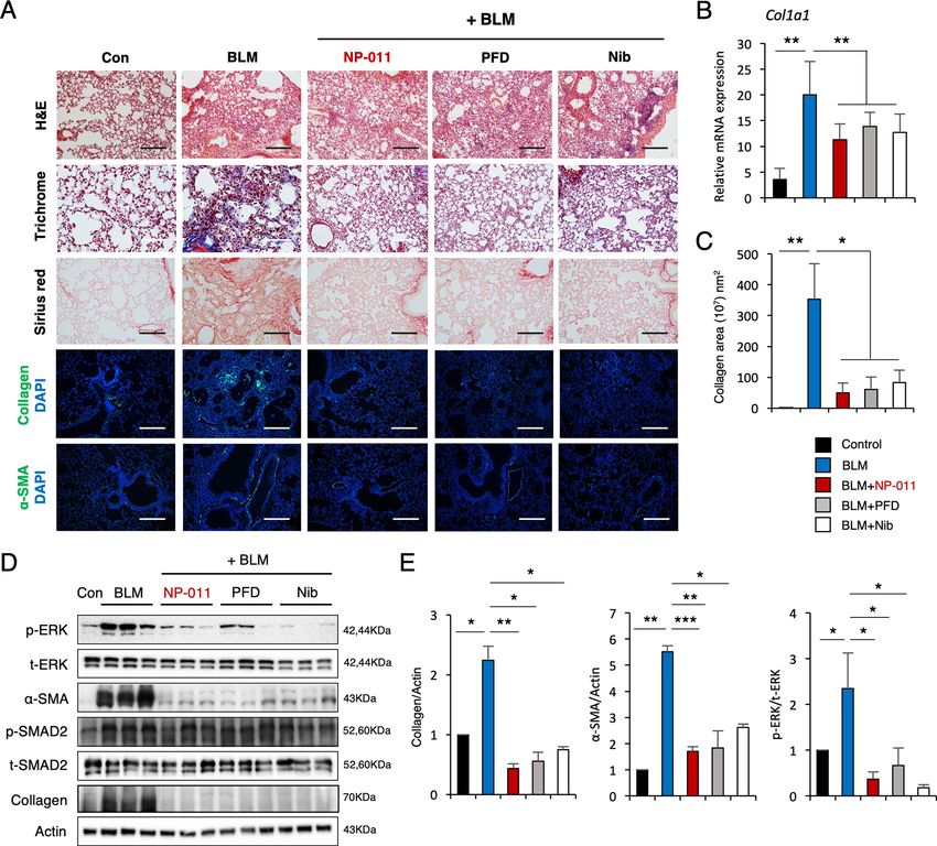
Kim et al. Cell Death Discovery (2021)7:48                                                                       Page 2 of 12

pulmonary epithelial cell line (A549) derived from a            mesenchymal support cells6. By taking advantage of this
human pulmonary adenocarcinoma has been widely used             protocol, two-dimensional cultures of emerging distal
instead of pAECs to model the alveolar epithelium for           alveolar cell types (SOX9, ID2, SFTPB, and SFTPC)20 on
biopharmaceutical research and to evaluate the pulmo-           day 21 of differentiation were dissociated to single cells to
toxicity of suspected harmful materials; however, these         form uniform aggregates by forced aggregation (Supple-
cells respond to toxins differently and exhibit altered         mentary Fig. 1A). The aggregates were transferred into
phenotypic, genetic, and functional properties compared         AEC maturation medium and cultured for 6 days to
with primary pAECs6–8. Alternatively, primary pAECs             establish AOs, which exhibited an alveolar sac-like
from other laboratory animals can be used, but species-         structure with multiple alveoli and layers of epithelial
specific differences in molecular mechanisms of fibrosis          cells (Supplementary Fig. 1B). Immunofluorescence
and responses to potential drugs may also limit applic-         staining showed that hPSC-derived AOs express markers
ability to humans9,10. Importantly, recent studies suggest      of AEPs (EPCAM, CPM, and NKX2.1), AEC1 (AQP5 and
that three-dimensional configurations, such as alveolar          T1α), AEC2 (SFTPC), and mesenchymal cells (VIMEN-
organoids (AOs) and spheroids, offer several advantages         TIN) (Supplementary Fig. 1C). Moreover, AEPs, AEC1,
compared to conventional two-dimensional pAEC                   AEC2, and mesenchymal cell-related genes were robustly
monolayer cultures for studying early lung development,         expressed in AOs compared to undifferentiated hPSC
modeling disease, and screening for novel drugs11–15.           cultures according to quantitative PCR (qPCR) analysis
Therefore, the development of an in vitro human AO              (Supplementary Fig. 1D). These observations confirmed
model using a reliable and renewable biological source to       that our method enables the generation of multicellular
generate pAECs that phenotypically and functionally             AOs containing AEPs, AEC1, AEC2, and mesenchymal
resemble primary pAECs offers great promise for PF              support cells from hPSCs.
modeling and drug screening.
   The ability to differentiate human pluripotent stem cells    Modified MFG-E8 protein (NP-011) ameliorates TGF-β1-
(hPSCs) into three germ layer lineages offers promise for       induced fibrotic changes in hPSC-derived AOs via
the development of in vitro three-dimensional organoids         suppression of ERK signaling
that recapitulate the complexity and functions of in vivo         Given the presence of multiple cell types in AOs, we
tissues16. In the lungs, hPSCs have been successful             investigated whether AOs are a useful in vitro model for
employed to generate several organoids that represent           studying PF. To evaluate the capacity of AOs to respond
different respiratory compartments, such as proximal and        to a fibrogenic stimulus, we treated AOs with 25 ng/ml
distal airways and alveoli11–15. More recently, hPSC-           TGF-β1 for 72 h and analyzed fibrotic changes at both the
derived alveolar and airway organoids with genetic defects      transcript and protein levels. We found that treatment of
associated cystic fibrosis, surfactant deficiency, and            AOs with TGF-β1 significantly induced extracellular
impaired surfactant secretion have been generated to            matrix (ECM) (COL1A1 and COL1A2), mesenchymal
support studies to understand the role of specific cell          (VIMENTIN) and FMT (α-SMA, CTNNB1, TWIST1, and
types during pathological development11,17–19. These            SNAIL1)-related genes (Fig. 1A). Furthermore, histo-
previously reported organoids, however, have not been           chemical and immunofluorescence staining showed an
employed for evaluation of the therapeutic potential of         increase in the fibrotic area (Sirius red staining) and
novel drugs for the treatment of PF.                            expression of α-SMA in AOs treated with TGF-β1 com-
   In the present study, we generated hPSC-derived mul-         pared to untreated controls (Fig. 1B). At the protein level,
ticellular AOs composed of functional AEC1, AEC2,               TGF-β1 induced increased expression of collagen, fibro-
alveolar progenitor cells (AEPs) and mesenchymal cells.         nectin, phosphorylation of SMAD2 and SMAD3 (hence-
These AOs exhibit phenotypic and genetic resemblance to         forth SMAD2/3) and extracellular signal-related kinase
in vivo human alveolar tissues and recapitulate critical PF     (ERK) signaling, as assessed by western blotting (Fig. 1C,
pathological features, including inflammation, fibrosis,          D). Altogether, these results suggest that AOs are able to
and collagen accumulation, following transforming               respond fibrogenic stimuli and support the potential of
growth factor (TGF)-β1 treatment. We further evaluated          AOs as an in vitro PF model for evaluating therapeutic
these fibrotic AOs for their use in evaluation of the            efficacy of novel drugs.
therapeutic efficacy of novel drugs for the treatment of PF.       Evidence from a recent study suggests an anti-fibrotic
                                                                property of milk fat globule-epidermal growth factor 8
Results                                                         (MFG-E8)21–23, and our collaborator, NEXEL Co., Ltd.,
Generation of multicellular AOs from hPSCs                      recently developed several modified forms of MFG-E8
  We have previously reported efficient, reproducible, and       protein, including NP-011 for evaluation of their anti-
stepwise alveolar epithelium differentiation from hPSCs,        fibrotic power compared to MFG-E8. The therapeutic
which not only generate functional AECs but also                efficacy of NP-011 was tested in our TGF-β1-induced

Official journal of the Cell Death Differentiation Association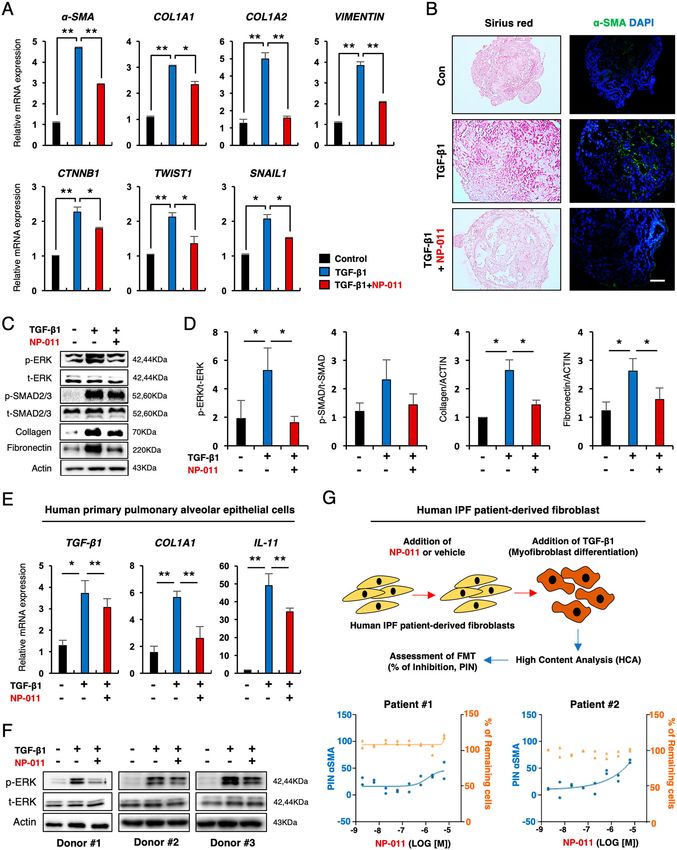
Kim et al. Cell Death Discovery (2021)7:48                      Page 3 of 12

  Fig. 1 (See legend on next page.)

Official journal of the Cell Death Differentiation Association
Kim et al. Cell Death Discovery (2021)7:48 Page 4 of 12
(see figure on previous page)
Fig. 1 NP-011 reduces TGF-β1-induced fibrosis in AOs. A qPCR of the indicated fibrosis-related genes in control AOs and AOs with TGF-β1 (25 ng/
ml)-induced fibrosis. B Representative images show Sirius red and α-SMA staining of AO sections from the indicated groups. Scale bars, 100 μm. C, D
Western blotting (C) and subsequent quantification of p-ERK, p-SMAD2/3, and Collagen (D) in AOs from the indicated groups. Actin was used as a
loading control. E qPCR analysis for expression of TGF-β1, COL1A1 and IL-11 from the indicated groups. F Western blot analysis for p-ERK in human
pAECs incubated with TGF-β1 (25 ng/ml) or TGF-β1 and NP-011 (500 ng/ml) for 72 h. Actin was used as loading control. G FMT assay using
pulmonary fibrosis patient-derived fibroblasts. The upper panel shows the overall procedure for FMT assay, and the low panel indicates the quantified
data from high-content analysis (HCA). Left axis and blue dots indicate the percentage of inhibition (PIN) of TGF-β1-mediated α-SMA expression in
patient fibroblasts by NP-011 treatment, and the right axis and orange dots indicate the percentage of remaining cells after NP-011 treatment in
patient fibroblast. Data are presented as means ± SD from three independent experiments. *p < 0.05, **p < 0.01 (ANOVA).
fibrosis AO model. Quantitative reverse transcription fibrotic markers (collagen and α-SMA; Supplementary
PCR (RT-qPCR) demonstrated that increased transcript Fig. 2D). Based on these observations, mice challenged
levels of the ECM (COL1A1 and COL1A2) and FMT (α- with BLM were intravenously treated with two differ-
SMA, CTNNB1, TWIST1, and SNAIL1) markers in ent doses of NP-011 (80 and 160 μg/kg) upon reaching
fibrotic AOs were significantly reduced by NP-011 a moderate stage of PF (day 5 post-BLM treatment).
(500 ng/ml) treatment (Fig. 1A). Sirius red and immuno- Importantly, BLM-induced fibrotic features were
fluorescence staining revealed that elevated expression of obviously alleviated at both the transcript and protein
collagen and α-SMA in TGF-β1-treated AOs were levels after NP-011 administration (Supplementary Fig.
markedly diminished by NP-011 treatment (Fig. 1B). In 3); however, there were no significant differences in the
addition, significant reduction of collagen and fibronectin reduction of fibrotic area and fibrosis markers between
in NP-011-treated AOs was observed by western blot (Fig. the two doses of NP-011. We further compared the
1C, D). We further determined whether the anti-fibrotic therapeutic efficacy of IV and IT administration of NP-
effects of NP-011 against TGF-β1-induced fibrosis in the 011 to determine optimal delivery route. Both admin-
AO model are mediated by canonical or non-canonical istration routes led to a similar reduction in expression
SMAD pathways. Interestingly, we found that NP-011 levels of fibrotic markers as well as fibrotic area via
suppressed the phosphorylation of non-canonical ERK in suppression of ERK signaling, but neither inhibited the
fibrotic lung tissues but had less inhibitory effect on phosphorylation of canonical SMAD2/3 (Fig. 2A–D).
phosphorylation of canonical SMAD2/3 (Fig. 2C, D). The The therapeutic role of NP-011 via suppression of ERK
anti-fibrotic effect via suppression of ERK signaling by signaling was further confirmed by two different GSEA,
NP-011 was also recapitulated in human pAECs (Fig. 1E, which showed suppression of non-canonical ERK sig-
F). Furthermore, NP-011 treatment significantly inhibited naling gene sets in NP-011-administered group com-
the TGF-β1-induced FMT in 2 different IPF patient- pared to BLM-treated group (Fig. 2E). Furthermore,
derived fibroblast (Fig. 1G). These results suggest that the transcriptomic analysis of RNA-Seq revealed the
hPSC-derived multicellular AOs serve as a model of potential target genes associated with anti-fibrotic
alveolar tissues and PF pathology and may facilitate the effects of NP-011 on the PF (Supplementary Fig. 4).
discovery of effective treatments for this disease. These findings suggest the predictive value of the
in vitro fibrosis AO model for identification of the
NP-011 reverses existing PF in mice preclinical therapeutic efficacy of novel drugs for PF.
We next sought to determine whether hPSC-derived
AOs could be used as a platform for predicting or Anti-fibrotic effects of NP-011 are comparable to those
validating drug efficacy in preclinical in vivo animal observed following administration of PFD and Nib
models. In order to validate the therapeutic effects of As NP-011 was found to exert anti-fibrotic effects in
NP-011 in vivo, we first observed the development of a mouse model of PF in vivo, we compared the anti-
PF in mice after treatment with BLM (3 mg/kg) to fibrotic effects of NP-011 with those of PFD and Nib,
determine the optimal timing of the administration of which are currently approved by the US Food and
NP-011 for anti-fibrotic evaluation (Supplementary Drug Administration for treatment of human IPF.
Fig. 2A). Levels of TGF-β1 pro-inflammatory cytokine Histochemical analysis demonstrated that adminis-
and expression levels of fibrosis-related genes (Col1a1, tration of either of these drugs resulted in effective
Mmp12, and Il-6) were increased on day 3 after BLM reduction of PF in mice compared with the untreated
treatment, exhibited considerable enhancement on day control mice (Fig. 3A). Immunofluorescence staining
7, and maintained a stable plateau until day 14 (Sup- confirmed the decrease in collagen deposition as well
plementary Fig. 2B, C). Histochemical analysis also as the expression of α-SMA (Fig. 3A). Correspond-
showed a similar temporal increase of fibrotic areas and ingly, quantification of collagen transcripts and
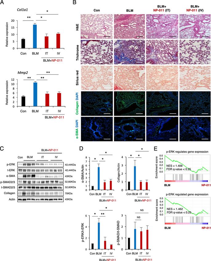
Official journal of the Cell Death Differentiation AssociationKim et al. Cell Death Discovery (2021)7:48 Page 5 of 12 Fig. 2 NP-011 reversed existing PF via suppression of ERK signaling. A The mRNA levels of Col1a1 and Mmp2 in lungs from the indicated groups of mice were detected by qPCR. B Representative images show H&E, Masson’s trichrome, Sirius red, Collagen, and α-SMA staining of lung sections from the indicated groups of mice. Scale bars, 100 μm. C, D Western blotting (C) and subsequent quantification of p-ERK, p-SMAD2/3, α-SMA, and Collagen (D) in whole-lung homogenates of mice from the indicated groups. Actin was used as a loading control. NS indicates a nonsignificant difference. E Two different GSEA data showing the significant suppression of non-canonical ERK signaling gene sets in NP-011-administered group compared to BLM-treated group in PF model. Data are presented as means ± SD; n = 10 (control), n = 11 (BLM), n = 5 (BLM + NP-011 IT), n = 7 (BLM + NP-011 IT). *p < 0.05, **p < 0.01 (ANOVA). Official journal of the Cell Death Differentiation Association
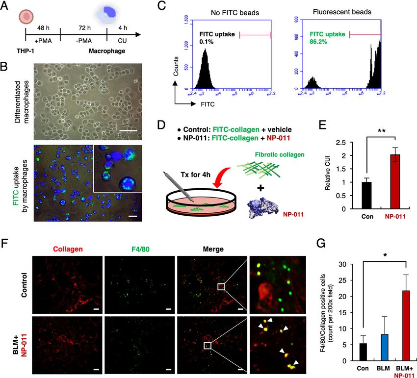
Kim et al. Cell Death Discovery (2021)7:48                                                                                               Page 6 of 12

  Fig. 3 Comparison of the therapeutic effects of NP-011, PFD, and Nib on BLM-induced PF in mice. A H&E, Masson’s trichrome, and Sirius red
  staining of representative lung sections from each group of treated mice. The effects of NP-011, PFD and Nib on the expression of collagen and α-
  SMA in lung tissues of BLM-induced PF in mice. Scale bars, 100 μm. B qPCR analysis for Col1a1 expression. C Quantification of collagen-positive areas
  as measured by confocal microscopy. D Whole lungs from control or BLM-challenged mice treated with NP-011, PFD, and Nib were used for Western
  blotting using p-ERK, Collagen, α-SMA, and a loading control (Actin). E Quantification of p-ERK, Collagen, and α-SMA. Data are presented as means ±
  SD; n = 6 (control), n = 8 (BLM), n = 7 (BLM + NP-011), n = 6 (BLM + PFD), n = 8 (BLM + Nib). *p < 0.05, **p < 0.01 (ANOVA).

fibrotic area showed a significant decrease in the lung                         NP-011 facilitates macrophage-mediated collagen uptake
tissues treated with either NP-011, PFD, or Nib com-                          in lung fibrosis
pared with the untreated control mice (Fig. 3B, C).                             Elimination of accumulated collagen by immune
Furthermore, western blot analysis demonstrated a                             response is another key factor in resolving fibrosis as seen
significant reduction of collagen and α-SMA levels in                          in PF mouse model24. In order to assess this, THP-1
NP-011-, PFD-, and Nib-treated mice, and these                                monocytes were differentiated into macrophages by PMA
effects were mediated by suppression of non-canonical                         treatment (Fig. 4A), and the phagocytic capability of dif-
ERK signaling (Fig. 3D, E). These results clearly                             ferentiated macrophages was confirmed by fluorescence
demonstrate that NP-011 accelerates fibrosis resolu-                           and flow cytometry analysis (Fig. 4B, C). Collagen uptake
tion comparable to that induced by administration of                          assay revealed that green fluorescence-labeled collagen
PFD and Nib.                                                                  was significantly up-taken by differentiated macrophages

Official journal of the Cell Death Differentiation Association
Kim et al. Cell Death Discovery (2021)7:48                                                                                                Page 7 of 12

  Fig. 4 NP-011 facilitates macrophage-mediated collagen uptake in lung tissues. A Overall schematic schedule for macrophage differentiation
  from monocyte by treatment of PMA. B Representative images for differentiated macrophages (upper panel) and FITC-labeled beads uptake by
  differentiated macrophages (lower panel). Scale bar, 10 μm. C Flow cytometry results showed that the FITC beads were uptaken by differentiated
  macrophages. D Graphical description of fibrotic collagen uptake assay by differentiated macrophages in the absence and presence of NP-011.
  E Quantitative analysis of the relative CUI (collagen uptake index) for control and NP-011 treatment. Bars represent the means ± SD from three
  replicates in each group. **P < 0.01 (Student t-test). F Representative images of immunostaining for mouse collagen and mouse F4/80 in the lung
  tissues of BLM-induced PF model and NP-011 administrated BLM-induced PF model. Scale bar, 50 μm. G Quantification of Collagen+F4/80+ cells in
  the lung tissues. The positive cells were counted in three non-repetitive fields of each stained section. Data presented as means ± SD (n = 3 mice per
  group). *p < 0.05 (Student t-test).

in the presence of NP-011 unlike the control culture (Fig.                    rich regions of fibrosis to facilitate uptake of deposited
4D, E). In fact, the engulfment of collagen by macro-                         collagen in the lung.
phages was demonstrated by immunostaining for collagen
and F4/80 after NP-011 administration in BLM-induced                          Discussion
PF model (Fig. 4F, G). Taken together, these results sug-                       Previous studies reported the hPSC-derived lung orga-
gest that NP-011 could not only reverse fibrosis by inhi-                      noids that represent different respiratory compartments
biting of mesenchymal transition in lung fibrosis, but also                    and utilized these models for gaining a deeper under-
resolve fibrosis by recruiting macrophages in the collagen                     standing of cell-to-cell communications during early lung

Official journal of the Cell Death Differentiation Association
Kim et al. Cell Death Discovery (2021)7:48                                                                    Page 8 of 12

development and pathogenesis of alveolar diseases, such         which is positioned within C2 domain28. Medin is aortic
as Hermanski-Pudlak syndrome and Bochdalek con-                 medial amyloid that occurs in people older than 60 years,
genital diaphragmatic hernia11–15,25. However, the appli-       and accumulated medin is engaged in Alzheimer’s disease
cation of hPSC-derived lung organoids that recapitulate         and type 2 diabetes29. Therefore, through the structural
the complexity and function of the native lung tissue for       truncation of C2 domain from MFG-E8, NP-011 might be
disease modeling and drug screening has remained chal-          free for concerning from side effects that possibly cause
lenging. Here, we describe a protocol for the generation of     Alzheimer’s disease and diabetes upon repeated admin-
multicellular AOs, which contain both functional AECs           istrations into patients. Second, NP-011 might exhibit
and mesenchymal support cells derived from hPSCs. Our           better binding affinity against collagen by removing gly-
method is based on optimized stepwise direct hPSC dif-          cosylation site of MFG-E8. MFG-E8 contains glycosyla-
ferentiation via mimicking of developmental cues in a           tion sites in C2 domain. Thus, NP-011 has no
temporally controlled manner6,26. Notably, we showed for        glycosylation that might impede the binding of discoidin
the first time that hPSC-derived AOs treated with TGF-β1         domain (C1 domain) of MFG-E8 to accumulated collagen
exhibited significant fibrogenic responses, which were            in PF. The well-known discoidin domain receptor (DDR)
effectively ameliorated by a drug candidate (NP-011).           has discoidin domain, stalk region, and transmembrane
Furthermore, we observed the anti-fibrotic effects of NP-        domain in its structure30. Although the mechanism of
011 in an in vivo mouse model of PF, underscoring the           collagen binding of DDR has not been identified, the
translational potential of hPSC-derived AOs as a robust         binding of discoidin domain of DDR to collagen has been
in vitro system for respiratory disease modeling and drug       reported31. Interestingly, the discoidin domain of DDR
testing.                                                        has no glycosylation, suggesting that discoidin domain of
   Human MFG-E8 consists of epidermal growth factor-            NP-011 might show better similarity to discoidin domain
like domain that bears an arginine-glycine aspartate            of DDR than MFG-E8, possibly facilitating the efficient
(RGD) motif, followed by two C domains (C1 and C2).             binding to accumulated collagen in the lung.
MFG-E8 modulates inflammatory responses by RGD                     While the promising data presented in this study and
motif-mediated binding to immune cells and engulfs              multicellular composition of hPSC-derived AOs is a
apoptotic cells by recognizing phosphatidylserine via C2        major advantage, one of the main limitations of this
domain. Although anti-fibrotic and anti-inflammatory              system, similar to other hPSC-derived organoid systems,
functions of MFG-E8 are well defined in several in vitro         is that the AOs remain incomplete as they lack of key
and in vivo experimental systems, including myocardial          components of the native alveolar tissue, including
infarction and skin and liver fibrosis21–23,27, it is unclear    macrophages and microvasculature. Macrophages are
how MFG-E8 is responsible for the anti-fibrotic effects.         remarkably plastic cells that can transform from the
Thus, we assumed that the roles of the C1 and C2                pro-inflammatory M1 phenotype to the anti-
domains of MFG-E8 might be different, and modulation            inflammatory M2 phenotype and vice versa32. Internal
of these two domains could improve the anti-fibrotic             and external stimuli induce dynamic changes in mac-
effect of MFG-E8 protein. Our collaborator newly syn-           rophage phenotype, which is closely associated with
thesized the C2 domain-truncated form of MFG-E8 (NP-            either exacerbation or prevention of IPF progres-
011), which expected to have the enhanced anti-fibrotic          sion33,34. Furthermore, a fine mesh of capillaries wraps
power in a fibrosis model compared to a full length of           around each alveolus and covers about 70% of its surface
MFG-E8 and the C1 domain-truncated form (NP-012).               area for gas exchange. Therefore, the development of
Based on these findings, we tested the anti-fibrotic effects      AOs closer to the native alveolar tissue architecture and
of NP-011 in both the in vitro AO system and the in vivo        function by the incorporation of these missing compo-
PF model. Our study revealed that NP-011 effectively            nents will provide more accurate and robust in vitro
reverses fibrosis via inhibition of ERK signaling, consistent    platform for studying PF and associated therapeutics.
with effects of administration of PFD and Nib. Although         Although the ability to differentiate hPSCs into three
these two medications are the only US Food and Drug             germ layer lineages presents great promise for the
Administration-approved drugs for the treatment of IPF,         development of various organoid models that recapitu-
these drugs do not work for all patients3,4. Therefore, NP-     late the complexity and functions of in vivo tissues, the
011 may provide a new alternative for IPF patients who do       variations in the differentiation potential among hPSC
not respond to existing drugs and may enhance response          lines due to genetic background, culture conditions and
in combination with PFD or Nib as well.                         differentiation protocols may interfere with organoid
   Besides the anti-fibrotic effects of NP-011 for amelior-      quality, reproducibility and faithfulness. Thus, the field
ating PF, NP-011 might have clinical benefits in com-            needs a standardized differentiation protocol with gold
parison with MFG-E8 by truncation of C2 domain. First,          standard hPSC lines, which may able to diminish off-
MFG-E8 contains an amyloid fragment (known as medin),           target cells and generate highly homogenous organoids.

Official journal of the Cell Death Differentiation Association
Kim et al. Cell Death Discovery (2021)7:48                                                                    Page 9 of 12

Materials and methods                                           Committee of Kangwon National University (IACUC NO.
Maintenance of hPSCs                                            KW-180913-1). Sample size estimation was calculated by
  Human PSCs (CHA15 and iPS-NT4-S1) were kindly                 IACUC and online free software (Open Epi, http://www.
provided by CHA University, South Korea and maintained          openepi.com/OE2.3/Menu/OpenEpiMenu.htm).              Male
as previously described35. Briefly, the cells were cultured      mice (C57BL/6), 8 to 10 weeks of age were purchased
under xeno-, serum- and feeder-free conditions using E8         from the Dooyeol Laboratory. Lung fibrosis was induced
medium (STEMCELL Technologies) on dishes coated                 by intratracheal (IT) administration of 50 μl of bleomycin
with vitronectin (STEMCELL Technologies). Cells were            (BLM) (3 mg per kg body weight, mg/kg) and the mice
subcultured at 80% confluency and passaged every 5 days          were randomly grouped in a blinded manner. On day 5
by mechanical dissociation. All cells were incubated at         after BLM administration, mice were given an intravenous
37 °C in a humidified atmosphere with 5% CO2.                    (IV) or IT injection of NP-011 (80 and 160 μg/kg), PFD
                                                                (300 mg/kg) and Nib (60 mg/kg). On day 3 after drug
Cell cultures                                                   administration, mice were sacrificed and lung tissues were
  Human pAECs were purchased from Sciencell Research            examined.
Laboratories, Inc. and cultured in alveolar epithelial cell
medium (#3201, Sciencell, Carlsbad, CA, USA) at 37 °C,          RNA extraction and quantitative real-time PCR (qPCR)
5% CO2. Human pAECs were treated with 5 ng/ml TGF-                Total RNA was extracted from mouse lung tissues,
β1 and 500 ng/ml NP-011 for 48 h and harvested for              AOs, pAECs and undifferentiated hPSC cultures using an
further analysis.                                               RNeasy Mini kit (Qiagen, Duesseldorf, Germany) and
                                                                complementary DNA was synthesized using TOPscripTM
Stepwise differentiation of hPSCs into AECs                     RT DryMIX (Enzynomics, Daejeon, Korea). PCR ampli-
  A stepwise direct AEC differentiation was performed as        fication was performed using a Step One Plus real-time
previously described6,36. Briefly, undifferentiated hPSCs        PCR system (Applied Biosystems, Warrington, UK) with
were dissociated and then plated in dishes coated with          TOPrealTM qPCR 2X PreMIX (Enzynomics). All the
vitronectin at a density of 1 × 10⁵ cells/cm2. After over-      mRNA expression was normalized to an internal control
night incubation, AEC differentiation was initiated with        GAPDH. The primer sequences for human and mouse
exposure to stepwise induction medium.                          genes are listed in Supplementary Tables 1 and 2,
                                                                respectively.
Generation of multicellular AOs from hPSCs
  Generation of multicellular AOs was performed by com-         Immunohistochemistry for paraffin section
bination of previously reported protocols with minor mod-         AOs and lung tissues were paraffin-embedded, sec-
ifications36–38. Briefly, the AEC cultures were dissociated on    tioned at a thickness of 5 μm and mounted on slide glass.
day 21 of AEC differentiation with 0.4 U/ml collagenase B       The slides were rinsed in xylenes and with decreasing
(Roche) for 2 h in a 37 °C incubator, followed by treatment     concentration (100% to 80%) of ethanol to deparaffinize
with cell dissociation buffer (Gibco) for 10 min in a 37 °C     and rehydrate. The slides were subjected to hydrated
water bath. The single-cell suspension was then passed          autoclaving using an automated antigen-retrieval a citrate
through a 70-μm cell strainer (BD Bioscience) and seeded        buffer (0.01 M sodium citrate, pH 6) for 121 °C for 1 min
into 96-well round-bottom plates (Corning, 5 × 10⁴ cells per    and cooled for 30 min before immunostaining. They were
well) containing AEC maturation medium supplemented             then treated with Dako Real Peroxidase-blocking solution
with 10 μM Rho-associated protein kinase inhibitor              for 20 min at room temperature (RT) and washed with
(STEMCELL Technologies). After distribution, 1:15 diluted       cold phosphate-Buffered Saline with 0.1% Tween 20
Matrigel (150 μl per well) was added into each well to          (PBST) for 10 min at RT. The slides were blocked with
improve adhesion between cells. The plates were centrifuged     10% normal goat serum for 1 h at RT and incubated with
at 450 × g for 5 min and incubated overnight to allow           primary antibodies in blocking buffer overnight at 4 °C.
aggregation at 37 °C in a humidified atmosphere with 5%          The next day, goat anti-mouse IgG (H + L) Cross-
CO2. After overnight incubation, the aggregates were            Adsorbed Secondary Antibody, Alexa Fluor 488 (Invi-
transferred to 6-well low-attachment plates (Corning) con-      trogen, A11001) secondary antibody was applied for 1 h at
taining fresh AEC maturation medium and cultured for            RT in the dark. Eventually, the slides were washed with
6 days to establish AOs.                                        PBST (0.1% tween) and covered with FluoroshieldTM
                                                                mounting medium with 4’, 6-diamidino-2-phenylindole
Mouse model for bleomycin-induced lung fibrosis and              (DAPI). IHC images were captured under a fluorescence
drug treatments                                                 microscopy (IX-51, Olympus). To evaluate anti-fibrotic
  All animal experiments were approved and followed the         effect of NP-011 in vivo, three mice were used in each
regulations of the Institutional Animal Care and Use            group and data were obtained in at least three fields. The

Official journal of the Cell Death Differentiation Association
Kim et al. Cell Death Discovery (2021)7:48 Page 10 of 12
positive area of collagen of total area were measured by for 1 h at RT. The membranes were incubated with pri-
using LSM880 with Airscan. mary antibodies against anti-phospho-p44/42 MAPK,
anti-p44/42 MAPK, anti-alpha smooth muscle actin (α-
Immunofluorescence staining for frozen AO sections SMA), anti-phospho-SMAD2/3, anti-SMAD2/3, anti-
AOs were fixed with 4% paraformaldehyde for 24 h at Collagen1A and anti-Fibronectin overnight at 4 °C. The
RT, followed by dehydration using sucrose solution of a chemiluminescence signal was scanned with Chemi-
gradual gradient series. The fixed AOs were then DOCTM imaging system (Bio-Rad Laboratories, Hercules,
embedded in Tissue-Tek® cryomold (Sakura finetek) CA, USA). The antibodies are listed in Table S3.
covered with Tissue-Tek® OCTTM Compound (Sakura
finetek) rocking at 4 °C overnight and stored in −80 °C. Enzyme-linked immunosorbent assay (ELISA)
Frozen AO blocks were sectioned into 8 μm cryosections, Concentration of TGF-β1 in bronchoalveolar lavage
which were carefully mounted onto silane-coated micro fluid (BALF) was measured by ELISA kits (R&D, Min-
slides (Muto Pure Chemicals) and stored in −80 °C. The neapolis, MN, USA) according to the manufacturer’s
sections were blocked by 10% donkey serum (Jackson instructions.
ImmunoResearch Laboratories) in PBST for 1 h at RT and
probed with primary antibodies (EPCAM, CPM, HOPX, RNA-sequencing and analysis
SFTPC, AQP5, T1α and VIMENTIN) in 1% donkey Total RNA was extracted from lung tissues of control,
serum in PBST at 4 °C overnight. The next day, the sec- BLM-administered and NP-011-administered groups
tions were rinsed with PBST for 5 min, followed by using Trizol reagent (Invitrogen). RNA quality was
incubation with secondary antibodies for 30 min at RT. assessed by Agilent 2100 Bioanalyzer using the RNA 6000
The sections were counterstained using FluoroshieldTM Nano Chip (Agilent Technologies, Amstelveen, The
with DAPI histology mounting medium (Sigma-Aldrich). Netherlands), and RNA quantification was performed
IHC images were captured under a fluorescence micro- using ND-2000 spectrophotometer (Thermo Inc., DE,
scopy (IX-51, Olympus). Primary antibodies were omitted USA). Triplicate samples were prepared for each group.
in control immunohistochemical staining. The antibodies For control and test RNAs, the construction of library was
are listed in Supplementary Table 3. performed using QuantSeq 3’mRNA-Seq Library Prep Kit
(Lexogen, Inc., Austria) according to the manufacturer’s
Hematoxylin and eosin, Sirius red, and Masson’s trichrome instructions. In brief, each 500 ng total RNA were pre-
staining pared and an oligo-dT primer containing an Illumina-
Paraffin sections of AOs and lung tissues were depar- compatible sequence at its 5’end was hybridized to the
affinized by sinking in xylene and sequentially rehydrated RNA and reverse transcription was performed. After
through a gradual concentration series, starting from degradation of the RNA template, second strand synthesis
100% ethanol to 70% ethanol and ending with deionized was initiated by a random primer containing an Illumina-
water. The sections were stained to nucleus using compatible linker sequence at its 5’end. The double-
Hematoxylin (BBC biochemical) and rinsed by water, stranded library was purified by using magnetic beads to
followed by a short exposure of acidic ethanol. The sec- remove all reaction components. The library was ampli-
tions also were exposed by Eosin targeting to cytoplasm fied to add the complete adapter sequences required for
and dehydrated through a sequential concentration cluster generation. The finished library is purified from
change, starting from 70% ethanol to 100% ethanol. After PCR components. High-throughput sequencing was per-
exposure to xylene three times, the sections were moun- formed as single-end 75 sequencing using NextSeq 500
ted with Permount mounting medium (Fisher Scientific). (Illumina, Inc., USA). To analyze RNA-Seq, these samples
The fibrotic areas in AOs and lung sections were detected were sent to the BGI Tech Solutions Company (Hon-
using Masson’s trichrome (Empire Genomics, #BPK2916, gkong). Gene Set Enrichment Analysis (GSEA) was con-
USA) and Sirius red staining (Abcam, ab150681, UK) ducted using GSEAv17 (Broad Institute, Cambridge,
according to the manufacturer’s instructions. MA, USA).
Western blot analysis Fibroblast to myofibroblast transition (FMT) assay
Protein extracted from mouse lung tissues, AOs and FMT assay was performed by Charles River Labora-
human pAECs were lysed in protein lysis buffer and tories International. Briefly, lung fibroblast from two dif-
quantified using the bicinchoninic acid protein assay. The ferent Idiopathic PF (IPF) donors were seeded onto
20 μg of protein were separated by sodium dodecyl sulfate culture plates. IPF fibroblasts were cultured with eight
polyacrylamide gel electrophoresis (8–15%) gel and then different concentrations of NP-011 and then treated with
transferred to polyvinylidene fluoride membranes. Non- TGF-β1. α-SMA-positive cells were assessed by high-
specific binding proteins were blocked with 5% skim milk content analysis (HCA).
Official journal of the Cell Death Differentiation AssociationKim et al. Cell Death Discovery (2021)7:48 Page 11 of 12
THP-1 differentiation and fluorescent bead phagocytosis Author contributions
assay J.H.K., G.H.A., J.Y.K., R.R., W.J.K., X.J., D.H.W., C.H., and S.R.Y.: conception and
design, collection and assembly of data, data analysis and interpretation,
THP-1 cells were purchased from ATCC and cultured in manuscript writing, final approval of manuscript; S.H.H. and J.H.K.: conception
RPMI-1640 (Gibco, 31800-022) containing 10% of heat and design, interpretation, manuscript writing, final approval of manuscript.
inactivated fetal bovine serum (Gibco, 16000-044) and 50 μM
Conflict of interest
β-mercaptoethanol in 37 °C, 5% CO2 incubator. THP-1 cells The NP-011 and the use of NP-011 for treating liver and lung fibrosis is
were differentiated into macrophages using 200 ng/ml of protected by published and unpublished patents (KOR/10-1947902, PCT/
phorbol 12-myristate 13-acetate (PMA, Sigma, P8139) for KR2017/005150, EU/17870624.8, JP/2018-524465, US/15/994.323, CN/
201780004259.5, KOR/10-2018-0128625, and PCT/KR2018/012801), and these
48 h followed by 72 h in PMA-free medium. In phagocytosis intellectual property rights are belonging to NEXEL. Co., Ltd.
assay, THP-1 cells were seeded at 1 × 10⁵ cells/cm2.
Carboxylate-Modified Microspheres, 2.0 μm, yellow-green
fluorescent beads (Invitrogen, F8827) were washed in THP-1 Publisher’s note
Springer Nature remains neutral with regard to jurisdictional claims in
cell culture media and resuspended at a final dilution of 1:500 published maps and institutional affiliations.
in serum-free RPMI-1640. Cells were incubated with fluor-
escent beads for 4 h in 37 °C, 5% CO2 incubator. Cells were Supplementary information The online version contains supplementary
material available at https://doi.org/10.1038/s41420-021-00439-7.
detached with TrypLE (Gibco, 12604-021) and measured by
flow cytometry (Accuri C6 Plus).
Received: 2 December 2020 Revised: 19 January 2021 Accepted: 13
February 2021
Collagen uptake assay
THP-1 cells were differentiated into macrophages by
200 ng/ml of PMA treatment and incubated with 50 μg/
ml of fluorescein isothiocyanate (FITC)-conjugated type References
1. Wynn, T. A. & Ramalingam, T. R. Mechanisms of fibrosis: therapeutic translation
I collagen (Anaspec, AS-85111) in serum-free media
for fibrotic disease. Nat. Med. 18, 1028–1040 (2012).
with or without 15 μg/ml of NP-011 for 4 h at 37 °C. 2. Blackwell, T. S. et al. Future directions in idiopathic pulmonary fibrosis research.
After 4 h, differentiated macrophages were washed An NHLBI workshop report. Am. J. Respir. Crit. Care Med. 189, 214–222 (2014).
3. Harari, S. & Caminati, A. Idiopathic pulmonary fibrosis: from clinical trials to real-
several times to remove un-ingested collagen and fixed
life experiences. Eur. Respir. Rev. 24, 420–427 (2015).
for 20 min with 4% paraformaldehyde. The cells were 4. King, T. E. Jr. et al. A phase 3 trial of pirfenidone in patients with idiopathic
counterstained with DAPI. Internalization of collagen pulmonary fibrosis. N. Engl. J. Med. 370, 2083–2092 (2014).
was quantified by fluorescence microscopy and expres- 5. Hiemstra, P. S., Grootaers, G., van der Does, A. M., Krul, C. A. M. & Kooter, I. M.
Human lung epithelial cell cultures for analysis of inhaled toxicants: Lessons
sed as a collagen uptake index (CUI: Number of cells learned and future directions. Toxicol. Vitr. 47, 137–146 (2018).
with ingestion of FITC-collagen divided by the total 6. Heo, H. R. et al. Human pluripotent stem cell-derived alveolar epi-
thelial cells are alternatives for in vitro pulmotoxicity assessment. Sci.
number of cells counted).
Rep. 9, 505 (2019).
7. Heijink, I. H. et al. Characterisation of cell adhesion in airway epithelial cell
Statistical analysis types using electric cell-substrate impedance sensing. Eur. Respir. J. 35,
894–903 (2010).
Values for all measurements are presented as means ±
8. Sporty, J. L., Horalkova, L. & Ehrhardt, C. In vitro cell culture models for the
standard deviation (SD). Student’s t-test was used for assessment of pulmonary drug disposition. Expert Opin. Drug Metab. Toxicol. 4,
comparisons between two groups. One-way ANOVA was 333–345 (2008).
9. Ren, H., Birch, N. P. & Suresh, V. An optimised human cell culture model for
performed for multigroup comparisons. All test utilized
alveolar epithelial transport. PLoS ONE 11, e0165225 (2016).
one-sided methodology. A P-value ofKim et al. Cell Death Discovery (2021)7:48 Page 12 of 12
17. Leibel, S. L. et al. Reversal of surfactant protein B deficiency in patient specific 27. Nakaya, M. et al. Cardiac myofibroblast engulfment of dead cells
human induced pluripotent stem cell derived lung organoids by gene ther- facilitates recovery after myocardial infarction. J. Clin. Invest. 127,
apy. Sci. Rep. 9, 13450 (2019). 383–401 (2017).
18. Strikoudis, A. et al. Modeling of fibrotic lung disease using 3D organoids 28. Haggqvist, B. et al. Medin: an integral fragment of aortic smooth muscle cell-
derived from human pluripotent stem cells. Cell Rep. 27, 3709–3723 (2019). produced lactadherin forms the most common human amyloid. Proc. Natl
19. McCauley, K. B. et al. Efficient derivation of functional human airway epithe- Acad. Sci. USA 96, 8669–8674 (1999).
lium from pluripotent stem cells via temporal regulation of Wnt signaling. Cell 29. Westermark, G. T. & Westermark, P. Localized amyloids important in diseases
Stem Cell 20, 844–857 (2017). outside the brain-lessons from the islets of Langerhans and the thoracic aorta.
20. Gotoh, S. et al. Generation of alveolar epithelial spheroids via isolated pro- FEBS J. 278, 3918–3929 (2011).
genitor cells from human pluripotent stem cells. Stem Cell Rep. 3, 394–403 30. Johnston, I. G. & Jones, N. S. Evolution of cell-to-cell variability in stochastic,
(2014). controlled, heteroplasmic mtDNA populations. Am. J. Hum. Genet. 99,
21. Fujiwara, C. et al. Suppressive regulation by MFG-E8 of latent transforming 1150–1162 (2016).
growth factor beta-induced fibrosis via binding to alphav integrin: significance 31. Borza, C. M. & Pozzi, A. Discoidin domain receptors in disease. Matrix Biol. 34,
in the pathogenesis of fibrosis in systemic sclerosis. Arthritis Rheumatol. 71, 185–192 (2014).
302–314 (2019). 32. Zhang, L. et al. Macrophages: friend or foe in idiopathic pulmonary fibrosis?
22. An, S. Y. et al. Milk fat globule-EGF factor 8, secreted by mesenchymal stem Respir. Res. 19, 170 (2018).
cells, protects against liver fibrosis in mice. Gastroenterology 152, 1174–1186 33. Murray, P. J. & Wynn, T. A. Protective and pathogenic functions of macrophage
(2017). subsets. Nat. Rev. Immunol. 11, 723–737 (2011).
23. Brissette, M. J., Laplante, P., Qi, S., Latour, M. & Cailhier, J. F. Milk fat globule 34. Mosser, D. M. & Edwards, J. P. Exploring the full spectrum of macrophage
epidermal growth factor-8 limits tissue damage through inflammasome activation. Nat. Rev. Immunol. 8, 958–969 (2008).
modulation during renal injury. J. Leukoc. Biol. 100, 1135–1146 (2016). 35. Lim, J. J. et al. Maintenance of hPSCs under xeno-free and chemically defined
24. Atabai, K. et al. Mfge8 diminishes the severity of tissue fibrosis in mice by culture conditions. Int. J. Stem Cells 12, 484–496 (2019).
binding and targeting collagen for uptake by macrophages. J. Clin. Invest. 119, 36. Yamamoto, Y. et al. Long-term expansion of alveolar stem cells
3713–3722 (2009). derived from human iPS cells in organoids. Nat. Methods 14,
25. Kunisaki, S. M. et al. Human induced pluripotent stem cell-derived lung 1097–1106 (2017).
organoids in an ex vivo model of the congenital diaphragmatic hernia fetal 37. Rasaei, R. et al. Regulation of JAM2 expression in the lungs of streptozotocin-
lung. Stem Cells Transl. Med. 10, 98–114 (2021). induced diabetic mice and human pluripotent stem cell-derived alveolar
26. Heo, H. R. et al. Reprogramming mechanisms influence the maturation of organoids. Biomedicines 8, 346 (2020).
hematopoietic progenitors from human pluripotent stem cells. Cell Death Dis. 38. Hong, S. H. et al. Cell fate potential of human pluripotent stem cells is
9, 1090 (2018). encoded by histone modifications. Cell Stem Cell 9, 24–36 (2011).
Official journal of the Cell Death Differentiation AssociationYou can also read