Luteolin Orchestrates Porcine Oocyte Meiotic Progression by Maintaining Organelle Dynamics Under Oxidative Stress - Frontiers
←
→
Page content transcription
If your browser does not render page correctly, please read the page content below
ORIGINAL RESEARCH
                                                                                                                                             published: 15 June 2021
                                                                                                                                      doi: 10.3389/fcell.2021.689826

                                            Luteolin Orchestrates Porcine
                                            Oocyte Meiotic Progression by
                                            Maintaining Organelle Dynamics
                                            Under Oxidative Stress
                                            Soo-Hyun Park 1,2† , Pil-Soo Jeong 1† , Ye Eun Joo 1,2 , Hyo-Gu Kang 1,3 , Min Ju Kim 1,2 ,
                                            Sanghoon Lee 1 , Bong-Seok Song 1 , Sun-Uk Kim 1,4 , Seong-Keun Cho 5* and
                            Edited by:      Bo-Woong Sim 1*
                      Heide Schatten,
                                            1
  University of Missouri, United States      Futuristic Animal Resource and Research Center, Korea Research Institute of Bioscience and Biotechnology, Cheongju,
                                            South Korea, 2 Department of Animal Science, College of Natural Resources and Life Science, Pusan National University,
                      Reviewed by:
                                            Miryang, South Korea, 3 Department of Animal Science and Biotechnology, College of Agriculture and Life Science,
                            Shen Yin,
                                            Chungnam National University, Daejeon, South Korea, 4 Department of Functional Genomics, University of Science
Qingdao Agricultural University, China
                                            and Technology, Daejeon, South Korea, 5 Department of Animal Science, College of Natural Resources and Life Science, Life
                      Zhonghua Liu,
                                            and Industry Convergence Research Institute, Pusan National University, Miryang, South Korea
    Northeast Agricultural University,
                                 China
                  *Correspondence:          Increasing evidence has demonstrated that oxidative stress impairs oocyte maturation,
                       Bo-Woong Sim         but the underlying mechanisms remain largely unknown. Here, for the first time, we
                 embryont@kribb.re.kr
                     Seong-Keun Cho         examined the antioxidant role of luteolin in meiotic progression and the underlying
                   skcho@pusan.ac.kr        mechanisms. Supplementation of 5 µM luteolin increased the rates of first polar body
     † These  authors have contributed      extrusion and blastocyst formation after parthenogenetic activation, and the expression
    equally to this work and share first
                            authorship
                                            levels of oocyte competence (BMP15 and GDF9)-, mitogen-activated protein kinase
                                            (MOS)-, and maturation promoting factor (CDK1 and Cyclin B)-related genes were also
                    Specialty section:      improved. Luteolin supplementation decreased intracellular reactive oxygen species
         This article was submitted to
             Cell Growth and Division,
                                            levels and increased the expression levels of oxidative stress-related genes (SOD1,
                a section of the journal    SOD2, and CAT). Interestingly, luteolin alleviated defects in cell organelles, including
  Frontiers in Cell and Developmental
                                            actin filaments, the spindle, mitochondria, the endoplasmic reticulum, and cortical
                                Biology
                                            granules, caused by H2 O2 exposure. Moreover, luteolin significantly improved the
             Received: 01 April 2021
             Accepted: 19 May 2021          developmental competence of in vitro-fertilized embryos in terms of the cleavage rate,
            Published: 15 June 2021         blastocyst formation rate, cell number, cellular survival rate, and gene expression and
                              Citation:     markedly restored the competencies decreased by H2 O2 treatment. These findings
       Park S-H, Jeong P-S, Joo YE,
Kang H-G, Kim MJ, Lee S, Song B-S,
                                            revealed that luteolin supplementation during in vitro maturation improves porcine
     Kim S-U, Cho S-K and Sim B-W           meiotic progression and subsequent embryonic development by protecting various
 (2021) Luteolin Orchestrates Porcine
                                            organelle dynamics against oxidative stress, potentially increasing our understanding
       Oocyte Meiotic Progression by
     Maintaining Organelle Dynamics         of the underlying mechanisms governing the relationship between oxidative stress and
              Under Oxidative Stress.       the meiotic events required for successful oocyte maturation.
      Front. Cell Dev. Biol. 9:689826.
      doi: 10.3389/fcell.2021.689826        Keywords: luteolin, antioxidant, oxidative stress, organelle dynamics, in vitro maturation, porcine oocyte

Frontiers in Cell and Developmental Biology | www.frontiersin.org                 1                                            June 2021 | Volume 9 | Article 689826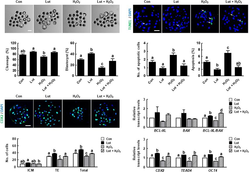
Park et al.                                                                                                 Luteolin Improves Oocyte Maturation

INTRODUCTION                                                             et al., 2016). In the present study, we explored the antioxidant
                                                                         effect of Lut supplementation on oocyte maturation and the
Understanding the in vitro maturation (IVM) of oocytes is                subsequent developmental competence of in vitro fertilization
important for developing assisted reproductive technology (ART)          (IVF) embryos. Given the important role of Lut in oocyte
and generating mature oocytes that are capable of successful             maturation, we also examined the role oxidative stress plays in
embryonic development (Hashimoto, 2009). However, only                   various changes in cell organelles during oocyte maturation.
a small percentage of immature oocytes can develop into
blastocysts and subsequently result in pregnancy (Kwak et al.,
2012). As low-quality oocytes resulting from improper IVM                MATERIALS AND METHODS
conditions are one of the main reasons for ART failure,
optimization of IVM conditions is vital for improving ART                Chemicals
(Lee et al., 2020).                                                      All chemicals and reagents were purchased from Sigma-
    Oocyte maturation involves both nuclear and cytoplasmic              Aldrich Chemical Co. (St. Louis, MO, United States) unless
maturation. Nuclear maturation mainly refers to chromosome               otherwise indicated.
segregation and reflects the capacity of the oocyte to resume
meiosis (Lopes et al., 2019). Cytoplasmic maturation involves the        IVM
accumulation of mRNA, proteins, and substrates required for              Porcine ovaries obtained from a local slaughterhouse were
subsequent fertilization ability and developmental competence            transported in 0.9% saline supplemented with 75 µg/mL
(Watson, 2007). Cell organelles including mitochondria, the              potassium penicillin G and 50 µg/mL streptomycin sulfate and
endoplasmic reticulum (ER), and microtubules play important              maintained at 37–38◦ C. COCs were aspirated from follicles (3–
roles in both nuclear and cytoplasmic maturation through                 6 mm in diameter) through a disposable 10-mL syringe with
the regulation of protein and ATP synthesis and chromosome               an 18-gauge needle. The COCs were washed in 0.9% saline
segregation (Mao et al., 2014). However, inadequate in vitro             containing 0.1% bovine serum albumin (BSA), and 40–50 oocytes
conditions lead to the abnormal behavior of cell organelles and          were matured in 500 µL of IVM medium in a four-well multi-dish
finally result in disrupted meiotic maturation (Ueno et al., 2005;       (Nunc, Roskilde, Denmark) at 38.5◦ C under 5% CO2 . For the
De los Reyes et al., 2011; De los Reyes et al., 2012).                   first 22 h, Tissue Culture Medium 199 supplemented with 10%
    In aerobic organisms, reactive oxygen species (ROS) including        porcine follicular fluid, 0.57 mM cysteine, 10 ng/mL epidermal
hydroxyl radicals, superoxide anions, and hydrogen peroxide              growth factor, 10 IU/mL pregnant mare serum gonadotropin
(H2 O2 ) are produced as byproducts of cell metabolism. However,         (PMSG), and 10 IU/mL human chorionic gonadotropin (hCG)
during IVM cultivation of cumulus oocyte complexes (COCs),               was used for maturation. During the second stage (22–44 h), the
the IVM medium has fewer antioxidant enzymes than does the               same medium was used without PMSG and hCG. After IVM,
in vivo environment comprising follicular and oviduct fluids             the cumulus cells were removed by repeated pipetting in 0.1%
provided by the mother (Ye et al., 2017). The imbalance between          hyaluronidase. Denuded oocytes were classified as immature,
ROS production and clearance caused by the lack of maternal              degenerate, or at metaphase II (MII; first polar body extrusion
antioxidants induces oxidative stress, which disrupts oocyte             visible) under a microscope (Nikon, Tokyo, Japan), and only MII
maturation and subsequent embryonic development (Khazaei                 oocytes were used for further experiments.
and Nematbakhsh, 2004). Numerous studies have shown that
antioxidant treatments such as adding quercetin, vitamin C,              Chemical Treatment
and resveratrol to IVM medium are helpful in improving                   A stock solution of 20 mM Lut was prepared with
oocyte quality (Sovernigo et al., 2017). Nevertheless, it is             dimethylsulfoxide and diluted in IVM medium to final
essential to elucidate the mechanisms underlying the relationship        concentrations of 0 (control), 1, 5, or 10 µM Lut. To
between oxidative stress and meiotic events for successful               demonstrate the protective effect of Lut against oxidative
oocyte maturation.                                                       stress, additional IVM experiments were performed in the
    Luteolin (3, 4, 5, 7-tetrahydroxyflavone; Lut) is a flavone,         absence or presence of 1 mM H2 O2 (Do et al., 2015), depending
a type of flavonoid, usually found in broccoli, rosemary, olive          on the experimental design.
oil, and peppermint. Accumulating studies have shown that
Lut possesses anti-inflammatory, anticancer, and cytoprotective          Parthenogenetic Activation
properties (Gupta et al., 2018). Especially, Lut functions as            Metaphase II oocytes were parthenogenetically activated in a 1-
an antioxidant, protecting various cell types against oxidative          mm gab wire chamber (CUY5000P1, Nepagene, Chiba, Japan)
stress (Xia et al., 2014; An et al., 2016). However, no studies          with 10 µL of 280 mM mannitol solution containing 0.1 mM
have examined the effects of Lut on mammalian oocytes. In                MgSO4·7H2 O, 0.1 mM CaCl2 ·2H2 O, 0.5 mM HEPES, and
the present study, we investigated the role of Lut on meiotic            1 mg/mL polyvinyl alcohol (PVA). Oocytes were activated with
progression using porcine oocyte because of the physiological            a 110-V direct current for 50 µs using an electro cell fusion
and phylogenetic similarities between porcine and human oocyte,          generator (LF101, Nepagene). Activated oocytes were transferred
such as oocyte diameter and time period of maturation, which             into in vitro culture (IVC) medium (porcine zygote medium-
means porcine oocyte can be used to reflect the reproduction             3 containing 4 mg/mL BSA) supplemented with 5 µg/mL
system of human (Day, 2000; Santos et al., 2014; Wang                    cytochalasin B and 2 mM 6-dimethylaminopurine for 4 h at

Frontiers in Cell and Developmental Biology | www.frontiersin.org    2                                     June 2021 | Volume 9 | Article 689826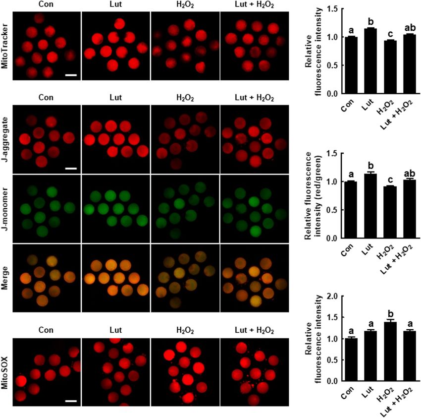
Park et al.                                                                                                 Luteolin Improves Oocyte Maturation

38.5◦ C under 5% CO2 . After 4 h, the oocytes were transferred to        blocked in blocking solution for 1 h at room temperature.
IVC medium at 38.5◦ C under 5% CO2 . Cleavage and blastocyst             Next, the oocytes were stained with 1 µg/mL anti-α-tubulin
formation were evaluated on days 2 and 6, respectively.                  antibody (Invitrogen) overnight at 4◦ C. The oocytes were
                                                                         washed in DPBS (Welgene) containing 0.05% (v/v) Tween
IVF                                                                      20 and then blocked again under the same conditions. The
In vitro fertilization was performed in modified Tris-buffered           oocytes were incubated for 1 h at room temperature with a
medium (mTBM) consisting of 113.1 mM NaCl, 3 mM KCl,                     conjugated secondary antibody-Alexa Fluor 488-labeled goat
7.5 mM CaCl2 ·2H2 O, and 20 mM Tris (crystallized free base;             anti-mouse IgG (1:200 in blocking solution) and washed in
Fisher Scientific, Waltham, MA, United States), 11 mM glucose,           DPBS (Welgene) containing 0.05% (v/v) Tween 20. Oocytes
5 mM sodium pyruvate, 2.5 mM caffeine sodium benzoate, and               were mounted on glass slides with 1.5 µg/mL DAPI and
1 mg/mL BSA. Ejaculated fresh swine semen was washed three               observed using a laser-scanning confocal fluorescent microscope
times by centrifugation for 3 min at 100g and room temperature           (LSM700; Zeiss).
with Dulbecco’s phosphate-buffered saline (DPBS; Gibco, Grand
Island, NY, United States) supplemented with 1 mg/mL BSA,                Analysis of Mitochondrial Distribution,
100 µg/mL penicillin G, and 75 µg/mL streptomycin sulfate.               Mitochondrial Membrane Potential, and
Washed spermatozoa were re-suspended in mTBM for 15 min.                 Mitochondrial ROS
Next, 2 µL of diluted spermatozoa was added to 48 µL of                  Mitochondrial distribution and the mitochondrial membrane
mTBM containing 10–15 oocytes, yielding a final concentration            potential (MMP) were detected using MitoTracker Deep Red
of 1.5 × 10⁵ spermatozoa/mL. The oocytes were co-incubated               (Invitrogen) and JC-1 (Cayman Chemical, Ann Arbor, MI,
with the spermatozoa for 6 h at 38.5◦ C under 5% CO2 . After             United States), with red fluorescence indicating the aggregated
6 h, spermatozoa covering the oocytes were stripped via gentle           form (J-aggregate) and green fluorescence indicating the
pipetting. Thereafter, the IVF embryos were incubated in IVC             monomer form (J-monomer). Additionally, mitochondrial ROS
medium at 38.5◦ C under 5% CO2 for 6 days. Cleavage and                  (mtROS) were detected using MitoSOX. MII oocytes were fixed
blastocyst formation were evaluated on days 2 and 6, respectively.       in formalin solution for 1 h at 38.5◦ C and then washed in DPBS–
                                                                         PVA. The oocytes were incubated with 200 nM MitoTracker
Measurement of Intracellular ROS Levels                                  in DPBS–PVA, JC-1 (1:100), or 10 µM MitoSOX at 38.5◦ C for
Oocytes from each treatment group were incubated for 10 min              30 min and then washed with DPBS–PVA. Fluorescence signals
in DPBS (Welgene, Gyeongsan, South Korea) supplemented with              were detected using a fluorescence microscope (DMi8; Leica).
1 mg/mL PVA (DPBS–PVA) mixed with 10 µM CM-H2DCFDA                       The fluorescence intensities of the oocytes were analyzed using
(Invitrogen, Paisley, United Kingdom). After incubation, the             ImageJ software (version 1.47) and normalized to those of the
oocytes were washed with DPBS–PVA, and fluorescence was                  control oocytes.
observed under a fluorescence microscope (DMI 4000B;
Leica, Wetzlar, Germany). The fluorescence intensities of the            Analysis of ER Distribution and
oocytes were analyzed using ImageJ software (version 1.47;               Cytoplasmic Calcium Concentration
National Institutes of Health, Bethesda, MD, United States) and          Metaphase II oocytes were fixed in formalin solution for 1 h.
normalized to those of the control oocytes.                              Washed oocytes were then incubated with 1 µM ER Tracker
                                                                         (Invitrogen) or 10 µM Fluo-3 (Invitrogen) dissolved in DMSO
Confocal Microscopy of Actin Filaments                                   plus 0.05% Pluronic F-127 for 30 min. After the oocytes were
Metaphase II oocytes were fixed in 10% neutral buffered formalin         washed in DPBS–PVA, fluorescence signals were detected using
solution overnight at 4◦ C. Fixed oocytes were permeabilized             a fluorescence microscope (DMi8; Leica). The fluorescence
in DPBS (Welgene) containing 0.5% (v/v). Triton X-100 for                intensities of the oocytes were analyzed using ImageJ software
30 min at room temperature and blocked in blocking solution              (version 1.47) and normalized to those of the control oocytes.
(DPBS–PVA supplemented with 2 mg/mL BSA) for 1 h at
room temperature. The oocytes were stained with 10 µg/mL                 Confocal Microscopy of Cortical
phalloidin-tetramethylrhodamine B isothiocyanate for 2 h at              Granules
room temperature. After washing in DPBS–PVA, the oocytes                 Metaphase II oocytes were fixed in formalin solution at
were mounted on glass slides with 1.5 µg/mL 4, 6-diamidino-              38.5◦ C for 3 h and then washed in DPBS–PVA supplemented
2-phenylindole (DAPI; Vector Laboratories, Inc., Burlingame,             with 3 mg/mL BSA and 100 mM glycine. The oocytes were
CA, United States) and observed using a laser-scanning confocal          incubated in DPBS containing 0.1% (v/v) Triton X-100 for
fluorescent microscope (LSM700; Zeiss, Oberkochen, Germany).             5 min at room temperature and then incubated with 10 µg/mL
                                                                         fluorescein isothiocyanate (FITC)-labeled peanut agglutinin
Confocal Microscopy of α-Tubulin                                         (Vector Laboratories, Inc.) for 30 min. Subsequently, the oocytes
After 28 h of IVM, denuded oocytes were fixed in formalin                were washed in DPBS–PVA supplemented with 3 mg/mL BSA
solution for at least 4 h at 38.5◦ C and permeabilized                   and 0.01% (v/v) Triton X-100. The oocytes were mounted with
in DPBS (Welgene) containing 0.5% (v/v) Triton X-100                     1.5 µg/mL DAPI and cortical granules (CGs) are observed using a
for 1 h at room temperature. Then, the oocytes were                      laser-scanning confocal fluorescent microscope (LSM700; Zeiss).

Frontiers in Cell and Developmental Biology | www.frontiersin.org    3                                     June 2021 | Volume 9 | Article 689826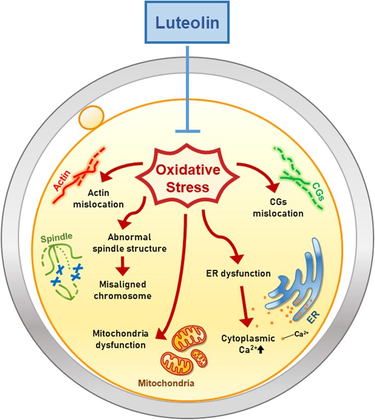
Park et al.                                                                                                  Luteolin Improves Oocyte Maturation

Terminal Deoxynucleotidyl                                               85◦ C for 5 s. The resulting cDNA was used as a template for
Transferase-Mediated                                                    polymerase chain reaction (PCR) amplification. The following
                                                                        PCR conditions were used: 95◦ C for 20 s and 60◦ C for 20 s. The
dUTP-Digoxygenin Nick End-Labeling                                      Mx3000P QPCR system (Agilent, Santa Clara, CA, United States)
Assay                                                                   and SYBR Premix Ex Taq (Takara Bio, Inc., Shiga, Japan) were
A terminal deoxynucleotidyl transferase-mediated dUTP-                  used for quantitative real-time (qRT)-PCR. The threshold cycle
digoxygenin nick end-labeling (TUNEL) assay was conducted               (Ct) is defined as the fractional cycle number at which the
using an In Situ Cell Death Detection kit (Roche, Basel,                fluorescence passes a fixed threshold above baseline. For the
Switzerland). On day 6, blastocysts were fixed in formalin              comparative analyses, mRNA expression levels were normalized
solution for 1 h at room temperature. The fixed blastocysts             to that of H2A and are expressed as in terms of fold-changes. The
were incubated in DPBS (Welgene) containing 1% (v/v) Triton             sample delta Ct (S1CT) value was calculated as the difference
X-100 for 1 h at room temperature, and stained with fluorescein-        between the Ct values of H2A and the target genes. The relative
conjugated dUTP and terminal deoxynucleotidyl transferase for           gene expression levels between the samples and control were
1 h at 38.5◦ C. As a negative control for the TUNEL reaction,           determined using the formula 2−(S1CT −C1CT) . The primers
a group of blastocysts was incubated in fluorescein-conjugated          used in the current study are listed in Supplementary Table 1.
dUTP in the absence of terminal deoxynucleotidyl transferase.
Thereafter, blastocysts were mounted on slides with 1.5 µg/mL           Statistical Analyses
DAPI, and DAPI-labeled or TUNEL-positive nuclei were                    All experiments were repeated at least three times, and
observed under a fluorescence microscope (DMi8; Leica).                 data are presented as the mean and standard error of the
                                                                        mean. The results were compared via one-way analysis of
Cdx2 Staining                                                           variance, followed by Tukey’s multiple-range tests, unless
On day 6, blastocysts were fixed in formalin solution for               Student’s t-test (Figures 1G,H) was indicated, using PASW
1 h at room temperature. The fixed blastocysts were then                Statistics for Windows, Version 18 (SPSS Inc., Chicago, IL,
washed and incubated in DPBS (Welgene) containing 1% (v/v)              United States). p-values less than 0.05 were considered to denote
Triton X-100 for 1 h at room temperature, washed in DPBS–               statistical significance.
PVA, and blocked in DPBS–PVA supplemented with 1 mg/mL
BSA (DPBS–PVA–BSA) at 4◦ C overnight. Next, the blastocysts
were blocked with 10% normal goat serum for 45 min and                  RESULTS
then incubated overnight at 4◦ C with the primary antibody-
mouse monoclonal anti-Cdx2 (undiluted solution; Biogenex                Lut Enhances Porcine Oocyte Quality by
Laboratories, Inc., San Ramon, CA, United States). Next, the            Reducing Intracellular ROS Levels
blastocysts were washed in DPBS–PVA–BSA and incubated for               To examine the effect of Lut on porcine oocyte meiotic
1 h at room temperature with the Alexa Fluor 488-labeled goat           maturation and subsequent embryonic development, we cultured
anti-mouse IgG conjugated secondary antibody (1:200 in DPBS–            the COCs in IVM medium supplemented with different
PVA–BSA). Finally, the blastocysts were washed in DPBS–PVA–             concentrations of Lut (0, 1, 5, and 10 µM). The proportion of MII
BSA, and DNA was stained with 1.5 µg/mL DAPI. DAPI-labeled              oocytes was significantly higher in the 1 and 5 µM Lut groups
or Cdx2-positive nuclei were observed using a fluorescence              than in the other groups (Figures 1A,B and Supplementary
microscope (DMi8; Leica).                                               Table 2). The blastocyst formation rate after parthenogenetic
                                                                        activation was significantly improved in the 5 µM Lut group
Quantitative Real-Time Polymerase                                       (Figures 1C–E and Supplementary Table 3). Based on these
Chain Reaction                                                          results, we used 5 µM Lut for the following experiments.
Poly(A) mRNAs were extracted from 10 MII oocytes or                        We measured intracellular ROS levels to determine the
blastocysts using the Dynabeads mRNA Direct Micro kit                   antioxidant activity of Lut in oocytes. ROS levels were
(Invitrogen, Paisley, United Kingdom). Samples were lysed in            remarkably decreased in the Lut group compared with the
100 µL of lysis/binding buffer at room temperature for 5 min,           control (Figures 1F,G). The expression levels of oxidative
and 30 µL of Dynabeads oligo (dT)25 was added to each sample.           stress (SOD1, SOD2, and CAT)-, oocyte competence (BMP15
The beads were separated from the binding buffer using a Dynal          and GDF9)-, mitogen-activated protein kinase (MAPK; MOS)-,
magnetic bar (Invitrogen). Bound poly(A) mRNAs and beads                and maturation promoting factor (CDK1 and Cyclin B)-related
were washed with buffers A and B and then separated by adding           genes in the Lut group were significantly higher than in the
7 µL of Tris buffer. Prior to reverse transcription, RNAase             control (Figure 1H). Next, we cultured COCs in IVM medium
contamination was removed with 3 µL of cleansing solution               supplemented with Lut and/or H2 O2 and recorded intracellular
containing genomic DNA (gDNA) Eraser and 5X gDNA Eraser                 ROS levels and the proportion of MII oocytes. The results showed
buffer. The resulting poly(A) mRNAs were reverse-transcribed            that H2 O2 -expose significantly increased intracellular ROS levels,
in 10-µL reactions containing Primescript RT Enzyme Mix,                and Lut supplementation reduced this increase to control level
5X Primescript buffer, and RT Primer Mix (Takara, Osaka,                (Supplementary Figure 1). Furthermore, the proportion of MII
Japan). The secondary RNA structure was denatured at room               oocytes was lower in H2 O2 -exposed oocytes, but this decrease
temperature for 5 min to facilitate cDNA production. The                was rescued by Lut supplementation (Supplementary Table 4).
reaction was terminated by incubation at 37◦ C for 15 min and           These results showed that Lut supplementation during IVM

Frontiers in Cell and Developmental Biology | www.frontiersin.org   4                                       June 2021 | Volume 9 | Article 689826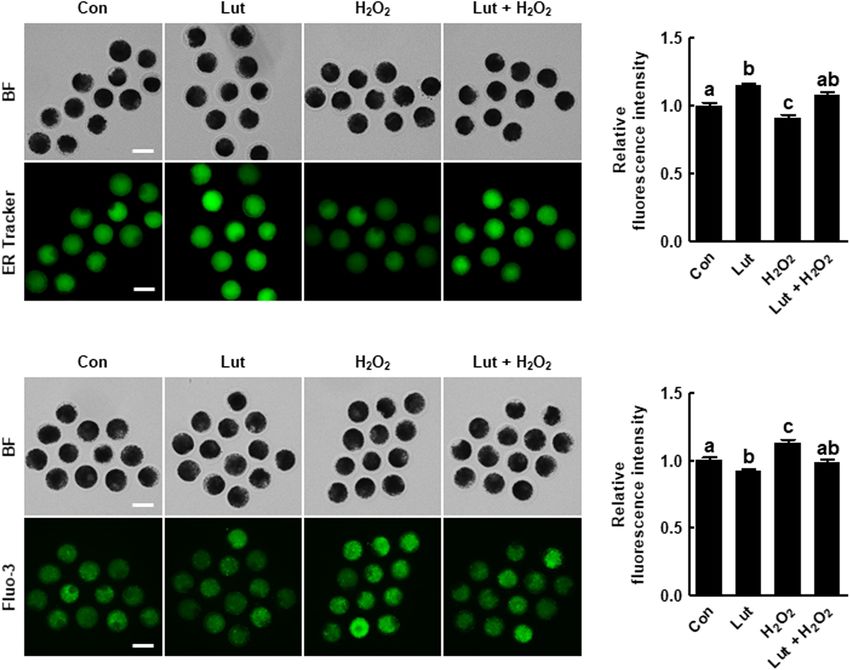
Park et al. Luteolin Improves Oocyte Maturation
FIGURE 1 | Effects of luteolin (Lut) on meiotic maturation and subsequent embryonic development in porcine embryos after parthenogenetic activation.
(A) Representative images of porcine oocytes with or without polar bodies. Scale bar = 50 µm. (B) Percentages of metaphase II (MII) oocytes after in vitro maturation
(IVM) in the indicated groups (0 µM, n = 182; 1 µM, n = 180; 5 µM, n = 178; 10 µM, n = 182). (C) Representative images of blastocyst formation in the indicated
groups. Scale bar = 100 µm. Rates of (D) cleavage and (E) blastocyst formation in the indicated groups (0 µM, n = 103; 1 µM, n = 103; 5 µM, n = 111; 10 µM,
n = 106). (F) Representative fluorescent images and (G) relative intensity levels of reactive oxygen species (ROS) in the indicated groups (control [Con], n = 28; Lut,
n = 28). Scale bar = 100 µm. (H) Relative expression of oxidative stress- and oocyte competence-related genes in the indicated groups. The data are from at least
three independent experiments, and the different superscript letters represent the significant difference (p < 0.05).
improves meiotic progression by reducing oxidative stress in control. Actin filament signals from the H2 O2 -exposed oocytes
porcine oocytes. were indicative of more abnormal morphology, such as a much
weaker or discontinuous distribution, but these were restored
by Lut supplementation (Figures 2A,B). The quantification
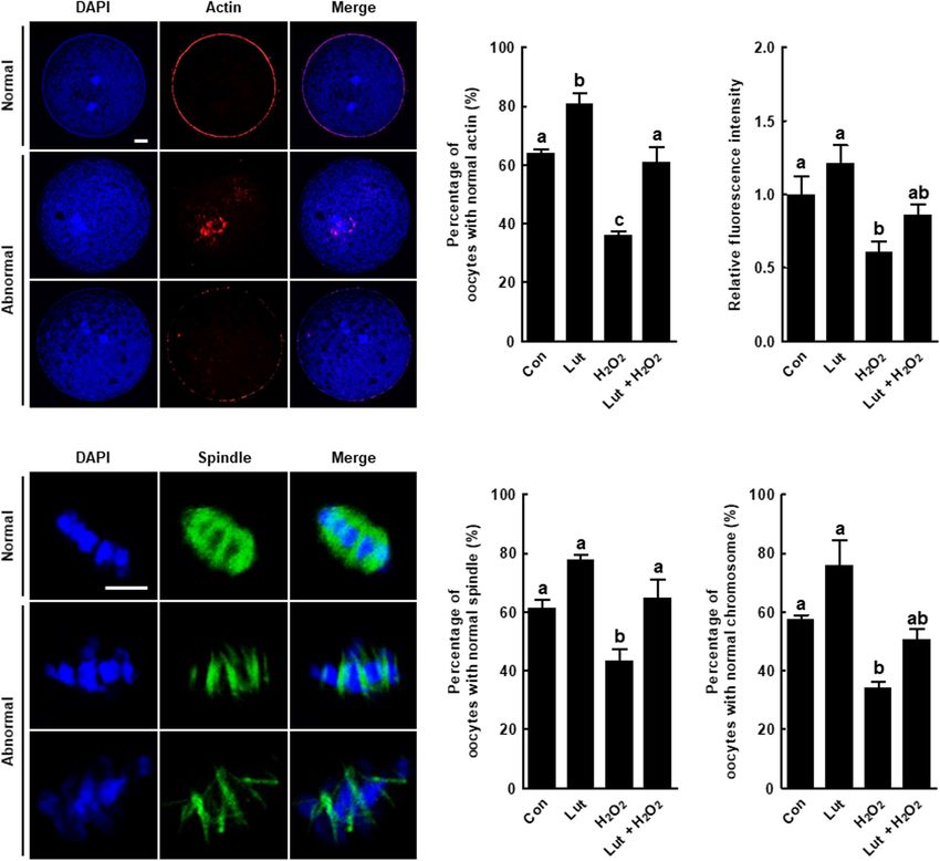
Lut Alleviates Oxidative Stress-Induced of actin fluorescence signals confirmed the observations. The
Actin and Spindle Defects in Porcine fluorescent intensity of actin was reduced in the oocytes
Oocytes exposed to H2 O2 , but the decrease was attenuated by Lut
We examined the actin and spindle morphology in Lut and/or supplementation (Figure 2C). Moreover, the proportion of
H2 O2 -treated oocytes. The percentage of oocytes with normal oocytes with well-organized spindle and chromosome structures
actin morphology, with the actin accumulating uniformly on was significantly lower in the H2 O2 group than in the other
the plasma membrane, was higher in the Lut group than in the groups, with the decrease restored by Lut supplementation
Frontiers in Cell and Developmental Biology | www.frontiersin.org 5 June 2021 | Volume 9 | Article 689826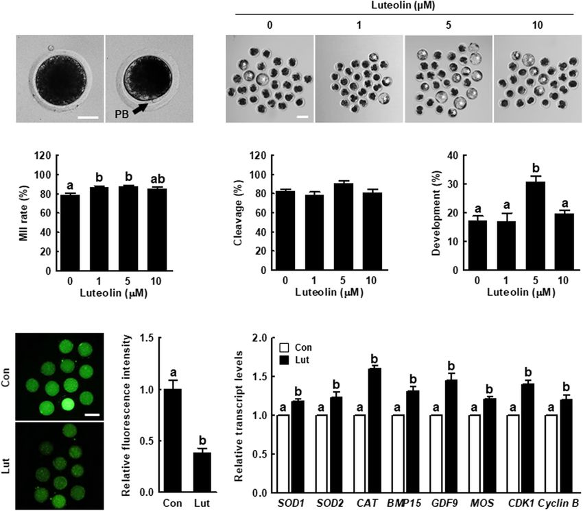
Park et al.                                                                                                                         Luteolin Improves Oocyte Maturation

  FIGURE 2 | Effects of Lut on actin and spindle defects induced by H2 O2 . (A) Representative images of normal and abnormal actin distribution in porcine oocytes.
  Scale bar = 50 µm. (B) Percentages of oocytes with normal actin morphology and (C) relative intensities of actin signals in the indicated groups (Con, n = 25; Lut,
  n = 25; H2 O2 , n = 25; Lut + H2 O2 , n = 25). (D) Representative images of a normal and abnormal spindle and chromosome structures. Scale bar = 10 µm.
  Percentages of oocytes with normal (E) spindle and (F) chromosome morphology in the indicated groups (Con, n = 32; Lut, n = 29; H2 O2 , n = 37; Lut + H2 O2 ,
  n = 29). The data are from at least three independent experiments, and the different superscript letters represent the significant difference (p < 0.05).

(Figures 2D–F). In short, Lut supplementation during IVM                                and MMP. Conversely, the fluorescence intensity of MitoSOX
improves cytoskeleton dynamics-mediated nuclear maturation                              was markedly higher in the H2 O2 -exposed oocytes than in the
by reducing oxidative stress.                                                           control and Lut-treated oocytes, but this increase was reduced
                                                                                        upon Lut treatment (Figures 3E,F). These results indicate that
Lut Recovers the Mitochondrial Content                                                  Lut supplementation during IVM improves mitochondrial
and MMP in Porcine Oocytes by                                                           function by alleviating oxidative stress.
Regulating the mtROS Level
We explored the mitochondrial content, MMP, and mtROS                                   Lut Restores Oxidative Stress-Induced
level in Lut- and/or H2 O2 -treated oocytes. MitoTracker                                ER Function Defects in Porcine Oocytes
intensity levels were higher in Lut-treated oocytes and                                 To investigate the effects of Lut on the ER contents and
lower in H2 O2 -treated oocytes compared with the control                               function in porcine oocytes, we evaluated the ER content and
(Figures 3A,B). Consistently, the ratio of fluorescence intensity                       intracellular calcium levels in Lut- and/or H2 O2 -treated oocytes.
of the J-aggregate (high membrane potential) to that of the                             The ER Tracker fluorescence level was significantly higher in
J-monomer (low membrane potential), which is an index of the                            the Lut group and lower in the H2 O2 group compared with the
MMP, was significantly increased in Lut-treated oocytes and                             control. Lut recovered the reduction in fluorescence intensity
decreased in H2 O2 -exposed oocytes compared with the control                           caused by H2 O2 exposure (Figures 4A,B). Additionally, the
(Figures 3C,D). Lut restored the reduced mitochondrial content                          intensity level of Fluo-3 was remarkably lower in Lut-treated

Frontiers in Cell and Developmental Biology | www.frontiersin.org                   6                                              June 2021 | Volume 9 | Article 689826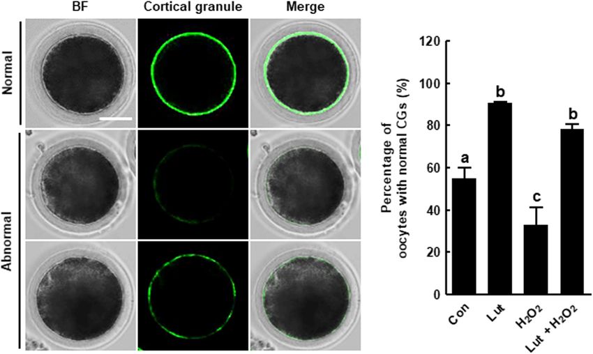
Park et al.                                                                                                                           Luteolin Improves Oocyte Maturation

  FIGURE 3 | Effects of Lut on H2 O2 -induced mitochondrial dysfunction and superoxide production. (A) Representative images of porcine oocytes stained with
  MitoTracker and (B) relative fluorescence intensities in the indicated groups (Con, n = 35; Lut, n = 35; H2 O2 , n = 35; Lut + H2 O2 , n = 35). Scale bar = 100 µm.
  (C) Representative images of porcine oocytes stained with JC-1 and (D) the ratios of red to green fluorescence intensity in the indicated groups (Con, n = 35; Lut,
  n = 35; H2 O2 , n = 35; Lut + H2 O2 , n = 35). Scale bar = 100 µm. (E) Representative images of porcine oocytes stained with MitoSOX and (F) relative fluorescence
  intensities in the indicated groups. Scale bar = 100 µm. (Con, n = 34; Lut, n = 34; H2 O2 , n = 34; Lut + H2 O2 , n = 34). The data are from at least three independent
  experiments, and the different superscript letters represent the significant difference (p < 0.05).

oocytes and higher in H2 O2 -exposed oocytes compared with the                           the H2 O2 group compared with the control. H2 O2 -exposed
control, but the increased fluorescence intensity due to H2 O2                           oocytes exhibited much weaker, discontinuous, or almost
exposure was altered to the control level by Lut supplementation                         completely absent subcortical localization. Interestingly, Lut
(Figures 4C,D). These results suggest that Lut supplementation                           supplementation remarkably restored these defects, indicating
during IVM maintains ER function in the face of oxidative stress.                        that Lut supplementation during IVM improves the dynamics of
                                                                                         CGs (Figure 5).
Lut Recovers the Distribution of CGs in
H2 O2 -Exposed Porcine Oocytes                                                           Lut Supplementation During IVM
We investigated the distribution of CGs in Lut- and/or H2 O2 -                           Improves the Developmental
treated oocytes using FITC-labeled peanut agglutinin staining.                           Competence of Porcine IVF Embryos
The percentage of oocytes with a normal CG distribution,                                 We assessed developmental competence in Lut- and/or H2 O2 -
which indicates localization in the cortex with a continuous                             treated oocytes following IVF. As shown in Figures 6A–C
and strong signal, was higher in the Lut group and lower in                              and Supplementary Table 5, the cleavage rate in the Lut

Frontiers in Cell and Developmental Biology | www.frontiersin.org                    7                                               June 2021 | Volume 9 | Article 689826
Park et al. Luteolin Improves Oocyte Maturation FIGURE 4 | Effects of Lut on endoplasmic reticulum (ER) defects induced by H2 O2 . (A) Representative images of porcine oocytes stained with ER Tracker and (B) relative fluorescence intensities in the indicated groups (Con, n = 40; Lut, n = 40; H2 O2 , n = 40; Lut + H2 O2 , n = 40). Scale bar = 100 µm. (C) Representative fluorescent images and (D) relative fluorescence intensities representing intracellular calcium levels in oocytes stained with Fluo-3 in the indicated groups (Con, n = 38; Lut, n = 38; H2 O2 , n = 38; Lut + H2 O2 , n = 38). Scale bar = 100 µm. The data are from at least three independent experiments, and the different superscript letters represent the significant difference (p < 0.05). and H2 O2 co-treatment group was significantly higher than these results showed that Lut treatment during IVM can improve that in the H2 O2 group. The blastocyst formation rate was the developmental competence of porcine embryos. significantly higher in the Lut group and lower in the H2 O2 group compared with the control. Lut supplementation recovered the reduced blastocyst formation rate to the control level. Next, DISCUSSION we characterized the blastocyst quality using Cdx2 and TUNEL analysis. The numbers of total blastocyst cells and trophectoderm In the present study, we investigated the effects of Lut on (TE) cells increased in the Lut group and decreased in the the IVM and subsequent developmental competence of porcine H2 O2 group, with Lut recovering the reductions in cell number oocytes. ROS are byproducts of metabolism, and excessive ROS (Figures 6D,E and Supplementary Table 6). The expression levels cause oxidative injury to cells that may result in DNA levels of inner cell mass (ICM)/TE differentiation-related genes damage, lipid peroxidation, mitochondrial defects, and cell death were significantly increased in the Lut group and decreased in (Kang et al., 2021). In oocytes, a disrupted ROS balance reduces the H2 O2 group. Lut supplementation restored the reductions developmental competence compared with in vivo oocytes, as due to H2 O2 (Figure 6F). Moreover, the apoptotic cell number shown by decreased rates of polar body extrusion, cleavage, and in the H2 O2 group was remarkably higher than those in the blastocyst formation in mammalian oocytes (Rizos et al., 2002; other groups (Figures 6G–I and Supplementary Table 7). The Qian et al., 2016). Thus, it is necessary to improve developmental rate of cell apoptosis was higher in the H2 O2 group and lower competence by investigating the regulation of oxidative stress and in the Lut group, with Lut supplementation alleviating the the mechanisms related to the effects of various antioxidants on increase in apoptosis. The ratio of BCL-XL to BAX expression oocyte maturation. was significantly higher in the Lut group and lower in the Flavonoids are polyphenols that protect plant cells against H2 O2 group compared with the control. Lut supplementation microorganisms, insects, and UV irradiation. Previous studies recovered the reduction due to H2 O2 (Figure 6J). Collectively, have shown that flavonoids such as kaempferol and quercetin Frontiers in Cell and Developmental Biology | www.frontiersin.org 8 June 2021 | Volume 9 | Article 689826
Park et al.                                                                                                                       Luteolin Improves Oocyte Maturation

  FIGURE 5 | Effects of Lut on H2 O2 -induced abnormal distribution of cortical granules (CGs). (A) Representative images of the normal and abnormal morphologies of
  CGs in porcine oocytes. Scale bar = 50 µm. (B) Percentages of oocytes with a normal CG distribution in the indicated groups (Con, n = 44; Lut, n = 43; H2 O2 ,
  n = 37; Lut + H2 O2 , n = 41). The data are from at least three independent experiments, and the different superscript letters represent the significant difference
  (p < 0.05).

exert antioxidant effects on mammalian oocytes (Cao et al.,                           signaling pathway and is an important signaling molecule,
2020; Zhao et al., 2020). Lut, a flavone in the flavonoid                             which functions microfilament or microtubule dynamics, during
group, also exhibits antioxidant activity. Lut can stabilize the                      oocyte maturation (Hatch and Capco, 2001). In addition,
radical group by donating hydrogen/electron of C2–C3 double                           phosphorylation of ERK1/2 (p-ERK1/2) at MII stage is
bond to radical and by blocking Fenton reaction using oxo                             considered to parameter of cytoplasmic maturation that affect
group at C4 that binds transitional metal ions including                              cleavage and the blastocyst formation rate after fertilization
iron and copper. These two structural features of Lut inhibit                         (Inoue et al., 1995). However, MAPK signaling pathway disorder
pro-oxidant enzymes such as xanthine oxidase, and induce                              is one of pathways that cause oxidative damage (Yao et al., 2019).
antioxidant enzymes (Gendrisch et al., 2021). In addition, Lut                        Previous studies reported that Lut upregulated the expression of
not only has its own antioxidant activity, but also interacts with                    p-ERK1/2 and MAPK, suggesting Lut alleviate oxidative stress
other antioxidants such as vitamins and cellular redox system                         by activating the MAPK signaling pathway (Wu et al., 2015;
(Gendrisch et al., 2021), indicating that Lut can synergistically                     Wang et al., 2020). In this study, Lut treatment increased the
enhance its antioxidant properties. Furthermore, some flavonoids                      developmental competence of PA and IVF embryos. Moreover,
including quercetin, genistein, and catechin induce DNA damage                        Lut treatment increased expression of oocyte competence-,
by pro-oxidative effect resulting in mutagenic and carcinogenic                       MAPK, and maturation promoting factor-related genes. These
activity (Kawanishi et al., 2005; Gendrisch et al., 2021), but                        results indicate Lut improves oocyte meiotic progression and
Lut has no report regarding these effects, suggesting that                            subsequent embryonic development against oxidative stress.
Lut can be expected as a comparatively safe antioxidant                                   A proper cytoskeletal system is crucial for meiotic maturation,
(Yamashita et al., 1999). In the present study, we demonstrated                       as the cytoskeleton controls chromosome condensation and
for the first time that Lut supplementation improves oocyte                           segregation, subsequent meiosis, fertilization, and further cell
maturation and subsequent embryonic development. Moreover,                            cleavage (Brunet and Maro, 2005). Within the cytoskeleton,
Lut supplementation decreased intracellular ROS levels and                            microfilaments are involved in chromosome migration, cortical
increased the expression levels of antioxidant-related genes.                         spindle anchorage, and first polar body emission, whereas
These results indicate that Lut exerts positive effects on IVM                        microtubules facilitate chromosome movement by organizing
porcine oocytes by reducing oxidative stress.                                         spindles (Sun and Schatten, 2006; Duan and Sun, 2019).
   The MAPK signaling pathway is crucial for the female                               However, previous studies showed that oxidative stress impaired
reproduction process including the embryo development and                             cytoskeletal dynamics, with the disrupted microfilament or
meiotic maturation (Chen et al., 2020). The extracellular                             microtubule dynamics resulting in defective polar body extrusion
signal-regulated kinase-1/2 (ERK1/2) is a member of MAPK                              (Zhang et al., 2018). Actin is a microfilament subunit,

Frontiers in Cell and Developmental Biology | www.frontiersin.org                 9                                              June 2021 | Volume 9 | Article 689826
Park et al.                                                                                                                         Luteolin Improves Oocyte Maturation

  FIGURE 6 | Effects of Lut during IVM on the developmental competence of in vitro fertilization (IVF) embryos. (A) Representative images of blastocyst formation at
  6 days after IVF in the indicated groups. Scale bar = 100 µm. Rates of (B) cleavage and (C) blastocyst formation in the indicated groups (Con, n = 144; Lut, n = 145;
  H2 O2 , n = 134; Lut + H2 O2 , n = 133). (D) Representative images of Cdx2- and DAPI-stained blastocysts in the indicated groups. Scale bar = 50 µm. (E) Number of
  inner cell mass (ICM), trophectoderm (TE), and total cells in the indicated groups (Con, n = 20; Lut, n = 20; H2 O2 , n = 20; Lut + H2 O2 , n = 20). (F) Relative
  expression of ICM/TE differentiation-related genes in the indicated groups. (G) Representative images of TUNEL- and DAPI-stained blastocysts in the indicated
  groups. Scale bar = 50 µm. (H) Number of apoptotic cells and (I) the apoptosis rate in the indicated groups (Con, n = 20; Lut, n = 20; H2 O2 , n = 20; Lut + H2 O2 ,
  n = 20). (J) Relative expression of apoptosis-related genes and the ratio of BCL-XL/BAX levels in the indicated groups. The data are from at least three independent
  experiments, and the different superscript letters represent the significant difference (p < 0.05).

and tubulin is a microtubule subunit. We investigated the                               intracellular Ca2+ homeostasis. Ca2+ is one of the major signal
dynamics of these subunits. Consistent with previous study, our                         molecules that regulate oocyte physiology, including cell cycle
results showed that oxidative stress caused by H2 O2 exposure                           resumption (Tiwari et al., 2017). Abnormally high concentrations
disrupted microfilament or microtubule dynamics resulting in                            of Ca2+ in the cytoplasm result in cell cycle arrest, disruption of
poor proportion of MII oocytes. Interestingly, Lut treatment                            fertilization ability, and apoptosis (Tiwari et al., 2015; Wang et al.,
completely or partially rescued these defects. Lut treatment                            2017). Oxidative stress causes Ca2+ influx into the cytoplasm
significantly recovered the decreased proportion of MII oocytes                         from the ER and subsequently increases the mitochondrial
and the disruption of actin and spindle structures caused by H2 O2                      Ca2+ concentration (Ermak and Davies, 2002). Mitochondrial
exposure, although actin amounts and chromosome structure                               Ca2+ overload is a critical sensitizing signal in the apoptosis
were not rescued to the control level. Thus, we suggest that                            pathway that causes embryonic developmental arrest and death
Lut enhances nuclear maturation by normalizing cytoskeletal                             (Marchi et al., 2018; Kim et al., 2020). Moreover, severe oxidative
dynamics against oxidative stress.                                                      stress triggers a decrease in cellular mitochondrial content
   Oocyte maturation includes a series of complex events, such                          through the suppression of mitochondrial biogenesis as well
as protein synthesis and the transcription of cytoplasmic RNA,                          as in mitochondrial function (Bouchez and Devin, 2019; Jeong
which require energy. The ER functions in protein folding                               et al., 2020). Our results showed that Lut protects the ER
and degradation, and mitochondria supply energy for protein                             and mitochondrial function against oxidative stress, with Lut
synthesis (Dumollard et al., 2007). The ER also acts as the                             supporting proper cytoplasmic maturation through regulation of
major storage area for calcium ions (Ca2+ ), thus regulating                            the ER and mitochondrial system.

Frontiers in Cell and Developmental Biology | www.frontiersin.org                  10                                             June 2021 | Volume 9 | Article 689826
Park et al.                                                                                                                      Luteolin Improves Oocyte Maturation

  FIGURE 7 | Hypothetical model of the antioxidant effect of Lut on porcine oocyte maturation. Lut improves the quality of porcine oocytes by alleviating oxidative
  damage. Especially, Lut recovers disrupted organelle dynamics including mislocation of actin, spindle, chromosome and CGs, and dysfunction of mitochondria and
  ER. These findings suggest that Lut improves porcine meiotic progression and subsequent embryonic development by protecting various organelle dynamics
  against oxidative stress.

   Finally, we investigated the fertilization ability and                             suggesting that Lut supplementation during IVM has positive
developmental competence of Lut-treated oocytes after IVF. CGs                        effects on the developmental competence of IVF embryos.
are organelles located in the subcortical region of oocytes, and the                      This is the first study to demonstrate the effects of Lut on
distribution of CGs is regarded as a marker of oocyte maturation.                     mammalian oocytes (Figure 7). Moreover, we especially focused
Additionally, as CGs are related to the blocking of polyspermy,                       on the dynamics of cell organelles during IVM under oxidative
proper CG dynamics are crucial for successful fertilization                           stress and subsequently determined the antioxidant activities of
(Burkart et al., 2012). In the present study, Lut normalized the                      Lut. Our study suggests that Lut improves the quality of porcine
distribution of CGs and enhanced developmental competence                             oocytes and subsequent embryonic development following IVF
in the face of oxidative stress, as shown in the cleavage and                         by alleviating oxidative damage to organelles. These findings
blastocyst formation rate, cell number, and apoptotic pattern,                        help raise awareness of the beneficial effects of Lut on IVM

Frontiers in Cell and Developmental Biology | www.frontiersin.org                11                                             June 2021 | Volume 9 | Article 689826
Park et al.                                                                                                                                      Luteolin Improves Oocyte Maturation

and elucidate how Lut supports proper oocyte maturation under                                  have read and agreed to the published final version of
oxidative stress.                                                                              this manuscript.

DATA AVAILABILITY STATEMENT                                                                    FUNDING
                                                                                               This study was supported by grants from the KRIBB
The original contributions presented in the study are included
                                                                                               Research Initiative Program (KGM4252122) and the Bio
in the article/Supplementary Material, further inquiries can be
                                                                                               and Medical Technology Development Program through the
directed to the corresponding authors.
                                                                                               National Research Foundation of Korea (NRF), funded by the
                                                                                               Ministry of Education, Science and Technology (MEST) (No.
                                                                                               2018M3A9H1023142), South Korea.
AUTHOR CONTRIBUTIONS
S-HP and P-SJ designed study, performed experiments, analyzed                                  SUPPLEMENTARY MATERIAL
data, and wrote the manuscript. YJ, H-GK, MK, SL, and
B-SS performed experiments and analyzed data. S-UK acquired                                    The Supplementary Material for this article can be found
financial and discussed study. S-KC and B-WS designed study,                                   online at: https://www.frontiersin.org/articles/10.3389/fcell.2021.
supervised the study, and wrote the manuscript. All authors                                    689826/full#supplementary-material

REFERENCES                                                                                     Gendrisch, F., Esser, P. R., Schempp, C. M., and Wolfle, U. (2021). Luteolin as
                                                                                                  a modulator of skin aging and inflammation. Biofactors 47, 170–180. doi:
An, F., Wang, S., Yuan, D., Gong, Y., and Wang, S. (2016). attenuation of oxidative               10.1002/biof.1699
   stress of erythrocytes by plant-derived flavonoids, orientin and luteolin. Evid.            Gupta, G., Tiwari, J., Dahiya, R., Kumar Sharma, R., Mishra, A., and Dua, K.
   Based Complement. Alternat. Med. 2016:3401269. doi: 10.1155/2016/3401269                       (2018). Recent updates on neuropharmacological effects of luteolin. EXCLI J.
Bouchez, C., and Devin, A. (2019). Mitochondrial biogenesis and mitochondrial                     17, 211–214. doi: 10.17179/excli2018-1041
   Reactive Oxygen Species (ROS): a complex relationship regulated by the                      Hashimoto, S. (2009). Application of in vitro maturation to assisted reproductive
   cAMP/PKA signaling pathway. Cells 8:287. doi: 10.3390/cells8040287                             technology. J. Reprod. Dev. 55, 1–10. doi: 10.1262/jrd.20127
Brunet, S., and Maro, B. (2005). Cytoskeleton and cell cycle control during meiotic            Hatch, K. R., and Capco, D. G. (2001). Colocalization of CaM KII and MAP kinase
   maturation of the mouse oocyte: integrating time and space. Reproduction 130,                  on architectural elements of the mouse egg: potentiation of MAP kinase activity
   801–811. doi: 10.1530/rep.1.00364                                                              by CaM KII. Mol. Reprod. Dev. 58, 69–77. doi: 10.1002/1098-2795(200101)58:
Burkart, A. D., Xiong, B., Baibakov, B., Jimenez-Movilla, M., and Dean, J. (2012).                13.0.CO;2-O
   Ovastacin, a cortical granule protease, cleaves ZP2 in the zona pellucida to                Inoue, M., Naito, K., Aoki, F., Toyoda, Y., and Sato, E. (1995). Activation of
   prevent polyspermy. J. Cell Biol. 197, 37–44. doi: 10.1083/jcb.201112094                       mitogen-activated protein kinase during meiotic maturation in porcine oocytes.
Cao, Y., Zhao, H., Wang, Z., Zhang, C., Bian, Y., Liu, X., et al. (2020). Quercetin               Zygote 3, 265–271. doi: 10.1017/S0967199400002665
   promotes in vitro maturation of oocytes from humans and aged mice. Cell                     Jeong, P. S., Lee, S., Park, S. H., Kim, M. J., Kang, H. G., Nanjidsuren, T., et al.
   Death Dis. 11:965. doi: 10.1038/s41419-020-03183-5                                             (2020). Butylparaben is toxic to porcine oocyte maturation and subsequent
Chen, L., Jiang, J. C., Dai, X. X., and Fan, H. Y. (2020). [Function and molecular                embryonic development following in vitro fertilization. Int. J. Mol. Sci. 21:3692.
   mechanism of mitogen-activated protein kinase (MAPK) in regulating oocyte                      doi: 10.3390/ijms21103692
   meiotic maturation and ovulation]. Sheng Li Xue Bao 72, 48–62.                              Kang, H. G., Lee, S., Jeong, P. S., Kim, M. J., Park, S. H., Joo, Y. E., et al.
Day, B. N. (2000). Reproductive biotechnologies: current status in porcine                        (2021). Lycopene improves in vitro development of porcine embryos by
   reproduction. Anim. Reprod. Sci. 60–61, 161–172. doi: 10.1016/S0378-4320(00)                   reducing oxidative stress and apoptosis. Antioxidants 10:230. doi: 10.3390/
   00079-8                                                                                        antiox10020230
De los Reyes, M., Palomino, J., Jofre, S., Villarroel, A., and Moreno, R. (2012). Golgi        Kawanishi, S., Oikawa, S., and Murata, M. (2005). Evaluation for safety of
   apparatus and endoplasmic reticulum dynamic during meiotic development in                      antioxidant chemopreventive agents. Antioxid. Redox. Signal. 7, 1728–1739.
   canine oocytes. Reprod. Domest. Anim. 47, 93–97. doi: 10.1111/rda.12014                        doi: 10.1089/ars.2005.7.1728
De los Reyes, M., Palomino, J., Parraguez, V. H., Hidalgo, M., and Saffie, P. (2011).          Khazaei, M., and Nematbakhsh, M. (2004). Coronary vascular and aortic
   Mitochondrial distribution and meiotic progression in canine oocytes during                    endothelial permeability during estrogen therapy: a study in DOCA-salt
   in vivo and in vitro maturation. Theriogenology 75, 346–353. doi: 10.1016/j.                   hypertensive ovariectomized rats. Physiol. Res. 53, 609–614.
   theriogenology.2010.09.005                                                                  Kim, M. J., Park, H. J., Lee, S., Kang, H. G., Jeong, P. S., Park, S. H., et al. (2020).
Do, L. T., Luu, V. V., Morita, Y., Taniguchi, M., Nii, M., Peter, A. T., et al. (2015).           Effect of triclosan exposure on developmental competence in parthenogenetic
   Astaxanthin present in the maturation medium reduces negative effects of heat                  porcine embryo during preimplantation. Int. J. Mol. Sci. 21:5790. doi: 10.3390/
   shock on the developmental competence of porcine oocytes. Reprod. Biol. 15,                    ijms21165790
   86–93. doi: 10.1016/j.repbio.2015.01.002                                                    Kwak, S. S., Cheong, S. A., Jeon, Y., Lee, E., Choi, K. C., Jeung, E. B., et al.
Duan, X., and Sun, S. C. (2019). Actin cytoskeleton dynamics in mammalian oocyte                  (2012). The effects of resveratrol on porcine oocyte in vitro maturation
   meiosis. Biol. Reprod. 100, 15–24. doi: 10.1093/biolre/ioy163                                  and subsequent embryonic development after parthenogenetic activation and
Dumollard, R., Duchen, M., and Carroll, J. (2007). The role of mitochondrial                      in vitro fertilization. Theriogenology 78, 86–101. doi: 10.1016/j.theriogenology.
   function in the oocyte and embryo. Curr. Top. Dev. Biol. 77, 21–49. doi:                       2012.01.024
   10.1016/S0070-2153(06)77002-8                                                               Lee, S., Kang, H. G., Jeong, P. S., Nanjidsuren, T., Song, B. S., Jin, Y. B., et al. (2020).
Ermak, G., and Davies, K. J. (2002). Calcium and oxidative stress: from cell                      Effect of oocyte quality assessed by Brilliant Cresyl Blue (BCB) staining on
   signaling to cell death. Mol. Immunol. 38, 713–721. doi: 10.1016/S0161-                        cumulus cell expansion and sonic hedgehog signaling in porcine during in vitro
   5890(01)00108-0                                                                                maturation. Int. J. Mol. Sci. 21:4423. doi: 10.3390/ijms21124423

Frontiers in Cell and Developmental Biology | www.frontiersin.org                         12                                                    June 2021 | Volume 9 | Article 689826
Park et al. Luteolin Improves Oocyte Maturation Lopes, J. S., Canha-Gouveia, A., Paris-Oller, E., and Coy, P. (2019). the fertilisation capacity and developmental ability of vitrified bovine oocytes. Supplementation of bovine follicular fluid during in vitro maturation Sci. Rep. 7:10652. doi: 10.1038/s41598-017-10907-9 increases oocyte cumulus expansion, blastocyst developmental Wang, T., Han, J., Duan, X., Xiong, B., Cui, X. S., Kim, N. H., et al. (2016). The kinetics, and blastocyst cell number. Theriogenology 126, 222–229. toxic effects and possible mechanisms of Bisphenol A on oocyte maturation of doi: 10.1016/j.theriogenology.2018.12.010 porcine in vitro. Oncotarget 7, 32554–32565. doi: 10.18632/oncotarget.8689 Mao, L., Lou, H., Lou, Y., Wang, N., and Jin, F. (2014). Behaviour of cytoplasmic Watson, A. J. (2007). Oocyte cytoplasmic maturation: a key mediator of oocyte and organelles and cytoskeleton during oocyte maturation. Reprod. Biomed. Online embryo developmental competence. J. Anim. Sci. 85, E1–E3. doi: 10.2527/jas. 28, 284–299. doi: 10.1016/j.rbmo.2013.10.016 2006-432 Marchi, S., Patergnani, S., Missiroli, S., Morciano, G., Rimessi, A., Wieckowski, Wu, P. S., Yen, J. H., Kou, M. C., and Wu, M. J. (2015). Luteolin and apigenin M. R., et al. (2018). Mitochondrial and endoplasmic reticulum calcium attenuate 4-hydroxy-2-nonenal-mediated cell death through modulation of homeostasis and cell death. Cell Calcium 69, 62–72. doi: 10.1016/j.ceca.2017. UPR, Nrf2-ARE and MAPK pathways in PC12 cells. PLoS One 10:e0130599. 05.003 doi: 10.1371/journal.pone.0130599 Qian, D., Li, Z., Zhang, Y., Huang, Y., Wu, Q., Ru, G., et al. (2016). Response of Xia, F., Wang, C., Jin, Y., Liu, Q., Meng, Q., Liu, K., et al. (2014). Luteolin mouse zygotes treated with mild hydrogen peroxide as a model to reveal novel protects HUVECs from TNF-alpha-induced oxidative stress and inflammation mechanisms of oxidative stress-induced injury in early embryos. Oxid. Med. via its effects on the Nox4/ROS-NF-kappaB and MAPK pathways. J. Atheroscler. Cell Longev. 2016:1521428. doi: 10.1155/2016/1521428 Thromb. 21, 768–783. doi: 10.5551/jat.23697 Rizos, D., Fair, T., Papadopoulos, S., Boland, M. P., and Lonergan, P. (2002). Yamashita, N., Tanemura, H., and Kawanishi, S. (1999). Mechanism of oxidative Developmental, qualitative, and ultrastructural differences between ovine and DNA damage induced by quercetin in the presence of Cu(II). Mutat. Res. 425, bovine embryos produced in vivo or in vitro. Mol. Reprod. Dev. 62, 320–327. 107–115. doi: 10.1016/S0027-5107(99)00029-9 doi: 10.1002/mrd.10138 Yao, X., Jiang, H., Li, Y. H., Gao, Q., Xu, Y. N., and Kim, N. H. (2019). Kaempferol Santos, R. R., Schoevers, E. J., and Roelen, B. A. (2014). Usefulness of bovine and alleviates the reduction of developmental competence during aging of porcine porcine IVM/IVF models for reproductive toxicology. Reprod. Biol. Endocrinol. oocytes. Anim. Sci. J. 90, 1417–1425. 10.1111/asj.13280 12:117. doi: 10.1186/1477-7827-12-117 Ye, R., Xu, S., Liu, Y., Pang, L., Lian, X., Zhong, Y., et al. (2017). Protective Sovernigo, T. C., Adona, P. R., Monzani, P. S., Guemra, S., Barros, F., Lopes, effect of icariin on the development of preimplantation mouse embryos F. G., et al. (2017). Effects of supplementation of medium with different against hydrogen peroxide-induced oxidative injury. Oxid. Med. Cell. Longev. antioxidants during in vitro maturation of bovine oocytes on subsequent 2017:2704532. doi: 10.1155/2017/2704532 embryo production. Reprod. Domest. Anim. 52, 561–569. doi: 10.1111/rda. Zhang, Y., Wang, H. H., Wan, X., Xu, Y., Pan, M. H., and Sun, S. C. (2018). 12946 Inhibition of protein kinase D disrupts spindle formation and actin assembly Sun, Q. Y., and Schatten, H. (2006). Regulation of dynamic events by during porcine oocyte maturation. Aging 10, 3736–3744. doi: 10.18632/aging. microfilaments during oocyte maturation and fertilization. Reproduction 131, 101667 193–205. doi: 10.1530/rep.1.00847 Zhao, Y., Xu, Y., Li, Y., Jin, Q., Sun, J., E, Z., et al. (2020). Supplementation Tiwari, M., Prasad, S., Shrivastav, T. G., and Chaube, S. K. (2017). Calcium signaling of kaempferol to in vitro maturation medium regulates oxidative stress and during meiotic cell cycle regulation and apoptosis in mammalian oocytes. J. Cell enhances subsequent embryonic development in vitro. Zygote 28, 59–64. doi: Physiol. 232, 976–981. doi: 10.1002/jcp.25670 10.1017/S0967199419000674 Tiwari, M., Prasad, S., Tripathi, A., Pandey, A. N., Ali, I., Singh, A. K., et al. (2015). Apoptosis in mammalian oocytes: a review. Apoptosis 20, 1019–1025. Conflict of Interest: The authors declare that the research was conducted in the doi: 10.1007/s10495-015-1136-y absence of any commercial or financial relationships that could be construed as a Ueno, S., Kurome, M., Ueda, H., Tomii, R., Hiruma, K., and Nagashima, H. (2005). potential conflict of interest. Effects of maturation conditions on spindle morphology in porcine MII oocytes. J. Reprod. Dev. 51, 405–410. doi: 10.1262/jrd.16091 Copyright © 2021 Park, Jeong, Joo, Kang, Kim, Lee, Song, Kim, Cho and Sim. Wang, H. R., Pei, S. Y., Fan, D. X., Liu, Y. H., Pan, X. F., Song, F. X., et al. This is an open-access article distributed under the terms of the Creative Commons (2020). Luteolin protects pheochromocytoma (PC-12) cells against abeta 25-35- Attribution License (CC BY). The use, distribution or reproduction in other forums induced cell apoptosis through the ER/ERK/MAPK signalling pathway. Evid. is permitted, provided the original author(s) and the copyright owner(s) are credited Based Complement. Alternat. Med. 2020:2861978. doi: 10.1155/2020/2861978 and that the original publication in this journal is cited, in accordance with accepted Wang, N., Hao, H. S., Li, C. Y., Zhao, Y. H., Wang, H. Y., Yan, C. L., et al. academic practice. No use, distribution or reproduction is permitted which does not (2017). Calcium ion regulation by BAPTA-AM and ruthenium red improved comply with these terms. Frontiers in Cell and Developmental Biology | www.frontiersin.org 13 June 2021 | Volume 9 | Article 689826
You can also read