RNase A Inhibits Formation of Neutrophil Extracellular Traps in Subarachnoid Hemorrhage
←
→
Page content transcription
If your browser does not render page correctly, please read the page content below
ORIGINAL RESEARCH
                                                                                                                                              published: 16 September 2021
                                                                                                                                           doi: 10.3389/fphys.2021.724611

                                                RNase A Inhibits Formation of
                                                Neutrophil Extracellular Traps in
                                                Subarachnoid Hemorrhage
                                                Anton Früh 1†, Katharina Tielking 1†, Felix Schoknecht 1, Shuheng Liu 1, Ulf C. Schneider 1,
                                                Silvia Fischer 2, Peter Vajkoczy 1* and Ran Xu 1
                                                1
                                                 Department of Neurosurgery, Charité – Universitätsmedizin Berlin, Corporate Member of Freie Universität Berlin,
                                                Humboldt-Universität zu Berlin, and Berlin Institute of Health, Berlin, Germany, 2 Department of Biochemistry,
                          Edited by:            Giessen University, Giessen, Germany
                     Jean-Luc Morel,
     Centre National de la Recherche
         Scientifique (CNRS), France            Background: Subarachnoid hemorrhage (SAH) caused by rupture of an intracranial
                      Reviewed by:              aneurysm, is a life-threatening emergency that is associated with substantial morbidity
               Erik Josef Behringer,
Loma Linda University, United States
                                                and mortality. Emerging evidence suggests involvement of the innate immune response
                       Md. A. Hakim,            in secondary brain injury, and a potential role of neutrophil extracellular traps (NETs) for
Loma Linda University, United States            SAH-associated neuroinflammation. In this study, we investigated the spatiotemporal
             Jose Javier Provencio,
      University of Virginia Hospital,          patterns of NETs in SAH and the potential role of the RNase A (the bovine equivalent to
                       United States            human RNase 1) application on NET burden.
                     *Correspondence:
                          Peter Vajkoczy
                                                Methods: A total number of n = 81 male C57Bl/6 mice were operated utilizing a filament
               peter.vajkoczy@charite.de        perforation model to induce SAH, and Sham operation was performed for the corresponding
     †
         These authors have contributed         control groups. To confirm the bleeding and exclude stroke and intracerebral hemorrhage,
                    equally to this work        the animals received MRI after 24 h. Mice were treated with intravenous injection of RNase
                      Specialty section:
                                                A (42 μg/kg body weight) or saline solution for the control groups, respectively. Quadruple-
            This article was submitted to       immunofluorescence (IF) staining for cell nuclei (DAPI), F-actin (phalloidin), citrullinated
                     Vascular Physiology,
                                                H3, and neurons (NeuN) was analyzed by confocal imaging and used to quantify NET
                  a section of the journal
                  Frontiers in Physiology       abundance in the subarachnoid space (SAS) and brain parenchyma. To quantify NETs in
           Received: 13 June 2021               human SAH patients, cerebrospinal spinal fluid (CSF) and blood samples from day 1, 2,
        Accepted: 10 August 2021                7, and 14 after bleeding onset were analyzed for double-stranded DNA (dsDNA) via
     Published: 16 September 2021
                                                Sytox Green.
                             Citation:
    Früh A, Tielking K, Schoknecht F,           Results: Neutrophil extracellular traps are released upon subarachnoid hemorrhage in
     Liu S, Schneider UC, Fischer S,
                                                the SAS on the ipsilateral bleeding site 24 h after ictus. Over time, NETs showed progressive
          Vajkoczy P and Xu R (2021)
        RNase A Inhibits Formation of           increase in the parenchyma on both ipsi- and contralateral site, peaking on day 14 in
     Neutrophil Extracellular Traps in          periventricular localization. In CSF and blood samples of patients with aneurysmal SAH,
          Subarachnoid Hemorrhage.
           Front. Physiol. 12:724611.
                                                NETs also increased gradually over time with a peak on day 7. RNase application
    doi: 10.3389/fphys.2021.724611              significantly reduced NET accumulation in basal, cortical, and periventricular areas.

Frontiers in Physiology | www.frontiersin.org                                          1                                        September 2021 | Volume 12 | Article 724611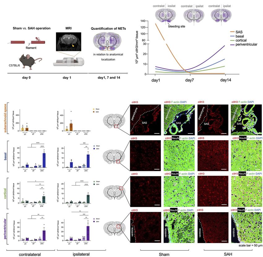
Früh et al.                                                                                                                     RNase A Reduces NETs in SAH

                                                Conclusion: Neutrophil extracellular trap formation following SAH originates in the
                                                ipsilateral SAS of the bleeding site and spreads gradually over time to basal, cortical, and
                                                periventricular areas in the parenchyma within 14 days. Intravenous RNase application
                                                abrogates NET burden significantly in the brain parenchyma, underpinning a potential role
                                                in modulation of the innate immune activation after SAH.
                                                Keywords: neutrophil extracellular traps, neutrophils, subarachnoid hemorrhage, hemorrhagic stroke,
                                                neuroinflammation, innate immune response, innate immune reaction

INTRODUCTION                                                                       homeostasis that can counteract exRNA when applied
                                                                                   exogenously (Fischer et al., 2011; Lasch et al., 2020; Preissner
Subarachnoid hemorrhage (SAH) caused by rupture of an                              et al., 2020). Recent studies have shown that NET-associated
intracranial aneurysm, is a life-threatening emergency that is                     RNA is a relevant NET component and its formation can
associated with substantial morbidity and mortality (Lawton                        occur also independently of DNA, which opened the hypothetical
and Vates, 2017; Van Lieshout et al., 2018). Approximately 5%                      avenue that RNase A (the bovine equivalent to human RNAse1)
of all strokes are caused by SAH and the incidence is estimated                    may also modify NET formation in vivo SAH models (Herster
to be nine per 100.000 person years (Etminan et al., 2019).                        et al., 2020). Moreover, preliminary data from our laboratory
Women are about 1.24 times more likely to be affected by                           show that RNase A modulates exRNA accumulation in SAH
the disease, and the incidence of SAH increases from the                           and the microglia-specific immune reaction after SAH.
age > 50 years and is higher in Japan and Finland compared                            In this study, with regard to the migratory characteristics
to other industrialized countries (Macdonald and Schweizer,                        of neutrophils, we investigated the spatial and temporal pattern
2017). Around one-third of SAH patients die within the first                       of NET formation in an in vivo model of SAH. We report
30 days after the initial bleeding event (Schertz et al., 2016).                   that NET accumulation begins in the ipsilateral side of bleeding,
Among survivors, secondary brain injury is described as the                        and spreads over time to the parenchyma on both hemispheres,
main cause of morbidity (Macdonald, 2014). Thereby, emerging                       peaking on day 14 after SAH. To confirm these findings in
evidence points toward involvement of inflammatory processes                       the human system, we also measured NET burden in
(Gris et al., 2019), particularly of innate immune cells                           cerebrospinal spinal fluid (CSF) and blood samples in SAH
(Schneider et al., 2015; Lucke-Wold et al., 2016; Zhang et al., 2020).             patients and show that NET accumulation occurs in both
    Among these immune reactions, recent studies suggest that                      compartments significantly. Furthermore, we report that RNase
neutrophils play a particular role in SAH induced inflammation                     significantly reduces NET formation in the parenchyma, thus
(Atangana et al., 2017). Neutrophil granulocytes are the first line                being an attractive mediator for evaluation in subsequent studies.
of the innate immune system to fight pathogens (Borregaard,
2010). During infectious processes, they exit blood vessel systems,
migrate to the site of infection and accumulate in high numbers                    MATERIALS AND METHODS
(Niemiec et al., 2015). Neutrophil infiltration into damaged brain
tissues has been shown after SAH, stroke, and traumatic brain                      Study Approval
injury (Liu et al., 2018; García-Culebras et al., 2019; Zeng et al.,               The analysis on human samples was approved by the local ethics
2021). Upon stimulation, neutrophils release DNA, granule proteins,                committee of Charité University Hospital (ethical approval number:
and histones (Brinkmann et al., 2004). These fibril matrixes are                   EA2/134/18). All patients or their authorized individuals gave
coined as neutrophil extracellular traps (NETs). NETs are involved                 written consent to the collection of blood and CSF samples. All
in various immune reactions and the pathophysiology of diverse                     animal experiments were approved by the Regional Office for
diseases (Papayannopoulos, 2018), including central nervous system                 Health and Social Affairs (Landesamt für Gesundheit und Soziales;
pathologies (Zhang et al., 2020). The impact of NET formation                      approval number: TVA 0063/18) and were performed in conformity
in SAH has not yet been studied extensively, but recent evidence                   with the German law of animal protection and the National
suggests that they may promote aneurysm rupture, and                               Institute of Health Guidelines for care and use of laboratory animals.
pharmacological removal of NETs can reduce the rate of aneurysm
rupture (Korai et al., 2021). Moreover, there is growing evidence                  Human Data
that NETs aggravate the inflammatory events after SAH, and                         Measurement of NET Surrogate Markers in CSF
impair revascularization and increase blood brain barrier (BBB)                    and Peripheral Blood in SAH Patients
damage after stroke (Kang et al., 2020).                                           Peripheral blood and CSF samples were collected from patients
    Next to extracellular DNA, other alarmins or so-called                         with aneurysmal SAH on day 1, 2, 7, and 14 after SAH onset.
danger-associated molecular pattern (DAMP) signals such as                         For each time point, 2 ml peripheral blood was drawn using
extracellular RNA (exRNA) have been described as a key player                      Ethylene Diaminetetraacetic acid (EDTA) tubes, and 2 ml CSF
in the involvement of central system pathologies, including                        was collected from the initial placement of an extraventricular
hemorrhagic stroke (Tielking et al., 2019). In this context,                       drain (EVD) at onset of SAH, and then further collected from
RNase is a natural enzyme involved in the regulation of vascular                   the existing EVD. Some patients also had additional lumbar

Frontiers in Physiology | www.frontiersin.org                                  2                                   September 2021 | Volume 12 | Article 724611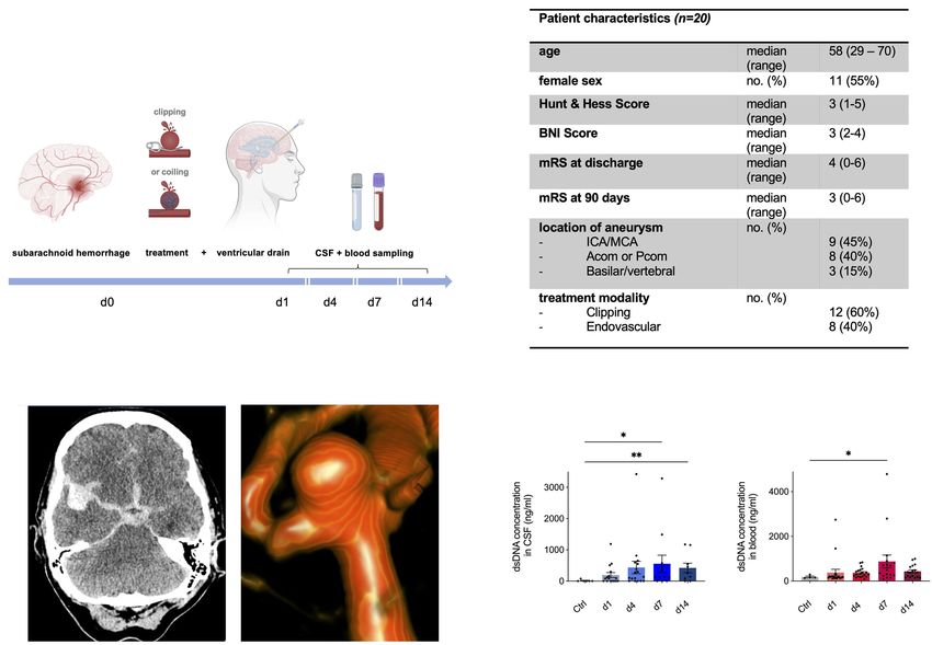
Früh et al.                                                                                                           RNase A Reduces NETs in SAH

drainage, but in this study only ventricular CSF was used.                 (Walberer et al., 2009). The first injection was administered
Blood and CSF samples were immediately placed on ice and                   perioperatively to ensure penetration to the CNS due to
spun down twice at 4°C at 500 g for 5 min, and supernatant                 breakdown of the BBB after SAH surgery. Due to anesthesia
was frozen at −80°C before further analysis. Double-stranded               of mice during operation as well their supine positioning, the
DNA (dsDNA) was quantified in CSF and plasma samples as                    first injection was applied via the sublingual vein. The further
previously published (Ondracek et al., 2020). In detail, samples           injections were repeated every 3 days in the tail vein until
were incubated with Sytox Green (Thermo Fisher), a fluorescent             scarification as previously reported (Fischer et al., 2013). The
dsDNA-binding dye, in a concentration of 1 μM for 5 min.                   total volume of injection was 100 μl at a flow rate of 20 μl/s.
Fluorescence intensities (excitation 480 nm, emission 520 nm)
were measured in 96-well microplates using a Tecan Infinite                Preparation of the Subarachnoid Space for
M200 reader. Values were normalized to a standard curve of                 Immunohistochemistry
dsDNA (Lambda DNA, Thermo Fisher).                                         To preserve the SAS, whole skulls were harvested and kept
                                                                           in an ascending concentration series of sucrose for proper
Animal Experiments                                                         dehydration (4 days at 20%, 4 days at 30%, and 4 days at 40%)
Male C57BL/6-mice were kept at the animal facility at the                  at 4°C and then snap frozen with isopentane. The skulls were
Neuroscience Research Center at Charité University (NWFZ,                  then embedded in Tissue-Tek O.C.T. Compound (4583, Sakura
Berlin). Their age ranged from 8 to 12 weeks and with a weight             Finetek) and carefully cut into 10 μm thin slices with Microm
range of 18–28 g. Animals underwent SAH operation, and sham                Sec35 blades (Termo APD Consumables) using a cryostat
operation (as the control condition), and were sacrificed at               (Thermo Fisher Scientific Inc. Microm HM 560).
three different time points (1, 7, and 14 days) after operation.
A total number of n = 81 mice were included in this study.                 Immunofluorescence
                                                                           Brain sections were blocked for 30 min with 5% bovine serum
Mouse Model of Subarachnoid Hemorrhage                                     albumin (BSA) in Tris-Buffered Saline with 0.05% Tween (TBST)
Subarachnoid hemorrhage was induced with a filament perforation            at room temperature. After that, primary antibodies against
model as described previously (Schneider et al., 2015). Briefly,           citrullinated H3 (rabbit, 1:30, ab5103, Abcam), NeuN (rabbit,
mice were anesthetized with an intraperitoneal ketamine/xylazine           1:200, ABN78, Millipore), and F-actin (Alexa Fluor 488 Phalloidin,
(70 mg resp. 16 mg/kg body weight) injection and placed in supine          1:200, A12379, Thermo Fisher Scientific) diluted in 5% BSA/
position. Starting with a midline neck incision, the carotid artery        TBST were added for incubation at 4°C over night. The next
was exposed and a 5–0 non-absorbable monofilament polypropylene            day, sections were washed three times in 5% BSA/TBST for 10 min,
suture inserted into the external carotid artery in a retrograde           followed by 1.5 h of incubation with the secondary antibodies
manner and advanced into the common carotid artery. In a                   Rhodamine Red-X-conjugated Donkey IgG anti-Mouse (1:200,
next step, the filament invaginated into the internal carotid artery       715-295-151, Dianova) and Alexa Fluor 647-conjugated Donkey
(ICA) and pushed forward to perforate the intracranial arterial            IgG anti-Rabbit IgG (1:200, 711-605-152, Dianova), diluted in
bifurcation. Mice were perfused intracardially with PBS.                   5% BSA/TBST. After secondary antibody incubation, specimens
                                                                           were washed three times in PBS for 5 min and mounted with
MRI                                                                        DAPI Mounting Medium (SCR-038448, Dianova). The sections
All SAH mice received an MRI 24 h after surgery to confirm                 were then imaged with confocal microscopy (Leica DM 6000/
the bleeding and animals who had a stroke or intracranial                  SP5) and analyzed with ImageJ. Amount of NETs was determined
hemorrhage (ICH) were excluded from the experiments                        by calculating the ratio area covered by citH3 over total area
(Figure 1A). The 1H magnetic resonance imaging was performed               covered by phalloidin, NeuN, and DAPI (Supplementary Figure S1).
on a PharmaScan 70/16 U (Bruker Corporation) with a field
strength of 7 Tesla by using the software Paravision 5.1                   Statistical Analysis and Figures
(Bruker Corporation). During the scans, mice received an                   Data were analyzed using GraphPad Prism for statistical analyses
O2/N2O + isoflurane gas anesthesia. The animals’ respiration was           (Graphpad Software, Version 6.0). ANOVA analyses were used
observed with Small Animal Monitoring System (SA Instruments,              to compare multiple, unpaired t-tests for the comparison of
Inc.), while their temperature was maintained via controlled               two groups. The values are displayed as means ± SEs and values
warming blankets. The presence of SAH was validated from                   of p < 0.05 were considered statistically significant. Elements
T2 weighted image. Here, SAH volume was estimated based                    of Figures 1A, 2A,B, 3A were composed with BioRender.com.
on the formula V = (A1 + A 2 +…+ A x )⋅ d, by which An
corresponds to the bleeding area on each coronal MRI and
d corresponds to the MRI slide thickness.                                  RESULTS
RNase Treatment                                                            NETs Are Released After Onset of
Both groups (Sham and SAH animals) received treatment with                 Subarachnoid Bleeding in an in vivo Mouse
RNase A (Thermoscientific EN0531), which was administered                  Model of Subarachnoid Hemorrhage
intravenously (42 μg/kg body weight) and intravenous application           To determine whether NETs are released upon subarachnoid
of saline solution was used as a control, as described previously          hemorrhage in vivo, we utilized a filament perforation model

Frontiers in Physiology | www.frontiersin.org                          3                                 September 2021 | Volume 12 | Article 724611
Früh et al. RNase A Reduces NETs in SAH
B
A
C D
E F
G H
I J
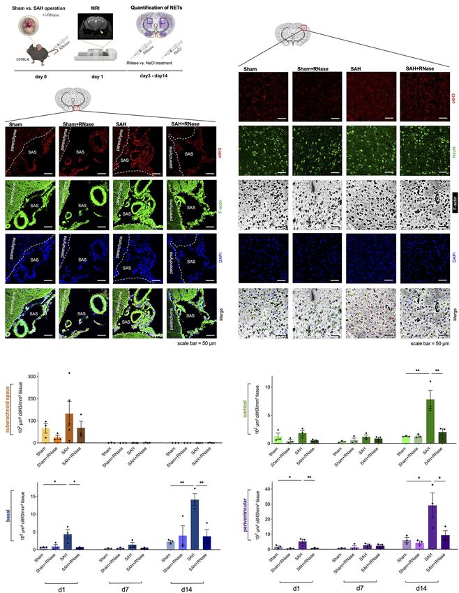
FIGURE 1 | Neutrophil extracellular traps (NETs) are increased in subarachnoid hemorrhage (SAH). (A) The filament perforation model was utilized to induce SAH in
C57BL/6 mice. MRI was done 24 h after SAH onset to verify bleeding and exclude stroke or intracerebral hemorrhage, and NETs were quantified via
immunofluorescence in relation to anatomical localization on three different time points (day 1, 7, and 14). (B) Cumulative data of NETs accumulation in SAH
condition over time. (C,D) Quantification of NETs in subarachnoid space (SAS) with representative immunofluorescence staining of extracellular citH3 (exCitH3),
F-actin, NeuN, and DAPI. Sham, n = 3 for each time point; SAH, n = 5 for each time point. (E–J) Quantification of NETs in basal, cortical, and periventricular
parenchyma with representative immunofluorescence staining of citH3 (citrullinated H3), F-actin, NeuN, and DAPI on day 1, 7, and 14. Sham, n = 3 for each time
point; SAH, n = 3 for each time point. contralat., contralateral; ipsilat., ipsilateral. Scale bar = 50 μm. *p < 0.05, **p < 0.01, ***p < 0.001, and ****p < 0.0001.
to induce subarachnoid hemorrhage in C57BL/6 mice, while marker for NET remnants (Wang et al., 2009). Indeed, increased
Sham operation was performed as a control condition. MRI levels of NET formation were found in SAH with the highest
was conducted 24 h after bleeding to verify SAH and exclude amount in the SAS on the ipsilateral side (where bleeding
other pathologies such as stroke or intracerebral hemorrhage was induced) on day 1 (Figures 1C,D), while after this time
(Figure 1A), and NETs quantified via immunofluorescence point they were essentially not detectable anymore in both
staining using citrullinated Histone (citH3), a known specific control and SAH conditions (Figure 1C). In contrast, in the
Frontiers in Physiology | www.frontiersin.org 4 September 2021 | Volume 12 | Article 724611Früh et al. RNase A Reduces NETs in SAH
A C
B
D E
F G
FIGURE 2 | RNase A treatment reduces accumulation of NETs. (A) Experimental setup for RNase treatment: during operative induction of SAH, mice were treated
with RNase A (42 μg/kg body weight) or sodium chloride (NaCl) as a control condition, and MRI to confirm bleeding. RNase A treatment was repeated every 3 days.
(B) Representative images of triple immunofluorescence staining with citH3, F-actin, and DAPI subarachnoid space of the four subgroups (Sham, n = 3;
Sham + RNase, n = 3; SAH, n = 3; and SAH + RNase, n = 3) on day 1, scale bar = 50 μm. (C) Representative images of quadruple immunofluorescence staining with
citH3, NeuN, F-actin, and DAPI of the corresponding four subgroups in cortical area on day 14, scale bar = 50 μm. (D–G) Quantification of NETs after RNase A
treatment in relation to localization (subarachnoid space, basal, cortical, and periventricular parenchyma) and time course (day 1, 7, and 14). SAS, subarachnoid
space. *p < 0.05 and **p < 0.01.
Frontiers in Physiology | www.frontiersin.org 5 September 2021 | Volume 12 | Article 724611Früh et al. RNase A Reduces NETs in SAH
B
A
C
D
FIGURE 3 | Neutrophil extracellular traps are increased in human cerebrospinal spinal fluid (CSF) and peripheral blood after aneurysmal SAH. (A) Study protocol for
collection of CSF and Ethylene Diaminetetraacetic acid (EDTA) blood; patients with aneurysmal SAH received either microsurgical clipping or endovascular
intervention (coiling) with placement of an extraventricular drain (EVD). In the course of 14 days, CSF and peripheral blood were collected in a prospective manner.
(B) Overview of patient characteristics. (C) Illustrative cases of SAH. CT imaging shows hyperdense blood collection in subarachnoid space and in the right sylvian
fissure of a 55-year old patient who experienced SAH from a right-sided MCA aneurysm who underwent microsurgical clipping (left). 3D reconstruction of angiogram
showing an ACom aneurysm of a 59-year old patient who was further treated via coiling. (D) Double-stranded DNA (dsDNA) concentration in CSF and peripheral
blood of control patients and SAH patients shows peak on day 7 after bleeding onset. Acom, anterior communicating artery; BNI score, barrow neurological institute
score; CSF, cerebrospinal fluid; ICA, internal carotid artery; MCA, middle cerebral artery; mRs, modified Rankin score; and Pcom, posterior communicating artery.
*
p < 0.05 and **p < 0.01.
parenchyma, the NETs showed a gradual increase over time including exRNA, and influence the microglia-specific immune
with the highest density on day 14 in all localizations reaction after SAH. Hence, we treated mice with intravenous
(Figures 1E–J). Interestingly, NET accumulation followed a application of RNase A and quantified NETs in both SAS and
spreading pattern within the parenchyma from basal to cortical parenchyma (Figure 2A). In SAS, RNase A decreased NET
over time, with the most significant increase in periventricular accumulation on day 1, but this was not statistically significant.
localization (Figure 1B). In contrast, in the parenchyma, RNase A reduced NET
accumulation significantly, specifically on day 14, where
Pharmacological Modulation With accumulation of citH3 peaked in all localizations (Figures 2B–G).
RNase A Reduces NET Formation
Since recent evidence suggests that NET-associated RNA is a NETs Are Increased in Both CSF and Blood
physiologically relevant NET component and its formation can of SAH Patients
occur also independently of the canonical NET component Next, we questioned whether this observed NET accumulation
DNA (Herster et al., 2020), we questioned whether RNase A after SAH is also relevant in the human system. To investigate
application can modify NET burden in SAH in vivo. RNase this, CSF and blood samples were collected within the scope
A seemed a feasible therapeutic approach since previous studies of a prospective observational study in aneurysmal SAH patients
in our group showed that RNase A can modulate other DAMPs (Figure 3A), and NETs were measured via quantification of
Frontiers in Physiology | www.frontiersin.org 6 September 2021 | Volume 12 | Article 724611Früh et al. RNase A Reduces NETs in SAH
dsDNA via Sytox Green. Figure 3B shows the baseline to reduce NET burden (Ondracek et al., 2020; Korai et al.,
characteristics of the SAH patients included in the study. Figure 3C 2021), there is mounting evidence that NET-associated RNA is
illustrates imaging from two exemplary cases of SAH patients: a relevant component of NET formation and can occur
on the left side, a CT scan of a 55-year old female patient is independently of DNA (Herster et al., 2020). Based on this
shown with SAH (Hunt&Hess grade II) with a ruptured right- finding and previously published data on the dampening effects
sided middle cerebral artery (MCA) aneurysm who underwent of RNase A on immune cells (Lasch et al., 2020), we questioned
microsurgical clipping; on the right side, a three-dimensional whether RNase A also modifies NET formation. Indeed, our
reconstruction of a ruptured anterior communicating artery data show that RNase A significantly reduces NET formation
(ACom) aneurysm of a 59-year old female (Hunt & Hess grade in all compartments of the brain. This specific effect on the
II) is shown who was further treated with aneurysm coiling. CNS may be explained by the blood brain barrier breakdown
The control patients comprised of patients in which lumbar after SAH with permeability to also bigger proteins such as
puncture was done to rule out meningitis or SAH without Evans Blue (70 kDa; Blecharz-Lang et al., 2018). In our experiments,
evidence of any intracranial pathology. Interestingly, dsDNA was we used a 13.7 kDa pancreatic RNase A for pre- and postoperative
significantly increased in both CSF and peripheral blood of SAH intravenous injections. These finding are particular compelling
patients compared to the control group, with a gradual increase since RNAse therapeutics have already been used to some degree
over time, peaking in both compartments on day 7 (Figure 3D). in clinical trials (Mikulski et al., 2002; Ardelt et al., 2007; Chang
et al., 2010; Squiquera et al., 2017). Therefore, it is tempting
to speculate that RNAse may be an accessible interesting potential
DISCUSSION therapeutic strategy for treatment of the underlying immune
reaction after SAH. However, the influence of i.v. application
Aneurysmal SAH remains a devastating pathology with high of RNAse on physiological processes such as regulation of cerebral
morbidity and mortality, and attempts to reduce secondary brain blood flow was not investigated in this study and should
damage have been made for decades, yet the exact pathomechanism be addressed in further studies.
contributing to the long-term damage is unclear (Black, 1986; The exact molecular mechanisms of NET formation are not
Peterson et al., 1990; Mathiesen et al., 1997; Sheehan et al., fully understood. SAH promotes generation of reactive oxygen
1999). Recent data suggests involvement of the immune system species (ROS; Ayer and Zhang, 2008), that have also been described
attributable to secondary brain damage, specifically through an as triggers of NET formation (Erpenbeck and Schön, 2017).
outside-in activation of neutrophil recruitment to endothelium, Therefore, one may pose the hypothesis that ROS-activation in
contributing to microglia activation and neuronal apoptosis the setting of SAH contributes NET accumulation, which then
(Schneider et al., 2015; Atangana et al., 2017). Among the may promote microglial activation leading to the activation of
classical immune defense mechanisms of neutrophils, consisting neuroinflammatory cascades. Furthermore, recent studies have
of engulfment of microbes and secretion of anti-microbials, described that NET formation in the context of SAH increases
recent data pinpoints to a novel function of neutrophils as part levels of cytokines such as IL-1β, IL-6, and TNF-α (Zeng et al., 2021).
of the innate immune response – the formation of NETs to Furthermore, in order to investigate whether NET burden is
kill extracellular pathogens (Brinkmann et al., 2004). also relevant in the human system, we measured circulating
In this study, we sought to investigate the potential role of NET abundance via the surrogate marker dsDNA and show
NET formation in SAH and describe the spatiotemporal patterns that increased levels are found in both CSF and peripheral blood
of NET accumulation after SAH. We demonstrate that in the of SAH patients. Here, we observed a peak of dsDNA 7 days
acute setting, a direct flood of NET formation occurs in the after the bleeding event, supporting the in vivo data from our
ipsilateral subarachnoid space (SAS), while in the parenchyma, SAH mouse model. Our findings are in line a recent study of
NET levels increase gradually over time in basal, cortical, and Zeng et al. (2021) showed that a significant increase of citH3 in
periventricular compartments distant to the region of bleeding. patients suffering SAH after 24 h of the bleeding event, which
These findings are supported by a recent study reporting NET correlated with the clinical Hunt and Hess score. Additionally,
accumulation after SAH as well as their involvement in we showed for the first time the further temporal dynamics of
neuroinflammatory events, albeit the time point of the peak NETs and their involvement in CSF in humans. The postponed
of NETs differed to some degree (Zeng et al., 2021). Interestingly, peak of NET burden potentially indicates a therapeutic window
in the study by Zeng et al. (2021), inhibition of NET formation after the bleeding event to attenuate secondary brain damage
via the PAD4 antagonist GSK484 as well as DNase I inhibited after SAH. The accumulation of NETs in the brain parenchyma
NET-associated neuroinflammation. Our data show that the also raises intriguing questions of its origin. Increasing levels
quantitatively most relevant NET formation occurred 14 days of neutrophils in the CSF after SAH have also been associated
after SAH specifically in periventricular localizations. This is with development of vasospasms (Provencio et al., 2010). Therefore,
intriguing as it raises the question whether NETs may also neutrophils may migrate in the brain parenchyma after SAH
be involved in CSF hydrodynamics after aneurysmal SAH, and produce NETs in situ as already described in other studies
especially since post-hemorrhagic hydrocephalus is a common in a model of ischemic stroke (Kang et al., 2020).
complication after SAH (Germanwala et al., 2010). Our study is limited by a small sample size, investigations
Moreover, while a direct inhibition of NETs via DNase has on the association of NET burden and clinical characteristics,
been postulated as a potential mechanistic treatment strategy and exploratory studies on cell-specific mechanism by which
Frontiers in Physiology | www.frontiersin.org 7 September 2021 | Volume 12 | Article 724611Früh et al. RNase A Reduces NETs in SAH
NETs may affect further inflammatory events in SAH. Albeit Charité – Universitätsmedizin, Berlin (Germany). The patients/
the range of the SAH patients in our small study cohort varies participants provided their written informed consent to participate
between 29 and 70, the median age of our study group in the in this study. The animal study was reviewed and approved by
patients is 58, which is fairly representative of typical age of Landesamt für Gesundheit und Soziales (LaGeSo) Berlin
aneurysm rupture in large cohorts (Rinkel et al., 1998; Shea (Germany).
et al., 2007; Jordan et al., 2009). In order to investigate potential
aging effects, further subgroup analyses on young and old mice
with larger study numbers are necessary. As we analyzed AUTHOR CONTRIBUTIONS
exclusively male mice, sex-related effects in the animal experiments
cannot be excluded. This study also did not investigate the KT, FS, AF, SL, and RX conducted the experiments and analyzed
impact of RNAse treatment on humans. As potential the data. RX, AF, and PV designed the study. All authors
neuroprotective effects of RNase in SAH patients are of pressing contributed to the article and approved the submitted version.
interest, a clinical study on the effect of RNase treatment in
hemorrhagic stroke patients are planned for the future.
In summary, our data reveal the spatiotemporal dynamics FUNDING
of NET accumulation after SAH in vivo with evidence for a
gradual increase of NET formation over time, both in a SAH RX was supported by the BIH-Charité Clinician Scientist
mouse model as well as in patients suffering aneurysmal SAH. Program funded by the Charité – Universitätsmedizin Berlin
Intriguingly, intravenous RNase A application abrogates NET and the Berlin Institute of Health. We acknowledge support
burden in the parenchyma, underpinning a potential role in from the German Research Foundation (DFG) and the Open
of RNase in the innate immune response after SAH. Further Access Publication Fund of Charité – Universitätsmedizin Berlin.
studies are needed to fully elucidate the exact nature of NET KT is supported by the Berlin Institute of Health (BIH) Research
formation and related immune cell-specific changes of RNase Stipend as well as the Sonnenfeld Foundation.
application after SAH.
ACKNOWLEDGMENTS
DATA AVAILABILITY STATEMENT
This paper is dedicated to the work of our dear colleague and
The raw data supporting the conclusions of this article will friend Klaus T. Preissner.
be made available by the authors, without undue reservation.
SUPPLEMENTARY MATERIAL
ETHICS STATEMENT
The Supplementary Material for this article can be found online
The studies involving human participants were at: https://www.frontiersin.org/articles/10.3389/fphys.2021.724611/
reviewed and approved by Ethikkommission Charité, full#supplementary-material
REFERENCES Borregaard, N. (2010). Neutrophils, from marrow to microbes. Immunity 33,
657–670. doi: 10.1016/j.immuni.2010.11.011
Ardelt, B., Juan, G., Burfeind, P., Salomon, T., Wu, J. M., Hsieh, T. C., Brinkmann, V., Reichard, U., Goosmann, C., Fauler, B., Uhlemann, Y., Weiss, D. S.,
et al. (2007). Onconase, an anti-tumor ribonuclease suppresses et al. (2004). Neutrophil extracellular traps kill bacteria. Science 303, 1532–1535.
intracellular oxidative stress. Int. J. Oncol. 31, 663–669. doi: 10.3892/ doi: 10.1126/science.1092385
ijo.31.3.663 Chang, C.-H., Gupta, P., Michel, R., Loo, M., Wang, Y., Cardillo, T. M.,
Atangana, E., Schneider, U. C., Blecharz, K., Magrini, S., Wagner, J., et al. (2010). Ranpirnase (frog rnase) targeted with a humanized,
Nieminen-Kelha, M., et al. (2017). Intravascular inflammation triggers internalizing, anti–trop-2 antibody has potent cytotoxicity against diverse
intracerebral activated microglia and contributes to secondary brain injury epithelial cancer cells. Mol. Cancer Ther. 9, 2276–2286. doi: 10.1158/
after experimental subarachnoid hemorrhage (eSAH). Transl. Stroke Res. 8, 1535-7163.MCT-10-0338
144–156. doi: 10.1007/s12975-016-0485-3 Erpenbeck, L., and Schön, M. (2017). Neutrophil extracellular traps:
Ayer, R., and Zhang, J. (2008). “Oxidative stress in subarachnoid haemorrhage: protagonists of cancer progression? Oncogene 36, 2483–2490. doi: 10.1038/
significance in acute brain injury and vasospasm,” in Cerebral Vasospasm. onc.2016.406
ed. T. Kiris (Springer), 33–41. Etminan, N., Chang, H.-S., Hackenberg, K., De Rooij, N. K., Vergouwen, M. D.,
Black, P. M. (1986). Hydrocephalus and vasospasm after subarachnoid hemorrhage Rinkel, G. J., et al. (2019). Worldwide incidence of aneurysmal subarachnoid
from ruptured intracranial aneurysms. Neurosurgery 18, 12–16. doi: hemorrhage according to region, time period, blood pressure, and smoking
10.1227/00006123-198601000-00003 prevalence in the population: a systematic review and meta-analysis. JAMA
Blecharz-Lang, K. G., Wagner, J., Fries, A., Nieminen-Kelha, M., Rosner, J., Neurol. 76, 588–597. doi: 10.1001/jamaneurol.2019.0006
Schneider, U. C., et al. (2018). Interleukin 6-mediated endothelial barrier Fischer, S., Gesierich, S., Griemert, B., Schanzer, A., Acker, T., Augustin, H. G.,
disturbances can be attenuated by blockade of the IL6 receptor expressed et al. (2013). Extracellular RNA liberates tumor necrosis factor-alpha to
in brain microvascular endothelial cells. Transl. Stroke Res. 9, 631–642. doi: promote tumor cell trafficking and progression. Cancer Res. 73, 5080–5089.
10.1007/s12975-018-0614-2 doi: 10.1158/0008-5472.CAN-12-4657
Frontiers in Physiology | www.frontiersin.org 8 September 2021 | Volume 12 | Article 724611Früh et al. RNase A Reduces NETs in SAH Fischer, S., Nishio, M., Dadkhahi, S., Gansler, J., Saffarzadeh, M., Shibamiyama, A., inflammation and infection. Front. Cell Dev. Biol. 8:619221. doi: 10.3389/ et al. (2011). Expression and localisation of vascular ribonucleases in endothelial fcell.2020.619221 cells. Thromb. Haemost. 105, 345–355. doi: 10.1160/TH10-06-0345 Provencio, J. J., Fu, X., Siu, A., Rasmussen, P. A., Hazen, S. L., and Ransohoff, R. M. García-Culebras, A., Durán-Laforet, V., Peña-Martínez, C., Moraga, A., (2010). CSF neutrophils are implicated in the development of vasospasm Ballesteros, I., Cuartero, M. I., et al. (2019). Role of TLR4 (toll-like receptor 4) in subarachnoid hemorrhage. Neurocrit. Care 12, 244–251. doi: 10.1007/ in N1/N2 neutrophil programming after stroke. Stroke 50, 2922–2932. doi: s12028-009-9308-7 10.1161/STROKEAHA.119.025085 Rinkel, G. J., Djibuti, M., Algra, A., and Van Gijn, J. (1998). Prevalence and Germanwala, A. V., Huang, J., and Tamargo, R. J. (2010). Hydrocephalus after risk of rupture of intracranial aneurysms: a systematic review. Stroke 29, aneurysmal subarachnoid hemorrhage. Neurosurg. Clin. N. Am. 21, 263–270. 251–256. doi: 10.1161/01.STR.29.1.251 doi: 10.1016/j.nec.2009.10.013 Schertz, M., Mehdaoui, H., Hamlat, A., Piotin, M., Banydeen, R., and Mejdoubi, M. Gris, T., Laplante, P., Thebault, P., Cayrol, R., Najjar, A., Joannette-Pilon, B., (2016). Incidence and mortality of spontaneous subarachnoid hemorrhage et al. (2019). Innate immunity activation in the early brain injury period in Martinique. PLoS One 11:e0155945. doi: 10.1371/journal.pone.0155945 following subarachnoid hemorrhage. J. Neuroinflammation 16:253. doi: 10.1186/ Schneider, U. C., Davids, A. M., Brandenburg, S., Muller, A., Elke, A., Magrini, S., s12974-019-1629-7 et al. (2015). Microglia inflict delayed brain injury after subarachnoid Herster, F., Bittner, Z., Archer, N. K., Dickhofer, S., Eisel, D., Eigenbrod, T., hemorrhage. Acta Neuropathol. 130, 215–231. doi: 10.1007/s00401-015-1440-1 et al. (2020). Neutrophil extracellular trap-associated RNA and LL37 enable Shea, A. M., Reed, S. D., Curtis, L. H., Alexander, M. J., Villani, J. J., and self-amplifying inflammation in psoriasis. Nat. Commun. 11:105. doi: 10.1038/ Schulman, K. A. (2007). Characteristics of nontraumatic subarachnoid s41467-019-13756-4 hemorrhage in the United States in 2003. Neurosurgery 61, 1131–1138. doi: Jordan, L. C., Johnston, S. C., Wu, Y. W., Sidney, S., and Fullerton, H. J. (2009). 10.1227/01.neu.0000306090.30517.ae The importance of cerebral aneurysms in childhood hemorrhagic stroke: a Sheehan, J. P., Polin, R. S., Sheehan, J. M., Baskaya, M. K., and Kassell, N. F. population-based study. Stroke 40, 400–405. doi: 10.1161/STROKEAHA.108.518761 (1999). Factors associated with hydrocephalus after aneurysmal subarachnoid Kang, L., Yu, H., Yang, X., Zhu, Y., Bai, X., Wang, R., et al. (2020). Neutrophil hemorrhage. Neurosurgery 45, 1120–1127. doi: 10.1097/00006123-199911000-00021 extracellular traps released by neutrophils impair revascularization and vascular Squiquera, L., Taxman, D. J., Brendle, S. A., Torres, R., Sulley, J., Hodge, T., remodeling after stroke. Nat. Commun. 11:2488. doi: 10.1038/s41467-020-16191-y et al. (2017). Ranpirnase eradicates human papillomavirus in cultured cells Korai, M., Purcell, J., Kamio, Y., Mitsui, K., Furukawa, H., Yokosuka, K., et al. (2021). and heals anogenital warts in a phase I study. Antivir. Ther. 22, 247–255. Neutrophil extracellular traps promote the development of intracranial aneurysm doi: 10.3851/IMP3133 rupture. Hypertension 77, 2084–2093. doi: 10.1161/HYPERTENSIONAHA.120.16252 Tielking, K., Fischer, S., Preissner, K. T., Vajkoczy, P., and Xu, R. (2019). Lasch, M., Kumaraswami, K., Nasiscionyte, S., Kircher, S., Van Den Heuvel, D., Extracellular RNA in central nervous system pathologies. Front. Mol. Neurosci. Meister, S., et al. (2020). RNase A treatment interferes with leukocyte recruitment, 12:254. doi: 10.3389/fnmol.2019.00254 neutrophil extracellular trap formation, and angiogenesis in ischemic muscle Van Lieshout, J. H., Dibué-Adjei, M., Cornelius, J. F., Slotty, P. J., Schneider, T., tissue. Front. Physiol. 11:576736. doi: 10.3389/fphys.2020.576736 Restin, T., et al. (2018). An introduction to the pathophysiology of aneurysmal Lawton, M. T., and Vates, G. E. (2017). Subarachnoid hemorrhage. N. Engl. J. subarachnoid hemorrhage. Neurosurg. Rev. 41, 917–930. doi: 10.1007/ Med. 377, 257–266. doi: 10.1056/NEJMcp1605827 s10143-017-0827-y Liu, Y.-W., Li, S., and Dai, S.-S. (2018). Neutrophils in traumatic brain injury Walberer, M., Tschernatsch, M., Fischer, S., Ritschel, N., Volk, K., Friedrich, C., (TBI): friend or foe? J. Neuroinflammation 15:146. doi: 10.1186/s12974-018-1173-x et al. (2009). RNase therapy assessed by magnetic resonance imaging reduces Lucke-Wold, B. P., Logsdon, A. F., Manoranjan, B., Turner, R. C., Mcconnell, E., cerebral edema and infarction size in acute stroke. Curr. Neurovasc. Res. 6, Vates, G. E., et al. (2016). Aneurysmal subarachnoid hemorrhage and 12–19. doi: 10.2174/156720209787466037 neuroinflammation: a comprehensive review. Int. J. Mol. Sci. 17:497. doi: Wang, Y., Li, M., Stadler, S., Correll, S., Li, P., Wang, D., et al. (2009). Histone 10.3390/ijms17040497 hypercitrullination mediates chromatin decondensation and neutrophil Macdonald, R. L. (2014). Delayed neurological deterioration after subarachnoid extracellular trap formation. J. Cell Biol. 184, 205–213. doi: 10.1083/jcb.200806072 haemorrhage. Nat. Rev. Neurol. 10, 44–58. doi: 10.1038/nrneurol.2013.246 Zeng, H., Fu, X., Cai, J., Sun, C., Yu, M., Peng, Y., et al. (2021). Neutrophil Macdonald, R. L., and Schweizer, T. A. (2017). Spontaneous subarachnoid extracellular traps may be a potential target for treating early brain injury haemorrhage. Lancet 389, 655–666. doi: 10.1016/S0140-6736(16)30668-7 in subarachnoid hemorrhage. Transl. Stroke Res. doi: 10.1007/s12975-021-00909-1 Mathiesen, T., Edner, G., Ulfarsson, E., and Andersson, B. (1997). Cerebrospinal [Epub ahead of print] fluid interleukin-1 receptor antagonist and tumor necrosis factor-alpha following Zhang, Z., Fang, Y., Lenahan, C., and Chen, S. (2020). The role of immune subarachnoid hemorrhage. J. Neurosurg. 87, 215–220. doi: 10.3171/jns.1997.87.2.0215 inflammation in aneurysmal subarachnoid hemorrhage. Exp. Neurol. Mikulski, S. M., Costanzi, J. J., Vogelzang, N. J., Mccachren, S., Taub, R. N., 336:113535. doi: 10.1016/j.expneurol.2020.113535 Chun, H., et al. (2002). Phase II trial of a single weekly intravenous dose of ranpirnase in patients with unresectable malignant mesothelioma. J. Clin. Conflict of Interest: The authors declare that the research was conducted in Oncol. 20, 274–281. doi: 10.1200/JCO.2002.20.1.274 the absence of any commercial or financial relationships that could be construed Niemiec, M. J., De Samber, B., Garrevoet, J., Vergucht, E., Vekemans, B., as a potential conflict of interest. De Rycke, R., et al. (2015). Trace element landscape of resting and activated human neutrophils on the sub-micrometer level. Metallomics 7, 996–1010. Publisher’s Note: All claims expressed in this article are solely those of the doi: 10.1039/C4MT00346B authors and do not necessarily represent those of their affiliated organizations, Ondracek, A., Hofbauer, T., Wurm, R., Arfsten, H., Seidl, V., Früh, A., et al. or those of the publisher, the editors and the reviewers. Any product that may (2020). Imbalance between plasma double-stranded DNA and be evaluated in this article, or claim that may be made by its manufacturer, is deoxyribonuclease activity predicts mortality after out-of-hospital cardiac not guaranteed or endorsed by the publisher. arrest. Resuscitation 151, 26–32. doi: 10.1016/j.resuscitation.2020.03.006 Papayannopoulos, V. (2018). Neutrophil extracellular traps in immunity and Copyright © 2021 Früh, Tielking, Schoknecht, Liu, Schneider, Fischer, Vajkoczy and disease. Nat. Rev. Immunol. 18, 134–147. doi: 10.1038/nri.2017.105 Xu. This is an open-access article distributed under the terms of the Creative Peterson, J. W., Kwun, B. D., Hackett, J. D., and Zervas, N. T. (1990). The Commons Attribution License (CC BY). The use, distribution or reproduction in role of inflammation in experimental cerebral vasospasm. J. Neurosurg. 72, other forums is permitted, provided the original author(s) and the copyright owner(s) 767–774. doi: 10.3171/jns.1990.72.5.0767 are credited and that the original publication in this journal is cited, in accordance Preissner, K. T., Fischer, S., and Deindl, E. (2020). Extracellular RNA as a with accepted academic practice. No use, distribution or reproduction is permitted versatile DAMP and alarm signal that influences leukocyte recruitment in which does not comply with these terms. Frontiers in Physiology | www.frontiersin.org 9 September 2021 | Volume 12 | Article 724611
You can also read