Penicillin Treatment Failure in Rabbit Syphilis Due to the Persistence of Treponemes (Treponema paraluisleporidarum Ecovar Cuniculus) in the Focus ...
←
→
Page content transcription
If your browser does not render page correctly, please read the page content below
ORIGINAL RESEARCH
                                                                                                                                                 published: 17 June 2021
                                                                                                                                         doi: 10.3389/fvets.2021.675631

                                             Penicillin Treatment Failure in Rabbit
                                             Syphilis Due to the Persistence of
                                             Treponemes (Treponema
                                             paraluisleporidarum Ecovar
                                             Cuniculus) in the Focus of Infection
                                             Vladimír Jekl 1,2† , Markéta Nováková 3,4† , Edita Jeklová 5 , Petra Pospíšilová 3 , Jitka Křenová 3 ,
                                             Martin Faldyna 5 , Miša Škorič 6 and David Šmajs 3*
                                             1
                                               Jekl and Hauptman Veterinary Clinic, Brno, Czechia, 2 Department of Pharmacology and Pharmacy, Faculty of Veterinary
                                             Medicine, Veterinary University Brno, Brno, Czechia, 3 Department of Biology, Faculty of Medicine, Masaryk University, Brno,
                                             Czechia, 4 Department of Botany and Zoology, Faculty of Science, Masaryk University, Brno, Czechia, 5 Department of
                            Edited by:       Infectious Diseases and Preventive Medicine, Veterinary Research Institute, Brno, Czechia, 6 Department of Pathological
                               Min Yue,      Morphology and Parasitology, Faculty of Veterinary Medicine, Veterinary University Brno, Brno, Czechia
              Zhejiang University, China

                        Reviewed by:
                           Fang Wang,
                                             Rabbit venereal spirochetosis, a disease caused by Treponema paraluisleporidarum
      Jiangsu Academy of Agricultural        ecovar Cuniculus (TPeC), affects both wild and pet rabbits, and is transmitted sexually
                      Sciences, China        and via direct contact among animals. Treatment of syphilis in pet rabbits requires
                           Alaa Sewid,
          The University of Tennessee,       administration of antibiotics, including penicillin G, chloramphenicol, or fluoroquinolones.
                         United States       The aim of this work was to elucidate the cause of penicillin treatment failure in rabbit
                   *Correspondence:          syphilis in a pet rabbit treated in Brno, Czech Republic, and to assess the phylogenetic
                         David Šmajs
                  dsmajs@med.muni.cz
                                             relatedness of the agent to previously characterized pathogenic treponemes. Following
                                             amputation of the infected digits, the second round of penicillin treatment using the
 † These   authors share first authorship
                                             same dosage and application route resulted in the disappearance of clinical symptoms
                    Specialty section:
                                             within a period of two weeks. The bacterium was successfully isolated from the claws,
         This article was submitted to       propagated in three experimental rabbits, and the resulting TPeC strain was designated
        Veterinary Infectious Diseases,
                                             as Cz-2020. Analysis of four genetic loci revealed that the Cz-2020 strain was similar but
                a section of the journal
        Frontiers in Veterinary Science      also clearly distinct from the only TPeC strain, which had been characterized in detail to
             Received: 03 March 2021         date, i.e., the Cuniculi A strain, which was isolated in North America. The strain Cz-2020
              Accepted: 25 May 2021          represents the first available viable TPeC strain of European origin. DNA sequences
             Published: 17 June 2021
                                             encoding five penicillin-binding proteins of the strain Cz-2020 were compared to those
                               Citation:
         Jekl V, Nováková M, Jeklová E,
                                             of Cuniculi A, which is known to be sensitive to penicillin. The sequences differed in six
 Pospíšilová P, Křenová J, Faldyna M,       nucleotides resulting in single amino acid changes in Penicillin-binding protein 1, 2, and
Škorič M and Šmajs D (2021) Penicillin
                                             3. Since the second round of treatment was successful, we conclude that the penicillin
   Treatment Failure in Rabbit Syphilis
Due to the Persistence of Treponemes         treatment failure in the first round resulted from the presence of infection foci in claws
       (Treponema paraluisleporidarum        where treponemes persisted.
     Ecovar Cuniculus) in the Focus of
  Infection. Front. Vet. Sci. 8:675631.      Keywords: rabbit, syphilis, Oryctolagus cuniculus, penicillin, sexually transmitted diseases, in vivo propagation,
       doi: 10.3389/fvets.2021.675631        dermatitis

Frontiers in Veterinary Science | www.frontiersin.org                               1                                              June 2021 | Volume 8 | Article 675631
Jekl et al. Rabbit Syphilis Penicillin Treatment Failure
INTRODUCTION The first difference includes the number of arp gene (TP0433)
repetitions. While there appear to be 25 repeats of a 60-bp long
Rabbit venereal spirochetosis, caused by Treponema segment in the arp gene in Cuniculi H, only 21 such repeats
paraluisleporidarum ecovar Cuniculus (TPeC), is transmitted have been described in the Cuniculi A strain (18). The second
sexually and via direct contact among infected animals (1). difference appears to be a deletion of TP0618 in Cuniculi M
While most cases of rabbit venereal spirochetosis are usually and Cuniculi H but not in Cuniculi A (19). To date, only a
sporadic, local epidemics are known to occur in commercial single strain (Cuniculi A) has been characterized at the genome
rabbit farms (2). TPeC infection commonly results in erythema, level (12, 16).
edema, and/or crusting ulcers in the genitoanal and/or The aim of this study was to determine the cause of
orofacial regions (3). Treatment of syphilis in pet rabbits penicillin treatment failure in rabbit syphilis in a pet rabbit,
consists of administration of penicillin G, chloramphenicol, or determine the phylogenetic position of the agent within the T.
fluoroquinolones (4). Penicillin is recommended for treatment pallidum/T. paraluisleporidarum cluster and establish a viable
of syphilis in both animals and humans (5, 6) and thus any isolate for further genomic or immunologic analyses. The
indications of resistance to this drug should be documented. isolate, designated as Cz-2020, is the first TPeC isolate of
The infecting organism causing rabbit syphilis in farmed European origin.
rabbits was initially described as Spirochaeta paralues-cuniculi
(7). Later, rabbit treponemal pathogens were referred to as
Treponema cuniculi or T. paraluiscuniculi (2, 8). In 2013,
Treponema paraluiscuniculi was reclassified as TPeC (9) and
MATERIALS AND METHODS
similar, widely spread treponemes causing infections in free A Case of Rabbit Syphilis in a Pet Rabbit
living hares (Lepus europaeus and Lepus timidus) have been A 10-month-old sexually intact female pet rabbit with
identified, i.e., T. paraluisleporidarum ecovar Lepus (TPeL) [e.g., dermatological changes in the nasal area was firstly brought
(9–11)]. Although being the closest known relatives to the to the clinic in December 2019. The lesions had been present
trio of human pathogenic T. pallidum subspecies, i.e., pallidum for more than 5 months and had already been unsuccessfully
(TPA), pertenue (TPE), and endemicum (TEN), the causative treated with crusty skin debridement and antiparasitic therapy
agents of syphilis, yaws and bejel, respectively (10), treponemes (two consecutive treatments of 7 days each, with subcutaneous
infecting lagomorphs are not pathogenic for humans (11). ivermectin 0.2 mg/kg of body weight). The animal was kept
There is, however, an overall high level of clinical manifestation in a single household, as such, cross contamination could
similarities among TPeC and human treponematoses, such as be excluded.
crusty sores in the genitoanal and orofacial areas resembling A clinical examination revealed the presence of severe crusty
human yaws or the sexual transmission route typical for skin changes with areas of proliferation in the area dorsal to the
human syphilis (12). Rabbits are susceptible not only to nostrils. Similar skin changes were observed on the skin around
rabbit (TPeC) but also to hare (TPeL) and human syphilis. the claws and on the skin of the toes on the right hindlimb;
Hence, laboratory rabbits played a fundamental role in no lesions were seen in the anogenital area. All lesions were
propagation and diagnostics of human pathogenic treponemes scaly and white. Skin scrapings and dermatophyte cultures were
for decades [e.g., (13, 14)]. While recent advance in in negative for parasites and dermatophytes. Since the skin lesions
vitro cultivation of TPA could lead to decreased need of were consistent with a diagnosis of rabbit syphilis (4), 14 doses of
animals for propagation, rabbit infection model will probably penicillin G (benzathine/procaine penicillin, 60,000 IU/kg) were
remain vital for a variety of biological experiments with administered intramuscularly every 12 h for 7 days. In February
treponemes (15). 2020, at the 8-week follow-up of the first penicillin treatment,
TPeC shows 98.1% identity to TPA and TPE on the genetic the nasal lesions had completely healed, however, the lesions
level (12, 16, 17) with the variability accumulated in several on the claws and on the toes persisted (Figures 1A,B), which
genes (e.g., tpr paralogous genes, and TP0136, TP0326, TP0488, indicated penicillin treatment failure. No other pathologies were
TP0548 loci). A part of these variable genes was found to be found. Clinical material sampled from the claws was used for
inactivated in TPeC strain Cuniculi A, which could explain the detection of the pathogen using PCR, dark field microscopy,
loss of infectivity to humans, and these genes represent promising histopathological staining, and experimental inoculation of
candidates for virulence factors of TPA (12). Hence, availability rabbits. After sampling, the rabbit claws, as well as the last digit
of higher number of treponemal isolates from lagomorphs of the right hindfoot, were amputated under general anesthesia
would be valuable to indentify the true genetic diversity of this due to severe claw changes, including fractures, defective growth,
bacterial species. and white scaly areas. At the same time, benzathine/procaine
The currently known TPeC isolates/strains include Cuniculi penicillin was administered for a second time, just as it had
A, Cuniculi H, and Cuniculi M, coming either from the been during the first round of treatment. A histopathological
Center for Disease Control and Prevention (the USA), i.e., examination of the amputated tissues revealed the presence of
Cuniculi A, or from a laboratory of S. Lukehart (University chronic purulent paronychia with reactive fibroblastic changes.
of Washington, Seattle, Washington, the USA), i.e., Cuniculi Follow-ups at the veterinary clinic at two weeks, 3 and 8 months
H and M. Minimal data on the genetic diversity within TPeC after the second round of antibiotics, found no disease symptoms,
are based on the three strains isolated in North America. which indicated successful penicillin treatment.
Frontiers in Veterinary Science | www.frontiersin.org 2 June 2021 | Volume 8 | Article 675631Jekl et al. Rabbit Syphilis Penicillin Treatment Failure
Experimental Inoculation
One male New Zealand White rabbit was inoculated in both
testes, within 2 h of sampling the pet rabbit, with 500 µl
of the suspension extracted from claws per one testicle. The
animal was clinically inspected daily, and the inflammatory
response (i.e., redness, swelling, and induration of the testes)
was monitored. The animal was placed under general anesthesia
using medetomidine (0.2 mg/kg IM; Cepetor, Werfft, Germany)
and ketamine (15 mg/kg i.m.; Narkamon, Bioveta, Czech
Republic), and the testes were aseptically removed, sliced
on Petri dishes, each testicle was placed in a 50-ml tube
containing 5 ml of sterile PBS and spirochetes were extricated
by agitation for 25 min at 100 cycles/min according to Lukehart
and Marra (20). The animal was then euthanized using 3 ml
IV of the T-61 solution (Intervet International, Netherlands).
Laboratory rabbits of the second and third passages were
intratesticularly inoculated with suspensions from both testes
obtained from the precedent passage (Supplementary Table 1)
and daily monitored. Viability and number of treponemes
per ml recovered from testicles was assessed by dark field
microscopy. Remaining testes suspensions were placed in 1.8 ml
Nunc cryogenic tubes [Thermo Fisher Scientific (Waltham, MA,
USA)] and stored at −80◦ C. Housing of all three experimental
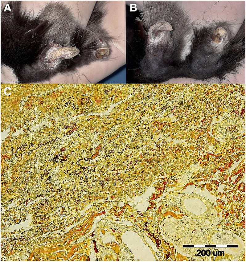
FIGURE 1 | Skin lesions on the right hindlimb of a pet rabbit with syphilis. (A,
animals was done in accordance with the Branch Commission
left) Detailed view of healthy (2nd) and syphilis affected (3rd) nail and toe. (B,
right) Note the fractured and deformed claws/nails on both the 3rd and 4th for Animal Welfare of the Ministry of Agriculture of the Czech
toes. White scaly lesions were seen on both affected claws/nails, the Republic (MZe 2085).
eponychium, and the distal parts of terminal phalanges. (C, bottom)
Wartin-Starry silver stain highlights spirochetes in dermis, magnification 200×.
Histopathological examination of the lesion in the area of claw and surrounding DNA Isolation
connective dermal tissue showed presence of proliferating fibrovascular tissue, DNA was isolated from 200 µl of sample material in PBS (i.e.,
moderate mixed inflammatory infiltrate with predominance of lymphocytes, material obtained from the infected claw of the pet rabbit or
plasma cells, and macrophages, with admixture of lesser number of
neutrophils. There was superficial erosion of epithelium, areas of serocellular
the testes of the laboratory rabbits) as described in Grillová
crusts, focally small hemorrhages and deposits of hemosiderin in dermis. et al. (21), using QIAamp DNA Blood Mini Kit (Qiagen, Hilden,
There were no visible bacteria seen in tissue sections stained with HE. Silver Germany). Isolation was performed within four hours of the
staining method (Warthin-Starry) revealed presence of numerous typical spiral samples being received. DNA samples were stored at −20◦ C prior
and thread-like organisms in epidermis and dermis within the area of to PCR analysis.
inflammatory reaction.
PCR Detection
The presence of treponemal DNA was examined using nested
Collection of Clinical Samples, Dark Field PCR detection of TP0105 (polA). A list of all primers is shown
Microscopy, and Histological Staining in Supplementary Tables 2, 3 (22–24). In the first step of the
Clinical samples were collected from the pet rabbit at the Jekl nested PCR, the final volume of the mixture (25 µl) contained
and Hauptman Veterinary Clinic, Brno, the Czech Republic, in 1 µl of DNA, 16.3 µl of water, 5 µl of GXL buffer, 0.095 µl
February 2020. The clinical material was taken from a crusty of each primer (100 pmol/µl), and 0.5 µl of Prime STAR GXL
lesion on a claw; it was crushed on a Petri dish, placed in a sterile polymerase (Takara Bio Europe, France). The touchdown PCR
1.5 ml Eppendorf tube, and immediately transported, on ice, to was performed at 94◦ C for 1 min; 8 cycles: 98◦ C for 10 s, 68◦ C
the animal facility at the Veterinary Research Institute, Brno, for 15 s (−1.0◦ C per each cycle from cycle 2–8), 68◦ C for 1 min
Czech Republic. 1.2 ml of sterile PBS was added and agitated for and 45 s; 35 cycles: 98◦ C for 10 s, 61◦ C for 15 s, 68◦ C for 1 min
25 min at 100 cycles/min to extricate spirochetes according to and 45 s; and 68◦ C for 7 min. The mixture for the second step
Lukehart and Marra (20). Five microliters of the suspension were was composed of 1 µl of product from the first step, 20.5 µl of
used for dark field microscopy to confirm presence and viability water, 2.5 µl of ThermoPol Reaction buffer, 0.5 µl of a 10 mM
of spirochetes, and to assess their number per ml. dNTP mixture, 0.25 µl of each primer (100 pmol/l), 0.1 µl of Taq
A part of the amputated claws was fixed in 10% buffered polymerase (5,000 U/ml; New England BioLabs, Ipswich, MA).
neutral formalin, dehydrated, and embedded in paraffin wax. PCR was performed at 94◦ C for 1 min; 40 cycles: 94◦ C for 30 s,
Tissue sections, each prepared on a microtome at a thickness of 48◦ C for 30 s (58◦ C for polA), 72◦ C for 1 min and 15 s; and 72◦ C
4 µm, were stained with Warthin–Starry silver staining according for 7 min. DNA from TPA strain Philadelphia 1 (10 pg/µl) was
to the manufacturer’s instructions (Diapath, Italy). used as a positive control; ddH2 O was used as a negative control.
Frontiers in Veterinary Science | www.frontiersin.org 3 June 2021 | Volume 8 | Article 675631Jekl et al. Rabbit Syphilis Penicillin Treatment Failure
The DNA from the third passage was subjected to molecular
typing PCR protocols developed for TPA molecular typing (i.e.,
TP0548, and TP0705) (25–28) together with TP0488 locus.
Penicillin-binding protein genes, namely TP0500,
TP0547, TP0574, and TP0760 were amplified
(Supplementary Tables 2, 3). The TP0705 gene, included
in the TPA molecular typing, encodes a penicillin-binding
protein as well.
PCR Product Purification, Sequencing, and
Sequence Analysis
PCR products were purified using QIAquick PCR
Purification Kits (Qiagen, Hilden, Germany) according to
the manufacturer’s instruction.
Sanger sequencing was performed at GATC Biotech AG
(Constance, Germany; Eurofins Genomics Company), and the
resulting sequencing reads were assembled and analyzed using
Lasergene software (DNASTAR v.7.1.0; Madison, WI, USA).
Sequences of the TP0548 locus, and concatenated sequences
of TP0105, TP0488, TP0548, and TP0705 loci were aligned FIGURE 2 | The course of infection in three experimental rabbits inoculated
using Muscle algorithm and subjected to maximum likelihood intratesticularly with the TPeC Cz-2020 strain. The cultivation of TPeC
Cz-2020 was achieved in three passages (psg), all of which resulted in swelling
(ML) method and Tamura-Nei model conducted using MEGA and induration of the testes and in the appearance of maculopapular rash on
v.7.0 (29). Node support was assessed by 1,000 non-parametric the infected testes. (A, top) The duration (in days) of inflammation (i.e., swelling
bootstrap replicates. The phylogenetic analysis of TP0548 and induration of testes) are illustrated by the length of the three line segments
was constructed using strains representing three T. pallidum delimited by •. The day of euthanasia of each animal is depicted by † . (B,
bottom) The duration of rash, for each passage, on scrotal skin is depicted by
subspecies, the only available TPeC strain Cuniculi A and the
the dash-dot lines.
only available TPeL strain Z27 A77/78 together with the newly
isolated TPeC strain Cz-2020. The ML phylogenetic analysis
based on concatenated TP0105–TP0488–TP0548–TP0705
all animals and the results from dark field microscopy are
included two T. pallidum subspecies and TPeC Cuniculi A and
summarized in Supplementary Table 1. The course of orchitis
Cz-2020, of which all four loci were available. Sequences of
is shown in Figure 2. The onset and duration of inflammatory
TPeC Cz-2020 generated in the present study are MW323406
symptoms on testes varied among the three laboratory animals.
(TP0105), MW323407 (TP0488), MW323408 (TP0548),
While the initially infected rabbit developed swelling and
MW323409 (TP0500), MW323410 (TP0547), MW323411
induration of testes after 13 days of intratesticular inoculation
(TP0574), MW323412 (TP0760), and MW323413 (TP0705).
and maculopapular rash appeared at day 25, swelling and
Accession numbers of sequences used for phylogenetic analyses
induration of testes started at day 5 in two subsequently
are CP004011 and CP004010 for TPA SS14 and Nichols,
inoculated rabbits and rash appeared before day 10. The resulting
respectively (30), CP002374 and CP021113 for TPE Samoa D
new TPeC strain was designated Cz-2020.
(17) and LMNP-1 (31), respectively, KY120800 for TEN 11q/j
(32), and CP002103 for TPeC Cuniculi A (12).
Phylogenetic Analysis of TPeC Cz-2020
A total of eight genomic loci from passage 3 of TPeC Cz-2020
RESULTS including TP0105 (polA), TP0488, TP0500, TP0547, TP0548,
Confirmation of Viable Treponemes in the TP0574, TP0705, and TP0760 were amplified and sequenced.
Infected Claw, Isolation, and Propagation Both phylogenetic trees (based on TP0548 and on concatenated
TP0105–TP0488–TP0548–TP0705 sequences) showed a clear
of Treponema paraluisleporidarum Ecovar clustering of the TPeC Cz-2020 with other strains isolated from
Cuniculus Cz-2020 Strain in Experimental lagomorphs (Figure 3), which confirms that the causative agent
Rabbits of crusty lesions in the pet rabbit was TPeC. The partial sequence
The presence of motile, flat, wave-shaped bacteria extracted (258 nt) of the locus TP0105 of TPeC Cz-2020 was identical to
from the claws was confirmed by dark field microscopy (4.25 × TP0105 of TPeC strain Cuniculi A with query coverage (QC)
104 /ml) and abundant spirochetes in claw tissue obtained from 100%. The sequence similarities of three remaining loci used
the pet rabbit were visualized by Warthin Starry histopathological for the phylogenetic analysis, i.e., TP0488, TP0548, and TP0705,
staining (Figure 1C). The initially infected laboratory rabbit were 1,018/1,023 (99.51%, QC 100%), 916/950 (96.42%, QC
was euthanized 28 days post-inoculation (p.i.). Two subsequent 99.68%), and 2,228/2,232 (99.82%, QC 100%), respectively.
passages were performed with a duration of 20 and 28 days DNA sequences of TP0105 and TP0548 amplified and
p.i. Inoculation doses, numbers of treponemes recovered from sequenced from the original claw and from passage 3 were
Frontiers in Veterinary Science | www.frontiersin.org 4 June 2021 | Volume 8 | Article 675631Jekl et al. Rabbit Syphilis Penicillin Treatment Failure
positions, resulting in a difference in three amino acids (one
amino acid difference in Pbp-1, one in Pbp-2, and one in Pbp-
3). Moreover, one of the amino acid replacements (543 H≥R in
TP0705) resembled the sequence seen in TPA strains.
DISCUSSION
Rabbit syphilis is most often seen in rabbit breeding colonies and
only occasionally seen in pet rabbits (4, 35). Prevalence of the
disease in pet rabbits has been reported in 35.0% (35/100) and
21.3% (26/122) animals in Japan and Korea, respectively (5, 36).
In other countries, syphilis in pet rabbits is described only in
0.6% (n = 343) (35). Direct inspection of rabbits may lead to
underestimation of the prevalence of the disease and suitable
serologic assays can be recommended. For example, most of the
wild hares infected with TPeL, which is closely related to TPeC,
had no visible symptoms (37). However, when treponemal and
non-treponemal serological tests were used, the prevalence of
TPeL was found to be reaching up to 55.2% among free living
brown hares in Europe (38–40). Lesions commonly occur in
the anal region, vulva, prepuce, nose, eyelids, and lips (3). Skin
changes other than facial or genital lesions have not hitherto
been described (4, 41). Interestingly, in the presented case, the
syphilitic lesions were also found on the keratinized surface
of rabbit claws/nails and the surrounding areas. The syphilis
pathogen was confirmed in these novel locations using PCR,
histopathological examination, and dark field microscopy of the
FIGURE 3 | The maximum likelihood phylogenetic analyses of TPeC strain
Cz-2020 and selected species of the genus Treponema. (A, top) The
claw. The spread of the infection to these novel locations is likely
phylogenetic tree is based on the sequence of TP0548 locus showing a result of habitual face cleaning behaviors.
clustering of TPeC Cz-2020 with TPeC Cuniculi A and with TPeL Z27 A77/78 Treatment of syphilis in pet rabbits consists of antibiotics
(the agent of hare syphilis) (9). Treponema pallidum ssp. endemicum (TEN) (4, 42). While treponemes are usually one of the most susceptible
11q/j (32–34) was used because other characterized TEN strains (i.e., Bosnia
bacteria to penicillin, with even small concentrations being
A and Iraq B) contain a putative recombination event at the TP0548 locus (32).
There was a total of 619 positions in the final dataset. All positions withJekl et al. Rabbit Syphilis Penicillin Treatment Failure
TABLE 1 | Sequences of TP0500, TP0547, TP0574, TP0705, and TP0760 loci that encode the penicillin-binding proteins in TPeC Cz-2020.
Locus* Protein Gene position and nucleotide difference Number of SNVs/total Protein position and amino acid
relative to TPeC Cuniculi A gene length [nt] difference relative to TPeC
Cuniculi A
TP0500 Pbp-1 55 T→C 2/1875 19 F→L
711 T→C No change
TP0547 LytB No change 0/1131 No change
TP0574 Carboxypeptidase 47 No change 0/1305 No change
kDa
TP0705 Pbp-2 1,628 A→G 1/2655 543 H→R**
TP0760 Pbp-3 412 C→T 3/1866 138 R→C
900 A→G No change
1,563 G→A No change
*Altogether, 8,832 bp were determined, covering the entire sequences of these loci.
**The amino acid change resulted in a residue that is present among TPA strains.
The determined sequences were compared to those found in TPeC Cuniculi A.
contained only a single amino acid replacement compared to apart lagomorph pathogenic species T. paraluisleporidarum from
Cuniculi A, which is susceptible to penicillin. Mutated penicillin- human pathogenic Treponema pallidum (TP) including all three
binding proteins, i.e., having a lower affinity for β-lactams, are TPA, TPE, and TEN subspecies (16, 49, 50). In addition, TP0548
known to encode partial resistance to β-lactam antibiotics (47) also detects variability among strains within subspecies (26).
and, therefore, each amino acid replacement could potentially We present that locus TP0548 is substantially variable among
encode partial resistance to penicillin. However, the amino acid all three available T. paraluisleporidarum strains (Figure 3A),
change in Pbp-2 resulted in a residue that is also present in i.e., the newly isolated TPeC Cz-2020, strain Cuniculi A that
TPA strains that are known to be fully susceptible to penicillin, also originated from a naturally infected rabbit (51), and TPeL
suggesting that the amino acid change in Pbp-2 does not strain Z27 A77/78 that was isolated from L. europaeus (9). A
encode partial penicillin resistance. Although a partial decrease larger number of samples obtained from both wild and domestic
in treponemal susceptibility due to mutations in Pbp-1 and Pbp- lagomorphs would help to understand the actual variability and
3 cannot be excluded in TPeC Cz-2020, the treatment failure phylogenetic relatedness of T. paraluisleporidarum populations
in the case of this pet rabbit case is consistent with penicillin infecting animals. In addition to Cz-2020, strain Cuniculi A is
treatment failure due to the presence of the focus of infection. The the only other fully characterized TPeC strain (12, 16, 51). Two
second round of penicillin treatment, initiated after elimination other TPeC strains have been partially described on the genetic
of the infected claws, led to complete cure of crusty lesions. No level. The first strain is Cuniculi H that appears to differ from
disease symptoms were found at 2 weeks, 3 and 8 months follow Cuniculi A in the number of 60-bp long repeats in the arp
ups. This study stresses the importance of infection location gene (25 repeats in Cuniculi H vs. 21 in Cuniculi A; (19)). The
and the possible reemergence of treponemal infections following second example, Cuniculi M, involves deletion of the TP0618
antibiotic treatment. Since the cornified surface of claws/nails gene, which is also deleted in the Cuniculi H strain, compared
is avascular, we suggest that penicillin did not reach sufficient to Cuniculi A (18). Although (18) stated that they determined
concentration in these locations to eliminate the pathogen. the sequence of the TP0618 locus in Cuniculi A, the whole-
In this study, we isolated a viable new strain of TPeC genome sequence of Cuniculi A, which was completed in 2011,
(i.e., Cz-2020) directly from the claw tissue. We observed revealed that the region comprising loci TP0618 through TP0620
that the bacterium retained pathogenicity to rabbits during was entirely deleted in this strain (12). It is therefore possible
three subsequent passages. The earlier onset of inflammatory that another paralogous genetic region was amplified by the
symptoms in passage animals 2 and 3, as shown in Figure 2, may primers designed for TP0618 amplification (18). Therefore, the
have been caused by one or more of these factors: (i) the initial only genetic difference among the TpeC Cuniculi strains/isolates
inoculation of the passage 1 rabbit was nearly 10-times lower than described so far is the putative difference in the number of arp
of passage 2 and 3 (Supplementary Table 1); (ii) shorter interval repetitions, in which the exact number of arp repetitions in
between sampling and inoculation (2 h for passage 1 vs. less than Cuniculi H is still not known precisely (18).
an hour for passage 2 and 3); (iii) the material sampled from the This work provides genetic evidence that TPeC Cz-2020,
pet rabbit was transported on ice while the testicular extracts were which is of European origin, is clearly different from TPeC
exposed to ambient temperature only. Cuniculi A, which is of North American origin. Moreover,
Routinely uncultivable pathogenic treponemes have highly since Cuniculi A was isolated before 1957 (19), the two strains
conserved genomes with a minimal number of variable loci were isolated more than 60 years apart. When a detailed
(48). Locus TP0548 was chosen for the phylogenetic analysis characterization of TpeC Cz-2020 is completed, it will provide
in this study since it carries a phylogenetic signal that enables valuable insight into the genetic divergence of the causative agent
determination on the species and subspecies level (26). It sets of rabbit syphilis. Since TPeC is not pathogenic to humans,
Frontiers in Veterinary Science | www.frontiersin.org 6 June 2021 | Volume 8 | Article 675631Jekl et al. Rabbit Syphilis Penicillin Treatment Failure
comparative analyses may determine which genes play the crucial Discussion. EJ supervised the experimental infection, prepared
role in pathogenesis of human syphilis. a part of the section Discussion. PP amplified and analyzed
genes for penicillin binding proteins and prepared Table 1. JK
DATA AVAILABILITY STATEMENT isolated DNA, amplified loci for molecular sequence typing
system, analyzed Sanger-sequenced traces. MŠ prepared and
The datasets presented in this study can be found in online analyzed histopathological staining. MF and DŠ designated
repositories. The names of the repository/repositories and the experiments and wrote initial manuscript. All authors
accession number(s) can be found below: NCBI GenBank contributed to revisions.
Nucleotide Accession numbers MW323406, MW323407,
MW323408, MW323409, MW323410, MW323411, MW323412, FUNDING
and MW323413.
This work was partially supported by funds provided by the
ETHICS STATEMENT Faculty of Medicine MU to junior researchers MN and PP, by the
Czech Science Foundation [grant number GC18-23521J] to DŠ,
The animal study was reviewed and approved by MZe 2085. and by the Ministry of Agriculture [grant number RO0518] to EJ
Written informed consent was obtained from the owners for the and MF.
participation of their animal in this study.
ACKNOWLEDGMENTS
AUTHOR CONTRIBUTIONS
We thank Thomas Secrest (Secrest Editing, Ltd.) for his assistance
VJ conducted treatment of the infected rabbit, noticed a with the English revision of the manuscript.
resistance to antibiotic therapy, collected samples, wrote the
corresponding part of the sections Materials and Methods, SUPPLEMENTARY MATERIAL
Results and Discussion. MN infected laboratory rabbits,
monitored the course of the experimental infection, prepared The Supplementary Material for this article can be found
phylogenetic trees, wrote the corresponding parts of the online at: https://www.frontiersin.org/articles/10.3389/fvets.
sections Introduction, Materials and Methods, Results and 2021.675631/full#supplementary-material
REFERENCES 12. Šmajs D, Zobaníková M, Strouhal M, Cejková D, Dugan-Rocha S. Pospíšilová
P, et al. Complete genome sequence of Treponema paraluiscuniculi, strain
1. Froberg MK, Fitzgerald TJ, Hamilton TR, Hamilton B, Zarabi M. Cuniculi A: the loss of infectivity to humans is associated with genome decay.
Pathology of congenital syphilis in rabbits. Infect Immun. (1993) 61:4743–9. PLoS One. (2011) 6:e20415. doi: 10.1371/journal.pone.0020415
doi: 10.1128/IAI.61.11.4743-4749.1993 13. Pereira LE, Katz SS, Sun Y, Mills P, Taylor W, Atkins P, et al. Successful
2. Smith JL, Pesetsky BR. The current status of Treponema cuniculi: review of the isolation of Treponema pallidum strains from patients’ cryopreserved
literature. Br J Vener Dis. (1967) 43:117–27. doi: 10.1136/sti.43.2.117 ulcer exudate using the rabbit model. PLoS One. (2020) 15:e0227769.
3. Cunliffe-Beamer TL, Fox RR. Venereal spirochetosis of rabbits: description doi: 10.1371/journal.pone.0227769
and diagnosis. Lab Anim Sci. (1981) 31:366–71. 14. Luo Y, Xie Y, Xiao Y. Laboratory diagnostic tools for syphilis: current
4. Varga M, Paterson S. Dermatological diseases of rabbits. In: Quessenberry status and future prospects. Front Cell Infect Microbiol. (2021) 10:574806.
KE, Orcutt CJ, Mans C, Carpenter JW, editors. Ferrets, Rabbits, and Rodents: doi: 10.3389/fcimb.2020.574806
Clinical Medicine and Surgery. St. Louis, MO: Elsevier (2020). p. 220–32. 15. Edmondson DG, Hu B, Norris SJ. Long-term in vitro culture of the syphilis
5. Kweon SJ, Kim SH, Park HJ, Seo KW, Song KH. Seroprevalence and treatment spirochete Treponema pallidum subsp. pallidum. mBio. (2018) 9:e01153–e18.
for skin lesions of rabbit syphilis in pet rabbits. J Vet Clin. (2014) 31:15–8. doi: 10.1128/mBio.01153-18
doi: 10.17555/ksvc.2014.02.31.1.15 16. Strouhal M, Šmajs D, Matějková P, Sodergren E, Amin AG. Howell JK, et al.
6. WHO. WHO Guidelines for the Treatment of Treponema pallidum (syphilis). Genome differences between Treponema pallidum subsp. pallidum strain
Geneva: WHO Document Production Services (2016). Nichols and T. paraluiscuniculi strain Cuniculi A. Infect Immun. (2007)
7. Jacobsthal E. Untersuchungen über eine syphilisähnliche Spontanerkrankung 75:5859–66. doi: 10.1128/IAI.00709-07
des Kaninchens (Paralues-cuniculi). Derm Wschr. (1920) 71:569–71 17. Cejková D, Zobaníková M, Chen L, Pospíšilová P, Strouhal M, Qin X, et al.
(in German). Whole genome sequences of three Treponema pallidum ssp. pertenue strains:
8. DiGiacomo RF, Talburt CD, Lukehart SA, Baker-Zander SA, Condon J. yaws and syphilis treponemes differ in less than 0.2% of the genome sequence.
Treponema paraluis-cuniculi infection in a commercial rabbitry: epidemiology PLoS Negl Trop Dis. (2012) 6:e1471. doi: 10.1371/journal.pntd.0001471
and serodiagnosis. Lab Anim Sci. (1983) 33:562–6. 18. Harper KN, Liu H, Ocampo PS, Steiner BM, Martin A, Levert K, et al. The
9. Lumeij JT, Mikalová L, Šmajs D. Is there a difference between hare syphilis sequence of the acidic repeat protein (arp) gene differentiates venereal from
and rabbit syphilis? Cross infection experiments between rabbits and hares. nonvenereal Treponema pallidum subspecies, and the gene has evolved under
Vet Microbiol. (2013) 164:190–4. doi: 10.1016/j.vetmic.2013.02.001 strong positive selection in the subspecies that causes syphilis. FEMS Immunol
10. Šmajs D, Norris SJ, Weinstock GM. Genetic diversity in Treponema Med Microbiol. (2008) 53:322–2. doi: 10.1111/j.1574-695X.2008.00427.x
pallidum: implications for pathogenesis, evolution and molecular 19. Harper KN, Ocampo PS, Steiner BM, George RW, Silverman MS, Bolotin S,
diagnostics of syphilis and yaws. Infect Genet Evol. (2012) 12:191–202. et al. On the origin of the treponematoses: a phylogenetic approach. PLoS Negl
doi: 10.1016/j.meegid.2011.12.001 Trop Dis. (2008) 2:e148. doi: 10.1371/journal.pntd.0000148
11. Graves S, Downes J. Experimental infection of man with rabbit- 20. Lukehart SA, Marra CM. Isolation and laboratory maintenance of
virulent Treponema paraluis-cuniculi. Br J Vener Dis. (1981) 57:7–10. Treponema pallidum. Curr Protoc Microbiol. (2007) 7:12A.1.1–18.
doi: 10.1136/sti.57.1.7 doi: 10.1002/9780471729259.mc12a01s7
Frontiers in Veterinary Science | www.frontiersin.org 7 June 2021 | Volume 8 | Article 675631Jekl et al. Rabbit Syphilis Penicillin Treatment Failure
21. Grillová L, Petrošová H, Mikalová L, Strnadel R, Dastychová E, Kuklová 36. Saito K, Tagawa M, Hasegawa A. RPR test for serological survey of
I, et al. Molecular typing of Treponema pallidum in the Czech Republic rabbit syphilis in companion rabbits. J Vet Med Sci. (2003) 65:797–9.
during 2011 to 2013: increased prevalence of identified genotypes and of doi: 10.1292/jvms.65.797
isolates with macrolide resistance. J Clin Microbiol. (2014) 52:3693–700. 37. Lumeij JT. Widespread treponemal infection of hare populations (Lepus
doi: 10.1128/JCM.01292-14 europaeus) in the Netherlands. Eur J Wildl Res. (2011) 57:183–6.
22. Liu H, Rodes B, Chen C-Y, Steiner B. New tests for syphilis: rational design doi: 10.1007/s10344-010-0428-3
of a PCR method for detection of Treponema pallidum in clinical specimens 38. Nováková M, Najt D, Mikalová L, Kostková M, Vrbová E, Strouhal, et al.
using unique regions of the DNA polymerase I gene. J Clin Microbiol. (2001) First report of hare treponematosis seroprevalence of European brown
39:1941–6. doi: 10.1128/JCM.39.5.1941-1946.2001 hares (Lepus europaeus) in the Czech Republic: seroprevalence negatively
23. Woznicová V, Šmajs D, Wechsler D, Matějková P, Flasarová M. Detection correlates with altitude of sampling areas. BMC Vet Res. (2019) 15:350.
of Treponema pallidum subsp. pallidum from skin lesions, serum, and doi: 10.1186/s12917-019-2086-3
cerebrospinal fluid in an infant with congenital syphilis after clindamycin 39. Hisgen L, Abel L, Hallmaier-Wacker LK, Lueert S, Siebert U, Faehndrich
treatment of the mother during pregnancy. J Clin Microbiol. (2007) 45:659–61. M, et al. High syphilis seropositivity in European brown hares (Lepus
doi: 10.1128/JCM.02209-06 europaeus), Lower Saxony, Germany. Transbound Emerg Dis. (2020) 67:2240–
24. Matějková P, Flasarová M, Zákoucká H, Borek M, Kremenová S, Arenberg 4. doi: 10.1111/tbed.13551
P, et al. Macrolide treatment failure in a case of secondary syphilis: a novel 40. Verin R, Pestelli M, Poli A. Treponemal infection in free-ranging European
A2059G mutation in the 23S rRNA gene of Treponema pallidum subsp. brown hares (Lepus europaeus) in Central Italy: serology and epidemiology. J
pallidum. J Clin Microbiol. (2009) 58:832–6. doi: 10.1099/jmm.0.007542-0 Wildl Dis. (2012) 48:1079–82. doi: 10.7589/2011-03-069
25. Grillová L, Bawa T, Mikalová L, Gayet-Ageron A, Nieselt K, Strouhal M, 41. Saito K, Hasegawa A. Clinical features of skin lesions in rabbit syphilis: a
et al. Molecular characterization of Treponema pallidum subsp. pallidum in retrospective study of 63 cases (1999-2003). J Vet Med Sci. (2004) 66:1247–9.
Switzerland and France with a new multilocus sequence typing scheme. PLoS doi: 10.1292/jvms.66.1247
One. (2018) 13:e0200773. doi: 10.1371/journal.pone.0200773 42. Nowland MH, Brammer DW, Garcia A, Rush HG. Biology and Diseases of
26. Pospíšilová P, Grange PA, Grillová L, Mikalová L, Martinet P, Janier M, rabbits. In: Fox JG, Anderson LC, Otto GM, Pritchett-Corning KR, Whary
et al. Multi-locus sequence typing of Treponema pallidum subsp. pallidum MT, editors. Laboratory Animal Medicine. American College of Laboratory
present in clinical samples from France: infecting treponemes are genetically Medicine (2015). p. 411–461. doi: 10.1016/B978-0-12-409527-4.00010-9
diverse and belong to 18 allelic profiles. PLoS One. (2018) 13:e0201068. 43. Eagle H, Fleischman R, Musselman AD. The effective concentrations of
doi: 10.1371/journal.pone.0201068 penicillin in vitro and in vivo for streptococci, pneumococci, Treponema
27. Vrbová E, Grillová L, Mikalová L, Pospíšilová P, Strnadel R, Dastychová pallidum. J Bacteriol. (1950) 59:625–43. doi: 10.1128/JB.59.5.625-643.1950
E, et al. MLST typing of Treponema pallidum subsp. pallidum in the 44. Gartlan WA, Rahman S, Reti K. Benzathine Penicillin. Treasure Island, FL:
Czech Republic during 2004-2017: clinical isolates belonged to 25 allelic StatPearls (2021).
profiles and harbored 8 novel allelic variants. PLoS One. (2019) 14:e0217611. 45. Jaslow BW, Ringler DH, Rush HG, Glosrioso JC. Pasteurella associated rhinitis
doi: 10.1371/journal.pone.0217611 of rabbits: efficacy of penicillin therapy. Lab Anim Sci. (1981) 31:382–5.
28. Zondag HCA, Bruisten SM, Vrbová E, Šmajs D. No bejel among Surinamese, 46. Jekl V, Hauptman K, Minarikova A, Kohutova S, Knotek Z, Gajdziok J,
Antillean and Dutch syphilis diagnosed patients in Amsterdam between et al. Pharmacokinetic study of benzylpenicillin potassium after intramuscular
2006-2018 evidenced by multi-locus sequence typing of Treponema pallidum administration in rabbits. Vet Rec. (2016) 179:18. doi: 10.1136/vr.103531
isolates. PLoS One. (2020) 15:e0230288. doi: 10.1371/journal.pone.0230288 47. Zapun A, Contreras-Martel C, Vernet T. Penicillin-binding proteins
29. Kumar S, Stecher G, Tamura K. MEGA7: molecular evolutionary genetics and beta-lactam resistance. FEMS Microbiol Rev. (2008) 32:361–85.
analysis version 7.0 for bigger datasets. Mol Biol Evol. (2016) 33:1870–4. doi: 10.1111/j.1574-6976.2007.00095.x
doi: 10.1093/molbev/msw054 48. Šmajs D, Strouhal M, Knauf S. Genetics of human and animal
30. Pětrošová H, Pospíšilová P, Strouhal S, Cejková D, Chen L. Mikalová uncultivable treponemal pathogens. Infect Genet Evol. (2018) 61:92–107.
L, et al. Resequencing of Treponema pallidum ssp. pallidum strains doi: 10.1016/j.meegid.2018.03.015
Nichols and SS14: correction of sequencing errors resulted in increased 49. Chuma IS, Roos C, Atickem A, Bohm T, Collins DA, Grillová L, et al. Strain
diversity of syphilis treponeme subclusters. PLoS One. (2013) 8:e74319. diversity of Treponema pallidum subsp. pertenue suggests rare interspecies
doi: 10.1371/journal.pone.0074319 transmission in African nonhuman primates. Sci Rep. (2019) 9:14243.
31. Knauf S, Gogarten JF, Schuenemann VJ, De Nys HM, Düx A, Strouhal M, doi: 10.1038/s41598-019-50779-9
et al. Nonhuman primates across sub-Saharan Africa are infected with the 50. Grillová L, Oppelt J, Mikalová L, Nováková M, Giacani L, Niesnerová A,
yaws bacterium Treponema pallidum subsp. pertenue. Emerg Microbes Infect. et al. Directly sequenced genomes of contemporary strains of syphilis reveal
(2018) 7:157. doi: 10.1038/s41426-018-0156-4 recombination-driven diversity in genes encoding predicted surface-exposed
32. Mikalová L, Strouhal M, Grange PA, Gaudin C, Janier M, Benhaddou antigens. Front Microbiol. (2019) 10:1691. doi: 10.3389/fmicb.2019.01691
N, et al. Human Treponema pallidum 11q/j isolate belongs to subsp. 51. Turner TB, Hollander DH. Biology of the treponematoses based on studies
endemicum but contains two loci with a sequence in TP0548 and carried out at the International Treponematosis Laboratory Center of
TP0488 similar to subsp. pertenue and subsp. pallidum, respectively. the Johns Hopkins University under the auspices of the World Health
PLoS Negl Trop Dis. (2017) 11:e0005434. doi: 10.1371/journal.pntd.00 Organization. Monogr Ser World Health Organ. (1957) 35:3–266.
05434
33. Grange PA, Allix-Beguec C, Chanal J, Benhaddou N, Gerhardt P, Morini JP, Conflict of Interest: The authors declare that the research was conducted in the
et al. Molecular subtyping of Treponema pallidum in Paris, France. Sex Transm absence of any commercial or financial relationships that could be construed as a
Dis. (2013) 40:641–4. doi: 10.1097/OLQ.0000000000000006 potential conflict of interest.
34. Mikalová L, Strouhal M, Grillová L, Šmajs D. The molecular typing
data of recently identified subtype 11q/j of Treponema pallidum subsp. Copyright © 2021 Jekl, Nováková, Jeklová, Pospíšilová, Křenová, Faldyna, Škorič
pallidum suggest imported case of yaws. Sex Transm Dis. (2014) 41:552–3. and Šmajs. This is an open-access article distributed under the terms of the Creative
doi: 10.1097/OLQ.0000000000000165 Commons Attribution License (CC BY). The use, distribution or reproduction in
35. Snook TS, White SD, Hawkins MG, Tell LA, Wilson LS, Outerbridge CA, other forums is permitted, provided the original author(s) and the copyright owner(s)
et al. Skin diseases in pet rabbits: a retrospective study of 334 cases seen at are credited and that the original publication in this journal is cited, in accordance
the University of California at Davis, USA (1984-2004). Vet Dermatol. (2013) with accepted academic practice. No use, distribution or reproduction is permitted
24:613–7, e148. doi: 10.1111/vde.12087 which does not comply with these terms.
Frontiers in Veterinary Science | www.frontiersin.org 8 June 2021 | Volume 8 | Article 675631You can also read