Key Role of Microglial Matrix Metalloproteinases in Choroidal Neovascularization - Frontiers
←
→
Page content transcription
If your browser does not render page correctly, please read the page content below
ORIGINAL RESEARCH
                                                                                                                                           published: 26 February 2021
                                                                                                                                       doi: 10.3389/fncel.2021.638098

                                           Key Role of Microglial Matrix
                                           Metalloproteinases in Choroidal
                                           Neovascularization
                                           Juhee Kim 1† , Jong-Heon Kim 2† , Ji Yeon Do 1 , Jung Yi Lee 1,3 , Ryoji Yanai 4 , In-kyu Lee 1,5,6 ,
                                           Kyoungho Suk 2,7 and Dong Ho Park 1,6,8 *
                                           1
                                            Leading-edge Research Center for Drug Discovery and Development for Diabetes and Metabolic Disease, Kyungpook
                                           National University Hospital, Daegu, South Korea, 2 Brain Science and Engineering Institute, Kyungpook National University,
                                           Daegu, South Korea, 3 R&D Center, JD Bioscience Inc., Gwangju, South Korea, 4 Department of Ophthalmology, Yamaguchi
                                           University Graduate School of Medicine, Ube, Japan, 5 Department of Internal Medicine, School of Medicine, Kyungpook
                                           National University, Kyungpook National University Hospital, Daegu, South Korea, 6 Research Institute of Aging and
                                           Metabolism, Kyungpook National University, Daegu, South Korea 7 Department of Pharmacology, School of Medicine,
                                           Kyungpook National University, Daegu, South Korea, 8 Department of Ophthalmology, School of Medicine, Kyungpook
                            Edited by:     National University, Kyungpook National University Hospital, Daegu, South Korea
                Małgorzata Kujawska,
          Poznan University of Medical
                    Sciences, Poland       Age-related macular degeneration (AMD), especially neovascular AMD with choroidal
                      Reviewed by:         neovascularization (CNV), is the leading cause of blindness in the elderly. Although
                       Yaohui Tang,
                                           matrix metalloproteinases (MMPs) are involved in pathological ocular angiogenesis,
Shanghai Jiao Tong University, China
                    Rosa Fernandes,        including CNV, the cellular origin of MMPs in AMD remains unknown. The present
     University of Coimbra, Portugal       study investigated the role of microglial MMPs in CNV. MMP activities were analyzed
                  *Correspondence:         by gelatin zymography in aqueous humor samples from patients with CNV and laser-
                       Dong Ho Park
               dongho_park@knu.ac.kr
                                           induced CNV mice. Active MMP-9 was increased in the aqueous humor samples
                                           from neovascular AMD patients compared with control subjects. In the retinal pigment
    †
        These authors have contributed
                   equally to this work
                                           epithelium (RPE)/choroid from CNV mice, active MMP-9 increased, beginning 1 h
                   Specialty section:      post-CNV induction, and remained upregulated until Day 7. In RPE/choroid from CNV
        This article was submitted to      mice, active MMP-9 was suppressed by minocycline, a known microglial inhibitor, at 6 h
             Cellular Neuropathology,
                a section of the journal
                                           and 1-day post-CNV induction. Flow cytometry revealed that the proportion of activated
    Frontiers in Cellular Neuroscience     microglia increased very early, beginning at 1 h post-CNV induction, and was maintained
                                           until Day 7. Similarly, immunohistochemistry revealed increased microglial activation and
        Received: 05 December 2020
         Accepted: 29 January 2021
                                           MMP-9 expression on CNV lesions at 6 h and 1-day post-CNV induction. SB-3CT, an
        Published: 26 February 2021        MMP inhibitor, decreased vascular leakage and lesion size in laser-induced CNV mice.
                             Citation:     These findings indicated nearly immediate recruitment of activated microglia and very
      Kim J, Kim J-H, Do JY, Lee JY,       early MMP-9 activation in the RPE/choroid. The present study newly identified a potential
 Yanai R, Lee I-k, Suk K and Park DH
  (2021) Key Role of Microglial Matrix
                                           role for early microglial MMP-9 expression in CNV, and furthermore that modulating
      Metalloproteinases in Choroidal      microglial MMP expression is a novel putative therapeutic for CNV.
                   Neovascularization.
    Front. Cell. Neurosci. 15:638098.      Keywords: age-related macular degeneration (AMD), choroidal neovascluarization, matrix metalloproteinase,
     doi: 10.3389/fncel.2021.638098        microglia, aqueous humor

Frontiers in Cellular Neuroscience | www.frontiersin.org                           1                                      February 2021 | Volume 15 | Article 638098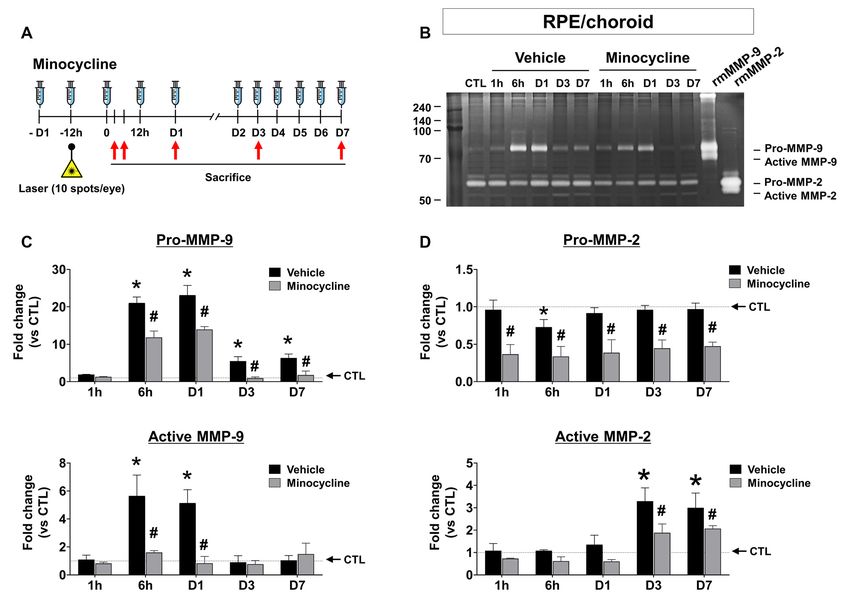
Kim et al.                                                                                                   Microglial MMPs in Neovascular AMD

INTRODUCTION                                                             macrophage infiltration (Huang et al., 2013), rather than MMPs.
                                                                         Moreover, although minocycline is a known microglial inhibitor
Age-related macular degeneration (AMD) is the leading cause              (Yrjanheikki et al., 1999), the mechanism behind this inhibition
of blindness in the elderly (Gehrs et al., 2006). Its prevalence         has not been fully elucidated, and the effect of minocycline
is expected to increase as aging populations expand, with                on the microglial MMP secretion has not yet been evaluated.
approximately 288 million people worldwide affected by AMD by            We, therefore, hypothesized in the present study that CNV
2040 (Wong et al., 2014). Neovascular AMD is characterized by            disease severity could be alleviated by suppressing microglial
the invasion of neovessels originating from the choroid through          MMP activity.
breaks in the Bruch’s membrane into the subretinal space in
the process of choroidal neovascularization (CNV). CNV occurs            MATERIALS AND METHODS
during late-stage advanced AMD and can lead to severe vision
loss (Ferris et al., 2013).
                                                                         Experimental Animals
    For pathological angiogenesis to occur, the basement
                                                                         All animal experiments were conducted following the guidelines
membrane surrounding endothelial tubes must be degraded
                                                                         of the Association for Research in Vision and Ophthalmology
to facilitate migration and proliferation of endothelial cells
                                                                         Statement for the Use of Animals in Ophthalmic and Vision
(Hanahan and Folkman, 1996), which is modulated in part
                                                                         Research and were approved by the Animal Care Committee
by matrix metalloproteinases (MMPs; Pepper, 2001). MMPs
                                                                         of Kyungpook National University (Approval No. 2019-0104-
are extracellular endopeptidases that cleave extracellular matrix
                                                                         01). C57BL/6J mice purchased from The Jackson Laboratory
proteins to regulate pathological tissue remodeling in disease
                                                                         were used for all in vivo experiments. Mice were allowed free
states, including inflammation (Nagase and Woessner, 1999).
                                                                         access to standard laboratory chow and tap water in a climate-
Prior studies reported that in normal tissues, basal MMPs
                                                                         controlled room with a 12 h light/dark cycle. Anesthesia was
are absent or weakly expressed (Pepper, 2001). MMPs are
                                                                         induced by i.p. injection of 250 mg/kg 2,2,2-tribromoethanol
upregulated in endothelial cells and immune cells in response
                                                                         (Sigma–Aldrich, St. Louis, MO, USA) at a dosage of 250 mg/kg in
to a variety of pathological events, including inflammation and
                                                                         survival procedures and 400 mg/kg in non-survival procedures.
angiogenesis (Lambert et al., 2002; O’Grady et al., 2007). Among
the large MMP family, MMP-2 and MMP-9 are of particular                  Minocycline and SB-3CT Treatment
interest to CNV, as their substrates include type IV collagen,           For in vivo studies, the minocycline treatment regimen was
a component of the Bruch’s membrane (Alcazar et al., 2007).              modified from a previously described study (Scholz et al.,
In the murine laser-induced CNV model, MMP-9 and MMP-2                   2015). Mice received i.p. injections of minocycline (45 mg/kg;
expression are upregulated on Days 3 and 5 post-CNV induction,           Sigma–Aldrich, M9511), SB-3CT (10 mg/kg; EMD Millipore,
when neovascular lesions are already present, and synergistically        Temecula, CA, USA, S1326), a selective MMP-2, 9 inhibitor,
promote CNV in this context (Lambert et al., 2002, 2003).                or normal saline vehicle with two initial twice-daily injections
However, potential early changes of MMP expression prior to              starting 1 day before CNV induction (Day -1), and one
3 days post-CNV, and therefore the role of MMPs in initiation            daily injection for 7 days beginning on the day of CNV
of the angiogenic response, have not been reported. Further, the         induction (Day 0).
cell types that secrete active MMP-9 during early CNV formation
have not been identified, which is paramount to the development          Laser-Induced CNV Model
of targeted approaches to treatment.                                     Eight-week-old male C57BL/6J mice were used for experiments.
    Microglia are resident immune cells in the retina that               A 532 nm OcuLight GLx Laser System (IRIDEX Corporation,
significantly contribute to the inflammatory processes of                Mountain View, CA, USA) was used to generate lesions.
age-related retinal pathologies and are known sources of MMPs            According to previous studies (Yanai et al., 2014; Hasegawa et al.,
(Chen and Xu, 2015). In the healthy retina, inactive microglia           2017), four lesions were induced for neovascular leakage and
retain a stable state characterized by a ramified morphology.            CNV size measurements, and 10 lesions were induced for gelatin
However, in response to injury or degeneration, microglia are            zymography. The laser was set to the following parameters:
activated, assuming an amoeboid morphology and migrating to              100 mW power, 100 µm spot size and 0.1-s pulse duration. The
disease lesions (Karlstetter et al., 2015). This has been observed       appearance of a bubble at the site of photocoagulation signified
in pathological retinal angiogenesis (Connor et al., 2007),              disruption of the Bruch’s membrane. After sacrificing mice, eyes
retinal detachment (Okunuki et al., 2018), retinal degeneration          were enucleated and fixed for 30 min. The whole retinas were
(Peng et al., 2014), and autoimmune uveitis (Okunuki et al.,             then separated from the underlying RPE/choroid for whole-
2019). Because activated microglia affect lesion sites by creating       mount. For gelatin zymography, unfixed retina and RPE/choroid
an inflammatory microenvironment, modulating microglial                  tissues were snap-frozen in liquid nitrogen for later analysis.
function is a potential therapeutic approach for inflammatory
chorioretinal diseases (Aslanidis et al., 2015).                         Choroidal Flat-Mount Preparation
    Although previous studies have reported the effect of                At 7 days after CNV induction (Day +7), eyes were enucleated
tetracycline derivatives, including doxycycline, in CNV,                 and fixed in 4% paraformaldehyde for 30 min. Retinas
these studies focused on FasL expression in the retinal                  were then removed from the choroid and sclera to generate
pigment epithelium (RPE; Roychoudhury et al., 2010) or                   choroidal flat mounts. Alexa Fluor 488-conjugated to isolectin

Frontiers in Cellular Neuroscience | www.frontiersin.org             2                                February 2021 | Volume 15 | Article 638098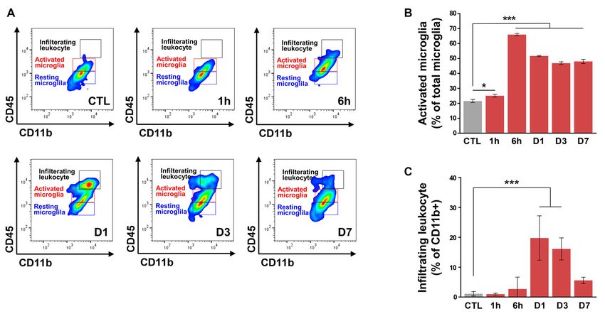
Kim et al.                                                                                                    Microglial MMPs in Neovascular AMD

B4 (1:100; Invitrogen, Carlsbad, CA, USA, I21411) was                      were then incubated for 48 h in developing buffer at 37◦ C.
used to stain the eyecups overnight at 4◦ C. Flat-mount                    After incubation, gels were stained with 0.5% Coomassie blue
images were captured using an LSM 800 fluorescence                         (Sigma–Aldrich) for 30 min and then destained for 1 h.
microscope with an Airyscan detector (Carl Zeiss, Oberkochen,
Germany). ImageJ software was used to measure CNV lesion                   Western Blotting
size, according to protocols described in previous studies                 Samples were lysed using radioimmunoprecipitation assay
(Yanai et al., 2014; Hasegawa et al., 2017).                               (RIPA) lysis buffer (Thermo Scientific, Waltham, MA, USA)
                                                                           with protease and phosphatase inhibitor cocktail (Roche
Immunohistochemical Staining                                               Holding AG, Basel, Switzerland) and centrifuged at 4◦ C
At 6 h and 1 day post-CNV induction, enucleated mouse eyes                 and 14,000 g for 20 min. The supernatant was collected,
were fixed in 4% paraformaldehyde for 1 h at room temperature,             and protein concentration was determined using a BCA kit
cryoprotected in 30% sucrose at 4◦ C overnight, and embedded in            (Thermo Scientific). The same amount of protein for each
OCT compound. Eyes were cryosectioned to 15 µm thickness.                  sample (20 µg) was loaded on a polyacrylamide gel (10%).
For immunofluorescence, sections were blocked in a blocking                Proteins were separated by SDS-PAGE and electro-transferred
buffer (0.5% Triton, 0.2% BSA, and 5% donkey serum in                      to PVDF membranes (Bio-Rad). Membranes were blocked
PBS) for 1 h at room temperature and subsequently incubated                using 5% skim milk in PBS with 0.25% Tween-20 (PBST),
with primary antibodies overnight at 4◦ C. After washing,                  and subsequently incubated at 4◦ C overnight with anti-MMP9
samples were incubated with secondary antibodies for 1 h at                (1:1,000; Abcam, ab38898) or β-actin (1:5,000; Thermo Scientific,
room temperature. Goat anti-Iba1 antibody (1:250; FUJIFILM                 MA5–15739) antibodies. After washing, membranes were
Wako Pure Chemical Corporation, Osaka, Japan, 011-27991),                  bound to HRP-conjugated secondary antibodies [Donkey IgG
rabbit anti-TMEM119 (1:500, Synaptic Systems, Göttingen,                   anti-rabbit (1:2,000; Cell Signaling Technology, Beverly, MA,
Germany, 400 002), rat anti-CD31 (1:100, BD Biosciences,                   USA, 7074s) or anti-mouse (1:2,000; Cell Signaling Technology,
NJ, USA, 553370), and rabbit anti-MMP-9 antibody (1:100;                   7076s)] and incubated at room temperature for 1 h. Blots
Abcam, Cambridge, MA, USA, ab38898) were used for primary                  were developed using enhanced chemiluminescence (ECL)
antibodies, and Alexa Fluor 488-conjugated donkey anti-goat                Western blotting detection reagent (Thermo Fisher Scientific)
antibody (1:500; Thermo Fisher Scientific, Waltham, MA,                    and analyzed using a MicroChemi system (DNR Bio-imaging
USA, A11055), Alexa Fluor 594-conjugated donkey anti-rabbit                Systems, Neve Yamin, Israel).
antibody (1:500; Thermo Fisher Scientific, A21207), and Alexa
Fluor 647-conjugated donkey anti-rat antibody (1:500; Jackson              Surface Marker Staining for Flow
Immuno Research Laboratories, INC., PA, USA, 712-605-15)                   Cytometry
were used for secondary antibodies.                                        For flow cytometric studies, eight retinas were pooled and
Fluorescein Angiography                                                    minced for digestion with 0.05% TrypLE (Thermo Fisher
A Micron IV Retinal Imaging Microscope (Phoenix Technology                 Scientific, 12604021) for 5 min at 37◦ C. The samples were
Group, Pleasanton, CA, USA) was used to capture fluorescein                filtered through a 40 µm cell strainer (SPL) and stained with
images. After anesthesia and pupil dilation, images were obtained          Zombie Aqua Kit (1:500; Biolegend, San Diego, CA, USA,
3–5 min (early phase) and 7–10 min (late phase) after i.p.                 423101). The cells were washed and suspended in cold 0.2%
injection of 0.1 ml 2% fluorescein sodium (Akorn, Lake Forest,             BSA (Thermo Fisher Scientific, A1000801) and blocked with Fc
IL, USA). A previously described scheme was used to grade the              BlockTM (0.01 µg/ml; BD, Franklin Lakes, NJ, USA, 553142)
hyperfluorescent lesions: faint or mottled fluorescence without            for 5 min at 4◦ C. Cell surface staining with CD45 V450
leakage was scored as 0 (no leakage); hyperfluorescence without            (0.3 µg/ml; BD, 560501) and CD11b PerCP-Cy5.5 (0.2 µg/ml;
any increase in size or intensity between early and late phases was        BD, 550993) was performed in the dark for 60 min at
scored as 1 (mild leakage); hyperfluorescence with constant size           4◦ C in buffer. Cells were detected using a BD LSR X-20
but increasing intensity was scored as 2A (moderate leakage); and          flow cytometer (BD) and analyzed with FlowJoTM V10 (BD).
hyperfluorescence with increasing size and intensity was scored            According to previous studies (Greter et al., 2015; Lee et al.,
as 2B (clinically significant leakage) (Yanai et al., 2014; Hasegawa       2019), CD11b and CD45 were used to identify the following cells:
et al., 2017).                                                             CD45low resting microglia (CD11b+ CD45low ), CD45intermediate
                                                                           activated microglia (CD11b+ CD45int ), and CD45high infiltrating
Gelatin Zymography                                                         leukocytes (CD11b+ CD45high ).
Retinal and RPE/choroid tissues were homogenized in lysis
buffer (25 mM Tris-HCl buffer, 100 mM NaCl, 1% Nonidet                     Patient Samples
P-40 (NP-40), and Complete Mini EDTA-free Protease Inhibitor               Institutional Review Board approval from the Kyungpook
Cocktail tablets; Sigma–Aldrich (11836170001) and centrifuged              National University School of Medicine was obtained for all
at 14,000 g for 10 min. Aliquots of supernatants containing                patient samples used in this study (Approval No. KNUH 2012-
equal amounts of protein (30 µg) were loaded without heating               04-006-020). The study was conducted following the tenets of the
onto 10% SDS-polyacrylamide gels containing 0.1% gelatin                   Declaration of Helsinki. Informed consent was obtained from all
(Sigma–Aldrich). Following electrophoresis, gels were washed               study participants. Aqueous humor samples were collected from
twice for 30 min in renaturing buffer at room temperature. Gels            consenting patients undergoing cataract surgery in the control

Frontiers in Cellular Neuroscience | www.frontiersin.org               3                               February 2021 | Volume 15 | Article 638098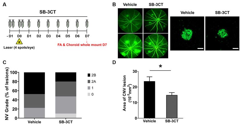
Kim et al. Microglial MMPs in Neovascular AMD
group and treatment-naïve patients with neovascular AMD in in the CNV mouse model (Figures 2C,D). In the RPE/choroid,
the disease group. minocycline significantly suppressed both pro and active MMP-9
(Figure 2C), as well as active MMP-2 (Figure 2D, lower panel) at
Statistical Analysis Days 3 and 7.
Statistical analyses were performed using Prism 6.0 (GraphPad
Software, San Diego, CA, USA). Results are expressed as Early Microglial Activation and MMP-9
mean ± SEM. Comparison of two groups was performed Expression in CNV Mice
using an unpaired non-parametric Mann–Whitney U-test. In addition to microglia, MMPs are also present in leukocytes
Multiple-group comparison was performed by Tukey’s multiple (Nuttall et al., 2007), although in the laser-induced CNV
comparison test. Statistical significance was defined as P < 0.05. model, leukocyte infiltration peaks at 2–3 days after CNV
induction (Sakurai et al., 2003; Tsutsumi et al., 2003). Thus,
RESULTS we evaluated time-dependent changes in a retinal population of
activated microglia and infiltrating leukocytes from CNV mice
Elevation of MMP-9 and MMP-2 Activities (Figure 3A). Resident resting microglia (CD11b+ CD45low ) were
abundant in control mice. The proportion of activated microglia
in Aqueous Humor From Neovascular AMD (CD11b+ CD45int ) began to increase very early, beginning by
Patients 1 h post-CNV induction, peaking at 6 h post-CNV, and
In our first series of experiments, the activities of MMP-9 maintaining elevation until Day 7 (Figure 3B). The above
and MMP-2 in aqueous humor samples from six patients with change was similar to our finding that active MMP-9 increased
neovascular AMD and six age-matched control subjects were in the RPE/choroid. However, the proportion of infiltrating
analyzed (Figure 1). Active MMP-9 (Figure 1B, left panel) and leukocytes (CD11b+ CD45high ) in CD11b+ cells increased at Day
active MMP-2 (Figure 1C, left panel) levels were increased in the 1 and Day 3 and subsequently decreased to basal levels by
neovascular AMD group relative to control. Furthermore, pro- Day 7 (Figure 3C). This finding is similar to a previous study
MMP-9 (Figure 1B, right panel) was increased in the neovascular demonstrating that recruitment of macrophages to CNV lesions
AMD group relative to control, but pro-MMP-2 was unchanged occurs 3 days post-CNV induction and subsequently decreases
(Figure 1C, right panel). (Jawad et al., 2013).
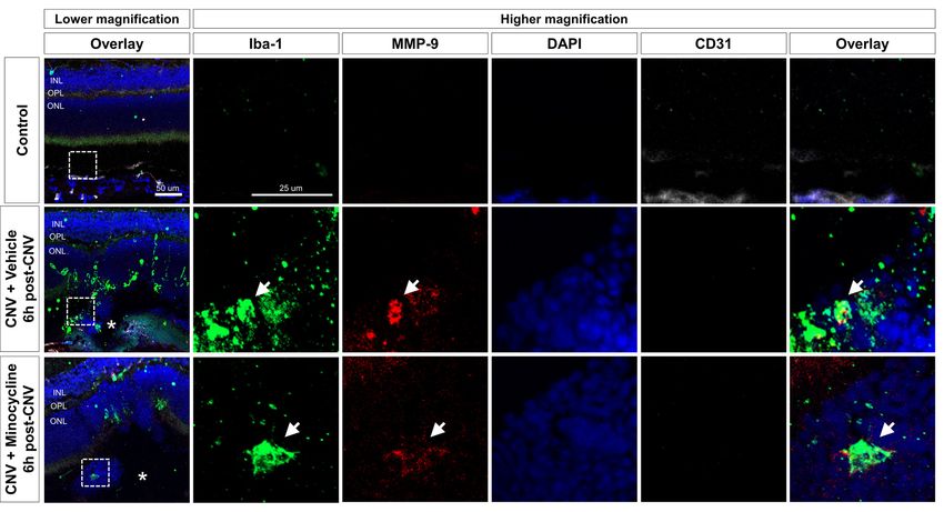
Next, to evaluate the localization of activated microglia
Early MMP-9 Activity in the CNV Mouse with upregulated MMP expression, we conducted
Model immunofluorescence staining of cryosections at 6 h (Figure 4)
Next, we investigated the spatial-temporal enzymatic activities of and 1-day post-CNV induction (Supplementary Figures 3, 4).
MMP-2 and -9 in the CNV mouse model using gel zymography. Activated microglia with amoeboid morphology were aggregated
In the RPE/choroid, the band intensities of pro-MMP-9 and on CNV lesions, and MMP-9 was increased in CNV lesions.
active MMP-9 dramatically increased 6 h after CNV induction, Activated microglia in CNV lesions were decreased in the
which was maintained through Day 1, followed by a subsequent minocycline group relative to the vehicle group. Furthermore,
decrease of active MMP-9 to basal levels by Day 3 post-CNV intracellular MMP-9 was present in Iba-1+ cells in CNV lesions,
induction (Figure 2C). Active MMP-2 increased in RPE/choroid which were CD31− (Figure 4). Also, all Iba-1+ cells localized to
tissue from CMV mice at Days 3 and 7, although pro- CNV lesions were positive for TMEM119, a specific microglial
MMP-2 was not significantly changed (Figure 2D). In retinas marker (Bennett et al., 2016; Supplementary Figure 4). Taken
from CNV mice, no active MMP-9 or MMP-2 was observed together, these findings suggested that microglial activation and
(Supplementary Figure 1). Although MMP-9 Western blots of MMP-9 expression occurred soon after CNV induction.
RPE/choroid tissue exhibited similar time-dependent patterns
to gelatin zymography (Supplementary Figure 2), separate pro- SB-3CT Alleviation of CNV Severity
and active MMP-9 bands could not be detected, as with gelatin To determine if MMP inhibition could alleviate CNV, we
zymography, which could be why prior studies used gelatin evaluated the effects of SB-3CT, a selective MMP-9 and
zymography instead of Western blotting in this context (Lambert MMP-2 inhibitor (Shin et al., 2016). To evaluate whether early
et al., 2002; Manabe et al., 2005; Kim et al., 2018). MMP-9 inhibition could affect CNV severity, the SB-3CT
Previous studies have demonstrated that MMP-9 amplifies administration started 1 day before CNV induction and was
pathological angiogenesis (Pepper, 2001) and inflammation maintained until Day 7 (Figure 5A). The proportion of grade
(Nagase and Woessner, 1999), and microglia are a known 2B lesions, defined as exhibiting clinically significant vascular
source of MMPs, contributing to the inflammatory process leakage, decreased in CNV mice treated with SB-3CT relative
(Lively and Schlichter, 2013; Rojewska et al., 2014). Furthermore, to vehicle-treated CNV mice (Figures 5B,C). Furthermore,
recruitment of activated microglia appears as early as 1 h after CNV lesion size decreased in mice treated with SB-3CT
CNV induction (Karlstetter et al., 2015). If microglia contribute compared with control mice, as assessed with choroidal
to the pathogenesis of CNV by secreting MMP early in the flat mounts (Figures 5B–D). This suggested that SB-3CT
CNV disease process, microglial inhibitors such as minocycline alleviated neovascular leakage and vascular outgrowth,
could suppress MMP-9 activities, suppressing the development which could have been modulated by early suppression of
of CNV. Thus, we confirmed the effect of minocycline over time MMP-9 from microglia. Similarly, minocycline, a known
Frontiers in Cellular Neuroscience | www.frontiersin.org 4 February 2021 | Volume 15 | Article 638098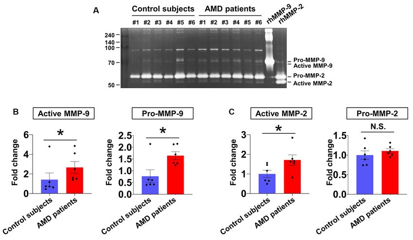
Kim et al.                                                                                                                  Microglial MMPs in Neovascular AMD

  FIGURE 1 | Aqueous humor matrix metalloproteinase (MMP) enzymatic activities in neovascular age-related macular degeneration (AMD) and senile cataract
  control subjects. (A) Gelatin zymography to detect MMP activity (pro-MMP-9, 95 kDa; active MMP-9, 82 kDa; pro-MMP-2, 62 kDa; active MMP-2, 56 kDa). (B)
  Relative abundances of pro-MMP-9 and active MMP-9 were quantified by densitometry. Active MMP-9 was elevated in aqueous humor samples from all six
  neovascular AMD patients compared with age-matched control subjects. *P < 0.05. (C) While active MMP-2 levels were increased in neovascular AMD group
  relative to control, pro-MMP-2 levels were unchanged. N.S.: not-significant.

microglial inhibitor (Yrjanheikki et al., 1999), also decreased                    consistent with our flow cytometry data. The proportion of
neovascular leakage and vascular outgrowth in CNV lesions                          activated microglia increased very early from 1 h post-CNV
(Supplementary Figure 5).                                                          induction and remained increased until Day 7. By contrast, only
   These findings suggested that suppression of MMP-9                              a small proportion of infiltrating leukocytes was detected before
secreted from microglia could attenuate neovascular lesion                         Day 1. These findings are consistent with a previous study,
formation in CNV. In particular, MMP-9 is involved in                              which demonstrated that CX3CR1-positive microglia migrate
the breakdown of the parenchymal basement membranes                                to laser-induced lesions as early as 1 h after CNV induction
surrounding blood vessels (Agrawal et al., 2006). This process                     (Karlstetter et al., 2015).
can damage the Bruch’s membrane and blood-retinal barrier,                            Regarding time-dependent changes of MMP activity in the
with accompanying neovascular outgrowth which occurs                               laser-induced CNV model, it is notable that active MMP-9
similarly in the blood-brain barrier (Rosenberg, 2002). Thus,                      increased at very early time points, beginning at 1 h post-CNV
modulation of microglial MMPs could be a targeted approach to                      induction in the RPE/choroid, which correlated with the
CNV treatment.                                                                     increase of microglial activation identified by our FACS data.
                                                                                   These results were consistent with a previous study, which
DISCUSSION                                                                         demonstrated intense MMP-9 staining in CNV lesions at Day 3,
                                                                                   although this study did not evaluate MMP-9 expression at early
MMP-9 and MMP-2 are essential to cleavage of collagen                              time points before Day 3 (Lambert et al., 2002).
type IV, the major component of the basement membrane,                                In the CNV model, active RPE/choroid MMP-2 was increased
and in the context of cancer biology, they are activated at                        by CNV in the later stages of the disease. Our in vivo
the onset of angiogenesis, resulting in invasive tumor growth                      experimental data are consistent with the results of other
(Pittayapruek et al., 2016). However, microglial MMP activity                      previous studies demonstrating that activated MMP-2 was not
has not previously been evaluated, particularly in the context                     present before Day 5 after CNV induction (Lambert et al.,
of CNV.                                                                            2003). Furthermore, Lively et al. reported differential expression
   The normal healthy retina lacks infiltrating leukocytes and                     of MMPs from microglia activated by different stimuli (Lively
has many resident microglia (Liyanage et al., 2016), which is                      and Schlichter, 2013). MMP-9 increased only in LPS-treated

Frontiers in Cellular Neuroscience | www.frontiersin.org                       5                                    February 2021 | Volume 15 | Article 638098
Kim et al. Microglial MMPs in Neovascular AMD
FIGURE 2 | Time-dependent changes of MMP activity and effect of minocycline on MMP activity inretinal pigment epithelium (RPE)/choroid tissues from
laser-induced choroidal neovascularization (CNV) mice. (A) Experimental design of minocycline injection and induction of CNV with laser photocoagulation. (B)
Representative image of gelatin zymography to visualize RPE/choroid MMP activity. Lane CTL, non-CNV control; 1 h, 6 h, D1, D3, and D7 after CNV induction. (C)
Quantification of gelatin zymography of pro-and active MMP-9 in panel (B). Enzymatic activity was measured by band intensity. (D) Quantification of gelatin
zymography of pro-and active MMP-2 in panel (B). Data are expressed as mean ± SEM. *P < 0.05 vs. CTL; # P < 0.05 vs. Vehicle. n = 6 eyes/group.
microglia, considered classical (M1) activation. Meanwhile, for CNV sprouting by degrading the Bruch’s membrane, which
MMP-2 increased only in IL-4-treated microglia, considered is attenuated by minocycline.
alternative (M2) activation. The above study can explain the There are several limitations of the present study, which
in vivo data showing that increased MMP-2 activity in the should be acknowledged to avoid its overinterpretation. First,
RPE/choroid occurred at Days 3 and 7, likely due to the our data are not sufficient to demonstrate that microglia
alternative activation of microglia. are the primary source of MMPs, as Iba-1 also labels
Minocycline, an antibiotic with known microglial inhibitory infiltrating leukocytes. However, immunofluorescence staining
activity (Yrjanheikki et al., 1999), is neuroprotective in models of demonstrated that Iba-1+ cells were also labeled with TMEM119,
light-induced retinal degeneration (Scholz et al., 2015), diabetic a microglia-specific marker, pointing to microglia as an
retinopathy (Krady et al., 2005), and ischemia-reperfusion injury important source of MMPs in this context. Furthermore, taken
(Ahmed et al., 2017). In addition to known neuroprotective together, the immunofluorescence and FACS data demonstrate
effects of minocycline, the current study demonstrated that that, at least in the early time points of CNV, microglia could
minocycline suppressed pathological neovascularization in laser- play an important role in angiogenesis by secreting MMPs.
induced CNV. Minocycline significantly suppressed microglial Second, because it is not possible to remove RPE/choroid tissues
MMP-9 secretion in the RPE/choroid, especially at early time from neovascular AMD patients without damaging vision, we
points. Furthermore, SB-3CT significantly alleviated disease used aqueous samples, which are commonly used to measure
severity, as demonstrated by decreased vascular leakage and biomarkers of neovascular chorioretinal diseases (Hsu et al.,
CNV lesion size. Active MMP-9 from activated microglia 2014). Because active MMP-9 in the aqueous humor could
could potentially provide an advantageous microenvironment originate from multiple sources, RPE/choroid tissues from
Frontiers in Cellular Neuroscience | www.frontiersin.org 6 February 2021 | Volume 15 | Article 638098Kim et al. Microglial MMPs in Neovascular AMD FIGURE 3 | Flow cytometry of retinal cells from control and choroidal neovascularization (CNV) retinal cells to detect microglial activation and infiltrating leukocytes. (A) Single live cells were selected and sorted according to their CD11b and CD45 status. Plot CTL, non-CNV control; plot 1 h, 6 h, D1, D3, and D7 after CNV induction. (B) The ratio of activated microglia (CD11b+ CD45int ) to total microglia started to increase very early after CNV induction at 1 h, peaked at 6 h, and remained significantly increased through D7. (C) Compared with the control, the proportion of infiltrating leukocytes (CD11b+ CD45high ) in CD11b+ cells increased on D1 and D3. *P < 0.05, ***P < 0.001 vs. CTL. n = 8 mice/experimental group in three independent experiments. FIGURE 4 | Activated microglia and upregulated MMP-9 expression at 6 h after CNV induction. Immunohistochemistry data of Iba-1+ cells (green), MMP-9 (red), and CD31 (white). At 6 h post-CNV induction, Iba-1+ amoeboid cells were suggestive of activated microglia aggregation on CNV lesions, indicated by asterisks. Fewer activated microglia were localized to CNV lesions in the minocycline group relative to vehicle control. Furthermore, at higher magnifications, intracellular MMP-9 signal was present in Iba-1+ cells, which were CD31− . Scale bar: 100 µm. INL, inner nuclear layer; OPL, outer plexiform layer; ONL, outer nuclear layer. Frontiers in Cellular Neuroscience | www.frontiersin.org 7 February 2021 | Volume 15 | Article 638098
Kim et al.                                                                                                                          Microglial MMPs in Neovascular AMD

  FIGURE 5 | Effect of SB-3CT on neovascular activity in the laser-induced CNV. (A) Experimental design of the SB-3CT injection and CNV induction with laser
  photocoagulation. (B) At 7 days after CNV induction, CNV lesion grading was conducted in control and SB-3CT groups. (C) The proportion of grade 2B lesions,
  which exhibit clinically significant leakage, decreased in mice treated with SB-3CT relative to the vehicle. (D) CNV lesion size decreased in mice treated with SB-3CT
  compared with vehicle, as assessed in choroidal flat mounts stained with fluorescent isolectin B4. *P < 0.05. n = 48 lesions/group. Scale bar: 100 µm.

human cadaver eyes would be optimal and will be investigated                             source of MMPs, macrophage markers did not increase until
in future studies. Third, because our data pointed to the early                          Day 3 after CNV induction (Zhou et al., 2017). Furthermore,
recruitment of microglia with the increase of active MMP-9,                              most MMP RNA levels, including MMP-9, are enriched in
we focused on the relationship between MMP-9 and microglia,                              microglia compared with peripheral leukocytes (Nuttall et al.,
which could be more influential in early CNV than MMP-2.                                 2007). In conclusion, the present study demonstrated a potential
Furthermore, because our data demonstrated that minocycline                              role of early microglial MMP-9 contributing to CNV, and
decreased pro-MMP-9 but not active MMP-9 from Day 3, we                                  that modulation of microglial MMP could be a novel putative
focused on early time points in the laser-induced CNV model. In                          therapeutic for CNV.
future studies, we will evaluate the roles of alternative microglial
activation, especially in later time points of CNV. Finally, as this                     DATA AVAILABILITY STATEMENT
is an observational study, the mechanism underlying MMP-9-
induced CNV will be further investigated in future studies.                              The original contributions presented in the study are included
    A previous pilot clinical trial reported that combined therapy                       in the article/Supplementary Material, further inquiries can be
with reduced-fluence photodynamic therapy (PDT), intravitreal                            directed to the corresponding author.
ranibizumab (0.3 mg), intravitreal dexamethasone, and oral
minocycline for neovascular AMD did not have favorable visual                            ETHICS STATEMENT
outcomes compared with outcomes of combination treatment
with PDT and intravitreal ranibizumab (0.5 mg) reported in                               The studies involving human participants were reviewed and
other studies (Sivaprasad et al., 2011). However, this pilot study                       approved by Institutional Review Board approval from the
evaluated only a single group and used a reduced dose of                                 Kyungpook National University School of Medicine. The
ranibizumab (0.3 mg). Thus, it is not clear whether that study                           patients/participants provided their written informed consent to
clearly evaluated the direct effect of minocycline and should                            participate in this study. The animal study was reviewed and
therefore be interpreted cautiously. Thus, our group is in the                           approved by Animal Care Committee of Kyungpook National
process of organizing a clinical trial to evaluate the effect of                         University (2019-0104-01).
minocycline in neovascular AMD.
    Taken together, our data demonstrated nearly immediate                               AUTHOR CONTRIBUTIONS
recruitment of activated microglia and MMP-9 activation in the
RPE/choroid, suggesting that activated microglia that aggregate                          JK, J-HK, JD, JL, KS, and DP: design and conduct of the study.
to CNV lesions could contribute to increased MMP-9 activity.                             JK, J-HK, JD, JL, KS, and DP: collection of data. J-HK, JL, RY, KS,
Although we cannot exclude infiltrating leukocytes as a potential                        and DP analysis and interpretation of data. J-HK, JD, JL, and DP:

Frontiers in Cellular Neuroscience | www.frontiersin.org                             8                                      February 2021 | Volume 15 | Article 638098
Kim et al. Microglial MMPs in Neovascular AMD
writing the manuscript. J-HK, I-kL, KS, and DP: critical revision program (IITP-2021-2020-0-01808) supervised by the IITP
of the manuscript. JK, J-HK, JD, JL, RY, I-kL, KS, and DP: final (Institute of Information & Communications Technology
approval of the manuscript. All authors contributed to the article Planning & Evaluation).
and approved the submitted version.
ACKNOWLEDGMENTS
FUNDING DP would like to thank Dr. Kip M. Connor in the Angiogenesis
Laboratory, Department of Ophthalmology, Massachusetts Eye
DP was financially supported by the Basic Science Research
and Ear Infirmary, Harvard Medical School for his guidance and
Program of the National Research Foundation of Korea (NRF),
training in the CNV model.
funded by the Korean government (Ministry of Science and
ICT; 2019R1A2C1084371) and the Korea Health Technology
R&D Project of the Korea Health Industry Development SUPPLEMENTARY MATERIAL
Institute (KHIDI), funded by the Ministry of Health and
Welfare, Republic of Korea (HI16C1501). DP was also supported The Supplementary Material for this article can be found
by the MSIT (Ministry of Science and ICT), Korea, under online at: https://www.frontiersin.org/articles/10.3389/fncel.
the ITRC (Information Technology Research Center) support 2021.638098/full#supplementary-material.
REFERENCES ophthalmologically relevant diseases via ultrahigh sensitive paper-based ELISA.
Biomaterials 35, 3729–3735. doi: 10.1016/j.biomaterials.2014.01.030
Agrawal, S., Anderson, P., Durbeej, M., van Rooijen, N., Ivars, F., Opdenakker, G., Huang, H., Parlier, R., Shen, J. K., Lutty, G. A., and Vinores, S. A. (2013).
et al. (2006). Dystroglycan is selectively cleaved at the parenchymal basement VEGF receptor blockade markedly reduces retinal microglia/macrophage
membrane at sites of leukocyte extravasation in experimental autoimmune infiltration into laser-induced CNV. PLoS One 8:e71808. doi: 10.1371/journal.
encephalomyelitis. J. Exp. Med. 203, 1007–1019. doi: 10.1084/jem.20051342 pone.0071808
Ahmed, A., Wang, L. L., Abdelmaksoud, S., Aboelgheit, A., Saeed, S., and Jawad, S., Liu, B., Li, Z., Katamay, R., Campos, M., Wei, L., et al. (2013).
Zhang, C. L. (2017). Minocycline modulates microglia polarization in ischemia- The role of macrophage class a scavenger receptors in a laser-induced
reperfusion model of retinal degeneration and induces neuroprotection. Sci. murine choroidal neovascularization model. Invest. Ophthalmol. Vis. Sci. 54,
Rep. 7:14065. doi: 10.1038/s41598-017-14450-5 5959–5970. doi: 10.1167/iovs.12-11380
Alcazar, O., Cousins, S. W., and Marin-Castano, M. E. (2007). MMP-14 and Karlstetter, M., Scholz, R., Rutar, M., Wong, W. T., Provis, J. M., and Langmann, T.
TIMP-2 overexpression protects against hydroquinone-induced oxidant injury (2015). Retinal microglia: just bystander or target for therapy. Prog. Retin. Eye
in RPE: implications for extracellular matrix turnover. Invest. Ophthalmol. Vis. Res. 45, 30–57. doi: 10.1016/j.preteyeres.2014.11.004
Sci. 48, 5662–5670. doi: 10.1167/iovs.07-0392 Kim, H. S., Vargas, A., Eom, Y. S., Li, J., Yamamoto, K. L., Craft, C. M., et al. (2018).
Aslanidis, A., Karlstetter, M., Scholz, R., Fauser, S., Neumann, H., Fried, C., Tissue inhibitor of metalloproteinases 1 enhances rod survival in the rd1 mouse
et al. (2015). Activated microglia/macrophage whey acidic protein (AMWAP) retina. PLoS One 13:e0197322. doi: 10.1371/journal.pone.0197322
inhibits NFkappaB signaling and induces a neuroprotective phenotype in Krady, J. K., Basu, A., Allen, C. M., Xu, Y., LaNoue, K. F., Gardner, T. W., et al.
microglia. J. Neuroinflammation 12:77. doi: 10.1186/s12974-015-0296-6 (2005). Minocycline reduces proinflammatory cytokine expression, microglial
Bennett, M. L., Bennett, F. C., Liddelow, S. A., Ajami, B., Zamanian, J. L., activation and caspase-3 activation in a rodent model of diabetic retinopathy.
Fernhoff, N. B., et al. (2016). New tools for studying microglia in the mouse and Diabetes 54, 1559–1565. doi: 10.2337/diabetes.54.5.1559
human CNS. Proc. Natl. Acad. Sci. U S A. 113, E1738–E1746. doi: 10.1073/pnas. Lambert, V., Munaut, C., Jost, M., Noel, A., Werb, Z., Foidart, J. M., et al. (2002).
1525528113 Matrix metalloproteinase-9 contributes to choroidal neovascularization. Am.
Chen, M., and Xu, H. (2015). Parainflammation, chronic inflammation and J. Pathol. 161, 1247–1253. doi: 10.1016/S0002-9440(10)64401-X
age-related macular degeneration. J. Leukoc. Biol. 98, 713–725. doi: 10.1189/jlb. Lambert, V., Wielockx, B., Munaut, C., Galopin, C., Jost, M., Itoh, T., et al. (2003).
3RI0615-239R MMP-2 and MMP-9 synergize in promoting choroidal neovascularization.
Connor, K. M., SanGiovanni, J. P., Lofqvist, C., Aderman, C. M., Chen, J., FASEB J. 17, 2290–2292. doi: 10.1096/fj.03-0113fje
Higuchi, A., et al. (2007). Increased dietary intake of omega-3-polyunsaturated Lee, S. W., de Rivero Vaccari, J. P., Truettner, J. S., Dietrich, W. D., and
fatty acids reduces pathological retinal angiogenesis. Nat. Med. 13, 868–873. Keane, R. W. (2019). The role of microglial inflammasome activation
doi: 10.1038/nm1591 in pyroptotic cell death following penetrating traumatic brain injury.
Ferris, F. L., Wilkinson, C. P., Bird, A., Chakravarthy, U., Chew, E., Csaky, K., J. Neuroinflammation 16:27. doi: 10.1186/s12974-019-1423-6
et al. (2013). Clinical classification of age-related macular degeneration. Lively, S., and Schlichter, L. C. (2013). The microglial activation state
Ophthalmology 120, 844–851. doi: 10.1016/j.ophtha.2012.10.036 regulates migration and roles of matrix-dissolving enzymes for invasion.
Gehrs, K. M., Anderson, D. H., Johnson, L. V., and Hageman, G. S. (2006). J. Neuroinflammation 10:75. doi: 10.1186/1742-2094-10-75
Age-related macular degeneration--emerging pathogenetic and therapeutic Liyanage, S. E., Gardner, P. J., Ribeiro, J., Cristante, E., Sampson, R. D.,
concepts. Ann. Med. 38, 450–471. doi: 10.1080/07853890600946724 Luhmann, U. F., et al. (2016). Flow cytometric analysis of inflammatory and
Greter, M., Lelios, I., and Croxford, A. L. (2015). Microglia versus myeloid resident myeloid populations in mouse ocular inflammatory models. Exp. Eye
cell nomenclature during brain inflammation. Front. Immunol. 6:249. Res. 151, 160–170. doi: 10.1016/j.exer.2016.08.007
doi: 10.3389/fimmu.2015.00249 Manabe, S., Gu, Z., and Lipton, S. A. (2005). Activation of matrix
Hanahan, D., and Folkman, J. (1996). Patterns and emerging mechanisms of the metalloproteinase-9 via neuronal nitric oxide synthase contributes to
angiogenic switch during tumorigenesis. Cell 86, 353–364. doi: 10.1016/s0092- NMDA-induced retinal ganglion cell death. Invest. Ophthalmol. Vis. Sci. 46,
8674(00)80108-7 4747–4753. doi: 10.1167/iovs.05-0128
Hasegawa, E., Inafuku, S., Mulki, L., Okunuki, Y., Yanai, R., Smith, K. E., et al. Nagase, H., and Woessner, J. F.Jr. (1999). Matrix metalloproteinases. J. Biol. Chem.
(2017). Cytochrome P450 monooxygenase lipid metabolites are significant 274, 21491–21494. doi: 10.1074/jbc.274.31.21491
second messengers in the resolution of choroidal neovascularization. Proc. Nuttall, R. K., Silva, C., Hader, W., Bar-Or, A., Patel, K. D., Edwards, D. R., et al.
Natl. Acad. Sci. U S A. 114, E7545–E7553. doi: 10.1073/pnas.1620898114 (2007). Metalloproteinases are enriched in microglia compared with leukocytes
Hsu, M. Y., Yang, C. Y., Hsu, W. H., Lin, K. H., Wang, C. Y., Shen, Y. C., and they regulate cytokine levels in activated microglia. Glia 55, 516–526.
et al. (2014). Monitoring the VEGF level in aqueous humor of patients with doi: 10.1002/glia.20478
Frontiers in Cellular Neuroscience | www.frontiersin.org 9 February 2021 | Volume 15 | Article 638098Kim et al. Microglial MMPs in Neovascular AMD O’Grady, A., Dunne, C., O’Kelly, P., Murphy, G. M., Leader, M., and in the S334ter-line3 retinitis pigmentosa model. PLoS One 11:e0167102. Kay, E. (2007). Differential expression of matrix metalloproteinase (MMP)- doi: 10.1371/journal.pone.0167102 2, MMP-9 and tissue inhibitor of metalloproteinase (TIMP)-1 and TIMP-2 Sivaprasad, S., Patra, S., DaCosta, J., Adewoyin, T., Shona, O., Pearce, E., in non-melanoma skin cancer: implications for tumour progression. et al. (2011). A pilot study on the combination treatment of reduced-fluence Histopathology 51, 793–804. doi: 10.1111/j.1365-2559.2007.02885.x photodynamic therapy, intravitreal ranibizumab, intravitreal dexamethasone Okunuki, Y., Mukai, R., Nakao, T., Tabor, S. J., Butovsky, O., Dana, R., et al. and oral minocycline for neovascular age-related macular degeneration. (2019). Retinal microglia initiate neuroinflammation in ocular autoimmunity. Ophthalmologica 225, 200–206. doi: 10.1159/000322363 Proc. Natl. Acad. Sci. U S A 116, 9989–9998. doi: 10.1073/pnas.1820 Tsutsumi, C., Sonoda, K. H., Egashira, K., Qiao, H., Hisatomi, T., Nakao, S., et al. 387116 (2003). The critical role of ocular-infiltrating macrophages in the development Okunuki, Y., Mukai, R., Pearsall, E. A., Klokman, G., Husain, D., Park, D. H., et al. of choroidal neovascularization. J. Leukoc. Biol. 74, 25–32. doi: 10.1189/jlb. (2018). Microglia inhibit photoreceptor cell death and regulate immune cell 0902436 infiltration in response to retinal detachment. Proc. Natl. Acad. Sci. U S A. 115, Wong, W. L., Su, X., Li, X., Cheung, C. M., Klein, R., Cheng, C. Y., et al. (2014). E6264–E6273. doi: 10.1073/pnas.1719601115 Global prevalence of age-related macular degeneration and disease burden Peng, B., Xiao, J., Wang, K., So, K. F., Tipoe, G. L., and Lin, B. (2014). Suppression projection for 2020 and 2040: a systematic review and meta-analysis. The Lancet of microglial activation is neuroprotective in a mouse model of human retinitis Global Health 2, e106–e116. doi: 10.1016/S2214-109X(13)70145-1 pigmentosa. J. Neurosci. 34, 8139–8150. doi: 10.1523/JNEUROSCI.5200- Yanai, R., Mulki, L., Hasegawa, E., Takeuchi, K., Sweigard, H., Suzuki, J., et al. 13.2014 (2014). Cytochrome P450-generated metabolites derived from omega-3 fatty Pepper, M. S. (2001). Role of the matrix metalloproteinase and plasminogen acids attenuate neovascularization. Proc. Natl. Acad. Sci. U S A 111, 9603–9608. activator-plasmin systems in angiogenesis. Arterioscler. Thromb. Vasc. Biol. 21, doi: 10.1073/pnas.1401191111 1104–1117. doi: 10.1161/hq0701.093685 Yrjanheikki, J., Tikka, T., Keinanen, R., Goldsteins, G., Chan, P. H., Pittayapruek, P., Meephansan, J., Prapapan, O., Komine, M., and and Koistinaho, J. (1999). A tetracycline derivative, minocycline, reduces Ohtsuki, M. (2016). Role of matrix metalloproteinases in photoaging and inflammation and protects against focal cerebral ischemia with a wide photocarcinogenesis. Int. J. Mol. Sci. 17:868. doi: 10.3390/ijms17060868 therapeutic window. Proc. Natl. Acad. Sci. U S A 96, 13496–13500. Rojewska, E., Popiolek-Barczyk, K., Jurga, A. M., Makuch, W., Przewlocka, B., doi: 10.1073/pnas.96.23.13496 and Mika, J. (2014). Involvement of pro- and antinociceptive factors in Zhou, Y., Yoshida, S., Kubo, Y., Yoshimura, T., Kobayashi, Y., Nakama, T., et al. minocycline analgesia in rat neuropathic pain model. J. Neuroimmunol. 277, (2017). Different distributions of M1 and M2 macrophages in a mouse model 57–66. doi: 10.1016/j.jneuroim.2014.09.020 of laser-induced choroidal neovascularization. Mol. Med. Rep. 15, 3949–3956. Rosenberg, G. A. (2002). Matrix metalloproteinases in neuroinflammation. Glia doi: 10.3892/mmr.2017.6491 39, 279–291. doi: 10.1002/glia.10108 Roychoudhury, J., Herndon, J. M., Yin, J., Apte, R. S., and Ferguson, T. A. (2010). Conflict of Interest: JL was employed by company JD Bioscience Inc. Targeting immune privilege to prevent pathogenic neovascularization. Invest. Ophthalmol. Vis. Sci. 51, 3560–3566. doi: 10.1167/iovs.09-3890 The remaining authors declare that the research was conducted in the absence of Sakurai, E., Anand, A., Ambati, B. K., van Rooijen, N., and Ambati, J. (2003). any commercial or financial relationships that could be construed as a potential Macrophage depletion inhibits experimental choroidal neovascularization. conflict of interest. Invest. Ophthalmol. Vis. Sci. 44, 3578–3585. doi: 10.1167/iovs. 03-0097 Copyright © 2021 Kim, Kim, Do, Lee, Yanai, Lee, Suk and Park. This is an Scholz, R., Sobotka, M., Caramoy, A., Stempfl, T., Moehle, C., and Langmann, T. open-access article distributed under the terms of the Creative Commons Attribution (2015). Minocycline counter-regulates pro-inflammatory microglia responses License (CC BY). The use, distribution or reproduction in other forums is permitted, in the retina and protects from degeneration. J. Neuroinflammation 12:209. provided the original author(s) and the copyright owner(s) are credited and that the doi: 10.1186/s12974-015-0431-4 original publication in this journal is cited, in accordance with accepted academic Shin, J. A., Kim, H. S., Vargas, A., Yu, W. Q., Eom, Y. S., Craft, C. M., practice. No use, distribution or reproduction is permitted which does not comply et al. (2016). Inhibition of matrix metalloproteinase 9 enhances rod survival with these terms. Frontiers in Cellular Neuroscience | www.frontiersin.org 10 February 2021 | Volume 15 | Article 638098
You can also read