Involvement of supralemniscal nucleus (B9) 5-HT neuronal system in nociceptive processing: a fiber photometry study
←
→
Page content transcription
If your browser does not render page correctly, please read the page content below
Moriya et al. Molecular Brain    (2020) 13:14
https://doi.org/10.1186/s13041-020-0553-1

 RESEARCH                                                                                                                                         Open Access

Involvement of supralemniscal nucleus (B9)
5-HT neuronal system in nociceptive
processing: a fiber photometry study
Shunpei Moriya1, Akira Yamashita1, Daiki Masukawa2, Yuki Kambe3, Junichi Sakaguchi1, Honami Setoyama1,
Akihiro Yamanaka4 and Tomoyuki Kuwaki1*

  Abstract
  Nociception is important perception that has harmful influence on daily life of humans. As to main pain
  management system, some descending pathways are called descending antinociceptive systems (DAS). As main
  pathways of DAS, it is well known that dorsal raphe (B6/B7) - rostral ventromedial medulla (B3) - spinal dorsal horn
  includes serotonergic system. However, possible role of supralemniscal (B9) serotonin (5-HT) cell group in pain
  management is still open question. In this study, we measured activities of B9 5-HT neuronal cell bodies and B9 5-
  HT neuron-derived axons located in the locus coeruleus (LC) and ventral tegmental area (VTA), which are also main
  players of pain management, using fiber photometry system. We introduced the G-CaMP6 in B9 5-HT neurons
  using transgenic mice carrying a tetracycline-controlled transactivator transgene (tTA) under the control of a
  tryptophan hydroxylase-2 (TPH2) promoter and site-specific injection of adeno associated virus (AAV-TetO(3G)-G-
  CaMP6). After confirmation of specific expression of G-CaMP6 in the target population, G-CaMP6 fluorescence
  intensity in B9 group and LC/VTA groups was measured in awake mice exposed to acute tail pinch and heat
  stimuli. G-CaMP6 fluorescence intensity rapidly increased by both stimuli in all groups, but not significantly reacted
  by nonnociceptive control stimuli. The present results clearly indicate that acute nociceptive stimuli cause a rapid
  increase in the activities of B9-LC/B9-VTA 5-HTergic pathways, suggesting that B9 5-HT neurons play important roles
  in nociceptive processing.
  Keywords: Nociception, B9 5-HT cell group, Locus coeruleus, Ventral tegmental area, Fiber photometry, G-CaMP6,
  TPH2-tTA mice

Introduction                                                                           tricyclic antidepressant (TCA) and anticonvulsant are
In clinical psychiatric medicine, clinicians take patients’                            prescribed [6]. Monoaminergic system of central nervous
various perceptions into consideration. Nociception is                                 system (CNS) is thought to be involved in the etiology
important perception that has harmful influence on daily                               of those diseases. As to main pain management system,
life of human [1]. Pain symptoms with some physical                                    there are some descending pathways [7, 8], which are
and mental disorders has increasingly become a major                                   called descending antinociceptive system (DAS). As
social problem. In the psychiatric field, pain symptom is                              main pathways of DAS, it is well known that the locus
frequently emerged in somatoform pain disorder [2],                                    coeruleus (LC) – spinal dorsal horn circuit includes the
major depressive disorder [3], neuropathic pain [4], and                               noradrenergic system, periaqueductal gray - rostral
sleep-related disorder [5]. As to drug therapy of these                                ventromedial medulla (RVM) - spinal dorsal horn in-
diseases, serotonin noradrenalin reuptake inhibitor                                    cludes serotonergic (5-HTergic) system [9, 10]. Also,
(SNRI), selective serotonin reuptake inhibitor (SSRI),                                 mesolimbic dopamine (DA) system affects regulating
                                                                                       nociceptive activation [11, 12]. Studies have demon-
* Correspondence: kuwaki@m3.kufm.kagoshima-u.ac.jp                                     strated that monoaminergic pathways play roles in pro-
1
 Department of Physiology, Kagoshima University Graduate School of                     cessing nociceptive information by electrophysiological
Medical and Dental Science, Kagoshima 890-8544, Japan
Full list of author information is available at the end of the article
                                                                                       methods. Activation of LC NA neurons and RVM

                                         © The Author(s). 2020 Open Access This article is distributed under the terms of the Creative Commons Attribution 4.0
                                         International License (http://creativecommons.org/licenses/by/4.0/), which permits unrestricted use, distribution, and
                                         reproduction in any medium, provided you give appropriate credit to the original author(s) and the source, provide a link to
                                         the Creative Commons license, and indicate if changes were made. The Creative Commons Public Domain Dedication waiver
                                         (http://creativecommons.org/publicdomain/zero/1.0/) applies to the data made available in this article, unless otherwise stated.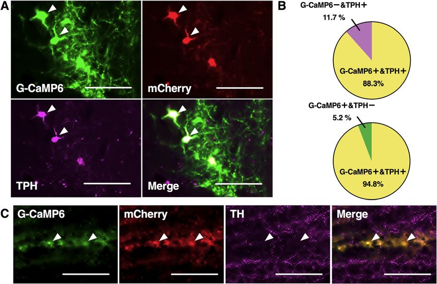
Moriya et al. Molecular Brain   (2020) 13:14                                                                  Page 2 of 12

serotonin (5-hydroxytryptamine, 5-HT) neurons in re-         light/dark cycle (7:00 AM to 7:00 PM), the temperature
sponse to nociceptive stimuli have been indicated by         of 24 ± 1 °C, food and water were available ad libitum.
some electrophysiological studies [13, 14]. Also, ventral    All efforts were made to minimize animal suffering and
tegmental area (VTA) DA neurons play roles in regulat-       discomfort; to reduce the number of animals used. All
ing nociceptive information [12, 15, 16]. We recently        experimental procedures were performed in accordance
certified that acute nociceptive stimuli rapidly increased   with the National Institute of Health Guide for the Care
the activities of LC noradrenalin (NA) neurons and           and Use of Laboratory Animals and approved by the
RVM 5-HT neurons and VTA DA neurons in awake                 Institutional Animal Use Committee of Kagoshima
mice by using fiber photometry system [17, 18].              University (MD17090).
  5-HT has extensive innervation in CNS [19] and sub-
sets of the 5-HT cells are designed as B1-B9 groups in a     Stereotaxic AAV injection
caudal to rostral direction [20]. The dorsal raphe nucleus   AAV vector production was performed by AAV Helper-
(DR: B7 and B6), median raphe nucleus (MR: B8 and            Free system (Agilent Technologies, Inc., Santa Clara,
B5), and RVM (B3) are well known as major parts. How-        CA, USA); their purification was as previously described
ever, supralemniscal (B9) 5-HT cell group located just       [17, 25, 26]. Mice were anesthetized with 2–3% isoflur-
dorsal to the medial lemniscus is less known [21] and        ane using a vaporizer for small animals and fixed with
has hardly been studied. In this study, we focused on the    stereotaxic instrument (ST-7, Narishige, Tokyo, Japan)
possible contribution of B9 5-HT system in pain pro-         with an aid of supportive ear bar (EB-6, Narishige) of
cessing because B9 5-HT cells project to LC and VTA          which touching surfaces to the animal were covered with
[22], which are important nuclei in pain processing (see     local anesthetic jelly (lidocaine, 2% Xylocaine AstraZe-
above). We have demonstrated fiber photometry system         neca). Both eyes were preserved with vaseline, head hair
[17, 18] for evaluating real time and cell type specific     was shaved using an electric hair shaver, and cranial
neuronal activity in awake mice with G-CaMP6 as a de-        dura mater was cut open with small scissors. We slowly
tector of Ca2+ concentration in the neuron of interest.      sucked up AAV into a glass micropipette (1B150F-3,
This system has high temporal resolution (< sec) and         World Precision Instrument, Inc., Sarasota, FL, USA),
unaffected by metabolism as in microdialysis, a different    which was connected to a nitrogen pressure source
character from other electrophysiological and chemical       through polyethylene tubing and to an injection manipu-
methods. Our study reported that acute nociceptive           lator (I-200 J, Narishige). In this study, AAV-TetO(3G)-
stimuli increased the activity of LC NA neurons or the       G-CaMP6 (Serotype: DJ; 1 μl/injection, 4 × 10¹³ copies/
activity of VTA DA neurons by adopting fiber photom-         ml) and AAV-TetO (3G)-mCherry (Serotype: DJ; 1 μl/in-
etry system. Therefore, we thought that it is meaningful     jection, 6 × 10¹² copies/ml) (Ohkura et al., 2012) (Fig. 1a)
to appraise the activities of B9-LC 5-HTergic pathway        were unilaterally injected into B9 site (Injection site was
and B9-VTA 5-HTergic pathway in response to acute            from bregma − 4.36 mm, lateral + 0.38 mm left side, and
nociceptive stimuli.                                         ventral − 5.08 mm from the cranium) (Fig. 1b). After
  We first introduced the G-CaMP6 in B9 5-HT neu-            AAV injection, the micropipette was left in place for
rons using transgenic mice carrying a tetracycline-          10 min before being slowly withdrawn; mice were given
controlled transactivator (tTA) transgene under the          an antibiotic, penicillin G (40,000 U kg-1) through sub-
control of a tryptophan hydroxylase-2 (TPH2) promoter        cutaneous injection. After operation, each mouse was in-
and site-specific injection of adeno associated virus        dividually kept for 14 days (2 weeks) in normal breeding
(AAV-TetO(3G)-G-CaMP6). We confirmed the specific            conditions (as mentioned in Animals section) because it
expression of G-CaMP6 in B9 neuronal cell body and           is needed for mice to recover and it takes about 2 weeks
axon located at the targeted sites (LC and VTA) using        for G-CaMP6 or mCherry to fully express (Fig. 1a).
an immunohistochemical method. We measured the
Ca2+ signal of G-CaMP6 in these sites of awake mice          In vivo fiber photometry system
while they were exposed to acute nociceptive stimuli.        We showed fiber photometry system in previous reports
                                                             [17, 18, 25, 26]. In this study, we adopted the fiber pho-
Material and methods                                         tometry system with two channels (Fig. 2a). In the first
Animals                                                      channel setting, the high-power LED driver (LEDD1B/
The tryptophan hydroxylase-2 tetracycline-controlled         M470F3, Thorlabs, Inc., Newton, NJ, USA) continuously
transactivator (TPH2-tTA) transgenic mice were used          produces blue excitation light (470 nm, 0.5 mW at the
[18, 23, 24] (Fig. 1a). We have already confirmed the        tip of the silica fiber) and the light passes through the
specificity of TPH2-tTA expression in the previous re-       excitation bandpass filter (475 ± 12.5 nm) and reflected
port [24]. Ten to fourteen-week-old mice were used in        by dichroic mirror-1; into a silica fiber (diameter:
this study. All mice were kept on a condition of 12 h        400 μm, numerical aperture = 0.6). The same fiber
Moriya et al. Molecular Brain   (2020) 13:14                                                                                    Page 3 of 12

 Fig. 1 Development of serotonin neuron specific expression of G-CaMP6/mCherry using tet system. a TPH2-tTA mouse was injected with AAV-
 TetO- G-CaMP6/mCherry and was individually kept for 14 days before the experiment. b AAV was unilaterally injected into B9 site

detects and collects the green fluorescent signal of G-                 New Zealand) and recorded by Labchart version-7 soft-
CaMP6. The signal passes through dichroic mirror-1                      ware (ADInstruments Inc.). Signals were collected at a
and reflected by dichroic mirror-2 and passes through                   sampling frequency of 100 Hz.
the bandpass emission filter (510 ± 12.5 nm) and guided
to a photomultiplier tube (PMTH-S1-1P28, Zolix Instru-                  Immunohistochemistry
ment, Beijing, China). At the second channel setting, the               To confirm AAV-induced expression of G-CaMP6 and
high-power LED driver continuously produces yellow                      mCherry in 5-HT neurons, after the experiments, mice
excitation light (590 nm) and the light passes through                  were processed for immunostaining. Mice were deeply
the excitation bandpass filter (590 ± 12.5 nm) and goes                 anesthetized with urethane (1.6 g/kg, i.p.) and transcar-
forward as well. The same fiber detects and collects the                dially perfused with 20 ml of phosphate buffered saline
red fluorescent signal of mCherry. The signal was for-                  (PBS) and 20 ml of 4%-paraformaldehyde in PBS (Naca-
ward and passed through the bandpass emission filter                    lai Tesque Inc., Kyoto, Japan). The brain was removed
(607 ± 12.5 nm) and guided to another photomultiplier                   and post fixed in the same paraformaldehyde solution
tube. First channel was adopted for detecting the neur-                 and soaked in 30% sucrose in PBS for 2 days. We formed
onal activity and the second channel was used as an                     serial 30 μm coronal sections including target sites (B9,
indicator of total stability of the fiber photometry system             LC, and VTA) with the cryostat (Cryotome FSE, Thermo
because mCherry fluorescence doesn’t reflect neuronal                   Scientific, Yokohama, Japan). Every third section was
activity [26]. Both signals were digitized by an A/D con-               adopted and floating immunohistochemical staining was
verter (PowerLab8/35, ADInstruments Inc., Dunedin,                      performed. The sections were soaked in blocking
Moriya et al. Molecular Brain     (2020) 13:14                                                                                           Page 4 of 12

 Fig. 2 Experimental procedures. a Schematic representation of the fiber photometry system with two channels. b Target sites in this study: B9, LC
 and VTA. c The fluorescence signal intensity was abruptly increased when the position of the optical fiber tip was placed just above target site. d
 Schematic representation of the procedure of the recording. Fiber implantation was performed under isoflurane anesthesia. We set 3 h before
 the beginning of experimental sessions so that anesthesia would not affect experimental sessions. Total four stimuli (two kinds of acute
 nociceptive stimuli and two kinds of noninvasive control stimuli) were set in order from weaker stimulus to stronger stimulus as follows; the first
 is low temperature heat stimulus at 25 °C, the second is a gentle touch, the third is acute heat stimulus at 55 °C, and the last is acute mechanical
 tail pinch stimulus at the force of 400 g. Inter-stimulus interval was set as 30 min to reduce possible carryover effect from the previous stimulus

solution (PBS containing 1% normal horse serum and                           guinea pig, Frontier Institute, Hokkaido, Japan, 1:200) in
0.3% Triton-X) for 1 h at room temperature and incu-                         blocking solution for overnight. The next day, the sec-
bated with anti-TPH antibody (AB1541, raised in sheep,                       tions were washed three times with PBS and incubated
EMD Millipore Corp., 1:1000) or anti-serotonin trans-                        with CF647 donkey anti-sheep IgG (20,284, Biotium,
porter (SERT) antibody (HTT-GP Af1400, raised in                             Inc., Fremont, CA, USA, 1:200) or CF647 donkey anti-
Moriya et al. Molecular Brain (2020) 13:14 Page 5 of 12
guinea pig IgG (20,837, Biotium, 1:200) in PBS for 2 h in tip was placed just above the target site (Fig. 2c). Open
a dark box. In some sections in LC, VTA, B9 were space around the optic fiber was covered with an oint-
treated with anti-tyrosine hydroxylase (TH) antibody ment to avoid possible drying. After the fiber was fixed
(AB152, Millipore, raised in rabbit, 1:500) and visualized and positioned to the optimal position, anesthesia was
with CF647 donkey anti-rabbit IgG (20,047, Biotium). turned off; each mouse recovered from anesthesia. We
After incubation, the sections were washed once with set 3 h before the beginning of experimental sessions so
PBS and mounted on microscope slides (PRO-02, Mat- that anesthesia would not affect experimental sessions.
sunami, Osaka, Japan) and covered with microcover The mice were divided into three groups; B9 group (n =
glasses (C024601, Matsunami). We observed and imaged 6), LC group (n = 6) and VTA group (n = 6). Experimen-
the sections under the fluorescence microscope (BZ- tal sessions of each mouse consist of two kinds of acute
X700, Keyence, Osaka, Japan), and analyzed the images nociceptive stimuli and two kinds of noninvasive control
with Adobe Photoshop CC (Adobe Systems, Inc., San stimuli (total four stimuli). To reduce the effect from the
Jose, CA, USA). G-CaMP6 and mCherry were visible previous stimulus, we set inter-stimulus intervals at
without immunostaining. 30 min; their stimuli were set in order of weaker to
stronger stimulus as follows; the first is low temperature
Acute nociceptive test heat stimulus at 25 °C, the second is a gentle touch, the
We applied two types of acute stress test showed in pre- third is acute heat stimulus at 55 °C; the last is acute
vious reports [17, 18]. We applied acute tail pinch mechanical tail pinch stimulus at the force of 400 g
stimulus using a pinch meter (PM-201, Soshin-Medic, (Fig. 2d). After experiments, mice were euthanized and
Chiba, Japan) and acute heat stimulus using a heating processed for immunostaining.
probe (5R7–570, Oven Industries, Inc., Mechanicsburg, The definition of neuronal activity characteristic index
PA, USA). Pinch stimulus was attached to the root of was set as follows: F: averaged fluorescent signal inten-
the tail for three seconds with a force of 400 g and heat sity value for three seconds just before each stimulus
probe set at 55 °C was attached to the root of the tail for and defined as 100%; ΔF: (maximum fluorescent signal
three seconds. We applied two noninvasive stimuli in intensity value during each stimulus) – F; onset latency:
the control group: gentle touch using a cotton stick and time from the start of stimulus to the time when the
low temperature heat stimulus set at 25 °C using the fluorescence signal intensity exceeded the maximum
same heating probe (5R7–570). value during the baseline period; peak latency: time from
the start of stimulus to the time when the fluorescence
Experimental protocol signal intensity reach the maximum value.
In this study, we recorded G-CaMP6/mCherry green/red
fluorescence intensity of B9 5-HT neuronal cell body Statistical analysis
and the axon located in LC/VTA (projecting sites of B9 Data analysis was conducted by two-way analysis of vari-
5-HT neurons, Fig. 2b) to acute nociceptive stimuli. ance (ANOVA) with Sidak’s test for post hoc analysis.
Each mouse was individually kept for at least 14 days Two factors on ΔF/F were modality (mechanical vs.
after injection of AAV (Fig. 1a). The mouse was again thermal) and intensity (nociceptive vs. gentle control).
anesthetized with 2–3% isoflurane using a vaporizer for Factors on latency were modality (mechanical vs. ther-
small animals and fixed with stereotaxic instrument (ST- mal) and brain area (B9, LC, or VTA). Values are
7) with an aid of supportive bar (EB-6) of which touch- expressed as the mean ± standard error of the mean
ing surfaces to the animal was covered with local (S.E.M). Probability values less than (p < 0.05) were con-
anesthetic jelly (AstraZeneca). Following experiments sidered statistically significant. The analyses were per-
were conducted in head-fixed condition. The head hair formed using GraphPad Prism version 7 (GraphPad
was shaved using electrical hair shaver; cranial dura software, San Diego, CA, USA).
mater was cut open with small scissors. A silica fiber
was slowly implanted into the places just above B9 Results
(bregma − 4.36 mm, lateral + 0.38 mm left, and ventral − Restricted expression of AAV-induced G-CaMP6/mCherry
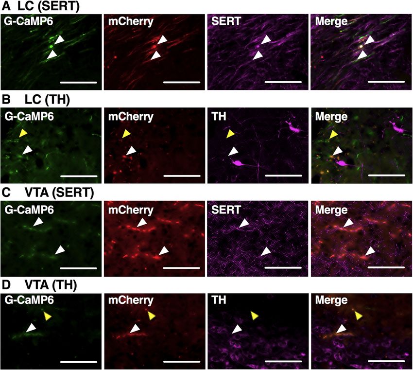
5.08 mm from the cranium), LC (bregma − 5.34 mm, Specific expressions of G-CaMP6/mCherry were con-
lateral + 0.80 mm left, and ventral − 2.60 mm from the firmed in B9 5-HT neuronal soma (Fig. 3a, b). We found
surface of the brain) and VTA (bregma − 3.15 mm, 23.3 ± 1.8 (n = 6, one representative slice per mouse)
lateral + 0.50 mm left, and ventral − 4.15 mm from the TPH-positive cells in the B9 and 88.3% of them also
surface of the brain) (Fig. 2b). We monitored the fluor- expressed G-CaMP6. All of the G-CaMP6 positive cells
escence signal intensity throughout fiber implantation also expressed mCherry and 94.8% expressed TPH. Al-
and confirmed that the fluorescence signal intensity in- though anti-TPH antibody we used (AB1541) sometimes
creased abruptly when the optimal position of the fiber binds TH, distribution of anti-TH-positive structure in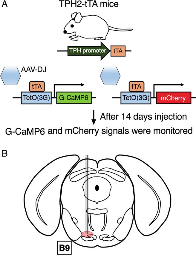
Moriya et al. Molecular Brain (2020) 13:14 Page 6 of 12
Fig. 3 Specific expressions of G-CaMP6/mCherry in B9 area. a Fluorescence from G-CaMP6 (green) and mCherry (red) overlapped on TPH-positive
cell soma (pink) in B9 area indicating specific expression of G-CaMP6/mCherry B9 5-HT neurons. Arrowheads show typical examples. b The
percentage of G-CaMP6+ and TPH+ double positive cells in total TPH+ cells (upper) and the percentage of G-CaMP6+ and TPH+ double positive
cells in total G-CaMP6+ cells (lower) (n = 6). c G-CaMP6/mCherry double positive cells did were not stained with anti-TH antibody
immunostaining. Scale bar length:100 μm
B9 did not overlap with that of G-CaMP6 (Fig. 3c). thermal stimuli (F(1, 5) = 21.9, p = 0.0054) whereas those
These expressions were also confirmed in B9 5-HT- in LC (F(1, 5) = 1.21, p = 0.3210) and VTA (F(1, 5) =
derived axons located in LC (Fig. 4a and b) and VTA 4.23, p = 0.0949) did not. Subsequent post hoc analysis
(Fig. 4c and d). G-CaMP6/mCherry positive soma and revealed that there was significant difference between
axons were hardly observed outside SERT positive struc- control vs. nociceptive stimulus in every combination of
tures (Fig. 4a and c). In LC and VTA, G-CaMP6/ brain area and stimulus modality (Fig. 6). Note that ΔF/
mCherry double positive fibers were found not only near F values during non-nociceptive control stimuli did not
TH positive cell body (white rectangles in Fig. 4b and d) exceed the fluctuation of fluorescence intensity during
but also TH negative areas (yellow rectangles) within the the baseline period; B9 group (1.12 ± 0.17%), LC group
nucleus. Therefore, fluorescence was detected in specific (1.47 ± 0.33%), VTA group (1.21 ± 0.23%).
manner in B9/LC/VTA sites. On the other hands, mCherry fluorescent intensity in
B9 group and LC/VTA groups was not significantly dif-
Effects of acute nociceptive stimulus on fluorescence ferent between stimulation intensities (nociceptive vs.
intensity of G-CaMP6 and mCherry gentle) and between modalities (mechanical and ther-
We confirmed proper positioning of the silica fiber on mal) (B9 group intensity: F(1, 5) = 0.3281, p = 0.5916; B9
B9, LC, and VTA by both physiological method (Fig. 2c) group modality: F(1, 5) = 0.00104, p = 0.9755; LC group
and histological method (Additional file 1: Figure S1). intensity: F(1, 5) = 0.1215, p = 0.7416; LC group modality:
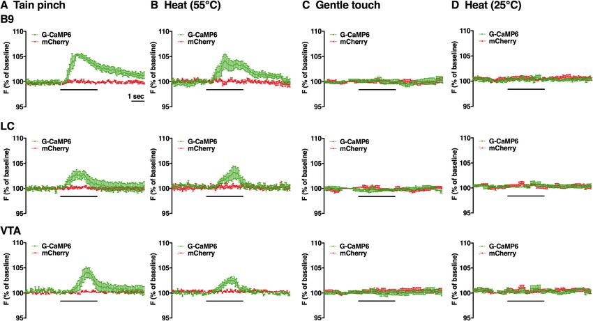
Figure 5 shows the trace of G-CaMP6/mCherry fluor- F(1, 5) = 0.5470, p = 0.4928; VTA group intensity: F(1,
escence intensity associated with acute nociceptive 5) = 0.0049, p = 0.9471, VTA group modality: F(1, 5) =
stimulus (Fig. 5a-d). G-CaMP6 fluorescence intensity in 0.09759, p = 0.767). ΔF/F values during both non-
B9 group and LC/VTA groups was rapidly increased by nociceptive control stimuli and nociceptive stimuli did
two acute nociceptive stimuli but not by non- not exceed the fluctuation of fluorescence intensity dur-
nociceptive control stimuli. Two factor ANOVA re- ing the baseline period; B9 group (1.27 ± 0.21%), LC
vealed that the increase in G-CaMP6 fluorescence was group (113 ± 0.23%), VTA group (1.05 ± 0.18%).
significantly different between stimulus intensities (gen- When comparing response characteristics, onset la-
tle vs. nociceptive) (B9 group: F(1, 5) = 31.1, p = 0.0026; tency was significantly different among 3 brain areas
LC group: F(1, 5) = 55.2, p = 0.0007; VTA group: F(1, (F(2, 15) = 57.19, p < 0.001) and also between modality
5) = 24.7, p = 0.0042). G-CaMP6 fluorescence in B9 also (F(1, 15) = 19.77, p = 0.0005). Sidak’s multiple compari-
showed significant difference between mechanical and son revealed that onset latency in B9 was significantlyMoriya et al. Molecular Brain (2020) 13:14 Page 7 of 12
Fig. 4 Expression of 5-HT fibers in LC and VTA. In LC (a, b) and VTA (c, d), triple positive axon fibers were confirmed (white rectangles). These
fibers represent B9 5-HT-derived axons since AAV was locally injected into B9. G-CaMP6/mCherry positive soma and axons were hardly observed
outside SERT positive structures. In LC (b) and VTA (d), G-CaMP6/mCherry double-positive fibers were found not only near TH positive cell body
but also TH negative areas (yellow rectangles) within the nucleus, indicating B9 5-HT neurons project to not only catecholaminergic neurons (NA
in LC and DA in VTA) but also other neurons in the target nuclei. Scale bar length:100 μm
shorter than that in LC and VTA in both pinch and heat report that measured the activities of B9 5-HT neurons
stimuli (Fig. 7a). during aversive stimuli and that showed possible role of
Although 2-way ANOVA revealed significant differ- B9 5-HT neurons in pain processing.
ence in peak latency among 3 brain areas (F(2, 15) = In addition, this is the first report that measured the ac-
7.483, p = 0.0056) and between modality (F(1, 15) = tivities of B9 5-HT nerve axons located in LC and VTA.
15.32, p = 0.0014), Sidak’s multiple comparison revealed The present results showed that the activity of B9-LC 5-HT
that there was significant difference between B9 and pathway and B9-VTA 5-HT pathway were rapidly in-
VTA when pinch stimulus was applied and that there creased by acute nociceptive stimuli. The results of onset
was no difference in other combinations (Fig. 7b). latency showed that in B9 was significantly shorter than
those in LC or VTA in both pinch and heat stimuli (Fig. 7a).
Discussion This result was in line with our hypothesis that the activ-
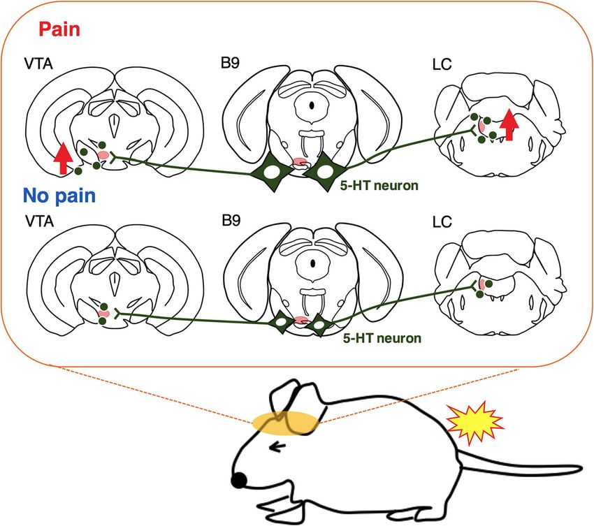
The results of this study clearly demonstrated that acute ities of B9 5-HT neuronal soma propagate to LC and VTA
nociceptive stimuli rapidly affected the activity of B9 5- through B9 5-HT-derived axons (Fig. 8). Our previous
HT neuronal cell bodies and B9 5-HT nerve axons studies using fiber photometry system showed that acute
located in LC and VTA in conscious mice adopting fiber nociceptive stimuli rapidly increased the activities of LC
photometry system. Recent tracer studies revealed B9- NA neurons and VTA DA neurons [17, 18]. Some studies
LC/B9-VTA 5-HT neuronal pathways [22]. B9 5-HT cell have reported the activation of LC NA neurons using mi-
group comprises approximately 20% of the total meso- crodialysis [14] or electrophysiological recording [28, 29].
pontine 5-HT neurons [21, 27], nevertheless has been Other studies have reported that nociceptive stimuli af-
much less studied compared to the wealth of studies on fected mesolimbic DA system [30, 31] and mesocortical
the DR, MR, and RVM groups. To our knowledge, our DA system [32, 33]. In this regard, it is considered that B9
data using the fiber photometry system are the first 5-HT neuronal projection to LC affected the activity of LCMoriya et al. Molecular Brain (2020) 13:14 Page 8 of 12 Fig. 5 Averaged traces of the fluorescence intensity of G-CaMP6 and mCherry. a Tail pinch, b Heat, c Gentle touch, d Low heat. Recordings taken from B9, LC, and VTA (from top to bottom). Horizontal bar shows time of stimulation. Each trace shows an average in 6 animals. Vertical bars indicate S.E.M NA neurons in pain processing system of DAS; in a similar results coincide with the previous studies showing dense manner, B9 5-HT neuronal projection to VTA affected the collections of 5-HT cells in B9 [21, 34]. Taken together, VTA DA neurons. This notion is supported by our histo- our method using AAV seemed applicable to study logical examination showing the close location of B9 5-HT activity of any 5-HT neurons in the CNS. axon near the NA neurons in LC (Fig. 4b) and the DA neu- Emerging evidence has pointed to anatomical and rons in VTA (Fig. 4d). Although we have revealed possible functional heterogeneity within the brainstem 5-HTergic flow of pain information from B9 to LC and VTA, we need cell groups [35, 36]. Nevertheless, the function of B9 5- more studies to reveal physiological importance and impact HT neurons has been largely unknown, except for stud- of this pathway in pain regulation. ies reporting that B9 5-HT neurons played prominent We confirmed the expression of G-CaMP6/mCherry roles in shaping aggression [37] and regulating affect in B9 5-HT neurons in TPH2-tTA mice injected with and stress responsivity [36, 38]. DAS, including LC, is AAV-tetO-GCaMP6/mCherry by immunohistochemical considered to be involved in pain-emotion symptoms method. In a previous study, we showed the expression [28, 39] and VTA involved in mood symptom [40, 41] of G-CaMP6/mCherry in RVM/DR 5-HT neurons in and fear [42]. An anatomical study demonstrated that TPH2-tTA mice using the same AAV [18]. Our present B9 5-HT neurons project to the hypothalamus, cortex, Fig. 6 Effects of aversive and control stimuli on G-CaMP6 fluorescence intensity. (a) B9 5-HT neuronal soma, (b) B9 5-HT-derived axon in LC, and (c) B9 5-HT-derived axon in VTA. Values are expressed as the mean ± S.E.M (n = 6, each). Statistical analysis was conducted by two-way ANOVA with Sidak’s test for post hoc analysis. P values by the Sidak’s post hoc test are shown in the figure
Moriya et al. Molecular Brain     (2020) 13:14                                                                                          Page 9 of 12

 Fig. 7 Characteristics of G-CaMP6 fluorescence response to aversive stimuli. a Onset latency; time from start of stimulus to the time when
 fluorescence signal intensity exceeded the maximum value during the baseline period. (ab) Peak latency; time from start of stimulus to the time
 when fluorescence signal intensity arrived at the maximum point. Values are expressed as mean ± S.E.M (n = 6, respectively). Statistical analysis
 was conducted by two-way ANOVA with Sidak’s test for post hoc analysis. P values by the Sidak’s post hoc test are shown in the figure

and hippocampus that are related to some psychiatric                        related drugs affect the state of monoamines in CNS.
symptoms [43]. Taking these observations together, our                      SNRI, SSRI and TCA for pain treatment act on synapses
results suggest that B9-LC/B9-VTA 5-HT neuronal                             of 5-HT neurons in CNS and it takes at least a few
pathways may be related to pain-emotion symptoms.                           weeks to relieve pain symptoms [44]. In case of patients
  In clinical psychiatric medicine, the center of thera-                    in the psychiatric field with pain symptoms receiving
peutic strategy is drug therapy and many psychiatric-                       these drug therapy, the state of neuronal activity in a
Moriya et al. Molecular Brain       (2020) 13:14                                                                                         Page 10 of 12

 Fig. 8 Schematic explanation of possible contribution of B9 5-HT neurons in pain processing

subset of 5-HTergic cell groups including B9 can be af-                   Abbreviations
fected. This possibility seems worth testing in the future                5-HTergic: Serotonergic; 5-hydroxytryptamine, 5-HTserotonin; AAV: Adeno
                                                                          associated virus; ANOVA: Analysis of variance; CNS: Central nervous system;
study.                                                                    DA: Dopamine; DAS: Descending antinociceptive system; DR: Dorsal raphe
   Limitations in this study are as follows. We did not                   nucleus; LC: Locus coeruleus; MR: Median raphe nucleus; NA: Noradrenalin;
adopt psychiatric-related drugs, therefore did not ap-                    PBS: Phosphate-buffered saline; RVM: Rostral ventromedial medulla;
                                                                          S.E.M: Standard error of the mean; SERT: Serotonin transporter;
praise how these drugs affect the activities of B9 5-HT                   SNRI: Serotonin noradrenalin reuptake inhibitor; SSRI: Selective serotonin
neurons. Also, our protocol is only acute nociceptive                     reuptake inhibitor; TCA: Tricyclic antidepressant; TH: Tyrosine hydroxylase;
system. Therefore, in the future, the protocol including                  TPH2: Tryptophan hydroxylase-2; tTA: Tetracycline-controlled transactivator
                                                                          transgene; VTA: Ventral tegmental area
psychiatric-related drugs and chronic nociceptive system
will be adopted. Fiber photometry system measures ac-                     Acknowledgements
tivities of ensemble average of the labeled neurons but                   We thank all the staff members of Institute of Laboratory Animal Sciences at
not single unit activity. Combination of advantages of                    Kagoshima University for keeping the animals in good condition. We
                                                                          acknowledge the Joint Research Laboratory, Kagoshima University Graduate
multiple methodologies are needed.                                        School of Medical and Dental Sciences, for the use of their facilities.
   In conclusion, the results of this study suggest that
acute nociceptive stimuli cause a rapid increase in the                   Authors’ contributions
activities of B9-LC/B9-VTA 5-HTergic pathways and                         SM designed the study; SM, A Yamashita, YK, JS, HS, A Yamanaka and TK
suggest that B9 5-HT neurons play important roles in                      conducted the study; SM, DM and TK analyzed the data and created the
                                                                          figure; and SM and TK wrote the manuscript. All authors approved the final
nociceptive processing in the CNS.                                        version of the manuscript.

Supplementary information                                                 Funding
Supplementary information accompanies this paper at https://doi.org/10.   This work was supported by JSPS KAKENHI Grants (19 K17093 to S.M.; 17
1186/s13041-020-0553-1.                                                   K14936 to A. Yamashita; 16H05130 to T.K.) and CREST JST (JPMJCR1656 to A.
                                                                          Yamanaka).

 Additional file 1: Figure S1. Confirmation of fiber implantation
 tracking. Fiber track was located just above B9, LC and VTA.             Availability of data and materials
                                                                          All data in this study are available upon requests.
Moriya et al. Molecular Brain (2020) 13:14 Page 11 of 12
Ethics approval 20. Dahlstrom A, Fuxe K. Localization of monoamines in the lower brain stem.
All experimental procedures were performed in accordance with the Experientia. 1964;20(7):398–9.
National Institute of Health Guide for the Care and Use of Laboratory 21. Vertes RP, Crane AM. Distribution, quantification, and morphological
Animals and approved by the Institutional Animal Use Committee of characteristics of serotonin-immunoreactive cells of the supralemniscal
Kagoshima University. nucleus (B9) and pontomesencephalic reticular formation in the rat. J Comp
Neurol. 1997;378(3):411–24.
Consent for publication 22. Muzerelle A, Scotto-Lomassese S, Bernard JF, Soiza-Reilly M, Gaspar P.
Not applicable. Conditional anterograde tracing reveals distinct targeting of individual
serotonin cell groups (B5-B9) to the forebrain and brainstem. Brain Struct
Competing interests Funct. 2016;221(1):535–61.
The authors declare that they have no competing interests. 23. Ohkura M, Sasaki T, Sadakari J, Gengyo-Ando K, Kagawa-Nagamura Y,
Kobayashi C, et al. Genetically encoded green fluorescent Ca2+ indicators
Author details with improved detectability for neuronal Ca2+ signals. PLoS One. 2012;
1
Department of Physiology, Kagoshima University Graduate School of 7(12):e51286.
Medical and Dental Science, Kagoshima 890-8544, Japan. 2Department of 24. Ikoma Y, Kusumoto-Yoshida I, Yamanaka A, Ootsuka Y, Kuwaki T. Inactivation
Molecular Pharmacology and Neurobiology, Yokohama City University of serotonergic neurons in the rostral medullary raphe attenuates stress-
Graduate School of Medicine, Yokohama 236-0004, Japan. 3Department of induced tachypnea and tachycardia in mice. Front Physiol. 2018;9:832.
Pharmacology, Kagoshima University Graduate School of Medical and Dental 25. Inutsuka A, Yamashita A, Chowdhury S, Nakai J, Ohkura M, Taguchi T, et al.
Science, Kagoshima 890-8544, Japan. 4Research Institute of Environmental The integrative role of orexin/hypocretin neurons in nociceptive perception
Medicine, Nagoya University, Nagoya 464-8601, Japan. and analgesic regulation. Sci Rep. 2016;6:29480.
26. Futatsuki T, Yamashita A, Ikbar KN, Yamanaka A, Arita K, Kakihana Y, et al.
Received: 3 October 2019 Accepted: 19 January 2020 Involvement of orexin neurons in fasting- and central adenosine-induced
hypothermia. Sci Rep. 2018;8(1):2717.
27. Baker KG, Halliday GM, Halasz P, Hornung JP, Geffen LB, Cotton RG, et al.
Cytoarchitecture of serotonin-synthesizing neurons in the pontine
References
tegmentum of the human brain. Synapse (New York). 1991;7(4):301–20.
1. Makris UE, Abrams RC, Gurland B, Reid MC. Management of persistent pain
in the older patient: a clinical review. JAMA. 2014;312(8):825–36. 28. Alba-Delgado C, Llorca-Torralba M, Horrillo I, Ortega JE, Mico JA, Sanchez-
2. Waller E, Scheidt CE. Somatoform disorders as disorders of affect regulation: Blazquez P, et al. Chronic pain leads to concomitant noradrenergic
a development perspective. Int Rev Psychiat. 2006;18(1):13–24. impairment and mood disorders. Biol Psychiatry. 2013;73(1):54–62.
3. Gebhardt S, Heinzel-Gutenbrunner M, Konig U. Pain relief in depressive 29. Voisin DL, Guy N, Chalus M, Dallel R. Nociceptive stimulation activates locus
disorders: a meta-analysis of the effects of antidepressants. J Clin coeruleus neurones projecting to the somatosensory thalamus in the rat. J
Psychopharmacol. 2016;36(6):658–68. Physiol. 2005;566(Pt 3):929–37.
4. Attal N, Bouhassira D. Translational neuropathic pain research. Pain. 2019; 30. Klitenick MA, Taber MT, Fibiger HC. Effects of chronic haloperidol on stress-
160(Suppl 1):S23–s8. 10. and stimulation-induced increases in dopamine release: tests of the
5. Morin CM, Drake CL, Harvey AG, Krystal AD, Manber R, Riemann D, et al. depolarization block hypothesis. Neuropsychopharmacol. 1996;15(4):424–8.
Insomnia disorder. Nat Rev Dis Primers. 2015;1:15026. 31. Naef L, Gratton A, Walker CD. Exposure to high fat during early
6. Lee YC, Chen PP. A review of SSRIs and SNRIs in neuropathic pain. Expert development impairs adaptations in dopamine and neuroendocrine
Opin Pharmacother. 2010;11(17):2813–25. responses to repeated stress. Stress. 2013;16(5):540–8.
7. Melzack R, Wall PD. Pain mechanisms: a new theory. Science. 1965; 32. Jedema HP, Grace AA. Chronic exposure to cold stress alters
150(3699):971–9. electrophysiological properties of locus coeruleus neurons recorded in vitro.
8. Millan MJ. Descending control of pain. Prog Neurobiol. 2002;66(6):355–474. Neuropsychopharmacol. 2003;28(1):63–72.
9. Yaksh TL, Hammond DL, Tyce GM. Functional aspects of bulbospinal 33. Butts KA, Phillips AG. Glucocorticoid receptors in the prefrontal cortex
monoaminergic projections in modulating processing of somatosensory regulate dopamine efflux to stress via descending glutamatergic feedback
information. Fed Proc. 1981;40(13):2786–94. to the ventral tegmental area. Int J Neuropsychopharmacol. 2013;16(8):
10. Ochi T, Goto T. The antinociceptive effect induced by FR140423 is mediated 1799–807.
through spinal 5-HT2A and 5-HT3 receptors. Eur J Pharmacol. 2000;409(2): 34. Hornung JP, Fritschy JM. Serotoninergic system in the brainstem of the
167–72. marmoset: a combined immunocytochemical and three-dimensional
11. Gear RW, Levine JD. Nucleus accumbens facilitates nociception. Exp Neurol. reconstruction study. J Comp Neurol. 1988;270(4):471–87.
2011;229(2):502–6. 35. Clark MS, McDevitt RA, Neumaier JF. Quantitative mapping of tryptophan
12. Taylor AM, Becker S, Schweinhardt P, Cahill C. Mesolimbic dopamine hydroxylase-2, 5-HT1A, 5-HT1B, and serotonin transporter expression across
signaling in acute and chronic pain: implications for motivation, analgesia, the anteroposterior axis of the rat dorsal and median raphe nuclei. J Comp
and addiction. Pain. 2016;157(6):1194–8. Neurol. 2006;498(5):611–23.
13. Chiang CY, Gao B. The modification by systemic morphine of the responses 36. Lowry CA, Hale MW, Evans AK, Heerkens J, Staub DR, Gasser PJ, et al.
of serotonergic and non-serotonergic neurons in nucleus raphe magnus to Serotonergic systems, anxiety, and affective disorder: focus on the
heating the tail. Pain. 1986;26(2):245–57. dorsomedial part of the dorsal raphe nucleus. Ann N Y Acad Sci. 2008;
14. Sajedianfard J, Khatami S, Semnanian S, Naghdi N, Jorjani M. In vivo 1148:86–94.
measurement of noradrenaline in the locus coeruleus of rats during the 37. Kerman IA, Clinton SM, Bedrosian TA, Abraham AD, Rosenthal DT, Akil
formalin test: a microdialysis study. Eur J Pharmacol. 2005;512(2–3):153–6. H, et al. High novelty-seeking predicts aggression and gene expression
15. Kalivas PW, Duffy P. Selective activation of dopamine transmission in the differences within defined serotonergic cell groups. Brain Res. 2011;
shell of the nucleus accumbens by stress. Brain Res. 1995;675(1–2):325–8. 1419:34–45.
16. Ungless MA, Magill PJ, Bolam JP. Uniform inhibition of dopamine neurons 38. Waselus M, Galvez JP, Valentino RJ, Van Bockstaele EJ. Differential
in the ventral tegmental area by aversive stimuli. Science. 2004;303(5666): projections of dorsal raphe nucleus neurons to the lateral septum and
2040–2. striatum. J Chem Neuroanat. 2006;31(4):233–42.
17. Moriya S, Yamashita A, Kawashima S, Nishi R, Yamanaka A, Kuwaki T. Acute 39. Borges G, Neto F, Mico JA, Berrocoso E. Reversal of monoarthritis-induced
aversive stimuli rapidly increase the activity of ventral tegmental area affective disorders by diclofenac in rats. Anesthesiol. 2014;120(6):1476–90.
dopamine neurons in awake mice. Neurosci. 2018;386:16–23. 40. Tidey JW, Miczek KA. Social defeat stress selectively alters
18. Moriya S, Yamashita A, Nishi R, Ikoma Y, Yamanaka A, Kuwaki T. Acute mesocorticolimbic dopamine release: an in vivo microdialysis study.
nociceptive stimuli rapidly induce the activity of serotonin and noradrenalin Brain Res. 1996;721(1–2):140–9.
neurons in the brain stem of awake mice. IBRO Rep. 2019;7:1–9. 41. Tye KM, Mirzabekov JJ, Warden MR, Ferenczi EA, Tsai HC, Finkelstein J, et al.
19. Jacobs BL, Azmitia EC. Structure and function of the brain serotonin system. Dopamine neurons modulate neural encoding and expression of
Physiol Rev. 1992;72(1):165–229. depression-related behaviour. Nature. 2013;493(7433):537–41.Moriya et al. Molecular Brain (2020) 13:14 Page 12 of 12
42. Abraham AD, Neve KA, Lattal KM. Dopamine and extinction: a convergence
of theory with fear and reward circuitry. Neurobiol Learn Memory. 2014;108:
65–77.
43. Vertes RP, Martin GF. Autoradiographic analysis of ascending projections
from the pontine and mesencephalic reticular formation and the median
raphe nucleus in the rat. J Comp Neurol. 1988;275(4):511–41.
44. Mitsi V, Zaxhariou V. Modulation of pain, nociception, and analgesia by the
brain reward center. Neurosci. 2016;338:81–92.
Publisher’s Note
Springer Nature remains neutral with regard to jurisdictional claims in
published maps and institutional affiliations.You can also read