Effects of Stabilization Exercise Using a Ball on Mutifidus Cross-Sectional Area in Patients with Chronic Low Back Pain
←
→
Page content transcription
If your browser does not render page correctly, please read the page content below
©Journal of Sports Science and Medicine (2013) 12, 533-541
http://www.jssm.org

    Research article

Effects of Stabilization Exercise Using a Ball on Mutifidus Cross-Sectional Area
in Patients with Chronic Low Back Pain

SinHo Chung 1 JuSang Lee                   2
                                               and JangSoon Yoon 3
1
 Department of Rehabilitation Medicine, Hanyang University Medical Center, Seoul, South Korea; 2 Department of
Physical Therapy, Hallym College, Gangwon-do, South Korea; 3 Department of Rehabilitation Medicine, Hallym Medi-
cal Center, Gangwon-do, South Korea

                                                                          that vary in method and intensity in order to improve or
Abstract                                                                  maintain their muscle strength and improve balance
The purpose of this study was to compare the effects of lumbar            (Cairns et al., 2006; Goldby et al., 2006; Kofotolis and
stabilization exercises using balls to the effects of general lum-        Kellis, 2006). Recently, stabilization exercises that focus
bar stabilization exercises with respect to changes in the cross          on core strengthening, mobility control, and muscle con-
section of the multifidus (MF), weight bearing, pain, and func-           trol have become a very important treatment method for
tional disorders in patients with non-specific chronic low back           low back pain (MacDonald et al., 2006; Richardson et al.,
pain. Twelve patients participated in either a 8 week (3 days per
week) stabilization exercise program using balls and control
                                                                          2002). These exercises can improve the functions of the
group (n = 12). The computer tomography (CT) was used to                  nervous and the muscular systems and thereby control and
analyze MF cross-sectional areas (CSA) and Tetrax balancing               protect the spine. In practice, these exercises enhance
scale was used to analyze left and right weight bearing differ-           control over the lumbar spine and the pelvis (Hodges,
ences. Both groups had significant changes in the CSA of the              2003), and can be performed in diverse body positions
MF by segment after training (p < 0.05) and the experimental              using the co-contraction of the abdominal and MF mus-
group showed greater increases at the L4 (F = 9.854, p = 0.005)           cles (Andrusaitis et al., 2011). The purpose of stabiliza-
and L5 (F = 39.266, p = 0.000). Both groups showed significant            tion exercises is to improve the activation patterns of
decreases in weight bearing, from 9.25% to 5.83% in the ex-               trunk muscles, in order to relieve lumbar pain and inca-
perimental group and from 9.33% to 4.25% in the control group
(p < 0.05), but did not differ significantly between the two
                                                                          pacity through trunk muscle contraction (Goldby et al.,
groups. These results suggests that stabilization exercises using         2006; Kavcic et al., 2004).
ball can increases in the CSA of the MF segments, improvement                     Unstable training devices, such as balls, can be
in weight bearing, pain relief, and recovery from functional              used to increase the difficulty of exercises employing
disorders, and the increases in the CSA of the MF of the L4 and           diverse body weight and free-weight resistance (Anderson
L5 segments for patients with low back pain.                              and Behm, 2005). Exercises that use balls use all regions
                                                                          of the body so that more extensive activities can occur
Key words: Stabilization exercise, ball, multifidus, cross-               than with exercises performed on fixed floors. The use of
sectional area, low back pain.                                            balls therefore can improve the dynamic balance ability,
                                                                          the flexibility and stability of the spine, and the sense of
                                                                          balance as ways to prevent damage (Marshall and Mur-
Introduction                                                              phy, 2005). Marshall and Murphy (2006) reported that a
                                                                          12 week regimen of spinal stabilization exercises using
Low back pain is the most representative musculoskeletal                  balls by low back pain patients brought about pain relief
system disorder (Chung et al., 2013; Kwon et al., 2011).                  and decreases in flexion-relaxation disorders as well as
Although this disorder can be remedied without any par-                   improvements in the ability to control balance through the
ticular treatment, chronic low back pain that persists for                strengthening of the MF muscle, which plays an impor-
three months or longer occurs in approximately 5~10% of                   tant role in spinal stability. However, other previous stud-
the patients and becomes a major cause that restricts pro-                ies reported that spinal motions did not change and re-
ductive lifestyle activities (Borenstein, 1996; Watson et                 vealed no increase in trunk muscle activity during core
al., 2000). Low back pain patients use movement strate-                   stability exercises on unstable surfaces (Drake et al.,
gies that differ from those of healthy persons (Grabiner et               2006; Freeman et al., 2006; Wahl and Behm, 2008).
al., 1992) because the onset time of their multifidus (MF)                        Although ball exercises are used for diverse pur-
and transverse abdominus (TrA), which are deep muscles,                   poses such as balance control and muscle strengthening,
are delayed and their ability to mobilize these muscles is                the beneficial effects of spinal stabilization exercises
reduced (Hodges and Richardson, 1999). In addition,                       using balls on lumbar vertebral segments remain to be
chronic low back patients frequently show poor balance                    verified. The purpose of the present study is the effects of
control (Harding et al., 1994) because they sway back-                    lumbar stabilization exercises using balls with respect to
ward to maintain their center of force (COF) when they                    changes in the cross-section area (CSA) of the MF,
balance themselves (Byl and Sinnott, 1991). Therefore,                    weight bearing, pain, and functional disorders in patients
low back pain patients are instructed to perform exercises                with non-specific chronic low back pain. The working

Received: 14 March 2013 / Accepted: 11 June 2013 / Available (online): 26 June 2013 / Published: 01 September 2013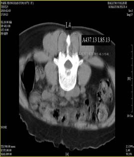
534                                                                                              Stabilization exercise with ball

hypothesis is that stabilization exercise programs will         group and 12 patients (5 males, 7 females; excluding two
increase trunk muscle activity and improve spinal stabil-       dropouts) in the control group. The experimental group
ity, thereby leading to pain relief, functional disorder        was divided into two treatment groups by randomly tak-
reduction, balance improvement, and increases in the            ing out pieces of paper with the treatment groups written
CSA of individual segments of the MF; i.e., greater im-         in them folded and put into an opaque envelope: group A
provement will be seen using stabilization exercise pro-        (Ball) or B (Mat). The CONSORT (consolidated stan-
grams using balls than with general stabilization exercise      dards of reporting trials) flowchart of the study is shown
programs.                                                       in Figure 1.
                                                                       The experimental group had a mean age of 35.20 ±
Methods                                                         10.01 years, height of 1.70 ± 0.01 m, weight of 64.40 ±
                                                                7.34 kg, and duration of illness of 12.76 ± 5.28 months
Subjects                                                        while the control group values were 41.32 ± 7.13 years,
The calculation of sample size was carried out with α =         1.66 ± 0.05 m, 62.54 ± 2.93 kg, and duration of illness of
0.05 (5% chance of type I error), 1- β = 0.80 (power            13.26 ± 7.13 months respectively. The experimental group
80%), and using the results of a previous study comparing       performed stabilization exercises using balls and the con-
between the Swiss ball and a stable surfaces because this       trol group performed general stabilization exercises. All
most closely resembles devices used in this study (Mar-         procedures were performed in accordance with the princi-
shall and Murphy, 2006), and a calculated effect size of δ      ples outlined in the Declaration of Helsinki, and partici-
= 1.21. This provided sample size of n = 20. This study         pants signed the appropriate informed consent form.
was large enough to consider drop out. The present study
was conducted with 28 patients of B Hospital who had no         Training program
particular anatomical or neurophysiological causes of           Warm-up and cool-down exercises consisted of walking
disease (X-ray and examination by a medical doctor) but         for 10 minutes each on a treadmill. The main exercises
had complained of low back pain for at least 12 weeks.          were performed three times per week for eight weeks
Subjects who had low back pain for the last six months,         using four different motions. Three times in the first
musculoskeletal system disorders, abnormal past histories       week, two physical therapists with five years or longer
in the spine (spondylitis, fracture), neurologic diseases, or   kinesiatrics careers educated the patients one by one by
functional restrictions on the upper or lower limbs were        explaining the purpose of the exercise therapy and dem-
excluded. Data were obtained from 12 patients (6 males, 6       onstrating the motions. Three times in the second and
females; excluding two dropouts) in the experimental            subsequent weeks, auditory feedback was provided by
Chung et al. 535
Figure 1. CONSORT flowchart of this study.
Figure 2. The stabilization exercise using a ball.
verbal instructions and tactile feedback was provided by 31-69 = moderate pain, 70-100 = severe pain (Kelly,
the therapist’s hand to guide the subjects in accurate exer- 2001). The inter-rater reliability of this tool is 0.55-0.97,
cise methods. Each exercise motion and the number of and the reliability within each rater is 0.69-0.91 (Taddio
times each was performed were recorded and the subjects et al., 2009).
were asked if they felt pain.
The stabilization exercise group that used balls per- Oswestry disability index (ODI)
formed as the following exercises: 1) In a supine position, For the functional assessment of low back pain, the Ko-
the subject placed a ball below the neck, bent the knees, rean version of the Oswestry disability index (ODI) ver-
and crossed and bent 90° the arms so that the crossed sion 2.0 developed by Fairbank et al. (2000) and trans-
arms came to the eye level. While breathing out, the sub- lated by Jeon et al. (2006) was used to measure low back
ject slowly raised each lower limb in turn. The subject pain. This tool was developed based on the 10-item ques-
performed the motion of bending the hip joint and the tionnaire of ODI to assess low back pain-related limita-
knee joint 90° five times, for 10 seconds each time; 2) In tions in daily life and requires the subject to select one of
a supine position, the subject placed a ball below the six points under each of the 10 items: the intensity of
pelvis, bent the knees, and crossed and bent 90° the arms pain, personal care, lifting, walking, sitting, standing,
so that the crossed arms came to the eye level. The subject sleeping, sex life, social life, and travelling (0–5 points).
performed the motion of pressing the ball below the pel- Functional disability scores were calculated as per-
vis slowly with the pelvis five times, for 10 seconds each centages by adding up the measured scores of individual
time; 3) In a crawling position, the subject placed a ball items, dividing the total score by the full score of the
below one knee and kept the toes away from contact with items, and multiplying the resultant value by 100. The
the floor. The subject balanced first to stabilize the pos- scores were presented as follows: 0-20 = mild disability,
ture and slowly raised the other lower limb. This exercise 20-40 = moderate disability, 40-60 = severe disability, 60-
was performed in turn for the two lower limbs 10 times, 80 = crippled, 80-100 = bed bound. This questionnaire is
for 10 seconds each time; and 4) After assuming a prone intended to examine disabilities resulting from pain rather
position, the subject placed a ball in front of the pelvis than the pain itself. This questionnaire provides supple-
and raised both lower limbs. As if kicking, the subjects mentary information for the results of the VAS. Cron-
repeatedly raised and lowered the two lower limbs alter- bach's alpha coefficient of 0.92, test-retest correlation
nately. The subject performed this exercise 10 times, for reliability was 0.93 (Jeon et al., 2006).
five sets, taking a rest of at least 15 seconds between each
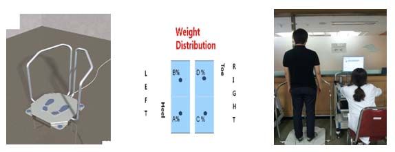
set, as shown in Figure 2. Weight bearing
The general stabilization exercise group performed Weight bearing was measured with a Tetrax Portable
the same motions on a mat. Multiple System (Tetrax, Sunlight Medical Ltd, Israel),
which enables implementation of biofeedback training
Procedures and instrumentation treatment while measuring balance. The degrees of pos-
Visual analog scale (VAS) tural sway for four placements of the feet can be meas-
In the present study, visual analog scales (VAS) were ured to indicate general stability indexes and an individ-
measured to assess pain. Each subject was instructed to ual’s control abilities and compensating postural changes
mark the intensity of his/her pain on a 100 mm stick can be monitored (Lee et al., 2012). Large changes in
without any gradation (Gould et al., 2001). Subjects body weight percentages at different foot sites and high
marked the intensity as 0 points when they experienced stability indexes indicate high instability (Kohen-Raz
no pain, and severe unendurable pain was given 10 points. et al., 1994: Kohen-Raz, 1991). This instrument consisted
The scores were presented as follows: 0-30 = mild pain, of separate force plates: two 12cm wide x 19cm long536 Stabilization exercise with ball
Figure 3. The balance measurement.
rectangular toe side foot force plates (left: B, right: D) and (Piview STAR, Infinitt Inc., Korea). The CSA were then
two 12cm wide x 12cm long square heel side foot force
measured and presented in Figure 4. The fat regions on
plates (left: A, right: C) and indicated weight indexes that
the innermost fascial borders of the MF and the erector
showed the proportion of weight distributed among the
spinae and on the MF fascial boundary were included.
four force plates. Information about the pressure imposed
The fat regions between the MF and the lamina were
on the force plates is amplified, filtered, and then deliv-
included in the CSA of the MF.
ered to the computer for analysis with the Tetrax software
In the present study, these four levels were ana-
program. The individual force plates measure changes in
lyzed to identify the most suitable level and to detect any
vertical pressure from the two toe side feet and heel side
systematic differences between different levels. Because
feet separately.
the L5s of many subjects were sharply angled, the lower
The subjects stood on the force plates, with arms
end plate of L4 was selected instead of the upper end
hanging down straight and their feet at shoulder width,
plate of L5 (Danneels et al., 2001; Keller et al., 1999). To
while viewing a mark 3m in front of them. The weight
reduce selective errors, the data were randomly measured
values on the two sides that were fixed after the subject
by the same specialist in radiology.
stood on the measuring instrument were selected as values
for the right and left sides, respectively. This procedure
Statistical analysis
was repeated three times for 30 seconds and the average
Statistical analyses were conducted using SPSS Windows
values were used as measurements (Kohen-Raz et al.,
(version 18.0). Data are presented as Mean ± Standard
1994) (Figure 3).
deviations. The normality of the independent variables
|(A % + B %) − (C % + D %)| = %
was tested by Kolmogrov-Smirnov test, indicating that all
Computed tomography (CT) of the multifidus muscle variables were confirmed normal distributions by the test.
Measuring method Demographic characteristics and dependent variables
The area of the lumbar MF was measured using CT scan were analyzed using Levene’s test of independent t-tests
(conditions: 120 kV, 160 mA, 0.6 s rotation time, 5 mm to verify homogeneity. Data showed that the normality
slice thickness, 5 mm reconstruction interval) using a and no violation of homogeneity of variance and thus that
Somatom Plus-4C (Siemens General Medical, Germany). parametrical tests. Paired t-tests were conducted to com-
The subject assumed a neutral position to avoid compres- pare differences in the CSA of the MF, weight bearing,
sion of the back muscle that occurs in a supine position. A pain, and functional disorders in the experimental group
pillow was placed below the abdomen to minimize lum- and the control group before and after the stabilization
bar lordosis and the subject was instructed to maintain a exercises. To compared over the training period in the
relaxed posture while being scanned. To confirm the experimental and control groups using two factor (group x
patient’s relaxed state, the tester palpated the patient’s time) repeated analysis of variance (ANOVA). The statis-
back muscle (Danneels et al., 2001). The iliac crest on tical significance level was set at 0.05.
both sides and the spinous process on L5 were identified
and their positions were marked on the skin. A longitudi- Results
nal scan was then performed toward the spine above the
spinous process. Changes in the CSA of the MF by segment pre and post
the experiment are showed significant increases at L2, L3,
Image analysis L4 and L5 of experimental and control groups, respec-
The CT images were analyzed after enlargement on the tively (p < 0.05). Between the two groups, in CSA of the
computer screen. On the sagittal plane of each MF, the MF at L2 (F = 2.236, p = 0.150) and L3 (F = 1.122, p
sizes of the MF at four levels from L2 to L5 were drawn =0.301) no statistically significant and greater increase in
clearly on the computer screen along the boundaries of the experimental group was statistically significant at L4
the muscles using the mouse cursor and the PACS (Pic- (F = 9.854, p = 0.005) and L5 (F = 39.266, p = 0.000)
ture Archiving and Communication System) program (Table 1). Both groups showed significant decreases inChung et al. 537
A B
C D
Figure 4. Cross-sectional images of the multifidus muscle. (A) L2 region. (B) L3 region. (C) L4 region. (D) L5 region.
weight bearing (p < 0.05). Comparison between the two the two groups indicated that the greater decrease in the
groups indicated no statistically significant (F = 2.512, p experimental group was statistically significant (F =
= 0.128). Decreases in pain were significant in both 5.256, p = 0.032) (Table 2).
groups (p < 0.05). Comparison between the two groups
indicated no statistically significant (F = 0.316, p = Discussion
0.580). Decreases in functional disorders were also sig-
nificant in both groups (p < 0.05). Comparison between The most important result of the present study is that
Table 1. Comparison of CSA within groups and between groups. Values are means (±SD).
Experimental (n=12) Control (n=12)
pre 351.62 (45.75) 355.37 (57.19)
L2 post 365.56 (44.39) * 362.04 (55.61) *
(mm2) change 13.94 (14.86) 6.67 (7.10)
(95% CI) (4.50-23.38) (2.16-11.17)
pre 578.67 (67.58) 586.03 (88.59)
L3 post 610.25 (59.52) * 601.60 (86.26) *
(mm2) change 31.58 (48.49) 15.57 (14.44)
(95% CI) (0.77-62.39) (6.39-24.74)
pre 913.92 (61.52) 919.75 (81.32)
L4 post 1085.57 (73.59) * 995.84 (117.59) *
(mm2) change 171.66 (46.95) † 76.09 (91.42)
(95% CI) (141.82-201.49) (18.00-134.17)
pre 1175.38 (164.74) 1187.14 (220.48)
L5 post 1454.82 (125.49) * 1290.48 (230.95) *
(mm2) change 279.43 (69.83) † 103.34 (70.17)
(95% CI) (235.06-323.80) (58.76-147.93)
CSA: Cross sectional area, CI: Confidence intervals. * p < 0.05 indicate dif-
ferences between pre- and post-training exercise groups. † p < 0.05 indicate
differences between experimental and control groups.538 Stabilization exercise with ball
Table 2. Comparison of VAS, ODI and WB within groups and between groups. Values are means (±SD).
Experimental (n=12) Control (n=12)
pre 9.25 (1.66) 9.33 (2.57)
WB post 5.83 (2.44) 4.25 (2.45)
(%) change -3.42 (2.97) -5.08 (3.00)
(95% CI) (-1.53 − -5.30) (-3.18 − -6.99)
pre 4.58 (.90) 4.92 (.90)
VAS post 2.75 (.75) * 3.08 (.79) *
(scores) change -1.83 (.58) -1.83 (.72)
(95% CI) (-1.47 − -2.20) (-1.38 − -2.29)
pre 26.33 (2.81) 25.25 (3.44)
ODI post 6.42 (2.23) * 11.00 (6.24)
(scores) change -19.92 (2.91) † -14.25 (7.49)
(95% CI) (-18.07 − -21.76) (-9.49 − -19.01)
VAS: Visual analog scale, ODI: Oswestry disability index, WB: Weight bearing,
CI: Confidence intervals. * p < 0.05 indicate differences between pre- and post-
training exercise groups. † p < 0.05 indicate differences between experimental
and control groups.
significant increases in the CSA of the MF of L4 and L5 change the activity of trunk muscles when compared to
and improvement in the functional disorder indexes were exercises on the floor. The authors measured muscle ac-
observed following stabilization exercises using balls for tivity in nine healthy subjects in their 20s after the sub-
eight weeks, when compared to general stabilization exer- jects had maintained each motion for three seconds and
cises. found no differences in the activity of the MF. Their ex-
Hides et al. (2001) indicated that the TrA and the planation was that global muscles were more involved in
MF (which is an erector muscle of the spine) play impor- trunk control than were local muscles. However, in the
tant roles in the stability of the trunk. The weakening of present study, the motions applied to the exercise program
the lumbar extensor muscles is also dominant over the using Swiss balls implemented by chronic low back pa-
weakening of the lumbar flexor muscles in chronic low tients with deep muscle atrophy were performed while the
back pain patients, so that strengthening of the extensor trunk was controlled using the upper extremity. The resul-
muscles is important (Mayer et al., 1985; 1989). Exercises tant low intensity shaking stimulated deep muscles and
on unstable surfaces provide stability to the spine due to led to the re-education and increased activity of the MF,
the co-activation of global and local muscles at the begin- resulting in increased CSA. The large changes at L4 and
ning of motor control (Carter et al., 2006). Instability L5 are considered attributable to the motion load imposed
training using Swiss balls mainly activates local stabiliz- on the lower lumbar spine due to the repetitive move-
ing muscles (Cooke, 1980), while the use of resistance to ments and resistance exercises applied to the lower ex-
body mass in unstable states without using external resis- tremity.
tance increases the integration and recruitment of global Beneck and Kulig (2012) indicated that decreases
and local muscles. The overall effect is an increase in in the volume of the MF would deteriorate lumbar stabil-
muscle activation and improvement in motor control, ity and cause painful structures or new injuries, thereby
which ultimately leads to increased muscle strength (Cug inducing pain and functional disorders. Hides et al. (1996)
et al., 2012). said that damage to the MF causing low back pain would
Danneels et al. (2001) reported a comparison of the not be naturally cured and the resultant lack of stability in
CSA of the MF after subjects had performed stabilization local regions was a factor that would increase the recur-
exercises and dynamic-static strengthening exercises and rence rate of low back pain. The instability of spinal seg-
showed that the L3 upper end plate increased in CSA 6.45 ments in lumbar skeletal structures without any deficit is a
%, the L4 upper end plate increased in CSA 6.29 %, and major cause of chronic low back pain (Long et al., 1996).
the L4 lower end plate increased in CSA 7.21 %. Hides et This instability induces pain, reduces endurance and
al. (2011) reported changes in the thicknesses of the MF flexibility, and restricts the range of motion of the lumbar
after subjects were allowed to take rests on tilting beds for joints (Gill et al., 1988). Therefore, the prevention of the
60 and then made to perform trunk flexor and strength recurrence of pain due to damage to the musculoskeletal
exercises for 14 days thereafter, and showed that L3 in- system and the improvement in the functions of decreased
creased in CSA 10.50 %, L4 increased in CSA 10.16 %, activities should be the goals of treatment (Jette, 1995).
and L5 increased in its CSA 7.88 %. They explained that França et al. (2012) showed that lumbar stabilization
L3 showed the largest change because of the position of exercises applied for six weeks resulted in a decrease in
the vertebral level to which exercise loads were imposed pain by 0.06 points (from 5.94 points) and a decrease in
during performance of exercises involving raising the the functional disorder index by 1.80 points (from 17.07
trunk and the lower limbs. On the other hand, Imai et al. points) and they advised that stabilization exercises were
(2010) reported that exercises using Swiss balls increased a good therapy. Sung (2003) reported that four weeks of
the activity of all trunk muscles compared to exercises on spinal stabilization exercise training resulted in improve-
the floor, whereas exercises on unstable surfaces using ment of functional disability conditions, while Sekendiz et
BOSU Balance Trainers, which have fixed floors, did not al. (2010) showed that 12 weeks of core strength trainingChung et al. 539
exercises using Swiss balls could improve abdominal that arise due to use of balance strategies that involve
endurance, lower back muscular endurance, and dynamic hyper-lordotic postures taken to reduce pain in standing
balance in female office workers. Marshall and Desai positions.
(2010) indicated that recreationally active participants Limitations of the present study include the small
who performed advanced Swiss ball exercises could ob- number of samples, the relatively short intervention pe-
tain good levels of physical fitness and strength. The riod of eight weeks, and the fact that stability in dynamic
present study also showed that the ball exercise group had conditions was not measured. The present study was also
a more significant functional improvement (a decrease in conducted with only some patients who met the study
pain from 4.58 to 2.75 points compared to 4.92 to 3.08 criteria; therefore, the results cannot be generalized to all
points for the control group) and decrease in ODI (from chronic low back pain patients. In addition, the changes in
26.33 to 6.42 points compared to 25.25 to 11.0 points in the sizes of muscles and the quality of muscles in relation
the control group). These low back pain relieving effects to age and muscle strength should be also studied. Since
are considered to have resulted by obtaining appropriate only the CSA of the MF was measured in the present
harmony among deep muscles through lumbar stabiliza- study, measuring and comparing the CSA of other sur-
tion exercises and decreases in stress imposed on the rounding muscles may generate different results. The
spine induced by the improvement of the stability of spi- effects of the exercises performed by more subjects for
nal segments. longer times should be examined in future studies and the
Nies and Sinnott (1991) indicated that low back correlation between changes in the size of the MF of low
pain patients showed severe back and forth swaying on back patients and changes in their functions should be
unstable surfaces and had poor balance when standing on examined through multilateral studies.
one foot. Stimuli necessary for balance control are deliv-
ered to the cerebrum and the cerebellum through central Conclusion
nerves that are linked to sight, vestibular senses, somes-
thesia, proprioceptive senses, and musculocutaneous and This study compared the CSA of the MF segments,
joint receptors. The central nerves integrate these stimuli weight bearing, pain, and functional disorders in patients
to control the joints and muscles and maintain balance (Di with chronic low back pain who performed stabilization
Fabio and Badke, 1990; Lacour et al., 2008). In low back exercises using balls vs. general stabilization exercises.
pain patients, inappropriate proprioceptive senses are The stabilization exercises resulted in increases in the
delivered to the central nervous system, which may re- CSA of the MF segments, improvement in weight bearing,
duce the ability to control postures (Gill and Callaghan, pain relief, and recovery from functional disorders, and
1998). Mechanical receptors in soft tissues around the the increases in the CSA of the MF of the L4 and L5
lumbar spine or synovial joints are affected by lumbar segments were greater in the experimental group that
damage. After the initial damage, changes occur in the performed exercises using balls. Future studies should
quantities or natures of proprioceptive inputs from the incorporate more subjects and longer intervention periods
muscle spindles, Golgi tendon organs, and joint/skin re- to compare the effects of exercises on the MF and sur-
ceptors. Therefore, somesthesia deteriorates due to inap- rounding muscles that contribute to spinal stabilization
propriate inputs on trunk positions in relation to the and to study the relationship between pain and functional
ground or gravity (Bennell and Goldie, 1994). Hamaoui et disorders.
al. (2004) advised that low back pain patients showed
increased anterio-posterior (A-P) postural sway, while References
Mientjes and Frank (1999) reported that chronic low back
Alexander, K.M. and LaPier, T.L. (1998) Differences in static balance
pain patients showed increased medio-lateral (M-L) direc- and weight distribution between normal subjects and subjects
tion balance sway. Rhee et al. (2012) reported that per- with chronic unilateral low back pain. Physical Therapy Journal
formance of stabilization exercises for four weeks de- of Orthopedic & Sports Physical Therapy 28(6), 378-383.
creased A-P sway but M-L sway was unaffected. In a Anderson, K. and Behm, D.G. (2005) Trunk muscle activity increases
with unstable squat movements. Canadian Journal of Applied
study comparing low back pain patients and healthy per- Physiology 30(1), 33-45.
sons, Alexander and LaPier (1998) reported no significant Andrusaitis, S.F., Brech, G.C., Vitale, G.F. and Greve, J.M.D. (2011)
differences in the states of static balance and the degrees Trunk stabilization among women with chronic lower back
of weight bearing by the two lower limbs when the sub- pain: a randomized, controlled, and blinded pilot study. Clinics
66(9), 1645-1650.
jects tilted their bodies forward, backward, and laterally, Beneck, G.J. and Kulig, K. (2012) Multifidus atrophy is localized and
with the eyes closed or open. In the present study, statisti- bilateral in active persons with chronic unilateral low back pain.
cally significant differences in weight bearing were ob- Archives of Physical Medicine & Rehabilitation 93(2), 300-306.
served between the left and right sides (a decrease from Bennell, K.L. and Goldie, P.A. (1994) The differential effects of exter-
nal ankle support on postural control. Physical Therapy Journal
9.25% to 5.83% in the experimental group and from of Orthopedic & Sports Physical Therapy 20(6), 287-295.
9.33% to 4.25% in the control group). Therefore, both Borenstein, D.G. (1996) Chronic low back pain. Rheumatic Diseases
exercises are considered effective lumbar stabilization Clinics of North America 22(3), 439-456.
therapies for developing the sense of balance since they Byl, N.N. and Sinnott, P. (1991) Variations in balance and body sway in
middle-aged adults: subjects with healthy backs compared with
improved muscle strength, endurance, and flexibility. subjects with low-back dysfunction. Spine 16(3), 325-330.
However, these authors agree with previous studies (Nies Cairns, M.C., Foster, N.E. and Wright, C. (2006) Randomized controlled
and Sinnott, 1991) indicating that balance impairments in trial of specific spinal stabilization exercises and conventional
low back pain patients are limited secondary problems physiotherapy for recurrent low back pain. Spine 31(19), E670-540 Stabilization exercise with ball
E681. ment at different speeds. Archives of Physical Medicine & Re-
Carter, J.M., Beam, W.C., McMahan, S.G., Barr, M.L. and Brown, L.E. habilitation 80(9), 1005-1012.
(2006) The effects of stability ball training on spinal stability in Imai, A., Kaneoka, K., Okubo, Y., Shiina, I., Tatsumura, M., Izumi, S.
sedentary individuals. The Journal of Strength & Conditioning and Shiraki, H. (2010) Trunk muscle activity during lumbar sta-
Research 20(2), 429-435. bilization exercises on both a stable and unstable surface. Physi-
Chung, S.H., Her, J.G., Ko, T., Ko, J., Kim, H., Lee, J.S. and Woo, J.H. cal Therapy Journal of Orthopedic & Sports Physical Therapy
(2013) Work-related musculoskeletal disorders among Korean 40(6), 369-375.
physical therapists. Journal of Physical Therapy Science 25(1), Jeon, C.H, Kim, D.J., Kim, S.K., Kim, D.J., Lee, H.M. and Park, H.J.
55-59. (2006) Validation in the cross-cultural adaptation of the korean
Cooke, J.D. (1980) The role of stretch reflexes during active move- version of the oswestry disability index. Journal of Korean
ments. Brain Research 181(2), 429-435. Medical Science 21(6), 1092–1097.
Cug, M., Ak, E., Ozdemir, R.A., Korkusuz, F. and Behm, D.G. (2012) Jette, A.M. (1995) Outcomes research: shifting the dominant research
The effect of instability training on knee joint proprioception paradigm in physical therapy. Physical Therapy 75(11), 965-
and core strength. Journal of Sports Science & Medicine 11(3), 970.
468-474. Kavcic, N., Grenier, S. and McGill, S.M. (2004) Quantifying tissue
Danneels, L.A., Vanderstraeten, G.G., Cambier, D.C., Witvrouw, E.E., loads and spine stability while performing commonly prescribed
Bourgois, J., Dankaerts, W. and De Cuyper, H.J. (2001) Effects low back stabilization exercises. Spine 29(20), 2319-2329.
of three different training modalities on the cross sectional area Keller, A., Johansen, J., Hellesnes, J. and Brox, J.I. (1999) Pridictors of
of the lumbar multifidus muscle in patients with chronic low isokinetic back muscle strength in patients with low back pain.
back pain. British Journal of Sports Medicine 35(3), 186-191. Spine 24(3), 275-280.
Di Fabio, R.P. and Badke, M.B. (1990) Relationship of sensory organi- Kelly, A. M. (2001). The minimum clinically significant difference in
zation to balance function in patients with hemiplegia. Physical visual analogue scale pain score does not differ with severity of
Therapy 70(9), 542-548. pain. Emergence Medicine Journal 18(3), 205-207.
Drake, J.D.M., Fischer, S.L., Brown, S.H.M. and Callaghan, J.P. (2006) Kofotolis, N. and Kellis, E. (2006) Effects of two 4-week proprioceptive
Do exercise balls provide a training advantage for trunk exten- neuromuscular facilitation programs on muscle endurance,
sor exercises? A biomechanical evaluation. Jounal of Manipu- flexibility, and functional performance in women with chronic
lative & Physiological Therapeutics 29(5), 354-362. low back pain. Physical Therapy 86(7), 1001-1012.
Fairbank, J.C.T. and Pynsent, P.B. (2000) The Oswestry Disability Kohen-Raz, R. (1991) Application of tetra-ataxiametric posturography
Index. Spine 25(22), 2940-2953. in clinical and developmental diagnosis. Perceptual & Motor
França, F.R., Burke, T.N., Caffaro, R.R., Ramos, L.A. and Marques, Skills 73(2), 635-656.
A.P. (2012) Effects of muscular stretching and segmental stabi- Kohen-Raz, R., Kohen-Raz. A., Erel, J., Davidson, B., Caine, Y. and
lization on functional disability and pain in patients with chronic Froom, P. (1994) Postural control in pilots and candidates for
low back pain: a randomized, controlled trial. Journal of Ma- flight training. Aviat, Space & Environmental Medicine 65(4),
nipulative & Physiological Therapeutics 35(4), 279-285. 323-326.
Freeman, S., Karpowicz, A., Gray, J. and McGill, S. (2006) Quantifying Kwon, B.K., Roffey, D.M., Bishop, P. B., Dagenais, S. and Wai. E.K.
muscle patterns and spine load during various forms of the (2011) Systematic review: occupational physical activity and
push-up. Medicine & Science in Sports & Exercise 38(3), 570- low back pain. Occupational Medicine-Oxford 61(8), 541-548.
577. Lacour, M., Bernard-Demanze, L. and Dumitrescu, M. (2008) Posture
Gill, K.P. and Callaghan, M.J. (1998) The measurement of lumbar control, aging, and attention resources: models and posture-
proprioception in individuals with and without low back pain. analysis methods. Neurophysiologie Clinique/Clinical Neuro-
Spine 23(3), 371-377. physiology 38(6), 411-421.
Gill, K.P., Krag, M.H., Johnson, G.B., Haugh, L.D. and Pope, M.H. Lee, J.W., Yoon, S.W., Kim, J.H., Kim, Y.P. and Kim, Y.N. (2012) The
(1988) Repeatability of four clinical methods for assessment of effect of ankle range of motion on balance performance of eld-
lumbar spinal motion. Spine 13(1), 50-53. erly people. Journal of Physical Therapy Science 24(10), 991-
Goldby, L.J., Moore, A.P., Doust, J. and Trew, M.E. (2006) A random- 994.
ized controlled trial investigating the efficiency of muscu- Long, D.M., BenDebba, M., Torgerson, W.S., Boyd, R.J., Dawson,
loskeletal physiotherapy on chronic low back disorder. Spine E.G., Hardy, R.W., Robertson, J.T., Sypert, G.W. and Watts, C.
31(10), 1083-1093. (1996) Persistent back pain and sciatica in the United States: pa-
Gould, D., Kelly, D., Goldstone, L., and Gammon, J. (2001) Examining tient characteristics. Journal of Spinal Disorders 9(1), 40-58.
the validity of pressure ulcer risk assessment scales: developing MacDonald, D.A., Moseley, G.L. and Hodges, P.W. (2006) The lumbar
and using illustrated patient simulations to collect the data. multifidus: Does the evidence support clinical beliefs? Manual
Journal of Clinical Nursing 10(5), 697-706. Therapy 11(4), 254-263.
Grabiner, M.D., Koh, T.J. and el Ghazawi, A. (1992) Decoupling of Maeshall, P.W. and Desai, I. (2010) Electromyographic analysis of
bilateral paraspinal excitation in subjects with low back pain. upper body, lower body, and abdominal muscles during ad-
Spine 17(10), 1219-1223. vanced swiss ball exercises. The Journal of Strength & Condi-
Hamaoui, A., Do, M.C. and Bouisset, S. (2004) Postural sway increase tioning Research 24(6), 1537-1545.
in low back pain subjects is not related to reduced spine range Marshall, P.W. and Murphy, B.A. (2006) Changes in muscle activity
of motion. Neuroscience Letters 357(2), 135-138. and perceived exertion during exercises performed on a swiss
Harding, V.R., Williamsa, A.C., Richardsonb, P.H., Nicholasa, M.K., ball. Applied Physiology, Nutrition & Metabolism 31(4), 376-
Jacksona, J.L., Richardsona, I.H. and Pithera, C.E. (1994) The 383.
development of a battery of measures for assessing physical Marshall, P.W. and Murphy, B.A. (2006) Evaluation of functional and
functioning of chronic pain patients. Pain 58(3), 367-375. neuromuscular changes after exercise rehabilitation for low
Hides, J.A., Jull, G.A. and Richardson, C.A. (2001) Long-term effects of back pain using a Swiss ball: a pilot study. Journal of Manipu-
specific stabilizing exercises for first-episode low back pain. lative & Physiological Therapeutics 29(7), 550-560.
Spine 26(11), E243-248. Marshall, P.W. and Murphy, B.A. (2005) Core stability exercises on and
Hides, J.A., Lambrecht, G., Richardson, C.A., Stanton, W.R., Arm- off a Swiss ball. Archives of Physical Medicine & Rehabilita-
brecht, G., Pruett, C., Damann, V., Felsenberg, D. and Belavý, tion 86(2), 242-249.
D.L. (2011) The effects of rehabilitation on the muscles of the Mayer, T.G., Smith, S.S., Keeley, J. and Mooney, V. (1985) Quantifica-
trunk following prolonged bed rest. European Spine Journal tion of lumbar function. Part 2: Sagittal plane trunk strength in
20(5), 808-818. chronic low-back pain patients. Spine 10(8), 765-772.
Hides, J.A., Richardson, C.A. and Jull, G.A. (1996) Multifidus muscle Mayer, T.G., Vanharanta, H., Gatchel, R.J., Mooney, V., Barnes, D.,
recovery is not automatic after resolution of acute, first-episode Judge, L., Smith, S. and Terry, A. (1989) Comparison of CT
low back pain. Spine 21(23), 2763-2769. scan muscle measurements and isokinetic trunk strength in
Hodges, P.W. (2003) Core stability exercise in chronic low back pain. postoperative patients. Spine 14(1), 33-36.
The Orthopedic Clinics of North America 34(2), 245-254. Mientjes, M.I. and Frank, J.S. (1999) Balance in chronic low back pain
Hodges, P.W. and Richardson, C.A. (1999) Altered trunk muscle re- patients compared to healthy people under various conditions in
cruitment in people with low back pain with upper limb move- upright standing. Clinical Biomechanics 14(10), 710-716.Chung et al. 541
Nies, N. and Sinnott, P.L. (1991) Variations in balance and body sway AUTHORS BIOGRAPHY
in middle-aged adults: subjects with healthy backs compared SinHo CHUNG
with subjects with low-back dysfunction. Spine 16(3), 325-330.
Rhee, H.S., Kim, Y.H. and Sung, P.S. (2012) A randomized controlled
Employment
trial to determine the effect of spinal stabilization exercise inter- Department of Rehabilitation Medicine,
vention based on pain level and standing balance differences in Hanyang University Hospital, Seoul, Repub-
patients with low back pain. Medical Science Monitor 18(3), lic of Korea
174-181. Degree
Richardson, C.A., Snijders, C.J., Hides, J.A., Damen, L., Pas, M.S. and PhD
Storm, J. (2002) The relation between the transversus abdominis Research interests
muscles, sacroiliac joint mechanics, and low back pain. Spine Rehabilitation in sport context and Biome-
27(4), 399-405.
Sekendiz, B. Cug, M. and Korkusuz, F. (2010) Effects of swiss-ball core
chanics of sports movements.
strength training on strength, endurance, flexibility, and balance E-mail: wwin72@empal.com
in sedentary women. The Journal of Strength & Conditioning JuSang LEE
Research 24(11), 3032-3040. Employment
Sung, P.S. (2003) Multifidi muscles median frequency before and after Department of Physical therapy, Hallym
spinal stabilization exercises. Archives of Physical Medicine & College, Gangwon-do, Republic of Korea
Rehabilitation 84(9), 1313-1318. Degree
Taddio, A., O'Brien, L., Ipp, M., Stephens, D., Goldbach, M. and Koren,
PhD
G. (2009) Reliability and validity of observer ratings of pain us-
ing the visual analog scale (VAS) in infants undergoing immu- Research interests
nization injections.. Pain 147(1-3), 141-146. Biomechanics of sports movements and
Wahl, M.J. and Behm, D.G. (2008) Not all instability training devices Anatomy.
enhance muscle activation in highly resistance-trained individu- E-mail: ljspt1004@empal.com
als. The Journal of Strength & Conditioning Research 22 (4), JangSoon YOON
1360-1370. Employment
Watson, D.J., Harper, S.E., Zhao, P.L., Quan, H., Bolognese, J.A. and Department of Rehabilitation Medicine,
Simon, T.J. (2000) Gastrointestinal tolerability of the selective
Hallym University Chuncheon Sacred Heart
cyclooxygenase-2 (COX-2) inhibitor rofecoxib compared with
nonselective COX-1 and COX-2 inhibitors in osteoarthritis. Ar- Hospital, Gangwon-do, Republic of Korea
chives of Internal Medicine 160(19), 2998-3003. Degree
PhD
Research interests
Biomechanics of human locomotor system
Key points and Rehabilitation in sport context.
E-mail: ptyjs@hanmail.net
• Compared with the stabilization exercise using a ball
and general stabilization exercise increased the CSA JuSang LEE, PhD, PT
of the MF, weight bearing, pain, and functional abili Department of Physical Therapy, Hallym College, Janghak-ri,
ty in patients with low back pain. Dong-myeon, Chuncheon-si, Gangwon-do, South Korea
• We verified that increases in the CSA of the MF of t
he L4 and L5 segments and functional ability during
the stabilization exercise using a ball.
• The stabilization exercise using a ball was shown to
be an effective exercise method for patients with low
back pain in a rehabilitation program by increasing
functional ability and the CSA of the MF.You can also read