Holoprosencephaly: A Guide to Diagnosis and Clinical Management - MedIND
←
→
Page content transcription
If your browser does not render page correctly, please read the page content below
R E V I E W ARTICLE

Holoprosencephaly: A Guide to Diagnosis and Clinical
Management
MANU S RAAM,† BENJAMIN D SOLOMON AND MAXIMILIAN MUENKE
From Medical Genetics Branch, National Human Genome Research Institute, National Institutes of Health, Bethesda, MD,
United States; and †HHMI-NIH Research Scholars Program, Howard Hughes Medical Institute, Chevy Chase, MD, United States.
Correspondence to: Maximilian Muenke, Building 35, Room 1B-203, 35 Convent Drive MSC 3717, Bethesda, MD 20892-3717,
United States. mamuenke@mail.nih.gov

Context: Holoprosencephaly affects 1 in 8,000 live births and is the most common structural anomaly of the developing
forebrain, resulting in facial dysmorphism, neurologic impairment, and additional clinical sequelae. Given the increasing
relative contribution of genetic diseases to perinatal morbidity and mortality in India, proper recognition and management
of holoprosencephaly can improve care for a significant number of affected Indian children.
Evidence Acquisition: We used the PubMed database (search terms: “holoprosencephaly,” “HPE,” “holoprosencephaly
India”) and cross-referenced articles regarding holoprosencephaly, using our research group’s extensive experience as a
guide for identifying seminal papers in the field.
Results: Holoprosencephaly is classified into four types based on the nature of the brain malformations as seen on
neuroimaging and/or pathologic examination, with typically recognizable craniofacial phenotypes. Despite the
identification of several genetic loci and other etiologic agents involved in pathogenesis, additional causes are elusive.
Moreover, satisfactory explanations for phenomena such as incomplete penetrance and variable expressivity are lacking.
Conclusions: For each patient, pediatricians should follow a diagnostic protocol including dysmorphology examination,
complete family history and ascertainment of risk factors, and neuroimaging. Many medical issues, including
hypothalamic dysfunction, endocrinologic dysfunction, motor impairment, respiratory issues, seizures, and
hydrocephalus should be prioritized in management. Pediatricians should work with genetic specialists to identify
syndromic forms and to perform cytogenetic investigation, molecular screening, and genetic counseling in order to fully
characterize prognosis and recurrence risk.
Key words: Diagnosis, Genetics, Holoprosencephaly, Management, Review.

H
            oloprosencephaly is the most common                 population structure, and perinatal morbidity and
            structural anomaly of the developing                mortality patterns indicate that proper recognition
            forebrain, resulting from incomplete                and management of congenital disorders like holo-
            midline cleavage of the prosencephalon              prosencephaly by pediatricians and medical gene-
and associated with neurologic impairment and                   ticists can improve healthcare for a sizeable number
dysmorphism of the brain and face. Studies in                   of Indian children.
humans and animals suggest that the defects
associated with holoprosencephaly occur at the                      Our research group, located at the National
human equivalent of approximately two to three                  Human Genome Research Institute (National
weeks post-conception [1], indicating that                      Institutes of Health) in the United States, has
holoprosencephaly is a disorder of gastrulation.                extensive clinical and research experience with
Holoprosencephaly occurs rather frequently, having              holoprosencephaly, and routinely works with
been observed in 1:250 conceptuses [2]; due to a                patients and families affected by holoprosencephaly,
high rate of fetal demise, the birth prevalence is              as well as with blood samples sent to us from within
1:8000 live births [3]. As subsequently discussed in            and outside the US. In the following text, we aim to
greater detail, India’s large population size, unique           provide the practicing Indian pediatrician with

INDIAN PEDIATRICS                                         457                             VOLUME 48__JUNE 17, 2011
RAAM, et al. HOLOPROSENCEPHALY
information regarding cardinal clinical and genetic tion; the semilobar form, characterized by non-
concepts regarding holoprosencephaly, with a separation of the frontal lobes; the lobar form,
special emphasis on clinical management and characterized by nonseparation of the basal aspect of
molecular diagnostic options available to enhance the frontal lobes; and the middle interhemispheric
care of Indian children with the condition. variant, characterized by nonseparation of the
posterior frontal and parietal lobes [10] (Fig.1).
EPIDEMIOLOGY AND IMPLICATIONS Additional nuances specific to each type are
Significant variation from the base prevalence of described in Table I. As further described in the
1:8000 live births has not been observed among section on clinical management, severity of cranio-
different international populations in several multi- facial malformations and prognosis tend to correlate
center studies. In the United States, seemingly higher with the degree of nonseparation: the alobar form is
prevalences have been reported in Hispanic, African- the most severe in terms of both craniofacial malfor-
American, and Pakistani ethnicities, likely attribu- mations and neurologic impairment; the semilobar
table to decreased prenatal diagnosis and termination form is characterized by milder or absent cranio-
rates in these groups [4]. This situation may be facial malformations, but persistence of severe
extrapolated to other countries, including India; as in motor abnormalities; and, the lobar and middle
any population, variable levels of knowledge regar- interhemispheric variant forms are comparatively
ding holoprosencephaly and reduced access to mild, both in terms of craniofacial malformations
prenatal healthcare in specific communities may and neurologic impairment [10]. Finally, very mildly
lead to higher apparent prevalences and suboptimal affected “microforms” have been described, wherein
clinical management. individuals may display subtle craniofacial features
including microcephaly, hypotelorism (closely
There is a paucity of specific information spaced eyes), and single maxillary central incisor but
regarding Indian patients with holoprosencephaly in typically do not demonstrate obvious radiologic
the literature; the largest case series of Indian evidence of nonseparation or severe neurologic
patients with holoprosencephaly consisted of 13 impairment [11].
patients and was described in 2004 [5].
CRANIOFACIAL FINDINGS
Nevertheless, the lack of such descriptions is not
likely to be due to a reduced number of Indian In most but not all cases, craniofacial manifestations
patients with the condition. In fact, large family sizes
and high rates of consanguineous marriages in India
lead one to expect increased occurrence of certain
genetic disorders [6], and the enormous Indian
population size translates to a large number of
infants (495,000 per year) who are affected by all
genetic disorders [7]. Given the increasing relative
contribution of genetic disease to perinatal morbidity
and mortality [7], it is reasonable to expect that an
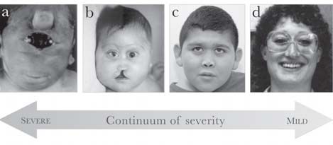
Indian pediatrician in a large city would encounter FIG. 2 Craniofacial phenotypes in patients with holo-
prosencephaly. From left to right: (a) synophthalmia and
and be required to significantly manage critically ill a proboscis in a patient with alobar holoprosencephaly;
patients with holoprosencephaly. (b) severe hypotelorism, flat nasal bridge, bilateral
colobomas, and midline cleft lip and palate in a patient
CLASSIFICATION SCHEMA with alobar holoprosencephaly; (c) hypotelorism, flat
nasal bridge, and closely spaced nostrils in a patient with
Holoprosencephaly is classically divided into four lobar holoprosencephaly; (d) hypotelorism, sharp nasal
types, based on the degree of nonseparation of the bridge, and single maxillary central incisor in an
prosencephalon [8,9]. These types, in order of individual with a microform of holoprosencephaly.
(Adapted from [20] and [25] with permission from Nature
increasing cortical separation, include the alobar Publishing Group and BMJ Publishing Group, Ltd.,
form, characterized by diffuse cortical nonsepara- respectively.)
INDIAN PEDIATRICS 458 VOLUME 48__JUNE 17, 2011RAAM, et al. HOLOPROSENCEPHALY
tend to follow DeMyer’s 1964 maxim, “the face
midline brain defects
No interhemispheric
predicts the brain” [12]. In other words, the severity
of the craniofacial phenotype tends to mirror the
May have subtle
subtle defects
severity of the brain malformations and correlates
Microform
May have
fusion inversely with survival [13] (Fig.2). The most
severe facial phenotypes include pronounced micro-
cephaly, cyclopia (single, centrally placed eye),
synophthalmia (partial union of the two eyes in the
Frequent fusion of thalami
Azygous anterior cerebral
center of the face), and a proboscis (a tube-like nasal
Gray matter heterotopias,
cortical dysplasia, and
appendage with a single nostril located above the
posterior frontal and
TABLE I DESCRIPTIONS OF TYPICAL BRAIN FINDINGS IN EACH OF THE TYPES OF HOLOPROSENCEPHALY
and caudate nuclei,
Absent body of the
ocular region) [13]. Less severe facial phenotypes
Nonseparation of
corpus callosum
can include microcephaly (except in cases of
parietal lobes
hydrocephalus, which can cause macrocephaly),
hypotelorism, midface hypoplasia with a flat nasal
MIHV*
artery
bridge, cleft lip and/or palate, ocular colobomas, and
a single maxillary central incisor [13]. Individuals
with microforms of holoprosencephaly, usually
Hypoplastic olfactory bulbs,
hypoplastic falx cerebri, and
identified as relatives of probands with frank
ventral frontal neocortex,
azygous anterior cerebral
holoprosencephaly, have isolated craniofacial find-
with hypoplastic falx
only the most rostral/
callosum in affected
ings without the classic clinical issues and
Nonseparation of
neurologic impairment seen in holoprosencephaly
Absent corpus
[11,13]. Conversely, individuals with mutations in
ZIC2, one of the genes implicated in select cases of
cerebri
region
Lobar
artery
holoprosencephaly, present an exception to the “face
predicts the brain” maxim, as these patients have
severe holoprosencephaly, neurologic impairment,
* MIHV: middle interhemispheric variant; Reproduced with permission from reference 10.
and absent anterior horns
of lateral ventricles and
No anterior separation,
fused deep gray nuclei,
Absent or hypoplastic
and characteristic clinical sequelae, but have a much
septum pellucidum
milder facial phenotype than that of other patients
corpus callosum
olfactory bulbs,
Absent anterior
some posterior
[13,14].
separation
Semilobar
ETIOLOGY AND MOLECULAR GENETICS
The etiology of holoprosencephaly is extremely
heterogeneous and is still being elucidated. With
Complete or near-complete
fused deep gray nuclei, and
varying levels of evidence, a number of environ-
Absent corpus callosum
Absent olfactory bulbs,
mental factors and teratogens have been suggested,
nonseparation, with
including maternal diabetes (infants born to diabetic
absent falx cerebri
mothers have a 200-fold risk of holoprosencephaly),
monoventricle
single midline
ALOBAR SEMILOBAR LOBAR MIHV
Alobar
Additional findings
Interhemispheric
Corpus callosal
characteristics
FIG 2. Axial sections through cranial MR images of patients
separation
with holoprosencephaly, distinguished by type. MIHV:
middle interhemispheric variant.
(Adapted from [32] with permission from Elsevier.)
INDIAN PEDIATRICS 459 VOLUME 48__JUNE 17, 2011RAAM, et al. HOLOPROSENCEPHALY
ethanol, cytomegalovirus infection, salicylates, anti- DIAGNOSIS
epileptic medications, retinoic acid, and maternal
hypocholesterolemia [15,16]. Genetic causes have A recommended protocol for clinical and molecular
also been implicated, based on familial occurrences diagnosis in patients with holoprosencephaly is
of holoprosencephaly, the presence of known provided in Fig. 3. The diagnostic process is
syndromes and associations including holoprosence- typically initiated by abnormal prenatal brain
phaly, and the nonrandom nature of chromosomal imaging, positive physical examination findings,
aberrations in patients with holoprosencephaly [16]. and/or positive family history. Whenever possible, a
Between 18%-25% of live births affected by thorough dysmorphology examination and an inter-
holoprosencephaly have a recognizable monogenic view to determine risk factors and family history
syndrome, including Smith-Lemli-Opitz syndrome should be obtained. Ascertainment of the specific
(MIM #270400), Pallister-Hall syndrome (MIM neurologic findings and holoprosencephaly type in
#146510), and Rubinstein-Taybi syndrome (MIM each patient, via brain imaging, is essential to proper
#180849) [16]. Chromosomal anomalies have been counseling of the patient and his/her family, given
implicated in 24-45% of live births affected by their effect on prognosis. MR (magnetic resonance)
holoprosencephaly [16-18], most fre-quently imaging provides the highest quality data for this
numeric anomalies in chromosomes 13, 18, and 21 purpose, allowing detailed analysis of cortical white
[19] and structural anomalies involving 13q, 18p, matter and structural abnormalities of the deep gray
7q36, 3p24-pter, 2p21, and 21q22.3 [16]. Intragenic nuclei [10], although logistic issues and the risks of
mutations in four genes have also been firmly the sedation required in neurologically impaired
established as increasing susceptibility to patients can make this impractical. If MR imaging
holoprosencephaly: SHH (7q36) (20-22), SIX3 cannot be performed, other options include ultra-
(2p21) (23-25), ZIC2 (13q32) (14, 26), and TGIF sound, which can be performed while the fontanelles
(18p11.3) [27]. While testing for mutations in these are patent, and CT (computed tomography) imaging,
four genes has led to significant diagnostic advance- which carries risks associated with radiation expo-
ments and implications for patient care, 75% of sure. If a patient is found to have microcephaly, a
chromosomally normal patients with holoprosence- large dorsal cyst, or rapidly enlarging head size,
phaly do not have identified mutations in any serial imaging is indicated [32].
screened genes [28], indicating the need to identify Prenatally, providing an early date of diagnosis is
additional susceptibility genes. important from both scientific and psychologic
points of view, because the severity of malfor-
The genetics of holoprosencephaly are such that mations leads to emotional effects among family
multiple affected individuals can present with members and may include consideration of preg-
holoprosencephaly within the same family, but nancy termination [33,34]. Prenatal ultrasound of the
incomplete penetrance and variable expressivity face and falx cerebri can be used to diagnose alobar
lead to tremendous intrafamilial phenotypic varia- and semilobar holoprosencephaly as early as the first
bility [29]. A related observation is that individuals trimester [10,33], while fetal MRI provides more
with certain chromosomal aberrations and intragenic sensitive diagnosis for milder forms of holopro-
mutations associated with holoprosencephaly may sencephaly during the third trimester [35]. Ultra-
not actually have holoprosencephaly in all cases: sound remains the gold standard due to its relative
only 50% of patients with deletions in 7q36, imperviousness to maternal obesity, fetal position,
including SHH, have holoprosencephaly, and only bone reverberation, and oligohydramnios [34]. In a
10% with deletions in 18p, including TGIF, do so recent study comparing ultrasound-based diagnosis
[30]. Thus, holoprosencephaly, like many other to postmortem autopsy findings, autopsy confirmed
entities considered to be “simple” Mendelian dis- the prenatal diagnosis of holoprosencephaly in 17/21
orders, is characterized by complex traits that are not cases, with two patients unable to receive a precise
reliably predicted by the presence of a single pathological diagnosis due to extensive severity of
mutation [31]. malformations, and two additional patients found to
INDIAN PEDIATRICS 460 VOLUME 48__JUNE 17, 2011Clinical diagnosis Neuroimaging Syndrome evaluation Cytogenetics Genetic counselling
Dysmorphology PRENATAL OPTIONS Smith-Lemli-Opitz syndrome: High-resolution karyotype
examination
RAAM, et al.
7-dehydroxycholesterol test
Identify Ultrasound Pallister-Hall syndrome: GL13 Fluorescent in situ
INDIAN PEDIATRICS
environmental risk molecular testing hybridization (FISH) of
factors known loci
Thorough family Fetal MRI Meckel-Gruber syndrome Molecular testing
history
POSTNATAL OPTIONS Other syndromic forms Sequence well-characterized
loci
(SHH, SIX3, ZIC2)
Ultrasound (if Sequence less common loci
fontanelles patent) (TFIF, GL12, others)
461
CT (radiation risk) Microarray analysis
MRI (slower,
sedation risks) Confirm test results in
parental samples to
determine inheritance
FIG 3 Recommended clinical protocol for diagnosing and elucidating causes of holoprosencephaly in patients. Each of the six major steps is medically indicated; within each step,
bolded items are medically indicated or preferred, while others are performed if suggested by the clinical characteristics of the patient or at the discretion of the clinical
laboratory. See text for more details.
HOLOPROSENCEPHALY
VOLUME 48__JUNE 17, 2011RAAM, et al. HOLOPROSENCEPHALY
have severe complex brain and facial malformations impairment observed in alobar and semilobar holo-
other than holoprosencephaly [34]. Ultrasound is not prosencephaly is less frequently seen in the lobar
completely accurate in determining holoprosence- type and the middle interhemispheric variant;
phaly type: in 7/17 cases, the holoprosencephaly patients with the latter forms may walk with
type determined through prenatal diagnosis differed assistance, adequately control their limbs, and even
from that determined via postmortem autopsy [34]. speak words or sentences [37]. The enhanced vocal
In families with an existing child with holo- communication in these patients may be explained
prosencephaly and an identified disease-causing by more complete separation of the deep gray nuclei,
mutation, prenatal molecular diagnosis is possible, but because separation of the deep gray nuclei does
although presence of the mutation does not not appear to correlate with social awareness, visual
necessarily portend holoprosencephaly [35]. attention, and auditory comprehension, differences
in those constructs may be caused by structural
PROGNOSIS changes in different regions [38]. The Carter
Neurocognitive Assessment (CNA) may be useful to
Survival rates vary in each type of holopro-
clinicians for assessing cognitive function in
sencephaly, but in general, mortality correlates
children with more severe impairment [38].
positively with the severity of the brain malfor-
mation and, by extension, severity of the facial CLINICAL MANAGEMENT
phenotype [13]. Of children with alobar holopro-
sencephaly, those with severe facial anomalies such Due to the medial and rostral location of the
as cyclopia and proboscis rarely survive the hypothalamus, nonseparation of the hypothalamus
immediate postnatal period, while those with less occurs frequently, leading to a variety of issues
severe facial malformations can survive for months involving homeostatic and hypothalamic-pituitary
or, in a minority of cases, longer than one year [36]. endocrine functions [39]. One disturbed homeostatic
In very rare instances, survival into the twenties has function is body temperature regulation, which is
been observed (authors’ own experience). In contrast significant for two reasons: first, ascertainment of
to most children with alobar holoprosencephaly, baseline body temperature helps identify abnormal
children with holoprosencephaly types other than deviations in temperature due to infections or other
alobar may more often survive into adulthood [36]. causes of morbidity; second, temperature instability
Frequent causes of death include respiratory in itself can cause morbidity and organ dysfunction if
infections, dehydration secondary to uncontrolled the core temperature falls below 34ºC or rises above
diabetes insipidus, intractable seizures, and sequelae 40ºC [37]. Other impaired homeostatic functions
of brainstem malfunction, including aberrant control include thirst, appetite, and sleep-wake cycles,
of respiration and heart rate [36]. disturbances of all of which can pose significant
problems for caregivers [37].
As with survival, developmental outcomes
generally correlate with the severity of the brain From an endocrinologic perspective, dysfunction
malformation, although again, tremendous varia- of the posterior pituitary, in the form of central
bility can occur. Children with alobar holopro- diabetes insipidus, is much more commonly obser-
sencephaly may develop to a stage equivalent to that ved than anterior pituitary insufficiency [39,40],
of a healthy, early infant: while they may track typically manifesting with polyuria, dehydration,
objects or sounds, they typically cannot speak words, hypernatremia, and decreased urine osmolarity [40].
sit without assistance, or reach for objects [37]. In The severity of diabetes insipidus generally
contrast, some children with semilobar holopro- correlates with the degree of hypothalamic nonsepa-
sencephaly can develop receptive language skills, ration but not with pituitary gland defects observed
and while speech is still frequently impaired, they via imaging [40]. Due to the high incidence of
can communicate through eye movements, gestures, posterior pituitary dysfunction, and because diabetes
or other non-verbal communication systems, and insipidus in these patients may be asymptomatic,
may be socially engaging [37]. The severe motor routine screening of electrolyte levels for evidence
INDIAN PEDIATRICS 462 VOLUME 48__JUNE 17, 2011RAAM, et al. HOLOPROSENCEPHALY
of posterior pituitary endocrinopathies is recommen- on electroencephalograms (EEGs) of some patients
ded in all patients, with repeated testing even in the without overt clinical seizures [41], suggesting that
event of an initial negative result and also in the routine EEG screening of patients may be useful. Of
acute setting [37,40]. Anterior pituitary issues, patients with recurring seizures, most are managed
occurring with lower frequency than posterior with one or two antiepileptic medications; intrac-
pituitary issues, include hypothyroidism, hypocorti- table seizures occur in one-third to one-half,
solism, growth hormone deficiency, and multiple typically in patients with more severe cortical
pituitary hormone deficiency [40]. As signs of malformations [37,39]. As seizure triggers can
hypothyroidism and hypocortisolism can be difficult include fluid and electrolyte imbalances from
to distinguish from those seen classically in holo- diabetes insipidus, proper management of seizures
prosencephaly, and because the effects of those requires consideration of endocrinologic issues [37].
endocrinologic deficiencies can be life-threatening, Hydrocephalus is another common finding that
we recommend basic screening evaluations in all depends on the specific brain malformation,
patients, but with in-depth stimulation tests only if correlating highly with thalamic nonseparation and
clinical suspicion is high. the presence of a dorsal cyst; it is thought to result
from blocked cerebrospinal fluid egress from the
Motor impairment in holoprosencephaly third ventricle [42]. Because holoprosencephaly
generally manifests as hypotonia, dystonia, and/or typically results in microcephaly, hydrocephalus
spasticity, frequently requiring pharmaceutical inter- should be suspected in patients with normal head
ventions such as intrathecal baclofen pumps and oral sizes or macrocephaly and followed using serial
trihexyphenidyl, as well as physical and occupatio- head circumference measurements and ultrasound
nal therapy and surgical interventions [37]. One of imaging [37]. Placing a cerebrospinal fluid shunt,
the most detrimental effects of motor impairment is while taking particular care to avoid overdrainage,
oromotor dysfunction, which significantly com- can improve developmental outcomes, improve
pounds the thirst and appetite disturbances resulting other issues, and reduce macrocephaly [37].
from hypothalamic dysfunction, and may also
exacerbate unique feeding challenges secondary to Thus, diverse clinical sequelae can result from a
cleft lip and palate [37]. Children with such issues primary insult of holoprosencephaly. Clinicians
frequently develop oropharyngeal dysphagia and should have a low threshold for testing for these
respiratory symptoms related to aspiration and diffi- sequelae, as specific abnormalities are difficult to
culty managing secretions, compromising oral predict in advance and may be challenging to
intake and increasing the risk of respiratory infec- diagnose.
tions. Additional respiratory issues can include
chronic lung disease with decreased pulmonary FURTHER STEPS TO ELABORATE GENETIC CAUSES
reserve and chronic inflammation. A gastrostomy AND INHERITANCE
tube is placed in many children with oromotor
Indian pediatricians are well-equipped to clinically
dysfunction to address these issues. Gastrointestinal
diagnose holoprosencephaly and to manage the
issues related to poor nervous regulation, including
clinical sequelae of the condition, but full benefit to
poor gastric and colonic motility and gastro-
the patient and his/her family cannot be achieved
esophageal reflux, can still impair feeding despite
without genetic investigation. We recognize that
placement of a gastrostomy tube, sometimes indi-
there are many barriers to the consistent application
cating medications and anti-reflux procedures [37].
of genetic testing and interpretation to each Indian
Finally, the nature of the brain malformation may patient with holoprosencephaly, as the current state
predispose patients to seizures and/or hydro- of medical genetics in India leaves many clinicians
cephalus. Seizures occur in approximately half of without formal training in genetics and easy access
the patients [39], most commonly complex partial to affordable genetic testing laboratories [43].
seizures, and typically develop during infancy [37]. Nevertheless, for a proper discussion with the family
In addition, “epileptiform” activity has been noted regarding etiology and recurrence risk, pediatricians
INDIAN PEDIATRICS 463 VOLUME 48__JUNE 17, 2011RAAM, et al. HOLOPROSENCEPHALY
KEY MESSAGES
• Holoprosencephaly is characterized by failure of the prosencephalon to divide into complete hemispheres, and
is associated with facial dysmorphism and neurologic impairment.
• Essential components of diagnosis include a thorough interview to determine family history and teratogenic
exposures, dysmorphology exam, and neuroimaging, which is critical for prognosis determination.
• Medical management should focus on hypothalamic and endocrinologic dysfunction, motor and developmental
impairment, respiratory issues, seizures, and hydrocephalus.
• Pediatricians should follow up medical management by collaborating with a genetic specialist, with the aim of
performing genetic testing, determination of associated syndromes, and genetic counseling.
should seek out genetic specialists within or outside (array CGH, or aCGH) and single nucleotide
India who are familiar with holoprosencephaly and polymorphism (SNP) arrays, is a relatively new
discuss the feasibility of genetic testing with them. molecular technique that allows for identification of
Here, we briefly discuss what is needed so that the deletions and duplications at resolutions far excee-
pediatrician may be familiar with the process. ding that of a karyotype, but currently, the novelty of
this technique indicates that logistical and financial
As previously mentioned, holoprosencephaly barriers, as well as the inadequacy of information
frequently occurs as part of a syndrome, and allowing us to separate benign copy number variants
additional diagnostic steps should be undertaken if from pathogenic deletions and duplications [46],
the patient is clinically suspected to be affected by may need to be addressed before the technique is
one of these syndromes. For instance, patients used more routinely.
suspected to have Smith-Lemli-Opitz syndrome
should have total cholesterol and 7-dehydro- All of the information gathered through the steps
xycholesterol levels checked for a decrease and an outlined above is necessary for proper genetic
increase outside the normal range, respectively [44]. counseling, the need for which is established by the
poor prognosis in the most severely affected patients
To determine genetic causes of holopro- and the relative uncertainty of each patient’s severity
sencephaly in each patient, a combination of a priori due to the extreme phenotypic variability of
cytogenetic and molecular testing is recommended. the condition. Effective genetic counseling takes into
Due to the high incidence of chromosomal ano- account the inconsistency of strict genotype-
malies, a high-resolution karyotype at the 550 band phenotype correlations for each identified genetic
level or greater is indicated in all patients. Direct variant, indicating the need for caution while inter-
DNA sequencing of SHH, ZIC2, and SIX3 is also preting molecular results. Although medical genetics
indicated, due to the high prevalence of intragenic may not be a particular physician’s area of expertise,
mutations in those genes [45]. DNA sequencing we urge pediatricians to familiarize themselves with
results should be compared to analyses of biologic the above recommendations and to correspond with
effects and results of functional studies [22,24] for medical geneticists, so that the quality of genetic
each potential mutation, which are essential to help counseling can be enhanced and further morbidity
determine the true pathogenic import of each variant. and mortality related to holoprosencephaly can be
Routine sequencing of minor loci is not performed ameliorated.
unless indicated by specific observations in a patient:
for instance, pituitary abnormalities in a patient with Contributors: All authors participated in the critical editing of
holoprosencephaly may warrant molecular testing of this review article.
Competing interests: No authors have any financial/personal
GLI2 [45] due to an emerging genotype/phenotype disclosures or competing interests.
correlation [13]. Microarray analysis, including Funding: Support was provided in part by Division of Intramural
array-based comparative genomic hybridization Research at the National Human Genome Research Institute
INDIAN PEDIATRICS 464 VOLUME 48__JUNE 17, 2011RAAM, et al. HOLOPROSENCEPHALY
(National Institutes of Health, Department of Health and Human York: McGraw-Hill; 2001. p.6203-30.
Services, United States of America). 17. Croen LA, Shaw GM, Lammer EJ. Holoprosencephaly:
epidemiologic and clinical characteristics of a California
REFERENCES population. Am J Med Genet. 1996; 64:465-72.
18. Olsen CL, Hughes JP, Youngblood LG, Sharpe-Stimac M.
1. O’Rahilly R, Müller F. Interpretation of some median Epidemiology of holoprosencephaly and phenotypic
anomalies as illustrated by cyclopia and symmelia. characteristics of affected children: New York State, 1984-
Teratology. 1989;40:409-21. 1989. Am J Med Genet. 1997;73:217-26.
2. Matsunaga E, Shiota K. Holoprosencephaly in human 19. Solomon BD, Rosenbaum KN, Meck JM, Muenke M.
embryos: epidemiologic studies of 150 cases. Teratology. Holoprosencephaly due to numeric chromosome
1977;16:261-72. abnormalities. Am J Med Genet C Semin Med Genet. 2010;
3. Leoncini E, Baranello G, Orioli IM, Annerén G, Bakker M, 154C:146-48.
Bianchi F, et al. Frequency of holoprosencephaly in the 20. Roessler E, Belloni E, Gaudenz K, Jay P, Berta P, Scherer
International Clearinghouse Birth Defects Surveillance SW, et al. Mutations in the human Sonic Hedgehog gene
systems: searching for population variations. Birth Defects cause holoprosencephaly. Nat Genet. 1996;14:357-60.
Res A. 2008;82:585-91. 21. Nanni L, Ming JE, Bocian M, Steinhaus K, Bianchi DW,
4. Orioli IM, Castilla EE. Epidemiology of holoprosen- de Die-Smulders C, et al. The mutational spectrum of the
cephaly: prevalence and risk factors. Am J Med Genet C Sonic Hedgehog gene in holoprosencephaly: SHH
Semin Med Genet. 2010;154C:13-21. mutations cause a significant proportion of autosomal
5. Thakur S, Singh R, Pradhan M, Phadke SR. Spectrum of dominant holoprosencephaly. Hum Mol Genet. 1999;8:
holoprosencephaly. Indian J Pediatr 2004;71:593-7. 2479-88.
6. Verma IC. Medical genetics in India. Indian J Pediatr. 22. Roessler E, El-Jaick KB, Dubourg C, Vélez JI, Solomon
1986;53:437-40. BD, Pineda-Álvarez DE, et al. The mutational spectrum of
7. Verma IC. Burden of genetic disorders in India. Indian J holoprosencephaly-associated changes within the SHH
Pediatr. 2000;67:893-8. gene in humans predicts loss-of-function through either
8. DeMyer W, Zeman W. Alobar holoprosencephaly key structural alterations of the ligand or its altered
(arhinencephaly) with median cleft lip and palate: clinical, synthesis. Hum Mutat. 2009;30:E921-35.
electroencephalographic and nosologic considerations. 23. Wallis DE, Roessler E, Hehr U, Nanni L, Wiltshire T,
Confin Neurol. 1963;23:1-36. Richieri-Costa A, et al. Mutations in the homeodomain of
9. Barkovich AJ, Quint DJ. Middle interhemispheric fusion: the human SIX3 gene cause holoprosencephaly. Nat Genet.
an unusual variant of holoprosencephaly. AJNR Am J 1999;22:196-8.
Neuroradiol. 1993;14:431-40. 24. Domené S, Roessler E, El-Jaick KB, Snir M, Brown JL,
10. Hahn JS, Barnes PD. Neuroimaging advances in Vélez JI, et al. Mutations in the human SIX3 gene in
holoprosencephaly: refining the spectrum of the midline holoprosencephaly are loss of function. Hum Mol Genet.
malformation. Am J Med Genet C Semin Med Genet. 2008;17:3919-28.
2010;154C:120-32. 25. Lacbawan F, Solomon BD, Roessler E, El-Jaick K,
11. Solomon BD, Lacbawan F, Jain M, Domené S, Roessler E, Domené S, Vélez JI, et al. Clinical spectrum of SIX3-
Moore C, et al. A novel SIX3 mutation segregates with associated mutations in holoprosencephaly: correlation
holoprosencephaly in a large family. Am J Med Genet A. between genotype, phenotype and function. J Med Genet.
2009;149A:919-25. 2009;46:389-98.
12. DeMyer W, Zeman W, Palmer CG. The face predicts the 26. Brown SA, Warburton D, Brown LY, Yu C-y, Roeder ER,
brain: diagnostic significance of median facial anomalies Stengel-Rutkowski S, et al. Holoprosencephaly due to
for holoprosencephaly (arhinencephaly). Pediatrics. 1964; mutations in ZIC2, a homologue of Drosophila odd-
34:256-63. paired. Nat Genet. 1998;20:180-3.
13. Solomon BD, Mercier S, Vélez JI, Pineda-Alvarez DE, 27. Gripp KW, Wotton D, Edwards MC, Roessler E, Ades L,
Wyllie A, Zhou N, et al. Analysis of genotype-phenotype Meinecke P, et al. Mutations in TGIF cause holoprosen-
correlations in human holoprosencephaly. Am J Med cephaly and link NODAL signalling to human neural axis
Genet C Semin Med Genet. 2010;154C:133-41. determination. Nat Genet. 2000;25:205-8.
14. Solomon BD, Lacbawan F, Mercier S, Clegg NJ, Delgado 28. Roessler E, Muenke M. The molecular genetics of
MR, Rosenbaum K, et al. Mutations in ZIC2 in human holoprosencephaly. Am J Med Genet C Semin Med Genet.
holoprosencephaly: description of a novel ZIC2-specific 2010;154C:52-61.
phenotype and comprehensive analysis of 157 individuals. 29. Odent S, Le Marec B, Munnich A, Le Merrer M, Bonaïti-
J Med Genet. 2010;47:513-24. Pellié C. Segregation analysis in nonsyndromic
15. Johnson CY, Rasmussen SA. Non-genetic risk factors for holoprosencephaly. Am J Med Genet. 1998;77:139-43.
holoprosencephaly. Am J Med Genet C Semin Med Genet. 30. Ming JE, Muenke M. Multiple hits during early embryonic
2010;154C:73-85. development: digenic diseases and holoprosencephaly. Am
16. Muenke M, Beachy PA. Holoprosencephaly. In: Scriver J Hum Genet. 2002;71:1017-32.
CR, Beaudet AL, Sly WS, Valle D, editors. The Metabolic 31. Dipple KM, McCabe ERB. Phenotypes of patients with
and Molecular Bases of Inherited Disease. 8th ed. New “simple” Mendelian disorders are complex traits:
INDIAN PEDIATRICS 465 VOLUME 48__JUNE 17, 2011RAAM, et al. HOLOPROSENCEPHALY
thresholds, modifiers, and systems dynamics. Am J Hum cephaly as predictor of function: beyond the face predicting
Genet. 2000;66:1729-35. the brain. Neurology. 2002;59:1058-66.
32. Hahn JS, Plawner LL. Evaluation and management of 40. Hahn JS, Hahn SM, Kammann H, Barkovich AJ, Clegg NJ,
children with holoprosencephaly. Pediatr Neurol. Delgado MR, et al. Endocrine disorders associated with
2004;31:79-88. holoprosencephaly. J Pediatr Endocr Met. 2005;18:935-
33. Joó GJ, Beke A, Papp C, Tóth-Pál E, Szigeti Z, Bán Z, et al. 41.
Prenatal diagnosis, phenotypic and obstetric characteristics 41. Hahn JS, Delgado MR, Clegg NJ, Sparagana SP, Gerace
of holoprosencephaly. Fetal Diagn Ther. 2005;20:161-6. KL, Barkovich AJ, et al. Electroencephalography in
34. Wenghoefer M, Ettema AM, Sina F, Geipel A, Kuijpers- holoprosencephaly: findings in children without epilepsy.
Jagtman AM, Hansmann H, et al. Prenatal ultrasound Clin Neurophysiol. 2003;114:1908-17.
diagnosis in 51 cases of holoprosencephaly: craniofacial 42. Simon EM, Hevner RF, Pinter JD, Clegg NJ, Delgado M,
anatomy, associated malformations, and genetics. Cleft Kinsman SL, et al. The dorsal cyst in holoprosencephaly
Palate Craniofac J. 2010;47:15-21. and the role of the thalamus in its formation.
35. Mercier S, Dubourg C, Belleguic M, Pasquier L, Loget P, Neuroradiology. 2001;43:787-91.
Lucas J, et al. Genetic counseling and “molecular” prenatal 43. Agarwal SS. Medical genetics in India – what needs to be
diagnosis of holoprosencephaly (HPE). Am J Med Genet C done? Indian J Med Res. 2009;130:354-56.
Semin Med Genet. 2010;154C:191-6. 44. Weaver DD, Solomon BD, Akin-Samson K, Kelley RI,
36. Barr Jr. M, Cohen Jr. MM. Holoprosencephaly survival Muenke M. Cyclopia (synophthalmia) in Smith-Lemli-
and performance. Am J Med Genet. 1999;89:116-20. Opitz syndrome: first reported case and consideration of
37. Levey EB, Stashinko E, Clegg NJ, Delgado MR. mechanism. Am J Med Genet C Semin Med Genet.
Management of children with holoprosencephaly. Am J 2010;154C:142-5.
Med Genet C Semin Med Genet. 2010;154C:183-90. 45. Pineda-Alvarez DE, Dubourg C, David V, Roessler E,
38. Roesler CP, Paterson SJ, Flax J, Hahn JS, Kovar C, Muenke M. Current recommendations for the molecular
Stashinko EE, et al. Links between abnormal brain evaluation of newly diagnosed holoprosencephaly patients.
structure and cognition in holoprosencephaly. Pediatr Am J Med Genet C Semin Med Genet. 2010;154C:93-101.
Neurol. 2006;35:387-94. 46. Sharp AJ. Emerging themes and new challenges in defining
39. Plawner LL, Delgado MR, Miller VS, Levey EB, Kinsman the role of structural variation in human disease. Hum
SL, Barkovich AJ, et al. Neuroanatomy of holoprosen- Mutat. 2009;30:135-44.
INDIAN PEDIATRICS 466 VOLUME 48__JUNE 17, 2011You can also read