Implantation technique and complications in the first 24
←
→
Page content transcription
If your browser does not render page correctly, please read the page content below
NEUROSURGICAL
            FOCUS                                                                                                              Neurosurg Focus 48 (4):E2, 2020

                          Robot-assisted stereoelectroencephalography exploration
                          of the limbic thalamus in human focal epilepsy:
                          implantation technique and complications in the first 24
                          patients
                          *Ganne Chaitanya, MBBS, PhD,1,2 Andrew K. Romeo, MD,3 Adeel Ilyas, MD,2,3 Auriana Irannejad,1,2
                          Emilia Toth, PhD,1,2 Galal Elsayed, MD,3 J. Nicole Bentley, MD,3 Kristen O. Riley, MD,3 and
                          Sandipan Pati, MD1,2
                          Department of Neurology, 2Epilepsy and Cognitive Neurophysiology Laboratory, and 3Department of Neurosurgery, University of
                          1

                          Alabama at Birmingham, Alabama

                          OBJECTIVE Despite numerous imaging studies highlighting the importance of the thalamus in a patient’s surgical prog-
                          nosis, human electrophysiological studies involving the limbic thalamic nuclei are limited. The objective of this study was
                          to evaluate the safety and accuracy of robot-assisted stereotactic electrode placement in the limbic thalamic nuclei of
                          patients with suspected temporal lobe epilepsy (TLE).
                          METHODS After providing informed consent, 24 adults with drug-resistant, suspected TLE undergoing evaluation with
                          stereoelectroencephalography (SEEG) were enrolled in the prospective study. The trajectory of one electrode planned
                          for clinical sampling of the operculoinsular cortex was modified to extend it to the thalamus, thereby preventing the need
                          for additional electrode placement for research. The anterior nucleus of the thalamus (ANT) (n = 13) and the medial
                          group of thalamic nuclei (MED) (n = 11), including the mediodorsal and centromedian nuclei, were targeted. The postim-
                          plantation CT scan was coregistered to the preoperative MR image, and Morel’s thalamic atlas was used to confirm the
                          accuracy of implantation.
                          RESULTS Ten (77%) of 13 patients in the ANT group and 10 (91%) of 11 patients in the MED group had electrodes ac-
                          curately placed in the thalamic nuclei. None of the patients had a thalamic hemorrhage. However, trace asymptomatic
                          hemorrhages at the cortical-level entry site were noted in 20.8% of patients, who did not require additional surgical inter-
                          vention. SEEG data from all the patients were interpretable and analyzable. The trajectories for the ANT implant differed
                          slightly from those of the MED group at the entry point—i.e., the precentral gyrus in the former and the postcentral gyrus
                          in the latter.
                          CONCLUSIONS Using judiciously planned robot-assisted SEEG, the authors demonstrate the safety of electrophysi-
                          ological sampling from various thalamic nuclei for research recordings, presenting a technique that avoids implanting ad-
                          ditional depth electrodes or compromising clinical care. With these results, we propose that if patients are fully informed
                          of the risks involved, there are potential benefits of gaining mechanistic insights to seizure genesis, which may help to
                          develop neuromodulation therapies.
                          https://thejns.org/doi/abs/10.3171/2020.1.FOCUS19887
                          KEYWORDS temporal lobe epilepsy; thalamus; stereotactic electroencephalography; anterior nucleus of the thalamus;
                          mediodorsal nucleus; centromedian nucleus

ABBREVIATIONS ANT = anterior nucleus of the thalamus; CeM = centromedian nucleus of the thalamus; DBS = deep brain stimulation; EZ = epileptogenic zone; MED =
medial group of thalamic nuclei; SEEG = stereoelectroencephalography; TLE = temporal lobe epilepsy.
ACCOMPANYING EDITORIAL DOI: 10.3171/2020.1.FOCUS2069.
SUBMITTED November 17, 2019. ACCEPTED January 24, 2020.
INCLUDE WHEN CITING DOI: 10.3171/2020.1.FOCUS19887.
* G.C., A.K.R., and A. Ilyas contributed equally to this work.

©AANS 2020, except where prohibited by US copyright law                                                       Neurosurg Focus Volume 48 • April 2020         1

                                                                                                               Unauthenticated | Downloaded 10/22/20 09:55 PM UTC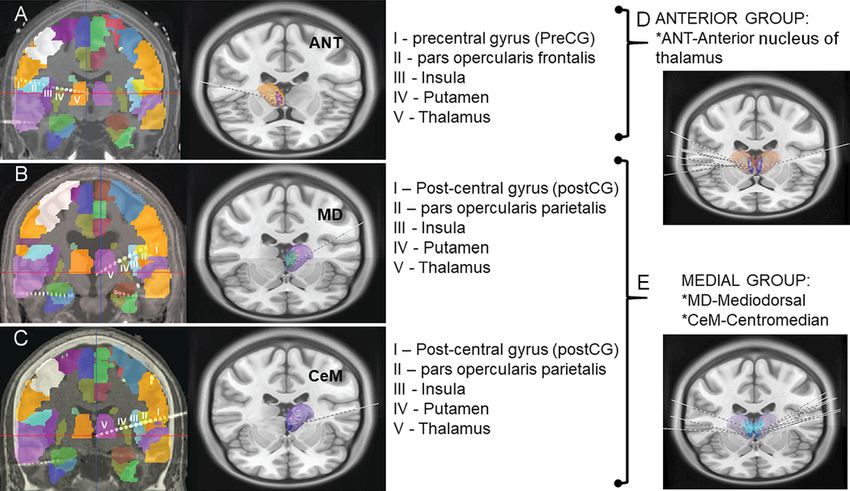
Chaitanya et al.
A
ccurate localization of the epileptogenic zone be highly informed by understanding thalamotemporal
(EZ) in temporal lobe epilepsy (TLE) is para- causal interactions during seizures. Here, we present the
mount to optimizing outcomes following resec- technical nuances, safety details, and accuracy data from
tion or ablation.44 However, despite significant technologi- stereoelectroencephalography (SEEG) implantation of
cal advancements in imaging and surgical tools, seizure- electrodes into the limbic thalamus during the presurgical
freedom outcomes have plateaued at 45%–65% over the evaluation of patients with suspected TLE.
last decade, necessitating a continued investigation of
how distributed brain regions interact to cause seizures.39 Methods
Growing evidence suggests that networks of functionally
and structurally connected areas, both within and out- Patient Selection
side of mesial temporal structures, contribute to EZs.4,10, Enrolled patients included those deemed eligible for
19,32
Among extratemporal structures implicated in seizure SEEG after consensus recommendation from a multidis-
inception, increased thalamotemporal structural and func- ciplinary epilepsy conference consisting of neurologists,
tional connectivity independently predicts poor surgical neurosurgeons, neuropsychologists, and nurses. Adults
outcomes.17 Experimental studies in preclinical models of with drug-resistant, suspected TLE undergoing SEEG
limbic epilepsy support the hypothesis that the thalamus evaluation were eligible to participate in the study. Sur-
can regulate limbic seizures, and the stage at which ic- geons modified the trajectory of one electrode planned for
togenesis is regulated (i.e., initiation, propagation, or ter- clinical sampling to extend to the thalamus, obviating the
mination) depends on the functional connectivity of the need to implant an additional electrode for thalamic sam-
thalamic nuclei with limbic structures.6,9,36 Furthermore, pling. A 5-stage evaluation process helped streamline the
modulation of the “limbic” thalamic nuclei (i.e., the an- process of recruiting considerably homogeneous groups
terior, midline and mediodorsal, and intralaminar centro- of patients with suspected TLE (mesial and/or temporal
median [CeM] nuclei) through chemical, optogenetic, or plus) who would receive the thalamic implant (Fig. 1). The
electrical perturbation can interrupt focal seizures.6,16,25,37 investigators approached the potential candidates for tha-
However, despite numerous preclinical and imaging stud- lamic SEEG at the outpatient follow-up clinic visit, and
ies highlighting the importance of the thalamus in surgi- written informed consent was obtained in accordance
cal prognosis,17 there are very limited human electrophysi- with protocols approved by the University of Alabama at
ological studies targeting the limbic thalamus. Birmingham Institutional Review Board.
Over the last decade, clinicians have implanted subdu-
ral grids, hybrid macro-micro depth electrodes, and lami- Thalamic Trajectory
nar electrodes for high-density intracranial recordings In this study, the limbic thalamic nuclei that were tar-
from both the superficial and deep cortexes.41,43 Although geted were as follows: 1) anterior nucleus of the thalamus
the thalamus is likely a rich source of data about seizure (ANT), including anterior ventral, anterior dorsal, and an-
regulation, propagation, and sleep dysfunction,12,15 prog- terior medial subnuclei; and 2) medial group of thalamic
ress in this area is relatively slow due to ethical and safety nuclei (MED), including mediodorsal and CeM subnuclei.
concerns. However, future therapeutic developments may Neurosurgeons planned trajectories using T1-weighted
FIG. 1. Patient selection and SEEG implantation: a multistage process to obtain informed consent from eligible patients scheduled
to undergo SEEG investigation for localization of epilepsy. EMU = epilepsy monitoring unit.
2 Neurosurg Focus Volume 48 • April 2020
Unauthenticated | Downloaded 10/22/20 09:55 PM UTC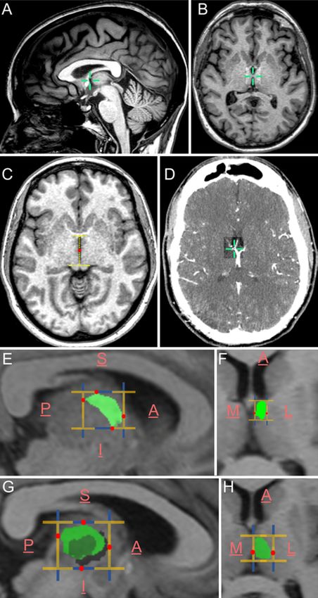
Chaitanya et al.

MRI sequences with gadolinium contrast using robotic
stereotactic planning software (ROSA; Medtech). MRI
scans were acquired using the epilepsy protocol in a Phil-
ips Achieva 1.5T scanner (matrix size 384 × 384 mm/32 ×
432 mm, slice thickness 1.2 mm, TR 7 msec, TE 3 msec,
interslice gap 1.2 mm, flip angle 8°), Philips Achieva 3T
scanner (matrix size 256 × 256 mm/528 × 528 mm, slice
thickness 1 mm/1.2 mm, TR 6 msec, TE 3 msec/4 msec,
interslice gap 1 mm/1.2 mm, flip angle 8°/9°), Philips In-
genia 1.5T scanner (matrix size 512 × 512 mm/432 × 432
mm, slice thickness 1 mm, TR 7 msec, TE 3 msec, inter-
slice gap 1 mm, flip angle 8°), Philips Ingenia 3T scanner
(matrix size 528 × 528 mm/432 × 432 mm, slice thickness
1.2 mm, TR 7 msec, TE 3 msec, interslice gap 1.2 mm, flip
angle 9°), or SIEMENS Skyra 3T scanner (matrix size 256
× 256 mm, slice thickness 0.9 mm, TR 7 msec, TE 3 msec,
interslice gap 0 mm, flip angle 8°).
    Based on a visual reference to Morel’s thalamic atlas,
the nuclei were determined to be located in relation to the
following landmarks21 while planning the trajectory. The
ANT was identified by its close relationship to the fora-
men of Monro and the venous angle formed by the conflu-
ence of thalamostriate and septal veins lying immediately
posterior and lateral (Fig. 2). Extending a trajectory that
incorporated the frontal operculum and insula, the tha-
lamic region of interest was targeted without requiring an
additional electrode. For the MED group of thalamic nu-
clei, neurosurgeons targeted the ventral midline thalamus
near the massa intermedia, providing recordings from the
centromedial or mediodorsal nuclei. The broadest midline
nuclear segment was anterior and ventral to the massa in-
termedia, where the reuniens and central median nuclei
are located.33 The massa intermedia could be visualized
in 5 of the 8 MED thalamic groups of patients. When not
distinct, the anteroposterior midpoint at the level of the
anterior commissure–posterior commissure plane was
chosen.5 The inferior and nearly parallel relationship with
the ANT provided further anatomical confirmation.
    Following SEEG implantation, patients were initially
monitored in the neurosurgical ICU for 24 hours. High-
resolution postimplantation head CT scans were obtained
within 24 hours. Subsequently, the patients were trans-
ferred to the epilepsy monitoring unit for seizure localiza-   FIG. 2. Landmarks for targeting the thalamic nuclei. The foramen of
tion and mapping.                                              Monro as the anteromedial extent (A), the massa intermedia as the
                                                               medial extent (B), the midpoint (red dot) of the anteroposterior length
Measuring Accuracy                                             (yellow line) of the third ventricle in axial view as the inferior extent (C)
                                                               (measured at the level of the superior colliculus and the venous angle
    Postimplantation CT scans were acquired using Phil-        as the anteromedial extent), and the confluence of the anterior septal
ips Brilliance64 and Brilliance16P scanners (matrix size       vein, thalamostriate vein, and internal cerebral vein (D) were the major
512 × 512 mm, slice thickness 1 mm, 120 kVp [peak kilo-        landmarks used while targeting the electrodes. Green crosshairs in A, B,
voltage], interslice gap 1 mm, exposure time 1550 sec/727      and D indicate the coordinates of the aforementioned landmarks. E and
sec). The CT scans were coregistered to the preoperative       F: The probable extents of ANT as per Morel’s atlas. G and H: The
MR images using Advanced Normalization Tools (Fig.             probable extents of MED (CeM + mediodorsal subnuclei) as per Morel’s
3).3 Electrodes were localized using Lead-DBS software         atlas. The yellow brackets indicate the anteroposterior extent of nuclei
(www.lead-dbs.org), and trajectories were mapped us-           in E and G and the medial to lateral extent in F and H. The red dots in-
                                                               dicate the farthest extent of the thalamic nucleus in any given direction.
ing iElectrodes software.7,18 Coregistered images were         The blue brackets indicate the superoinferior extent of the nuclei. A =
normalized to ICBM152–2009b nonlinear asymmetri-               anterior; I = inferior; L = lateral; M = medial; P = posterior; S = superior.
cal atlases using nonlinear diffeomorphic normalization
algorithms, and brain shift corrections were performed
using Schönecker normalizations, providing refined reg-
istration of subcortical structures.35 Registrations were
checked manually for errors in 3D Slicer. The data were

                                                                                          Neurosurg Focus Volume 48 • April 2020          3

                                                                                          Unauthenticated | Downloaded 10/22/20 09:55 PM UTC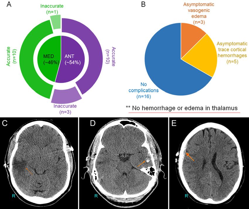
Chaitanya et al.
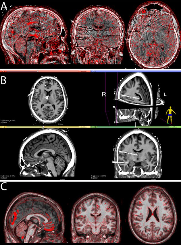
FIG. 3. Coregistration strategy. A: The postimplantation CT scan was coregistered onto the preoperative MR image to determine
the postoperative target of the thalamic targets using Advanced Normalization Tools in Lead-DBS. B: Following coregistration, a
manual verification of the anatomical landmarks and the overlap of the inner and outer tables of the MR image and CT scan were
used to ensure accurate coregistration. In particular, the most probable location of the thalamic electrode was confirmed. C: The
coregistered MR image and CT scan were then normalized to the MNI (Montreal Neurological Institute) space. The coregistered
images were then resolved for finer registration and brain shifts using the Schönecker normalization algorithm to improve the
registration of the subcortical structure (3D Slicer).
then visualized using Morel’s atlas.21 To map the trajecto- accurate location of the thalamic target as well as the lin-
ries, the coregistered and normalized CT and MRI scans ear component distances along the x, y, and z axes and
were imported along with the corresponding CT mask the Euclidean distances from the landmarks. Postexplan-
into iElectrodes and registered with the AAL2 atlas to tation CT scans were also obtained to look for postop-
identify their cortical-subcortical locations.28,40 As a final erative hemorrhage or edema. If found, hemorrhage was
step in establishing the accuracy of the implant strategy graded using McGovern’s SEEG hemorrhage grading
from our experience, we estimated the postimplantation system.23
4 Neurosurg Focus Volume 48 • April 2020
Unauthenticated | Downloaded 10/22/20 09:55 PM UTC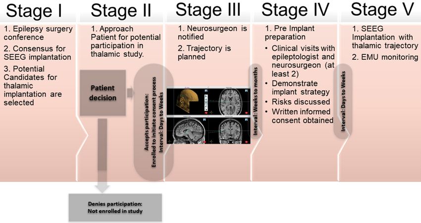
Chaitanya et al.

TABLE 1. Clinical presentation of the 24 participants and summary of post-SEEG localization of the seizure pathology and the treatment
strategy
  Thalamic                 Age at     Duration No. of                      Laterality
  Nucleus &         Age Epilepsy of Epilepsy Failed Preimplant   No. of   of Thalamic
Participant No. Sex (yrs) Onset (yrs)  (yrs)   AEDs    MRI     Electrodes Electrode                                     Post-SEEG EZ                  Treatment
     ANT
       1            F     42         38              3.5          5        HS+              9             Lt        Lt TLE subtype*            Pt deferred surgical
                                                                                                                                                  therapy
        2           M     24          6            18.5           2          N             14             Rt        Rt MTLE                    Rt ATL
        3           F     47         41             6.2           7          N             14             Rt        Bilat TLE                  Bilat HG RNS
        4           M     37         29             8.0           4         EH             10             Rt        Rt TLE plus†               Rt HG & OrbF RNS
        5           F     57         53             4.2           9         N              13             Rt        Rt TLE: Tp subtype         Rt ATL
        6           F     51         40            11.0           3         N              11             Rt        Rt TLE plus†               Rt ATL extended to Ins
                                                                                                                                                  & OperG
        7           F     59         16            43.2         10          EH             14             Rt        Multifocal: rt TLE,        Palliative rt HG LiTT
                                                                                                                       parietal
        8           M     34         10            23.9          11         EH             20             Rt        Multifocal: rt TLE,        Palliative rt ATL
                                                                                                                       frontal
        9           M     46          6            39.9           4        HS+             15             Rt        Rt MTLE                    Rt ATL
        10          F     36         31             4.5           6         N              16             Lt        Lt TLE plus,† rt MTLE      Bilat HG RNS
        11          M     29          8            20.6           4         N              12             Lt        Lt TLE plus†               Mesial OrbF, ACingG
                                                                                                                                                  resected, planned
                                                                                                                                                  HG RNS
       12           F     48         41             7.5           5        HS+             14             Rt        Rt TLE plus†               Rt ATL extended to Ins
       13           M     61         40            21.4           6         N              20             Rt        Rt MTLE                    Rt LiTT HG
     MED
       14           F     43         39              3.9          4          N             15             Lt        Lt MTLE               Scheduled for lt HG
                                                                                                                                             RNS
        15          M     30          11           18.6           8         EH             15             Lt        Nonlocalized          Offered DBS (failed
                                                                                                                                             VNS)
        16          F     23         13              9.5          5          N              8             Lt        Lt MTLE               Lt HG, subtemporal
                                                                                                                                             RNS
        17          F     40         14            25.7           7         N               8             Rt        Rt MTLE               Rt ATL
        18          F     42          8            34.0          12        HS+             18             Lt        Lt TLE plus,† rt MTLE Palliative lt HG LiTT
        19          M     46         41             4.7           3        HS              16             Rt        Rt TLE: Tp subtype    Rt ATL
        20          M     27         13            13.6           5        HS              14             Lt        Lt MTLE               Lt ATL
        21          M     34         20            13.9           5        EH              15             Rt        Rt lat TLE            Rt lat resection
        22          F     40          8            32.0           8        HS              16             Lt        Lt TLE plus†          Scheduled palliative lt
                                                                                                                                             HG LiTT
        23          M     24         16              8.1          3        HS+             15             Rt        Bilat TLE             Bilat HG RNS
        24          F     44         33             11.0          8        HS+             13             Lt        Lt TLE plus†          Scheduled palliative lt
                                                                                                                                             HG LiTT
ACingG = anterior cingulate gyrus; AEDs = antiepileptic drugs; ATL = anterior temporal lobectomy; EH = extrahippocampal pathology; EZ = epileptogenic zone; HG =
hippocampal gyrus; HS = hippocampal sclerosis; HS+ = hippocampal sclerosis along with additional temporal lobe pathology; Ins = insula; LiTT = laser interstitial ther-
mal therapy; MTLE = mesial TLE; N = normal; OperG = opercular gyrus; OrbF = orbitofrontal; pt = patient; RNS = responsive neuronal stimulation; Tp = temporopolar;
VNS = vagal nerve stimulation.
* Mesial to lateral temporal subtype of TLE.
† “TLE plus” indicates multiple seizure onset sites including the temporal lobe.

Results                                                                                 (54.2%), and the MED group nuclei were targeted in 11
Patient Demographics                                                                    patients (45.8%) (male/female ratio 11:13, mean age at im-
   Details regarding patient demographics are shown                                     plantation 40 ± 11 years). Brain MR images were negative
in Table 1. A total of 24 patients underwent thalamic                                   for any epileptogenic lesions in 10 patients (41.7%), while
SEEG implantation. The ANT was targeted in 13 patients                                  4 patients (16.7%) had hippocampal sclerosis, 5 (20.8%)

                                                                                                                     Neurosurg Focus Volume 48 • April 2020               5

                                                                                                                     Unauthenticated | Downloaded 10/22/20 09:55 PM UTC
Chaitanya et al.

           FIG. 4. Thalamic implant and complication rates. A: Pie chart showing the number of patients receiving ANT or MED thalamic
           implants. B: Pie chart demonstrating the number of patients who had postoperative complications in terms of asymptomatic
           vasogenic edema and asymptomatic hemorrhages in the proximity of the entry site of the electrodes. C: Postexplantation CT scan
           showing asymptomatic vasogenic edema (arrow) in the right temporal lobe. D: Postexplantation CT scan demonstrating an asymp-
           tomatic left temporal intracranial hematoma (arrow). E: Postexplantation CT scan showing an asymptomatic right frontal extradural
           hematoma (arrow).

had hippocampal sclerosis with additional extrahippocam-                       we found no significant difference in sex, age, or number
pal pathology, and the remaining 5 (20.8%) had extrahip-                       of electrode implants for patients with and without hemor-
pocampal pathology only.                                                       rhage (male/female ratio 4:1, t = 3.8, p = 0.051; age of pa-
                                                                               tients in the hemorrhage group 42.7 ± 7 years and age of pa-
SEEG Implantation and Complications                                            tients in the no-hemorrhage group 39.4 ± 11 years, t = 0.58,
    On average, each patient received a median of 172 con-                     p = 0.56; number of electrodes in the hemorrhage group 14
tacts (range 102–274 contacts). SEEG implantation and                          ± 4 and number of electrodes in the no-hemorrhage group
outcome data are presented in Table 1. None of the patients                    14 ± 3, t = 0.34, p = 0.73). Asymptomatic vasogenic edema
had thalamic hemorrhage or edema. Asymptomatic sub-                            in the temporal or parietal lobe was noted in 3 of the 24
arachnoid, subdural, and intracranial hemorrhages were                         patients. Overall, 34% of patients had asymptomatic hem-
noted close to the entry site of the electrodes in 5 (21%) of                  orrhage or edema shown on the postexplantation CT scans.
24 patients (Fig. 4B). All hemorrhages were grade 1–2 ac-                      Follow-up CT scans showed resolution of these findings.
cording to McGovern’s SEEG hemorrhage grading23 (con-                          None of the patients had any symptomatic hemorrhage or
sisting of a small intracranial bleed, either close to or away                 required any surgical interventions to treat hemorrhage.
from eloquent cortex); these were low-grade hemorrhages
with a lower probability of being symptomatic. On evaluat-                     Targeting Accuracy
ing the 3 major risk factors as identified by McGovern et al.,                    Of the 13 patients who underwent planned ANT im-
6        Neurosurg Focus Volume 48 • April 2020

                                                                                                      Unauthenticated | Downloaded 10/22/20 09:55 PM UTC
Chaitanya et al.
TABLE 2. Measurements of electrode targets to anatomical landmarks
Target (mm) FM (mm) MI (mm) TV (mm)
X Y Z X Y Z X Y Z X Y Z
ANT
Location
Lt (n = 1) −5 −5 8 −4 1 5 0 −12 10 0 −12 −4
Rt (n = 9) 2±5 −6 ± 2 6±7 2±1 0±1 3±2 0±0 −11 ± 2 2±5 0±0 −12 ± 1 −4.1 ± 1
Target to FM Target to MI Target to TV
Linear component distance
Lt 1 6 3 5 7 2 5 7 12
Rt 3±4 6±2 6±3 5±2 5±2 7±5 5±2 7±2 11 ± 5
to FM to MI to TV
Euclidian distance
Lt 7 9 15
Rt 10 ± 3 11 ± 3 15 ± 2
Target (mm) FM (mm) MI (mm) TV (mm)
X Y Z X Y Z X Y Z X Y Z
MED
Location
Lt (n = 8) −3 ± 1 −10 ± 3 2±4 −3 ± 1 −1 ± 1 2±2 0±0 −10 ± 2 2±5 0±0 −12 ± 1 −4 ± 1
Rt (n = 2) 5±3 −12 ± 1 1±4 2±1 0±2 3±4 0±0 −10 ± 1 −2 ± 1 0±0 −13 ± 2 −51
Target to FM Target to MI Target to TV
Linear component distance
Lt 0±1 9±3 4±2 2±1 2±3 2±3 2±1 3±3 7±3
Rt 4±3 12 ± 2 3±2 5±3 2±1 3±3 5±3 1±2 6±5
to FM to MI to TV
Euclidian distance
Lt 10 ± 2 5±3 8±4
Rt 13 ± 2 7±2 9±3
FM = foramen of Monro; MI = massa intermedia; TV = anteroposterior midpoint of third ventricle.
The measurements (in mm) between the electrode target and the anatomical landmarks are provided in terms of actual 3D coordinates of the electrodes, their linear
components vectorized on the x, y, and z axes, and, finally, the 3D euclidean distance in space between the target tip and the anatomical landmark. Values are pre-
sented as mean ± SD.
plantation, 10 (77%) had confirmed localization in the MED group at the entry point, which was in the precentral
ANT (Fig. 4A). In one of the patients, the electrode passed gyrus in the former and in the postcentral gyrus in the lat-
through the ipsilateral ANT and crossed the midline to the ter (Fig. 5).
contralateral thalamus, traversing through the mediodor- Once the implantation process was completed, SEEG
sal nucleus. In another patient, the electrode target stopped data were recorded continuously. The thalamic SEEG
short by 4 mm and was situated in the ventral lateral nu- signals in all patients were interpretable and comparable
cleus of the thalamus. In the third patient, the electrode to those of the cortical channels. However, the local field
target was situated 3 mm anterior to the ANT in the ante- potentials of the thalamus were of a lower amplitude than
rior fornix. those of the cortical channels (Fig. 6). The electrophysi-
For the 11 patients who underwent MED implantation, ological data thus obtained were analyzed and published
the massa intermedia was used for localization. Ten elec- previously, addressing key clinical questions.26,27,29,40
trodes (91%) were situated in the MED (CeM and medio-
dorsal). In one patient, the electrode target stopped short
by 8 mm from the midline and was situated in the ventro- Discussion
medial thalamic nucleus. Stereotactic procedures targeting the thalamus date
Postimplantation accuracies on the x, y, and z axes and back to the mid-20th century when Spiegel and Wycis
euclidean errors are presented in Table 2. The trajecto- reported on thalamotomy for several psychiatric indica-
ries for the ANT group differed slightly from those of the tions,33 and these authors were also the first to record sei-
Neurosurg Focus Volume 48 • April 2020 7
Unauthenticated | Downloaded 10/22/20 09:55 PM UTCChaitanya et al.
FIG. 5. Implant registration accuracy: Lead-DBS was used to reconstruct the target location of the thalamic electrodes, while
the trajectories were reconstructed, and the cortical contacts were identified using iElectrodes. A: ANT implantation followed
the trajectory of the precentral gyrus, pars opercularis frontalis, insula, putamen, and thalamus. B and C: MED group implanta-
tion followed a similar trajectory except that the entry point was the postcentral gyrus. D and E: The group implants showing the
trajectories of all ANT and MED group implants, respectively.
zure activity from this location.38 Since then, numerous to what was done by McGovern et al. An electrophysiolog-
thalamic stereotactic procedures have been performed for ical sampling of the thalamus during SEEG investigation
a wide variety of indications. However, by far the most has been reported, but complications were not studied in
significant experience in thalamic stereotaxy has been the detail.1,12,15,30,31
implantation of deep brain stimulation (DBS) electrodes in Although the thalamus is successfully and safely tar-
the ventral intermediate thalamus for the management of geted in DBS, SEEG requires quite different trajectory
essential tremor. The published complication rates of these planning and implantation techniques. In contrast to DBS,
DBS procedures mirror those of SEEG overall (range 1%– where there is considerable flexibility in choosing the en-
1.3%).8,11,14 In one pivotal study, McGovern et al. reported try point, SEEG entry points and the target, as well as the
an overall hemorrhage rate of 19.1%,23 while in our study structures along the path to the target, are frequently con-
the hemorrhage rate was 20.8%, with all hemorrhages oc- strained by MRI abnormalities, magnetoencephalography
curring at the entry site of the electrodes. Contrary to the or PET findings, intervening sulci, and vasculature. Also,
common belief that thalamic implantation is associated thalamic DBS affords the opportunity, in many cases, to
with a high bleed rate, in our study we noted no thalamic fully visualize the cortex if the dura is opened fully, where-
bleeds. In the pivotal trial for ANT DBS, 4.5% of the 110 as SEEG electrodes are placed via a small craniostomy in
patients had incidental asymptomatic intracerebral hemor- which direct visualization is not possible. Furthermore, a
rhage, but it is unclear if these bleeds were around the en- rigid cannula is passed either to the target or just shy of
try site of the DBS electrodes or if they were thalamic.13,34 the target to guide placement of DBS electrodes, whereas
Overall, SEEG requires an increased number of brain SEEG electrodes are placed without a cannula and are
penetrations compared to DBS; however, the electrodes more prone to deviations. Finally, SEEG surgical plan-
are smaller, and the procedure is very well tolerated, with ning requires achieving adequate coverage of the putative
a reported hemorrhage rate of 1%–4%.8,11,14,24 The higher epileptogenic areas with a limited number of electrodes.
complication rate reported in the study may be due to dif- In suspected TLE, anatomical sampling with SEEG often
ferences in reporting. Prior SEEG studies have reported includes extratemporal structures—including the insular
hemorrhage rates based on evaluating postimplantation operculum and the orbitofrontal, parietal, and cingulate
brain CT scans, while in the present study, postexplanta- regions—to rule out a lesion mimicking TLE.2,20 Numer-
tion CT scans were used to estimate complications, similar ous stereotactic techniques have been described for depth
8 Neurosurg Focus Volume 48 • April 2020
Unauthenticated | Downloaded 10/22/20 09:55 PM UTCChaitanya et al.
FIG. 6. Seizure recording from the thalamus. A depiction of seizures recorded in the thalamus. The identification of the unequivo-
cal electrographic onset was done by the clinician in the seizure onset channels (SOZ). Subsequent review of the thalamic channel
showed that shortly after the seizure was noted in the SOZ, there was an evolving ictal rhythm noted in the thalamic channels. This
EEG activity change was of a lower amplitude compared to that of the cortical channels and had a different morphology. A: An
evolving ictal rhythm in a thalamic channel recording from ANT during a focal seizure originating in the ipsilateral hippocampus.
B: Similar ictal activity recorded from the CeM nucleus during a focal seizure originating from the ipsilateral hippocampus.
electrode placement, including frame-based, frameless, DBS system. Systematic evaluation of complex thalamo-
and robotic methods, each with its own relative advantages cortical interactions will eventually help in the develop-
and disadvantages. Many surgeons have recently gravitated ment of such neuromodulation interventions in patients
toward robotic methods due to the precision and speed of- with drug-resistant epilepsies. Current thalamic DBS strat-
fered. We do not endorse any particular technique; in fact, egies are based mostly on a one-size-fits-all model without
we believe thalamic depth electrode implantation is likely knowledge of the thalamocortical interactions specific to
safe when using any modern stereotactic system that has an a given patient. Estimating patient-specific inherent thala-
accuracy error of approximately 1 mm or less. We maintain mocortical frequency interactions can help in tailoring the
that safety is more a factor of careful trajectory planning, stimulation parameters and developing DBS systems to
taking care to avoid cortical, sulcal, or deep vasculature optimize clinical response, which could significantly im-
and to ensure accurate image registration and cautious sur- prove their clinical outcomes.
gical technique that avoids common complications, such as
drill skiving, drill plunging, and human measurement er- Limitations
rors. These fundamentals are critical for the safety of all From our collective experience, we highlight some of
stereotactic procedures. We utilized a single technique and the challenges and future perspectives about thalamic
therefore cannot directly address the safety or accuracy dif- sampling during SEEG investigation. First, target selec-
ferences of the various stereotactic methods. tion was performed directly on a 3T standard T1-weighted
Despite these myriad technical constraints, here our gadolinium-enhanced MRI sequence, which is challeng-
results demonstrate that it is safe to extend clinically in- ing since both the ANT and MED are not well visual-
dicated trajectories, specifically those through the frontal ized. While used as an external visual reference, Morel’s
operculum or insula, for accurate targeting of the limbic thalamic atlas overlay or its coordinate system21 has not,
thalamus. The overall complication rates are low and to date, been integrated into the robotic navigation sys-
comparable to those of electrodes placed in any other lo- tems, and we did not use specialized MRI sequences to
cation. With these results, we propose that if patients are visualize landmarks such as the mammillothalamic tract
fully informed of the risks involved, there are significant on an FGATIR (fast gray matter acquisition T1 inversion
benefits to obtaining robust signals from thalamic nuclei recovery) sequence. With experience and with potential
involved in seizure networks, which may help guide fu- incorporation of deformable atlases, we anticipate that
ture therapies.26,27,29 Some of the early studies by our group our targeting process will become more precise. Second,
have shown the following: 1) periictal electrophysiological although no hemorrhagic or focal neurological complica-
changes occurring in the thalamus during the seizures, 2) tions were noted, detailed neuropsychiatric examinations
cortical responsiveness to thalamic stimulation, and 3) a were not performed to assess whether the routine place-
temporal predictive model to determine ictal and interictal ment of electrodes produces damage that results in cogni-
thalamic states in TLE. In concordance with the growing tive decline.42 A randomized trial of ANT DBS did not
evidence from various centers around the world, there is a find any cognitive decline associated with the placement
possibility to envisage a more patient-oriented closed-loop of the ANT DBS system. However, the transventricular
Neurosurg Focus Volume 48 • April 2020 9
Unauthenticated | Downloaded 10/22/20 09:55 PM UTCChaitanya et al.
trajectory utilized in that study is different from the lateral 8. Cardinale F, Cossu M, Castana L, Casaceli G, Schiariti MP,
trajectory used in the current study, making direct com- Miserocchi A, et al: Stereoelectroencephalography: surgical
parison difficult. Variable neuropsychological changes methodology, safety, and stereotactic application accuracy in
500 procedures. Neurosurgery 72:353–366, 2013
have been reported following ventral intermediate DBS 9. Cassidy RM, Gale K: Mediodorsal thalamus plays a critical
that, when present, are thought to be primarily stimulation role in the development of limbic motor seizures. J Neurosci
related and not the result of a lesion.22,45 18:9002–9009, 1998
10. Chabardès S, Kahane P, Minotti L, Tassi L, Grand S, Hoff-
mann D, et al: The temporopolar cortex plays a pivotal role in
Conclusions temporal lobe seizures. Brain 128:1818–1831, 2005
The therapeutic potential and prognostic role of the 11. Cossu M, Cardinale F, Castana L, Citterio A, Francione S,
thalamus in focal epilepsy are well established in pre- Tassi L, et al: Stereoelectroencephalography in the presurgi-
clinical and clinical imaging studies. However, the lack cal evaluation of focal epilepsy: a retrospective analysis of
of electrophysiological studies limits our knowledge of 215 procedures. Neurosurgery 57:706–718, 2005
its involvement and may potentially hinder the develop- 12. Evangelista E, Bénar C, Bonini F, Carron R, Colombet B,
Régis J, et al: Does the thalamo-cortical synchrony play a
ment of therapies. Using robot-assisted SEEG, we have role in seizure termination? Front Neurol 6:192, 2015
demonstrated the safety of electrophysiological sampling 13. Fisher R, Salanova V, Witt T, Worth R, Henry T, Gross R, et
from various thalamic nuclei for research recordings and al: Electrical stimulation of the anterior nucleus of thalamus
have presented a technique that avoids implanting addi- for treatment of refractory epilepsy. Epilepsia 51:899–908,
tional depth electrodes or compromising clinical care. We 2010
state with utmost caution that the current results should not 14. González-Martínez J, Bulacio J, Thompson S, Gale J,
be mistaken for a safety blanket; instead, safe deep brain Smithason S, Najm I, et al: Technique, results, and complica-
tions related to robot-assisted stereoelectroencephalography.
structure implantation should be judiciously performed Neurosurgery 78:169–180, 2016
only after the development of meticulous anatomical tar- 15. Guye M, Régis J, Tamura M, Wendling F, McGonigal A,
get strategies and robotic planning to avoid untoward com- Chauvel P, et al: The role of corticothalamic coupling in hu-
plications during the SEEG procedure. man temporal lobe epilepsy. Brain 129:1917–1928, 2006
16. Hamani C, Ewerton FI, Bonilha SM, Ballester G, Mello LE,
Lozano AM: Bilateral anterior thalamic nucleus lesions and
Acknowledgments high-frequency stimulation are protective against pilocar-
We would like to acknowledge the contribution of patients and pine-induced seizures and status epilepticus. Neurosurgery
their family members without whom this study would be incom- 54:191–197, 2004
plete. Ms. Jennifer Mahaffey and Ms. Cynthia Stover, Department 17. He X, Doucet GE, Pustina D, Sperling MR, Sharan AD, Tracy
of Neurology, University of Alabama at Birmingham (UAB), have JI: Presurgical thalamic “hubness” predicts surgical outcome
extensively coordinated and organized the initial administrative in temporal lobe epilepsy. Neurology 88:2285–2293, 2017
components of the study. We would also like to recognize the 18. Horn A, Li N, Dembek TA, Kappel A, Boulay C, Ewert S, et
continued enthusiasm and support from the EEG technicians at al: Lead-DBS v2: Towards a comprehensive pipeline for deep
the UAB Epilepsy Center. S.P. and G.C. would like to acknowl- brain stimulation imaging. Neuroimage 184:293–316, 2019
edge support from NIH (1RF1MH117155-01), and S.P. and A. 19. Isnard J, Guénot M, Ostrowsky K, Sindou M, Mauguière F:
Irannejad would like to acknowledge support from the EPSCoR The role of the insular cortex in temporal lobe epilepsy. Ann
program at the National Science Foundation (NSF RII-2FEC Neurol 48:614–623, 2000
OIA1632891). 20. Kahane P, Barba C, Rheims S, Job-Chapron AS, Minotti L,
Ryvlin P: The concept of temporal ‘plus’ epilepsy. Rev Neu-
rol (Paris) 171:267–272, 2015
References 21. Krauth A, Blanc R, Poveda A, Jeanmonod D, Morel A,
1. Arthuis M, Valton L, Régis J, Chauvel P, Wendling F, Nac- Székely G: A mean three-dimensional atlas of the human
cache L, et al: Impaired consciousness during temporal lobe thalamus: generation from multiple histological data. Neuro-
seizures is related to increased long-distance cortical-subcor- image 49:2053–2062, 2010
tical synchronization. Brain 132:2091–2101, 2009 22. Loher TJ, Gutbrod K, Fravi NL, Pohle T, Burgunder JM,
2. Aupy J, Noviawaty I, Krishnan B, Suwankpakdee P, Bulacio Krauss JK: Thalamic stimulation for tremor. Subtle changes
J, Gonzalez-Martinez J, et al: Insulo-opercular cortex gener- in episodic memory are related to stimulation per se and not
ates oroalimentary automatisms in temporal seizures. Epi- to a microthalamotomy effect. J Neurol 250:707–713, 2003
lepsia 59:583–594, 2018 23. McGovern RA, Ruggieri P, Bulacio J, Najm I, Bingaman
3. Avants BB, Tustison N, Song G: Advanced Normalization WE, Gonzalez-Martinez JA: Risk analysis of hemorrhage
Tools (ANTs). Insight J 2:1–35, 2009 in stereo-electroencephalography procedures. Epilepsia
4. Barba C, Rheims S, Minotti L, Guénot M, Hoffmann D, 60:571–580, 2019
Chabardès S, et al: Temporal plus epilepsy is a major deter- 24. Mullin JP, Shriver M, Alomar S, Najm I, Bulacio J, Chauvel
minant of temporal lobe surgery failures. Brain 139:444– P, et al: Is SEEG safe? A systematic review and meta-analysis
451, 2016 of stereo-electroencephalography-related complications. Epi-
5. Baydin S, Gungor A, Baran O, Tanriover N, Rhoton AL: The lepsia 57:386–401, 2016
double massa intermedia. Surg Neurol Int 7:30, 2016 25. Paz JT, Davidson TJ, Frechette ES, Delord B, Parada I, Peng
6. Bertram EH, Zhang D, Williamson JM: Multiple roles of K, et al: Closed-loop optogenetic control of thalamus as a
midline dorsal thalamic nuclei in induction and spread of tool for interrupting seizures after cortical injury. Nat Neu-
limbic seizures. Epilepsia 49:256–268, 2008 rosci 16:64–70, 2013
7. Blenkmann AO, Phillips HN, Princich JP, Rowe JB, Bekin- 26. Pizarro D, Ilyas A, Chaitanya G, Toth E, Irannejad A, Romeo
schtein TA, Muravchik CH, et al: iElectrodes: a comprehen- A, et al: Spectral organization of focal seizures within the
sive open-source toolbox for depth and subdural grid elec- thalamotemporal network. Ann Clin Transl Neurol 6:1836–
trode localization. Front Neuroinform 11:14, 2017 1848, 2019
10 Neurosurg Focus Volume 48 • April 2020
Unauthenticated | Downloaded 10/22/20 09:55 PM UTCChaitanya et al.
27. Pizarro D, Ilyas A, Toth E, Romeo A, Riley KO, Esteller R, et 40. Toth E, Chaitanya G, Pati S: Mapping short-latency cortical
al: Automated detection of mesial temporal and temporope- responses to electrical stimulation of thalamic motor nuclei
risylvian seizures in the anterior thalamic nucleus. Epilepsy by increasing sampling rate—a technical report. Clin Neuro-
Res 146:17–20, 2018 physiol 131:142–144, 2020
28. Rolls ET, Joliot M, Tzourio-Mazoyer N: Implementation of a 41. Tóth E, Fabó D, Entz L, Ulbert I, Erőss L: Intracranial neuro-
new parcellation of the orbitofrontal cortex in the automated nal ensemble recordings and analysis in epilepsy. J Neurosci
anatomical labeling atlas. Neuroimage 122:1–5, 2015 Methods 260:261–269, 2016
29. Romeo A, Issa Roach AT, Toth E, Chaitanya G, Ilyas A, 42. Tröster AI, Meador KJ, Irwin CP, Fisher RS, SANTE Study:
Riley KO, et al: Early ictal recruitment of midline thalamus Memory and mood outcomes after anterior thalamic stimula-
in mesial temporal lobe epilepsy. Ann Clin Transl Neurol tion for refractory partial epilepsy. Seizure 45:133–141, 2017
6:1552–1558, 2019 43. Van Gompel JJ, Stead SM, Giannini C, Meyer FB, Marsh
30. Rosenberg DS, Mauguière F, Catenoix H, Faillenot I, Magnin WR, Fountain T, et al: Phase I trial: safety and feasibility of
M: Reciprocal thalamocortical connectivity of the medial intracranial electroencephalography using hybrid subdural
pulvinar: a depth stimulation and evoked potential study in electrodes containing macro- and microelectrode arrays.
human brain. Cereb Cortex 19:1462–1473, 2009 Neurosurg Focus 25(3):E23, 2008
31. Rosenberg DS, Mauguière F, Demarquay G, Ryvlin P, Isnard 44. Wiebe S, Blume WT, Girvin JP, Eliasziw M: A randomized,
J, Fischer C, et al: Involvement of medial pulvinar thalamic controlled trial of surgery for temporal-lobe epilepsy. N Engl
nucleus in human temporal lobe seizures. Epilepsia 47:98– J Med 345:311–318, 2001
107, 2006 45. Woods SP, Fields JA, Lyons KE, Koller WC, Wilkinson
32. Ryvlin P, Kahane P: The hidden causes of surgery-resistant SB, Pahwa R, et al: Neuropsychological and quality of life
temporal lobe epilepsy: extratemporal or temporal plus? changes following unilateral thalamic deep brain stimulation
Curr Opin Neurol 18:125–127, 2005 in Parkinson’s disease: a one-year follow-up. Acta Neurochir
33. Rzesnitzek L, Hariz M, Krauss JK: The origins of human (Wien) 143:1273–1278, 2001
functional stereotaxis: a reappraisal. Stereotact Funct Neu-
rosurg 97:49–54, 2019
34. Salanova V, Witt T, Worth R, Henry TR, Gross RE, Nazzaro Disclosures
JM, et al: Long-term efficacy and safety of thalamic stimula- The authors report no conflict of interest concerning the materi-
tion for drug-resistant partial epilepsy. Neurology 84:1017– als or methods used in this study or the findings specified in this
1025, 2015 paper.
35. Schönecker T, Kupsch A, Kühn AA, Schneider GH, Hoff-
mann KT: Automated optimization of subcortical cerebral Author Contributions
MR imaging-atlas coregistration for improved postoperative
electrode localization in deep brain stimulation. AJNR Am J Conception and design: Pati, Chaitanya, Romeo, Riley. Acquisi-
Neuroradiol 30:1914–1921, 2009 tion of data: Pati, Romeo, Ilyas, Irannejad, Elsayed, Bentley, Riley.
36. Sherdil A, Coizet V, Pernet-Gallay K, David O, Chabardès S, Analysis and interpretation of data: Pati, Chaitanya, Irannejad,
Piallat B: Implication of anterior nucleus of the thalamus in Toth. Drafting the article: Pati, Chaitanya, Romeo, Ilyas. Criti-
mesial temporal lobe seizures. Neuroscience 418:279–290, cally revising the article: Pati, Chaitanya, Romeo, Bentley, Riley.
2019 Reviewed submitted version of manuscript: Chaitanya, Toth.
37. Sloan DM, Bertram EH III: Changes in midline thalamic Approved the final version of the manuscript on behalf of all
recruiting responses in the prefrontal cortex of the rat dur- authors: Pati. Study supervision: Pati.
ing the development of chronic limbic seizures. Epilepsia
50:556–565, 2009 Correspondence
38. Spiegel E, Wycis H: Thalamic recordings in man with special Sandipan Pati: Cognitive Neurophysiology Laboratory, University
reference to seizure discharges. Electroencephalogr Clin of Alabama at Birmingham, AL. spati@uabmc.edu.
Neurophysiol 2:23–27, 1950
39. Thom M, Mathern GW, Cross JH, Bertram EH: Mesial tem-
poral lobe epilepsy: how do we improve surgical outcome?
Ann Neurol 68:424–434, 2010
Neurosurg Focus Volume 48 • April 2020 11
Unauthenticated | Downloaded 10/22/20 09:55 PM UTCYou can also read


