Imaging of Small Airways Disease - SYMPOSIA
←
→
Page content transcription
If your browser does not render page correctly, please read the page content below
SYMPOSIA

                               Imaging of Small Airways Disease
                         Gerald F. Abbott, MD,* Melissa L. Rosado-de-Christenson, MD,w zy
                                  Santiago E. Rossi, MD,J and Saul Suster, MDz

                                                                                 SECONDARY PULMONARY LOBULE
Abstract: Small airways disease includes a spectrum of inflammatory            The secondary pulmonary lobule (SPL) is a key
and fibrotic pulmonary diseases centered on the small conducting
                                                                         structure in the lung anatomy and is distinguished as the
airways. High-resolution computed tomography plays a key role in
the detection and classification of small airways disease and, when       smallest functioning subunit of lung that is bound by
combined with relevant clinical and pathologic findings, leads to a       connective tissue septa, supplied by a lobular bronchiole
more accurate diagnosis. The imaging manifestations of small airways     and arteriole, and drained by veins and lymphatics in the
disease on high-resolution computed tomography may be direct or          interlobular septa. Each SPL measures 1 to 2.5 cm and
indirect signs of small airway involvement and include centrilobular     contains 3 to 12 acini. The SPL are better formed and more
nodules and branching nodular (tree-in-bud) opacities, or the            easily recognized in the peripheral subpleural lung and
demonstration of mosaic attenuation that is typically exaggerated on     are smaller and less regular in the central lung. The SPL are
expiratory computed tomography. This article reviews the normal          not normally visible on radiography or computed tomo-
anatomy and histology of bronchioles and the clinical, pathologic, and
                                                                         graphy (CT)/HRCT (Fig. 1). The small airways are arrayed
imaging features of small airways diseases.
                                                                         within the SPL, branching successively from a central,
Key Words: small airways disease, bronchioles, bronchiolitis,            lobular bronchiole (1 mm diameter) that in turn supplies
constrictive bronchiolitis                                               3 to 12 terminal bronchioles, respiratory bronchioles, and
                                                                         alveolar ducts.3,4
(J Thorac Imaging 2009;24:285–298)

T   he term bronchiolitis has been used to refer to a broad
    spectrum of inflammatory and fibrotic pulmonary
diseases centered on the small conducting airways.1 Several
                                                                                               BRONCHIOLES
                                                                              The axial pathway of human conducting airways
                                                                         extends from the main bronchus of each lung to the
classification schemes of bronchiolitis have been proposed.               terminal bronchiole and may contain as many as 25 air-
Some of these are based on clinical features and take into               way generations or as few as 5. The number of generations
consideration the clinical setting and the etiologic factors             may vary among different pulmonary lobes and seg-
associated with bronchiolitis. Others are based on histo-                ments according to their proximity or distance from the
logic features and others on high-resolution computed                    pulmonary hilum. Small bronchi give rise to membranous
tomography (HRCT) findings of small airways disease.                      bronchioles, so-called because cartilage disappears from the
HRCT plays a key role in the detection and classification                 airway wall at that level, and the airway diameter decreases
of small airways disease and, when combined with relevant                to approximately 1 mm. The last conducting membranous
clinical and pathologic findings, leads to a more accurate                bronchiole, the terminal bronchiole, leads into the pulmo-
diagnosis. This article will review the normal anatomy and               nary acinus, a structural unit of lung distal to the terminal
histology of bronchioles and the clinical, pathologic, and               bronchiole, supplied by first-order respiratory bronchioles,
imaging features of small airways diseases.                              which in turn supply alveolar ducts, alveolar sacs, and
                                                                         alveoli. The acinus is the largest unit of lung in which all
                   NORMAL ANATOMY                                        airways participate in the gas exchange.
      The designation ‘‘small airways’’ refers to the membra-                 The membranous bronchioles, including the termimal
nous and respiratory bronchioles. In addition to their small size        bronchiole, are lined by epithelial cells consisting of ciliated
(r2 mm internal diameter), the small airways are also dis-               columnar cells and nonciliated Clara cells (Fig. 2). Elastic
tinguished by their lack of cartilage and submucosal glands.2,3          fibers attached to the adventitia of bronchioles from
The terms bronchioles and small airways are used here                    adjacent alveoli provide mechanical support and prevent
interchangeably, although they are not strictly synonymous.              small airway collapse in the final phase of expiration.3,4
                                                                              Respiratory bronchioles arise from the distal aspects
                                                                         of terminal bronchioles or as branches of other respiratory
From the *Department of Radiology, Massachusetts General Hospital,
   Boston, MA; yDepartment of Radiology and Nuclear Medicine,
                                                                         bronchioles; they are partially alveolated, transitional
   Uniformed Services University of the Health Sciences, Bethesda,       airways—both conductive and respiratory—and measure
   MD; wDepartment of Radiology, Saint Luke’s Hospital of Kansas         in the range of 0.5 mm in diameter. The respiratory
   City; zDepartment of Radiology, University of Missouri-Kansas         bronchioles branch into multiple alveolar ducts, character-
   City, Kansas City, MO; JDepartment of Radiology, Centro de
   Diagnóstico Dr. Enrique Rossi, Buenos Aires, Argentina; and
                                                                         ized by walls that are totally alveolated and terminate
   zDepartment of Pathology and Laboratory Medicine, Medical             in a semicircular blind end called the alveolar sac, each
   College of Wisconsin, Milwaukee, WI.                                  surrounded by 4 or more alveoli.
Reprints: Gerald F. Abbott, MD, Department of Radiology, Massa-               The canals of Lambert are direct, epithelial-lined
   chusetts General Hospital, Thoracic Radiology-FND 202, 55 Fruit
   Street, Boston, MA 02114-2696 (e-mail: GABBOTT@PARTNERS.
                                                                         channels between membranous bronchioles and adjacent
   ORG).                                                                 alveoli that provide an alternate route for collateral
Copyright r 2009 by Lippincott Williams & Wilkins                        ventilation and allow passage of macrophages from the

J Thorac Imaging        Volume 24, Number 4, November 2009                                        www.thoracicimaging.com |       285
Abbott et al                                                               J Thorac Imaging      Volume 24, Number 4, November 2009

FIGURE 1. Normal anatomy. A, Illustration depicts normal CT anatomy showing polyhedral secondary pulmonary lobules that are
better defined peripherally and less regular centrally. Gray lines outline the boundaries of the secondary pulmonary lobules, the
interlobular septa. The small airways are not visible. B, Illustration depicts the typical findings on normal chest CT. Note that the
interlobular septa are not normally visible.

alveolus to the respiratory and terminal bronchioles where             associated lymphoid tissue reaction, designated as chronic
ciliated cells can clear them from the lungs.4                         bronchiolitis. When germinal centers are also present, the
                                                                       condition is termed follicular bronchiolitis (FB).2,3

       RELATIONSHIP WITH VASCULAR AND
            LYMPHATIC STRUCTURES                                             IMAGING OF SMALL AIRWAYS DISEASE
     Throughout its course, each bronchiole is paired with                  Small airways disease is difficult to detect by conven-
a homologous arteriole of equivalent size to form a                    tional radiographic imaging and physiologic testing until
bronchovascular bundle that is further supplied by                     widespread involvement has occurred. In recent decades,
accompanying lymphatic vessels until reaching the alveo-               the utilization of thin-section and high-resolution CT has
lar-capillary network. In chronic inflammatory processes,               advanced our understanding of small airways diseases and,
lymphoid follicles along small airways form the mucosa-                when correlated with clinical features and pathologic

FIGURE 2. Microscopic anatomy of normal small airways. A, Intermediate power photomicrograph (hematoxylin and eosin stain) shows
a normal membranous bronchiole lined by a single layer of low-cuboidal epithelium with a circumferential layer of smooth muscle and
no cartilage in the airway wall. B, High-power photomicrograph (hematoxylin and eosin stain) shows a terminal bronchiole with
surrounding normal alveoli. The bronchiole is partially lined by a single layer of low-columnar epithelium in continuity with the alveolar
walls. Note the absence of smooth muscle surrounding the bronchiole and its communication with adjacent respiratory bronchioles.

286 | www.thoracicimaging.com                                                                     r   2009 Lippincott Williams & Wilkins
J Thorac Imaging      Volume 24, Number 4, November 2009                                               Imaging of Small Airways Disease

FIGURE 3. Direct signs of small airways disease. A, Illustration depicts some of the direct signs of small airways disease including
centrilobular ground-glass nodules (A), solidnodules (B), and branching and tree-in-bud opacities (C) in the center of secondary
pulmonary lobules, outlined by gray lines that correspond to the anatomic location of the interlobular septa. B, Because interlobular
septa are not normally visible on HRCT, the location of centrilobular nodules and tree-in-bud opacities is inferred by their distance from
adjacent pleural surfaces (0.5 to 1.0 cm) and occasionally by their separation from lobular boundaries.

findings, has greatly enhanced diagnostic accuracy in the               visible at the center of the SPL when there is abnormal
evaluation of patients suspected of having small airways               increased soft-tissue density in or around the bronchiole
disease.                                                               and thickening of the bronchiolar wall. The soft tissue
     The imaging evaluation of patients suspected of                   density may form direct CT signs of small airways disease
having small airways disease should include inspiratory                as centrilobular nodules and/or V-shaped or Y-shaped
and expiratory HRCT obtained using thin collimation                    branching linear opacities that resemble the early seasonal
(1.25 mm or less) acquired as either noncontiguous images              appearance of a budding tree in spring time (‘‘tree-in-bud’’
at 1 cm or 2 cm intervals, or reconstructed from a spiral/             pattern) (Fig. 3). Poorly defined centrilobular nodules,
helical volumetric acquisition. Volumetric image acquisi-              often of a CT attenuation less than that of soft tissue, may
tion requires a higher radiation dose, but allows for                  occur when inflammatory cellular infiltrates involve the
evaluation of larger airways for the presence of bronch-               peribronchiolar alveoli [eg, hypersensitivity pneumonitis
iectasis and optimizes the detection and characterization of           (HP) or respiratory bronchiolitis (RB)].9 A less common
airway abnormalities by utilization of reconstructed multi-            direct sign of bronchiolitis is the presence of bronchiolec-
planar or postprocessed images.5                                       tasis, representing dilated bronchioles that are usually
     Multidetector row CT of the chest enables produc-                 associated with a chronic, fibrotic process and are most
tion of multiplanar volume reconstructions that may be                 easily recognized in the subpleural lung.
manipulated to encode the minimum-intensity or max-
imum-intensity voxels of a scanned volume of tissue
onto a 2-dimensional image. Minimum-intensity projection
(MinIP) techniques project voxels with the lowest attenua-             Air Trapping and Mosaic Attenuation
tion value, improving the detection of subtle areas of low                  The CT findings of mosaic attenuation and/or air
attenuation and the conspicuity of regional heterogeneity              trapping may be indirect signs of small airways disease. The
(mosaic attenuation) of lung parenchyma that may be                    term mosaic attenuation refers to a patchwork of regions of
manifestations of small airways disease. Projection of                 differing attenuation detected on inspiratory CT images,
voxels with the highest attenuation values produces maxi-              and may represent obliterative small airways disease,
mum-intensity projection (MIP) images that facilitate the              patchy interstitial disease, or occlusive vascular disease.
recognition of small centrilobular nodules, tree-in-bud                Air trapping is seen on end-expiration CT scans as
opacities, and poorly defined centrilobular nodules.6–8                 parenchymal areas with less than the normal increase in
                                                                       attenuation and a lack of volume reduction and manifests
                                                                       on expiratory HRCT as sharply defined geographic areas of
          DIRECT AND INDIRECT CT SIGNS                                 low attenuation with contours that follow the outlines
            OF SMALL AIRWAYS DISEASE                                   of the underlying polyhedral SPLs (Fig. 4).6,10 Subtle or
                                                                       diffuse areas of air trapping may be more easily differ-
Centrilobular Nodules and Tree-in-bud Opacities                        entiated by comparison between inspiratory and expiratory
    The small airways are not normally visible on HRCT.                CT scans and thus it is important to include expiratory
Their location may be inferred from visualization of their             imaging in the CT evaluation of individuals suspected of
accompanying small lobular arteries. They may become                   having small airways disease.5

r   2009 Lippincott Williams & Wilkins                                                            www.thoracicimaging.com |         287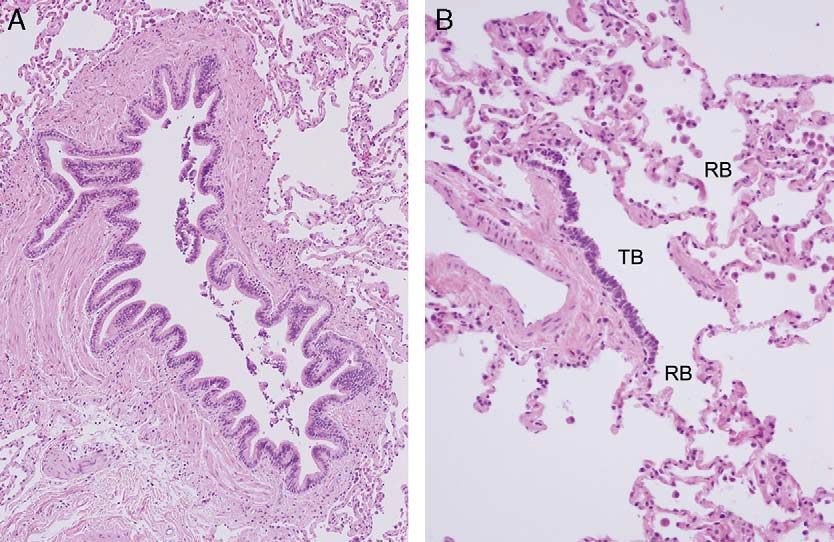
Abbott et al                                                                  J Thorac Imaging      Volume 24, Number 4, November 2009

FIGURE 4. Indirect signs of small airways disease. A, Illustration depicts mosaic attenuation/air trapping characterized by sharply defined
geographic areas of low attenuation (darker lung) with contours that follow the polyhedral outlines of the underlying secondary pulmonary
lobules, depicted by gray lines. B, Illustration depicts the same findings as in A without the underlying pulmonary architecture and, therefore
more closely approximates the findings seen on CT and HRCT.

               SMALL AIRWAYS DISEASE                                      iolitis correlates most directly with the imaging features of
                                                                          small airways disease.9,12 This article will emphasize the
Definition                                                                imaging appearances of bronchiolitis and review those
      The term small airways disease refers to pathologic                 lesions that produce direct and indirect imaging features of
conditions that involve the bronchioles primarily, or as                  small airways disease.
components of interstitial or alveolar lung disease. Bronch-                    Cellular bronchiolitis refers to bronchiolitis in which
iolitis occurs in a heterogeneous group of lesions that vary              inflammatory cells (acute, chronic, or acute and chronic)
in their etiology, clinical settings, and pathologic features             are the predominant histopathologic finding and is a
but are centered on small conducting airways.1                            common pattern found in many clinicopathologic settings.
                                                                          Bronchioles are not visible on normal HRCT but may
Historic Perspective                                                      become visible at the center of the SPL as a direct sign of
      The term ‘‘small airway disease’’ was first used by                  bronchiolitis when their walls are thickened by inflamma-
Hogg and colleagues11 in 1968, in a study, that showed that               tory cell infiltrates. Cellular infiltrates in the peribronchio-
the main site of airflow resistance in patients with chronic               lar alveoli may manifest as poorly defined centrilobular
airflow obstruction was in airways less than 2 mm in                       nodules that may be of soft tissue or ground glass
diameter. They also confirmed in humans what Macklem                       attenuation.2,9,13
and Mead had shown in dogs—that the peripheral airways                          Constrictive bronchiolitis (syn. bronchiolitis obliter-
normally contribute only 10% to 20% of total airway                       ans) is a purely bronchiolar lesion with luminal narrowing
resistance because the total cross-sectional area of the small            by collagenous fibrosis and scarring that may be subtle
airways is much greater than the total cross-sectional area               or result in complete luminal obliteration. Constrictive
of the central airways. When diseased, however, the small                 bronchiolitis can be seen in a variety of disorders and in
airways contribute disproportionately to increased airway                 some specific clinical settings.2
resistance.11 As a consequence, measurement of airway
resistance might be normal in the setting of considerable
obstruction in the peripheral airways. For this reason, the                              CELLULAR BRONCHIOLITIS
small airways have been described as the ‘‘silent zone’’ of
the lungs, and diseases affecting these structures may not be              Infectious Bronchiolitis
evident on pulmonary function tests until late in their                   General Features
course.11                                                                       The imaging features of bronchiolitis are relatively
                                                                          common on chest CT but are often nonspecific, typically
Classification of Small Airways Disease                                   manifesting as centrilobular nodules and branching nodular
     Small airways disease (bronchiolitis) may be classified               (tree-in-bud) opacities, and have been described in viral
according to its clinical setting, its histologic pattern, or on          infection, bacterial pneumonia, tuberculosis, nontuber-
the basis of HRCT imaging findings. The clinical classifica-                culous mycobacterial infection, and aspergillosis. Some
tion is on the basis of the proven or presumed etiology, or               disease entities, however, have associated findings that
on associated systemic conditions. Histologic classification               provide clues to the diagnosis. Detection of associated
into proliferative (cellular) or fibrotic (constrictive) bronch-           cavitary lesions, for instance, suggests the diagnosis of

288 | www.thoracicimaging.com                                                                        r   2009 Lippincott Williams & Wilkins
J Thorac Imaging Volume 24, Number 4, November 2009 Imaging of Small Airways Disease
FIGURE 5. Microscopic features of cellular bronchiolitis. A, Intermediate power photomicrograph (hematoxylin and eosin stain) shows
dilated branching bronchioles surrounded by a dense acute and chronic inflammatory cellular infiltrate. B, High-power
photomicrograph (hematoxylin and eosin stain) shows the inflammatory cellular infiltrate involving the bronchiolar epithelium and wall.
Mycobacterium tuberculosis, atypical mycobacterial, or as hyperinflation; patchy bilateral consolidation often
fungal infection,14 whereas findings of bronchiolitis asso- indicates the presence of bronchopneumonia.
ciated with bronchiectasis, particularly with involvement
of the middle lobe and lingula, suggest the diagnosis of CT/HRCT Features
atypical mycobacterial infection.15 The combination of In adults, infectious bronchiolitis typically manifests
centrilobular nodules and lobular areas of ground-glass on HRCT as well-defined centrilobular nodules and tree-in-
opacity on chest CT is suggestive of Mycoplasma pneumo- bud opacities that may be patchy and unilateral or bilateral
niae pneumonia.16 and asymmetric. The presence of tree-in-bud nodules is
Acute infectious bronchiolitis most commonly affects highly suggestive of infectious bronchiolitis but may be a
children and is most often caused by viruses (respiratory manifestation of noninfectious conditions (eg, aspiration,
syncytial virus, adenoviruses, parainfluenza virus, influenza cystic fibrosis) (Figs. 6–9).17
virus, and human metapneumovirus) and M. pneumoniae
pneumonia. Less common etiologies include chlamydia, Hypersensitivity Pneumonitis
bacteria, and fungi (eg, aspergillus in immunocompromised Cellular bronchiolitis is a prominent feature of
individuals).8,17 hypersensitivity pneumonitis (HP) (syn. extrinsic allergic
alveolitis), an allergic lung disease caused by the inhalation
Clinical Presentation of organic or inorganic agents and of some chemicals. In
The clinical presentation of adults with infectious affected individuals, the small organism or protein complex
bronchiolitis is typically less severe than that of affected lodges in the terminal and respiratory bronchioles and
infants and children, but may sometimes be severe and
fatal.17 Children affected by infectious bronchiolitis often
present with symptoms of an upper respiratory tract
infection followed 2 to 3 days later by the abrupt onset of
dyspnea, tachypnea, and fever.1,8
Pathology
Histologically, infectious bronchiolitis is characterized
by the presence of inflammatory cells, mainly neutrophils,
in the walls of airways and inflammatory exudates in
the airway lumens (Fig. 5).2 In some instances, specific
histologic or microbiologic findings may indicate a specific
diagnosis (eg, hyphae in aspergillus infection, observation
of acid-fast bacilli in M. tuberculosis infection).
Imaging Findings
Radiography FIGURE 6. Purulent bronchiolitis. Unenhanced axial thin-section
Chest radiographs may reveal bilateral, often subtle, chest CT (lung window) demonstrates diffuse bilateral centrilob-
nodular, or reticulonodular opacities. In children, airway ular nodules and tree-in-bud opacities consistent with the cellular
wall thickening and peribronchial consolidations are bronchiolitis characteristically seen in bacterial infections. Note
common. Partial small airway obstruction may manifest the centrilobular nodules do not abut the pleural surfaces.
r 2009 Lippincott Williams & Wilkins www.thoracicimaging.com | 289Abbott et al J Thorac Imaging Volume 24, Number 4, November 2009
Aspiration Bronchiolitis
General Features
Diffuse aspiration bronchiolitis (DAB) is a recently
described clinical entity characterized by chronic inflamma-
tion of bronchioles as a result of recurrent aspiration of
foreign material.23
Clinical Presentation
Half of all the patients affected with DAB have
oropharyngeal dysphagia; many are elderly or bedridden
patients, or individuals with neurologic disorders or
dementia. Many present with bronchorrhea, bronchos-
pasm, wheezing, and dyspnea—signs and symptoms that
are often associated with oral food intake. Patients with
DAB often present with a persistent respiratory illness and
radiographic findings of diffuse small nodular opacities.23,24
FIGURE 7. Viral bronchiolitis. Unenhanced coronal chest CT MIP
image (lung window) of a 45-year-old man with cough and fever Pathology
demonstrates profuse bilateral centrilobular ground-glass nodu- DAB is characterized histologically by chronic mural
lar and tree-in-bud opacities surrounded by uninvolved normal
lung within each secondary pulmonary lobule.
inflammation of bronchioles with foreign body reaction.
Evidence of cryptogenic organizing pneumonia or broncho-
pneumonia may be seen within associated patchy areas of
alveoli, and induces an alveolitis and inflammatory consolidation.24
granulomatous bronchiolitis of variable severity.18–21
Patients with subacute HP may experience an insidious
onset of symptoms over a period of weeks or months and Imaging Findings
may present with cough and dyspnea on exertion, or
experience loss of appetite, weight loss, and fatigue.22 Radiography
Acute HP manifests as pulmonary edema and diffuse Chest radiographs demonstrate diffuse small (J Thorac Imaging Volume 24, Number 4, November 2009 Imaging of Small Airways Disease
FIGURE 9. Bronchiolitis in Mycobacterium tuberculosis infection. A, Contrast-enhanced thin-section chest CT (lung window) of a 39-
year-old man with cough demonstrates a cavitary lesion in the superior segment of the left lower lobe, patchy consolidation in the left
upper lobe, and centrilobular nodular and tree-in-bud opacities in the left upper and lower lobes. B, Contrast-enhanced coronal chest
CT MIP image (lung window) of the same patient shows bilateral pulmonary involvement with two cavitary lesions in the right upper
lobe, bilateral upper lobe consolidations, and centrilobular nodular and tree-in-bud opacities affecting both upper lobes and the
superior segments of both lower lobes.
Follicular Bronchiolitis immunoglobulin deficiencies. However, FB may also be
General Features found in patients with collagen vascular disease (ie, rheuma-
toid arthritis, Sjögren syndrome), systemic hypersensitivity
Follicular bronchiolitis (FB) (syn. pulmonary lymphoid
reactions, infection, lymphoproliferative disease, and diffuse
hyperplasia, hyperplasia of the bronchus-associated lym-
panbronchiolitis (DBP), and as a reactive process distal to
phoid tissue, or hyperplasia of the mucosa-associated
bronchiectasis or in association with middle-lobe syndrome
lymphoid tissue) is a cellular bronchiolitis characterized
(Fig. 13).2,17,26
histologically by peribronchial and peribronchiolar lymphoid
follicles. The detection of FB in lung-biopsy specimens
suggests the possibility of congenital or acquired immuno- Clinical Presentation
deficiency syndromes, including HIV infection and acquired FB occurs most commonly in middle-aged adults but
may occasionally be found in children. Affected patients
typically present with progressive shortness of breath and
cough. Fever, recurrent pneumonia, and weight loss are less
common clinical manifestations of FB. Pulmonary function
tests may reveal obstruction, restriction, or a mixed pattern
of functional abnormalities.26,27
Clinical conditions associated with FB are similar to
those associated with lymphoid interstitial pneumonia and
FIGURE 10. Subacute hypersensitivity pneumonitis. Unenhanced FIGURE 11. Subacute hypersensitivity pneumonitis. Contrast-
coronal chest CT MinIP image (lung window) of a 54-year-old enhanced thin-section chest CT (lung window) of a 49-year-old
woman with pet birds demonstrates bilateral patchy and geo- man with dyspnea demonstrates bilateral centrilobular nodules
graphic areas of low attenuation (air trapping) in both upper lobes, of soft tissue and ground-glass attenuation diffusely involving
and diffuse centrilobular ground-glass nodules and patchy areas of both upper-lung zones, and a focal geographic area of low
ground-glass attenuation involving both lungs. attenuation (air trapping) in the left upper lobe.
r 2009 Lippincott Williams & Wilkins www.thoracicimaging.com | 291Abbott et al J Thorac Imaging Volume 24, Number 4, November 2009
FIGURE 12. Bronchiolitis in aspiration. Unenhanced thin-section FIGURE 14. Follicular bronchiolitis. Contrast-enhanced thin-
chest CT (lung window) of a 79-year-old woman with neurologic section chest CT (lung window) of a 42-year-old HIV-positive
deficits demonstrates secretions in the lumen of the left lower man with immunodeficiency demonstrates numerous small
lobe bronchus, multifocal consolidation in the lingula and left bilateral centrilobular nodules and tree-in-bud opacities.
lower lobe, and scattered centrilobular nodules and tree-in-bud
opacities affecting both lungs.
include: (1) immunological disorders (Sjögren syndrome, cirrhosis, and myasthenia gravis), (2) immunodeficiency
Hashimoto thyroiditis, pernicious anemia, autoimmune (HIV, AIDS), (3) allergy, including asthma, and (4)
hemolytic anemia, chronic active hepatitis, primary biliary allogeneic bone-marrow transplantation.27
Pathology
Histologically, FB is characterized by abundant
lymphoid follicles with abundant germinal centers in the
walls of bronchioles and, less extensively, along bronchi,
interlobular septa, and the pleura.17,27 Obstructive pneu-
monia may be seen as a result of airway compression.27
Imaging Findings
Radiography
FB manifests on chest radiography as bilateral
reticular or reticulonodular opacities. However, the findings
may be subtle and in some cases the radiograph may be
interpreted as normal.8,26
CT/HRCT Features
CT/HRCT demonstrates small, poorly defined centri-
lobular nodules that are diffusely distributed. Additional
but variable findings include peribronchial and subpleural
nodules, patchy areas of ground-glass attenuation, bron-
chial wall thickening, and patchy areas of low attenua-
tion.8,28 In 1 study of 12 patients with FB, HRCT findings
included bilateral centrilobular nodules (100%), patchy
ground-glass opacities (75%), peribronchial nodules (42%),
and subpleural nodules (25%) (Fig. 14).28
Diffuse Panbronchiolitis
General Features
Diffuse panbronchiolitis (DPB) is a progressive form of
cellular bronchiolitis that occurs almost exclusively in adults of
Japanese heritage and is associated with chronic inflammation
FIGURE 13. Microscopic features of follicular bronchiolitis. of the paranasal sinuses. The imaging features of DPB may
Intermediate power photomicrograph (hematoxylin and eosin have considerable overlap with those of other inflammatory
stain) shows interstitial chronic inflammation and a hyperplastic airway diseases, including cystic fibrosis, inflammatory bowel
lymphoid follicle with prominent germinal center adjacent to disease, idiopathic bronchiectasis, constrictive bronchiolitis
small bronchioles. (CB), and FB.2
292 | www.thoracicimaging.com r 2009 Lippincott Williams & WilkinsJ Thorac Imaging Volume 24, Number 4, November 2009 Imaging of Small Airways Disease
Clinical Presentation
Patients with DPB typically present with cough
and dyspnea on exertion and may have symptoms of
sinusitis. Pseudomonas aeruginosa frequently colonizes the
respiratory tract of affected patients. Pulmonary function
tests reveal marked obstruction and mild restrictive disease.8
Pathology
The gross pathologic features of DPB are yellow 1 to
3 mm nodules. Histologically, there is transmural infiltration
of the bronchiolar interstitium and peribronchiolar tissue by
foamy macrophages, lymphocytes, and plasma cells. The
involved small airways may include respiratory bronchioles,
terminal (membranous) bronchioles, and alveolar ducts.
Terminal bronchioles may be ectatic. Follicular bronchiolitis
may be present, and acute inflammation may occur in
bronchiolar lumens. Superimposed acute or organizing
pneumonia and bronchiectasis may also occur.2,13
Imaging Findings
Radiography
Chest radiography reveals diffuse nodules (Abbott et al J Thorac Imaging Volume 24, Number 4, November 2009
infection, transplantation, collagen vascular disorders,
inhalational lung injury, and ingested toxins.
Infection
Childhood infection is a rare cause of CB. The causa-
tive infection typically occurs in children under 8 years of
age.31 Typical infections are viral (respiratory syncytial
virus, parainfluenza virus, adenovirus), but M. pneumoniae
has also been implicated.30 Some affected patients may go
on to exhibit imaging features of the Swyer-James
syndrome or MacLeod syndrome as adults. It is postulated
that viral bronchiolitis of childhood may interfere with the
normal development of the affected lung with resultant CB.
These patients may be asymptomatic or may present with
cough, recurrent infection, or hemoptysis.32 CB may also
be seen in cases of cystic fibrosis and is felt to represent
the sequela of recurrent episodes of pulmonary infection
(Figs. 16, 17).32
Transplantation
Lung Transplantation
CB is a known complication of lung and heart-lung
transplantation that impacts the morbidity and limits the
survival of affected patients.33 CB is considered a manifes-
tation of chronic allograft dysfunction or chronic rejec- FIGURE 17. Constrictive bronchiolitis in cystic fibrosis. Unenhanced
tion.29,34 The major risk factor for CB is acute rejection, coronal chest CT MIP image (lung window) of a 25-year-old
particularly when severe or recurrent.29,33 Severe infection, woman with cystic fibrosis demonstrates airway wall thickening,
particularly cytomegalovirus pneumonia is also associated bronchiectasis, nodular, and tubular areas of mucoid impaction,
with the development of CB.33 CB is a late complication of and patchy areas of mosaic attenuation (air trapping) that diffusely
transplantation that occurs at least 3 months (median 16 to involve both lungs but predominantly involve the upper lung
zones.
20 mo) after the transplant.29,30 The onset of symptoms
may be insidious or rapid. The mechanism of rejection is
related to the activity of the recipient immune system
against the transplanted lung tissue.29
The International Society of Heart Lung Transplantation
has defined the BOS based on deterioration of pulmonary
function, specifically, the forced expiratory volume in one
second (FEV1) when compared with baseline. A clinical
diagnosis of BOS is based on documentation of a decline in
FEV1 of more than 20% during the posttransplantation
period, after exclusion of other causes of allograft dysfunc-
tion.35 The category of ‘‘potential’’ BOS or stage BOS 0-p is
based on a decrease in forced expiratory flow in the mid-
expiratory phase (forced expiratory flows between 25% and
75% of forced vital capacity), but its prognostic usefulness is
under debate.34,36 Although air trapping found on expiratory
thin section CT is considered the most sensitive and accu-
rate imaging indicator of CB in lung transplant recipients
(Fig. 18),37,38 some investigators have found a weak correlation
between the BOS stage and the severity of bronchial dilatation,
bronchial wall thickening, air trapping and mosaic attenuation
seen on CT, particularly in single lung transplant recipients.35
In addition, CT may be insensitive to the diagnosis of early CB
and to the detection of early stage BOS.36,39 The prevalence
of CB in lung transplant recipients surviving at 5 years is
approximately 50% with a 5-year survival after the onset of
FIGURE 16. Postinfectious constrictive bronchiolitis. Contrast- disease of approximately 30% to 40%.30
enhanced coronal chest CT MIP image (lung window) of a 56-
year-old man with a history of recent lower respiratory tract
infection shows bilateral geographic areas of mosaic attenuation
Hematopoietic Stem Cell Transplantation
and air trapping in the right lung with hyperinflation of the right CB is a known complication of hematopoietic stem cell
lower lobe and multiple centrilobular nodules and small foci of (HSC) transplantation with a prevalence of approximately
consolidation in both upper lobes. 5%. It is one of the late pulmonary complications of HSC
294 | www.thoracicimaging.com r 2009 Lippincott Williams & WilkinsJ Thorac Imaging Volume 24, Number 4, November 2009 Imaging of Small Airways Disease
FIGURE 18. Constrictive bronchiolitis after lung transplantation. A, Unenhanced inspiratory thin-section chest CT (lung window) of a
52-year-old woman after left lung transplantation demonstrates subtle heterogeneity (mosaic attenuation) of the left lung parenchyma.
B, Expiratory thin-section chest CT (lung window) of the same patient shows patchy areas of air trapping.
transplantation and typically develops more than 100 days opment of CB of variable severity.29 Inhalation of fire
(median 400 to 450 d) after the transplant.29,30 It is thought fumes (Fig. 19) and the butter-flavoring ingredient acetyl in
to result from graft-versus-host disease induced by the popcorn plant workers have also been associated with the
donor cells.30 However, although it is typically described development of CB.30,41
in patients undergoing allogeneic transplants, it is also
reported in those undergoing autologous transplants. Risk
factors for the development of CB include older age, chronic
graft-versus-host disease and methotrexate therapy. The
histologic diagnosis may be difficult to make on transbron-
chial lung biopsy due to the patchy nature of the disease.29
The prevalence of CB in allogeneic HSC transplant
recipients is estimated at approximately 9% with a range of
up to 48%.29 The mortality of affected patients reported in
the literature is variable with reports of 12% to 27% 5-year
mortalities in one series and survival of only 10% of
patients with CB at 5 years in another.29
Connective Tissue Disease
CB may occur in the setting of rheumatoid arthritis
with or without penicillamine treatment. Affected patients
are typically women in the fifth or sixth decades of life, and
most have long-standing disease. The small airways disease
may rarely antedate the rheumatologic manifestations
of the disease.40 It should be noted that FB (a cellular
bronchiolitis) has also been described in patients with
rheumatoid arthritis. CB has also been described in a few
patients with systemic lupus erythematosus.29
Inhalational Lung Disease
Although several chemicals have been implicated, the
best understood diseases are those related to inhalation of
oxides of nitrogen. Silo filler’s disease is related to nitrogen
dioxide (NO2) and nitric oxide (NO), which are produced
during the anaerobic fermentation of silage. Nitrous acids
and nitric oxides produce severe injury to tissues. The
presentation of exposed individuals will vary with the
degree of exposure. Clinical presentation may be accom-
FIGURE 19. Constrictive bronchiolitis after smoke inhalation.
panied by mild symptoms that may progress to pulmonary A, Unenhanced inspiratory thin-section chest CT (lung window)
edema and acute respiratory distress syndrome. Those of a 45-year-old woman after smoke inhalation in a house fire
patients who recover may become asymptomatic for a short demonstrates mild mosaic attenuation and patchy ground-glass
period of time, and subsequently present with progressive opacity in the left upper lobe. B, Expiratory thin-section CT
cough, dyspnea, and hypoxemia, secondary to the devel- demonstrates bilateral scattered areas of air trapping.
r 2009 Lippincott Williams & Wilkins www.thoracicimaging.com | 295Abbott et al J Thorac Imaging Volume 24, Number 4, November 2009
Miscellaneous Conditions
CB has been reported in association with ingestion of
Sauropus androgynous, a weight control preparation used
in Asia. Other associations include inflammatory bowel
disease,43 paraneoplastic pemphygus and gold and penicil-
lamine therapy.40
Pathology
CB is characterized by fibrosing bronchiolitis of
membranous and respiratory bronchioles with submucosal
and peribronchiolar concentric fibrosis and little active
inflammation or granulation tissue (Fig. 21). Early disease
is characterized by a predominantly lymphocytic inflam-
matory cellular infiltrate affecting the lumen, mucosa,
submucosa, and the tissues surrounding the bronchioles.
This progresses to concentric peribronchiolar fibrosis with-
out associated fibroblastic proliferation.29 The airway
FIGURE 20. Small airways disease in diffuse idiopathic pulmo- involvement tends to be circumferential and results in
nary neuroendocrine cell hyperplasia. Unenhanced expiratory bronchiolar luminal narrowing and obstruction. Advanced
thin-section chest CT (lung window) of a 49-year old woman disease may be difficult to diagnose histologically as the
with diffuse idiopathic pulmonary neuroendocrine cell hyperpla- bronchioles may become completely obliterated and may be
sia demonstrates small bilateral pulmonary nodules and patchy
inconspicuous due to the absence of surrounding inflam-
geographic areas of air trapping.
mation.1 In these cases, elastic stains may be helpful for the
identification of affected small airways.29 Furthermore, the
patchy nature of the disease may result in sampling errors
Diffuse Idiopathic Pulmonary Neuroendocrine on transbronchial biopsy.29 Thus, there may be subtle
Cell Hyperplasia pathologic abnormalities in spite of severe symptoms and
Proliferation of pulmonary neuroendocrine cells is extensive imaging abnormalities.
typically confined to the bronchial and bronchiolar
epithelium. These proliferations may take the form of
tumorlets, which can be diffusely distributed through- Imaging Findings
out the lung. Nodular neuroendocrine cell prolifera- Radiography
tions measuring more than 5 mm in diameter are usually Patients with CB typically have normal or near normal
classified as carcinoid tumors. Affected patients are chest radiographs. Previously reported radiographic ab-
typically women who may exhibit obstructive and/or normalities include hyperinflation, air trapping, and per-
restrictive lung function or may be asymptomatic. These ipheral attenuation of pulmonary vascular markings.29,32
patients may exhibit HRCT evidence of small airways Nodular and reticular pulmonary opacities have also been
disease in which multifocal pulmonary nodules may be described.32
associated with mosaic lung attenuation and air trapping Patients with Swyer-James syndrome or MacLeod
(Fig. 20).42 syndrome may exhibit a unilateral hyperlucent lung with
FIGURE 21. Microscopic features of constrictive bronchiolitis. A, Intermediate power photomicrograph (hematoxylin and eosin stain)
shows a small terminal bronchiole surrounded by concentric fibrosis. B, High-power photomicrograph (hematoxylin and eosin stain)
shows luminal narrowing of a small terminal bronchiole surrounded by concentric fibrosis.
296 | www.thoracicimaging.com r 2009 Lippincott Williams & WilkinsJ Thorac Imaging Volume 24, Number 4, November 2009 Imaging of Small Airways Disease
FIGURE 22. Constrictive bronchiolitis in Swyer-James syndrome or MacLeod syndrome. A, PA chest radiograph of a 33-year-old man
with Swyer-James syndrome or MacLeod syndrome demonstrates subtle hyperlucency in the right upper lobe. B. Unenhanced
expiratory thin-section chest CT (lung window) of the same patient shows bilateral areas of geographic air trapping that are most
evident in the right upper lobe. C, Unenhanced coronal expiratory chest CT (lung window) of the same patient shows bilateral areas of
air trapping that are most evident in the right upper lobe but also affect the left upper and left lower lobes. Note central bronchiectasis
and bronchial wall thickening in the left lung.
decreased pulmonary vascularity and a small ipsilateral typically exhibit air trapping and hyperlucency in other
hilum. The affected lung parenchyma may exhibit normal lobes and in the contralateral lung, which is not readily
or decreased lung volume during inspiration and expiratory appreciated at radiography.43,50 Bronchiectasis and bron-
air trapping (Fig. 22).32 chial wall thickening may also be observed.9
CT/HRCT Features
The classically described CT feature of CB is a pattern
of mosaic pulmonary attenuation characterized by geo- SUMMARY
graphic clusters of lung lobules that exhibit alternating Small airways disease includes a spectrum of inflam-
increased and decreased lung attenuation (Fig. 4). The matory and fibrotic pulmonary diseases centered on
mosaic attenuation is typically accentuated on expiratory the small conducting airways. The imaging manifestations
imaging where air trapping manifests as areas of persistent of small airways disease on CT/HRCT may be direct or
hyperlucency highlighted by increased pulmonary attenua- indirect signs of small airway involvement and include
tion in surrounding normal lung. Within low-attenuation centrilobular nodules and branching nodular (tree-in-bud)
areas of air trapping there is often a decrease in the caliber opacities, or the demonstration of mosaic attenuation that
of vascular structures that is likely secondary to hypoxic is typically exaggerated on expiratory CT. Some disease
vasoconstriction. In addition, there is no decrease in the entities have distinguishing clinical, pathologic, and/or
cross-sectional area of the affected hyperlucent lung on imaging features that facilitate a more accurate diagnosis.
expiration.6 The areas of increased attenuation typically
reflect normal lung parenchyma, which exhibits a relative
increase in vessel caliber and blood flow. Expiratory thin- REFERENCES
section CT facilitates detection of areas of air trapping. The 1. Visscher DW, Myers JL. Bronchiolitis: the pathologist’s
patchy distribution of airway involvement gives rise to perspective. Proc Am Thorac Soc. 2006;3:41–47.
mosaic lung attenuation and perfusion. The areas of 2. Couture C, Colby TV. Histopathology of bronchiolar dis-
abnormal attenuation may exhibit well or poorly defined orders. Semin Respir Crit Care Med. 2003;24:489–498.
contours.44 It should be noted that mild-mosaic attenuation 3. Abbott G. Airway structure. In: Federle MP, Rosado-de-
and air trapping on expiratory CT has also been described Christenson ML, Woodward PJ, et al. Diagnostic and Surgical
in healthy subjects.45–47 In addition, mosaic attenuation Imaging Anatomy: Chest, Abdomen, Pelvis. Salt Lake City:
may occur in patients with occlusive vascular disease and Amirsys; 2006:64–87.
in association with infiltrative lung disease. Patients with 4. Tomashefski JF, Farver CF. Anatomy and histology of the
lung. In: Tomashefski JF, ed. Dail and Hammar’s Pulmonary
diffuse severe disease may exhibit diffuse decreased lung Pathology. 3rd ed. New York: Springer; 2008:20–48.
attenuation that may be subtle.6,48,49 5. Lynch DA. Imaging of small airways disease and chronic
Associated findings include bronchial dilatation, obstructive pulmonary disease. Clin Chest Med. 2008;29:165–179.
bronchiectasis, and bronchial wall thickening.9,29 Other 6. Hansell DM. Small airways disease: detection and insights with
findings include centrilobular nodules. If pulmonary no- computed tomography. Eur Respir J. 2001;17:1294–1313.
dules are seen, diffuse idiopathic neuroendocrine cell hyper- 7. Biegelman-Aubry C, Hill C, Guibal A, et al. Multi-detector
plasia should be considered. row CT and postprocessing techniques in the assessment of
Swyer-James syndrome or MacLeod syndrome refers diffuse lung disease. Radiographics. 2005;25:1639–1652.
to predominant involvement of one lobe or lung with 8. Naidich DP, Webb WR, Grenier PA, et al. Small airway
disease. In: Naidich DP, Webb WR, Grenier PA, et al. Imaging
findings of CB.9 Affected patients typically have radio- of the Airways. Philadelphia: Lippincott Williams & Wilkins;
graphic evidence of focal lung hyperlucency and decreased 2005:146–176.
vascularity with normal or decreased volume of the affected 9. Pipavath SJ, Lynch DA, Cool C, et al. Radiologic and
lung. Expiratory HRCT typically demonstrates air trapping pathologic features of bronchiolitis. Am J Roentgenol. 2005;
of the affected lung lobe. However, affected patients 185:354–363.
r 2009 Lippincott Williams & Wilkins www.thoracicimaging.com | 297Abbott et al J Thorac Imaging Volume 24, Number 4, November 2009
10. Hansell DM, Bankier AA, MacMahon H, et al. Fleischner 31. Chiu CY, Wong KS, Huang YC, et al. Bronchiolitis obliterans
Society: glossary of terms for thoracic imaging. Radiology. in children: clinical presentation, therapy and long-term
2008;246:697–722. follow-up. J Paediatr Child Health. 2008;44:129–133.
11. Hogg J, Macklem P, Thurlbeck W. Site and nature of airway 32. Müller NL, Miller RR. Diseases of the bronchioles: CT and
obstruction in chronic obstructive lung disease. N Engl J Med. histopathologic findings. Radiology. 1995;196:3–12.
1968;278:1355–1360. 33. Collins J. Imaging of the chest after lung transplantation.
12. Poletti V, Costtable U. Bronchiolar disorders: classification and di- J Thorac Imaging. 2002;17:102–112.
agnostic approach. Semin Respir Crit Care Med. 2003;24:457–464. 34. de Jong PA, Dodd JD, Coxson HO, et al. Bronchiolitis
13. Cagle PT, Roggli VL. Pathology of small airways. In: obliterans following lung transplantation: early detection
Tomashefski JF, ed. Dail and Hammar’s Pulmonary Pathology. using computed tomographic scanning. Thorax. 2006;61:
3rd ed. New York: Springer; 2008:886–910. 799–804.
14. Leung AN. Pulmonary tuberculosis: the essentials. Radiology. 35. Choi YW, Rossi SE, Palmer SM, et al. Bronchiolitis obliterans
1999;210:307–322. syndrome in lung transplant recipients: correlation of com-
15. Aquino SL, Gamsu G, Webb WR, et al. Tree-in-bud pattern: puted tomography findings with bronchiolitis obliterans
frequency and significance on thin section CT. J Comput Assist syndrome stage. J Thorac Imaging. 2003;18:72–79.
Tomogr. 1996;20:594–599. 36. Konen E, Gutierrez C, Chaparro C, et al. Bronchiolitis
16. Reittner P, Müller NL, Heyneman L, et al. Mycoplasma obliterans syndrome in lung transplant recipients: can thin-
pneumoniae pneumonia: radiographic and high-resolution CT section CT findings predict disease before its clinical appear-
features in 28 patients. Am J Roentgenol. 2000;174:37–41. ance? Radiology. 2004;231:467–473.
17. Silva CIS, Müller NL. Bronchiolitis. In: Silva CIS, Müller NL, 37. Leung AN, Fisher K, Valentine V, et al. Bronchiolitis
Hansell DM. Imaging of the Chest. Philadelphia: Saunders; obliterans after lung transplantation: detection using expira-
2008:1071–1095. tory HRCT. Chest. 1998;113:365–370.
18. Hansell DM, Wells AU, Padley SPG. Hypersensitivity 38. Worthy SA, Park CS, Kim JS, et al. Bronchiolitis obliterans
pneumonitis: correlation of individual CT patterns with after lung transplantation: high-resolution CT findings in 15
functional abnormalities. Radiology. 1996;199:123–128. patients. Am J Roentgenol. 1997;169:673–677.
19. Lacasse Y, Selman M, Costabel U, et al. Clinical diagnosis 39. Lee ES, Gotway MB, Reddy GP, et al. Early bronchiolitis
of hypersensitivity pneumonitis. Am J Respir Crit Care Med. obliterans following lung transplantation: accuracy of expira-
2003;168:952–958. tory thin-section CT for diagnosis. Radiology. 2000;216:
20. Matar LD, McAdams HP, Sporn TA. Hypersensitivity 472–477.
pneumonitis. Am J Roentgenol. 2000;174:1061–1066. 40. Ryu JH, Myers JL, Swensen SJ. Bronchiolar disorders. Am
21. Silva CIS, Churg A, Müller NL. Hypersensitivity pneumonitis: J Respir Crit Care Med. 2003;168:1277–1292.
spectrum of high-resolution CT and pathologic findings. Am 41. Lockey JE, Hilbert TJ, Levin LP, et al. Airway obstruction
J Roentgenol. 2007;188:334–344. related to diacetyl exposure at microwave popcorn production
22. Morris DG, Gotway MB, Homer RJ, et al. Diffuse parench- facilities. Eur Respir J. 2009;34:63–71.
ymal and alveolar lung disease. In: George RB, Light RW, 42. Davies SJ, Gosney JR, Hansell DM, et al. Diffuse idio-
Matthay MA, et al. Chest Medicine. Essentials of Pulmonary pathic pulmonary neuroendocrine cell hyperplasia: an
and Critical Care Medicine. 5th ed. Philadelphia: Lippincott under-recognized spectrum of disease. Thorax. 2007;62:
Williams & Wilkins; 2005:234–288. 248–252.
23. Matsuse T, Fukuchi Y, Oka T, et al. Importance of diffuse 43. Waitches GM, Stern EJ. High-resolution CT of peripheral
aspiration bronchiolitis caused by chronic occult aspiration in airways diseases. Radiol Clin North Am. 2002;40:21–29.
the elderly. Chest. 1996;110:1289–1293. 44. Worthy SA, Müller NL. Small airway diseases. Radiol Clin
24. Barnes TW, Vassallo R, Tazelaar HD, et al. Diffuse North Am. 1998;38:163–173.
bronchiolar disease due to chronic occult aspiration. Mayo 45. Bankier AA, van Muylem A, Knoop C, et al. Bronchio-
Clin Proc. 2006;81:172–176. litis obliterans syndrome in heart-lung transplant reci-
25. Heyneman LE, Ward S, Lynch DA, et al. Respiratory pients: diagnosis with expiratory CT. Radiology. 2001;218:
bronchiolitis, respiratory bronchiolitis-associated interstitial 533–539.
lung disease, and desquamative interstitial pneumonia: differ- 46. Tanaka N, Matsumoto T, Miura G, et al. Air trapping at CT:
ent entities or part of the spectrum of the same disease process? high prevalence in asymptomatic subjects with normal
Am J Roentgenol. 1999;173:1617–1622. pulmonary function. Radiology. 2003;227:776–785.
26. Travis WD, Galvin JR. Non-neoplastic pulmonary lymphoid 47. Arakawa H, Kurihara Y, Sasaka K, et al. Air trapping on CT
lesions. Thorax. 2001;56:964–971. of patients with pulmonary embolism. Am J Roentgenol.
27. Corrin B, Nicholson AG, Burke MM. Diseases of the airways. 2002;178:1201–1207.
In: Corrin B, Nicholson AG, eds. Pathology of the Lungs. 2nd 48. Sherrick AD, Swensen SJ, Hartman TE. Mosaic pattern of
ed. Philadelphia: Churchill Livingstone; 2006:87–130. lung attenuation on CT scans: frequency among patients
28. Howling SJ, Hansell DM, Wells AU, et al. Follicular with pulmonary artery hypertension of different causes. Am J
bronchiolitis: thin-section CT and histologic findings. Radio- Roentgenol. 1997;169:79–82.
logy. 1999;212:637–642. 49. Teel GS, Engeler CE, Tashijian JH, et al. Imaging of small
29. Angel LA, Homma A, Levine S. Bronchiolitis obliterans. airways disease. Radiographics. 1996;16:27–41.
Semin Respir Crit Care Med. 2000;21:123–134. 50. Moore ADA, Goodwin JD, Dietrich PA, et al. Swyer-James
30. Cordier JF. Challenges in pulmonary fibrosis. 2: Bronchiolo- syndrome: CT findings in eight patients. Am J Roentgenol.
centric fibrosis. Thorax. 2007;62:638–649. 1992;158:1211–1215.
298 | www.thoracicimaging.com r 2009 Lippincott Williams & WilkinsYou can also read