Complications of antireflux surgery
←
→
Page content transcription
If your browser does not render page correctly, please read the page content below

REVIEW ARTICLE              1137

see related CME on page 1117

Complications of antireflux surgery

                                                                                                                                                               REVIEW ARTICLE
Rena Yadlapati, MD, MSHS1, Eric S. Hungness, MD2 and John E. Pandolfino, MD, MSCI2

Antireflux surgery anatomically restores the antireflux barrier and is a therapeutic option for proton pump inhibitor
(PPI)-refractory gastroesophageal reflux disease or PPI intolerance. Laparoscopic fundoplication is the standard
antireflux surgery, though its popularity has declined due to concerns regarding wrap durability and adverse events. As
the esophagogastric junction is an anatomically complex and dynamic area subject to mechanical stress, wraps are
susceptible to disruption, herniation or slippage. Additionally, recreating an antireflux barrier to balance bidirectional
bolus flow is challenging, and wraps may be too tight or too loose. Given these complexities it is not surprising
that post-fundoplication symptoms and complications are common. Perioperative mortality rates range from 0.1 to
0.2% and prolonged structural complications occur in up to 30% of cases. Upper gastrointestinal endoscopy with a
comprehensive retroflexed examination of the fundoplication and barium esophagram are the primary tests to assess
for structural complications. Management hinges on differentiating complications that can be managed with medical
and lifestyle optimization versus those that require surgical revision. Reoperation is best reserved for severe structural
abnormalities and troublesome symptoms despite medical and endoscopic therapy given its increased morbidity
and mortality. Though further data are needed, magnetic sphincter augmentation may be a safer alternative to
fundoplication.
Am J Gastroenterol (2018) 113:1137–1147. https://doi.org/10.1038/s41395-018-0115-7

IntroduCtIon                                                                         a posterior or anterior fundoplication [7, 8]. Success rates of
Gastroesophageal reflux disease (GERD) is defined as the pres-                       laparoscopic fundoplication range from 67 to 95%, and depend
ence of troublesome symptoms and/or complications that develop                       highly on surgical expertise, adequate preoperative evaluation and
due to retrograde reflux of gastric contents in the esophagus [1].                   appropriate patient selection [9–11]. A comprehensive diagnostic
GERD affects one in five Americans and is one of the most com-                       evaluation prior to antireflux surgery is requisite to ensure that the
mon diagnoses managed in the outpatient gastroenterology clinic                      patient has objective evidence of GERD, to confirm that ongoing
[1, 2]. Although the majority of patients with GERD derive symp-                     PPI-refractory GERD symptoms are due to GERD rather than
tom relief with proton pump inhibitors (PPIs), 30 to 40% do not                      non-GERD causes and to exclude contraindications to surgery.
respond adequately [3]. Various mechanisms can drive PPI non-                        Gastroenterologists commonly evaluate patients with PPI non-
response [4], one being PPI-refractory GERD. PPI-refractory                          response and function as the primary point of care for patients
GERD patients continue to experience troublesome symptoms                            following antireflux surgery. Since laparoscopic fundoplication
in relation to ongoing objective GERD despite optimized PPI                          is an invasive and irreversible intervention associated with non-
therapy. In the case of PPI-refractory GERD or PPI intolerance,                      negligible morbidity and mortality rates, it is essential that gastro-
antireflux surgery is a therapeutic option [5, 6].                                   enterologists are familiar with the evaluation and management of
   The objective of antireflux surgery is to anatomically restore the                complications following fundoplication [12]. This paper will focus
antireflux barrier and thereby reduce gastroesophageal reflux epi-                   on complications of antireflux surgery, and provide a matrix for
sodes. This is no easy task as the esophagogastric junction and its                  the evaluation and management of complications following antire-
antireflux mechanisms are complex and dynamic. The antireflux                        flux surgery.
barrier is composed of the lower esophageal sphincter, the extrin-
sic crural diaphragm and the flap valve configuration related to
supporting structures and orientation of the intrinsic and extrinsic                 LaparosCopIC fundopLICatIon
sphincters. The current standard for antireflux surgery is laparo-                   Trends in utilization
scopic fundoplication which seeks to repair a hernia, reposition                     Surgical Nissen fundoplication was first described in 1956 and
the sphincter within the abdomen and recreate a flap valve using                     regarded as a highly efficacious antireflux intervention, particu-

1
 University of Colorado, Anschutz Medical Campus, Aurora, CO, USA. 2Northwestern University, Chicago, IL, USA. Correspondence: R.Y.
(email: Rena.yadlapati@ucdenver.edu)
Received 3 February 2018; accepted 11 April 2018; Published online 14 June 2018

© 2018 The American College of Gastroenterology                                                              The American Journal of GASTROENTEROLOGY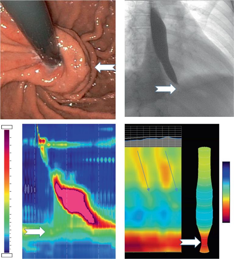
1138            Yadlapati et al.

                             a                                               b                                             c
                                                                                                                                   Scope

                                                      Scope                                                                                         Ant. groove
REVIEW ARTICLE

                                                         Ant. groove

                                                              Lip
                                                                                                            “Omega”-
                                                                                                                           No
                                                                                                            shaped lip
                              Post                       Body of value                                                     posterior
                             groove                      “stacked coils”                                                   groove

                 Fig. 1 Surgical fundoplication techniques. (a) A complete 360 degree Nissen fundoplication creates a nipple valve. On retroflexed endoscopic view the lip
                 of the valve should be thin, the body of the valve should have a “stacked coils” appearance in alignment with the long axis of the endoscope, and the valve
                 should adhere tightly to the scope. The posterior groove will be deep and anterior groove will be shallow. White lines depict the appropriate orientation of
                 the gastric folds as just below the diaphragm and directed perpendicular to the endoscope and parallel to the diaphragm. (b) The Toupet fundoplication
                 is a partial 270 degree posterior wrap which creates a flap valve. The lip of the valve should be thick and “omega” shaped and the valve should be mod-
                 erately adherent to the scope. Both the anterior and posterior groove should be shallow. (c) The Dor fundoplication is a partial 180 degree anterior wrap
                 which creates a flap valve. The lip of the valve should be wide and “S” shaped, and the valve should be moderately adherent to the scope. The anterior
                 groove should be shallow, and there is no posterior groove. Drawings borrowed from Jobe et al. [22]. Endoscopic images courtesy of the Esophageal Center
                 at Northwestern

                 larly in comparison to the pharmacologic alternative of hista-                  which creates a flap valve while leaving the anterior esophagus
                 mine-2 receptor antagonists at that time [13, 14]. In 1991, the                 exposed to allow for radial expansion (Fig. 1b), and a 180-degree
                 less invasive laparoscopic approach to fundoplication was intro-                anterior (Dor) fundoplication sutured to the esophagus and right
                 duced and quickly became the standard in antireflux surgery [8].                crus which restores the angle of His and creates a flap valve mech-
                 Accordingly, the volume of antireflux surgeries increased from 4.4              anism (Fig. 1c) [22, 23].
                 in 1990 to 15.7 in 1999 per 100,000 adults among the Nationwide
                 Inpatient Sample database [15–17]. However, as concerns regard-
                 ing the durability and side effects of laparoscopic fundoplica-                 prEopEratIvE EvaLuatIon
                 tion surfaced along with the increasing accessibility to PPIs, the              Appropriate patient selection for fundoplication is integral to
                 surge in antireflux surgery declined by 30% in the ensuing years                achieving positive postoperative outcomes (Table 1). Objective
                 [15, 18, 19]. Between 2005 and 2010, the annual volume of elec-                 documentation of pathologic acid reflux, either by endoscopic
                 tive open or laparoscopic fundoplication plateaued at approxi-                  evidence of erosive disease or pH monitoring, is essential [5, 6, 9].
                 mately 5.3 cases per 100,000 adults, equating to approximately                  The primary purposes of preoperative esophageal manometry
                 19,000 surgical fundoplications [20, 21].                                       are to rule out contraindications to antireflux surgery and to
                                                                                                 assess esophageal contractile vigor and reserve. There is no
                 Laparoscopic fundoplication technique                                           distinct cut-off for risk of dysphagia based on peristaltic function
                 The major tenants of surgical fundoplication are to return the                  after a fundoplication as most dysphagia is related to underly-
                 esophagogastric junction to an intraabdominal location, close                   ing anatomical and mechanical issues related to the wrap. That
                 the hiatal opening and create a one-way valve. Laparoscopic fun-                being said, absent contractility is an absolute contraindication
                 doplication may be performed via a complete or partial technique.               for a fundoplication as this creates pseudo-achalasia. Thus, the
                 Laparoscopic Nissen fundoplication is a 360-degree fundoplica-                  degree of ineffective peristalsis that is tolerable before fundopli-
                 tion after crural closure, and aims to circumferentially plicate the            cation should be based on a case-by-case evaluation. There are
                 stomach to the esophagus in order to strengthen the esophagogas-                some data supporting that augmentation after multiple rapid
                 tric junction valve (Fig. 1a) [22]. The two most common partial                 swallows and a lack of preoperative dysphagia are indicators of
                 laparoscopic fundoplication techniques include a 270-degree pos-                a reduced likelihood of dysphagia [9]. Therefore, a surgeon may
                 terior (Toupet) fundoplication sutured to the crura and esophagus               select a partial fundoplication technique in the setting of reduced

                 The American Journal of GASTROENTEROLOGY                                                                        VOLUME 113 | AUGUST 2018 www.nature.com/ajg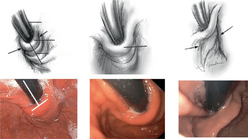
Complications of Antireflux Surgery         1139

 Table 1 Diagnostic evaluation prior to antireflux surgery

 Diagnostic testing           Required       Reason to test           Results in support of consid-        Recommending groups    Comments
                                                                      ering antireflux surgery

                                                                                                                                                                        REVIEW ARTICLE
 pH monitoring off of PPI     Yes            Document pathologic      Pathologic acid exposure; posi-      ACG, AGA, EDAP         Not needed if previous evidence
                                             acid reflux; associate   tive symptom-reflux association                             of Los Angeles Grade C or D
                                             reflux events with       in context of other abnormali-                              esophagitis or long-segment
                                             symptoms                 ties associated with pathologic                             Barrett’s esophagus
                                                                      reflux (e.g., large hiatal hernia)
 Upper GI endoscopy           Yes            Evaluate for signs       Los Angeles Grade C or D es-         ACG, AGA, EDAP
                                             of erosive disease;      ophagitis; long-segment Barrett’s
                                             assess EGJ anatomy;      esophagus
                                             exclude non-GERD
                                             etiologies
 Esophageal manometry         Yes            Exclude achalasia;       Defective antireflux barrier;        ACG, AGA, EDAP         Absent contractility is a contrain-
                                             assess esophageal        intact esophageal peristalsis                               dication to antireflux surgery
                                             peristaltic reserve
 Barium esophagram            No             Evaluate overall         Possibly a large hiatal hernia       EDAP
                                             anatomy
 Gastric emptying study       No             Evaluate for delayed     Normal gastric emptying                                     Should perform in the setting
                                             gastric emptying                                                                     of bloating and dyspeptic
                                                                                                                                  symptoms that are not otherwise
                                                                                                                                  explained, and when consider-
                                                                                                                                  ing surgical revision in setting of
                                                                                                                                  refractory GERD
 ACG American College of Gastroenterology, AGA American Gastroenterological Association, EDAP Esophageal Diagnostic Advisory Panel, GERD gastroesophageal reflux
 disease, GI gastrointestinal, PPI proton pump inhibitor

peristaltic vigor [5, 6, 9]. Barium esophagram is a useful preop-                        be separated into structural complications of the fundoplication
erative study to evaluate the foregut anatomy [9]. Additionally,                         or functional abnormalities.
a gastric emptying study should be performed if there is suspi-
cion of gastroparesis, as this will also tailor the type of antireflux                   Structural complications of fundoplication
intervention [9].                                                                        Structural complications following fundoplication occur in up to
                                                                                         30% of cases and are often related to surgical positioning or con-
                                                                                         struction of the wrap [27]. The esophagogastric junction is a com-
CoMpLICatIons of LaparosCopIC                                                            plex anatomical area that is subject to mechanical stress related
fundopLICatIon                                                                           to the gastroesophageal pressure gradient and its dynamic nature
Complication following laparoscopic fundoplication can occur in                          that allows it to move axially during swallowing and reflux. Thus,
both the acute and prolonged settings (Tables 2 and 3) [24].                             the durability of a fundoplication weakens over time, and is exac-
                                                                                         erbated by intermittent abdominal strain such as from nausea,
Acute complications of laparoscopic fundoplication                                       vomiting, coughing, trauma, abdominal exercises, heavy lifting
The reported 30-day surgical mortality rate of laparoscopic fun-                         and weight gain/obesity. These stressors increase susceptibility to
doplication ranges from 0.1% to 0.2% [24–27]. In a recent pop-                           wrap disruption, herniation or slippage. Additionally, recreation
ulation-based cohort study of 2655 patients who underwent a                              of an antireflux barrier that can balance antegrade and retrograde
primary laparoscopic fundoplication between 2005 and 2014,                               bolus flow is difficult and can result in either a tight or loose wrap.
4.1% of patients had a defined complication within 30 days of                            Given these anatomical and physiologic issues, it is not surprising
surgery, which included infection (1.1%), bleeding (0.9%) and                            that patients can present with post-fundoplication symptoms and
esophageal perforation (0.9%) [27]. Acute-onset dysphagia is also                        structural complications [24].
common, affecting approximately 50% of patients, and presumed                            Structural laxity of fundoplication. Laxity of the fundoplication
to be a consequence of edema and inflammation caused by the                              can progress to various structural dysfunctions of the fundoplica-
surgery. Acute postoperative dysphagia is managed with dietary                           tion (Fig. 2), as described by the Hinder and Horgan classifica-
modification and reassurance, and typically resolves within 3                            tions [31, 32]. A common dysfunction is hiatal herniation where
months [28–30].                                                                          the esophagogastric junction is displaced proximally through
                                                                                         the hiatus (Fig. 2c). Herniation can result from partial or com-
Prolonged complications of laparoscopic fundoplication                                   plete wrap disruption, or slippage of stomach proximal to the
Beyond the acute postoperative setting, patients are susceptible to                      fundoplication in the setting of an intact or partially disrupted
a multitude of prolonged complications which may impair quality                          wrap (Fig. 3). When the crural repair is disrupted the fundoplica-
of life [24]. Prolonged complications following fundoplication can                       tion itself may also herniate into the chest and redundance of a

© 2018 The American College of Gastroenterology                                                                     The American Journal of GASTROENTEROLOGY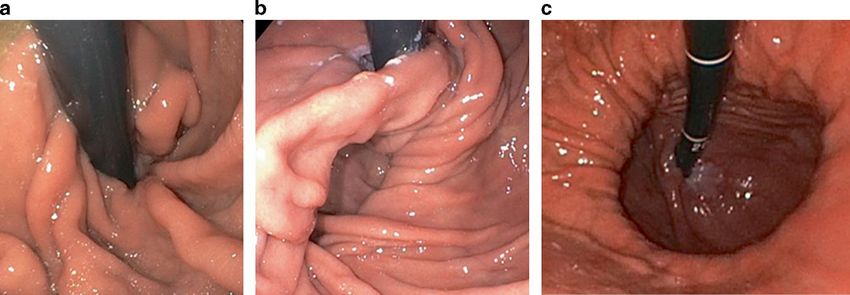
1140            Yadlapati et al.

                 Table 2 Rates of complications following antireflux surgery                       number of comorbidities [27, 33]. Patients with laxity of the fundo-
                                                                                                   plication and herniation may also present with obstructive symp-
                  Complication                                         Reported rates              toms related to compression within the hiatus or as a consequence
                  Primary fundoplication                               NIS database 2010           of the paraesophageal component causing extrinsic compression.
REVIEW ARTICLE

                                                                       n = 18,780                  It is not uncommon to have both GERD and obstructive symp-
                   Acute postoperative complications (within           4.1% [27] (n = 769)         toms with wrap herniation.
                  30 days)                                                                         Post-fundoplication stenosis. Persistent dysphagia due to fun-
                   30-Day surgical mortality                           0.1 to 0.2% [26, 27]        doplication-related stenosis without herniation may occur in 10%
                                                                       (n = 19 to 38)              of cases [33] and is secondary to a very tight or long fundoplica-
                   Infection                                           1.1% [27] (n = 207)         tion (Fig. 4) [23, 24, 29]. This is usually the result of poor position-
                   Bleeding                                            0.9% [27] (n = 169)         ing of the wrap or construction of a tense fundoplication wrap
                                                                                                   even when a bougie is used during the fundoplication compo-
                   Esophageal perforation                              0.9% [27] (n = 169)
                                                                                                   nent of the operation [27]. Other structural sources of persistent
                  Acute postoperative dysphagia                        50% [28, 30]
                                                                       (n = 9390)
                                                                                                   dysphagia include excessive angulation at the esophagogastric
                                                                                                   junction and intraluminal penetration of prosthetic surgical
                   Failure of fundoplication: wrap herniation,         2 to 23% [23, 27]
                  pouch formation, paraesophageal herniation           (n = 376 to 4319)
                                                                                                   material [23].
                                                                                                   Evaluation. Upper gastrointestinal endoscopy and barium
                  Post-fundoplication stenosis                         10% (n = 1878)
                                                                                                   esophagram are the primary diagnostic modalities used to evalu-
                  Post-fundoplication dilation rate                    2.8% [23] (n = 530)         ate for a structural complication following fundoplication (Fig. 5).
                  Gas-bloat syndrome                                   10 to 32% [23, 42, 43]      Endoscopy is useful in assessing for the presence of esophagitis
                                                                       (n = 1878 to 6010)          and also in appraising the location, orientation and integrity of
                  Esophageal dysmotility                                                           the post-fundoplication valve. When assessing the fundoplication
                  Chest pain                                                                       in the retroflexed position, the gastric folds of the fundoplication
                  Diarrhea                                             18 to 33% [40, 44]
                                                                                                   should be located just below the diaphragm and directed perpen-
                                                                       (n = 3380 to 6197)          dicular to the endoscope and parallel to the diaphragm (Fig. 1).
                  Secondary fundoplication                                                         The relationship between the fundoplication and crura is crucial
                                                                                                   in defining abnormalities of the fundoplication. Presence of gas-
                   Acute postoperative complications (within           23.4% [27]
                  30 days)                                                                         tric folds above the diaphragm indicates herniation and this may
                                                                                                   represent slippage of the stomach through the fundoplication or
                   30-Day surgical mortality                           1% [35]
                                                                                                   disruption of the wrap. Slippage can occur in the setting of an
                   Infection                                           6.5% [27]
                                                                                                   intact or partially disrupted wrap. Absence of fundoplication folds
                   Bleeding                                            5.2% [27]                   on retroflexion indicates total disruption of the fundoplication.
                   Esophageal perforation                              6.5% [27]                   Endoscopy may also be useful in assessing for a paraesophageal
                  Dysphagia                                            24.7% [27]                  hernia, where the herniated pouch of stomach will be seen next to
                                                                                                   the fundoplication folds. Retention of food and fluid in the esoph-
                  Magnetic sphincter augmentation
                                                                                                   agus, angulation at the distal esophagus and difficulty passing the
                  Acute postoperative complications                    0.1% [53–55]                endoscope may indicate proximal slippage of the fundoplication
                  Postoperative mortality                              0% [53–55]                  or stenosis at the wrap.
                  Device erosion                                       0.15% [53–55]                  Barium esophagram provides valuable and complimentary
                  Rate of device removal                               2.7% [56]
                                                                                                   information to endoscopy. Barium esophagram can help to define
                                                                                                   the anatomy and location of the fundoplication, better appreci-
                  Dysphagia                                            7.0% [57]
                                                                                                   ate a paraesophageal hernia and provide valuable information on
                  Bloating                                             10.0% [57]                  esophageal emptying [29]. Further body imaging with chest com-
                  The n values reported for primary fundoplication extrapolated from Nationwide    puted tomography scans and magnetic resonance imaging can be
                  Inpatient Sample (NIS) database volume of 18,780 elective surgical fundoplica-
                  tions performed in 2010 [16]                                                     helpful in defining anatomy, the degree of crural disruption and
                                                                                                   the extent and type of herniation. When persistent GERD is sus-
                                                                                                   pected, esophageal pH monitoring is useful to assess the pattern
                                                                                                   and burden of reflux, and to establish a congruence between reflux
                 fundoplication can also progress to a paraesophageal hernia com-                  episodes and symptoms as recurrent symptoms may not be recur-
                 ponent (Fig. 2b).                                                                 rent acid reflux. Endoscopy may not be able to determine whether
                   Patients with a failed fundoplication due to structural laxity will             the wrap is tight in the context of a normal-appearing wrap and
                 often present with recurrence of or persistent GERD symptoms                      the functional lumen imaging probe (FLIP) may be useful to docu-
                 including heartburn, regurgitation and erosive esophageal disease                 ment abnormal distensibility of the esophagogastric junction. In
                 such as peptic stricture formation or reflux esophagitis. Recurrent               addition, esophageal manometry may be useful to assess postoper-
                 GERD is the primary indication for reoperation and is more                        ative integrated relaxation pressures (Figs. 3 and 4). While there are
                 common among females, older patients and patients with a greater                  limited data on normal manometric values after fundoplication,

                 The American Journal of GASTROENTEROLOGY                                                                        VOLUME 113 | AUGUST 2018 www.nature.com/ajg
Complications of Antireflux Surgery 1141
Table 3 Mechanisms of post-fundoplication symptoms
Mechanism Post-fundoplication symptoms
Dysphagia Regurgitation Heartburn Esophagitis Chest Dyspepsia/ IBS/Diarrhea
REVIEW ARTICLE
stricture pain Gas-bloat
Mechanical
Tight fundoplication; no hernia ++ + − − ++ ++ −
Disrupted fundoplication/laxity; no hernia + ++ ++ ++ + − −
Slipped wrap with/without hernia ++ ++ + ++ ++ + −
Paraesophageal herniation type II/III/IV (new/recurrent) ++ ++ ++ ++ + + −
Esophageal dysmotility
Primary or pseudo-achalasia ++ ++ + − ++ + −
Hypercontractility/esophageal spasm + + + − + − −
Reduced preoperative esophageal peristaltic vigor + + +* +* +* − −
Functional
Hypersensitivity/functional esophageal syndrome + + + − + + +
Vagal nerve injury − +* +* +* +* ++ +
Accelerated gastric emptying − − − − − + ++
Association of mechanism with symptom if present: ++ strongly associated, + may be associated, − likely not associated
*May be associated if abnormal reflux is occurring due to a disrupted wrap, but if abnormal reflux is not occurring likely not associated
Fig. 2 Endoscopic views of fundoplication complications associated with disruption. Endoscopic Views of fundoplication complications associated with
disruption. a shows that the wrap is disrupted and the folds are more parallel with the endoscope. In b the wrap is partially intact and there is disruption of
the crural repair and a paraesophageal hernia tracking along side of the wrap and into the chest. c is a frank recurrence of the hernia with only a hint of the
remnant wrap noted deep in the type III hernia. Courtesy of the Esophageal Center at Northwestern
integrated relaxation pressure values are slightly higher after a Management. When post-fundoplication symptoms are due
normal fundoplication without symptoms [34]. Thus, the inter- to persistent gastroesophageal reflux, the management options
pretation of integrated relaxation pressure in the evaluation of include medical antireflux therapy, endoscopic dilation of pep-
post-fundoplication symptoms can be difficult and using cut-off tic stricture if present and surgical revision of the fundoplication
to diagnose esophagogastric junction outflow obstruction may be [29]. Surgical revision should be reserved as a last-resort option
problematic. However, if the values are very high with an elevated for patients with significant symptom burden that is not con-
intrabolus pressure, and the elevation in integrated relaxation trolled by PPI and/or endoscopic therapy and evidence of struc-
pressure persists despite position change, a diagnosis of obstruc- tural abnormality. If symptoms and complications are controlled
tion can be made. Distinguishing primary achalasia from pseudo- by PPI therapy, surgical revision may not be necessary unless there
achalasia from the fundoplication may be possible by evaluation of is an overt surgical indication. Surgical revisions are more com-
the preoperative and postoperative peristaltic pattern. In a report plex than the primary fundoplication, in part related to adhesion
from 1986, the amyl nitrite inhalation test was able to distinguish formation and altered anatomy, and the proportion of reoperation
mechanical from neurogenic obstruction [35]. interventions that can be performed laparoscopically declines
© 2018 The American College of Gastroenterology The American Journal of GASTROENTEROLOGY1142 Yadlapati et al.
a
c
EGJ
REVIEW ARTICLE
EGJ
b
d
Fig. 3 Slipped Nissen with frank herniation above the diaphragm and a partially intact wrap below the diaphragm depicted on esophagram (a), high-
resolution manometry (b), and Endoscopy (c-front view, d-Retrofelxed view. The red lines show the corresponding anatomic locations. a represents
the esophagram showing the herniation and the tight wrap [rotating blue arrow] with an elevated intrabolus pressure between the end of the distal
esophagus and the diaphragm. The wrap is located at the white arrow and the obstruction is at the diaphragm (black arrows showing crural contraction).
c and d are the endoscopic images. Note the esophagitis in this patient who presented with food impactions and reflux symptoms. Courtesy of the
Esophageal Center at Northwestern
with each subsequent reintervention. The reported success rate in cases with very short esophagus (for adequate mobilization)
of subsequent revisions is lower than for the primary fundoplica- or in a multiple redo situation (approaching the hernia through
tion, and continues to decrease with subsequent reoperations [31, “virgin” tissue planes). In cases of significantly reduced esopha-
36]. The mortality rate for reoperation is approximately 1%; how- geal peristalsis, esophagectomy may be considered as a last resort.
ever, complications of all types (perforation, postoperative leak, Roux-en-Y reconstruction may be an option after a failed primary
gastrotomy, vagal nerve injury and treatment failure) is signifi- or reoperative fundoplication (Table 4) [41]. It is also important to
cantly increased, particularly for multiple time redos [37, 38]. In a note that Roux-en-Y gastric bypass should be a primary antireflux
study of 940 patients undergoing primary or redo antireflux sur- intervention choice for patients with PPI-refractory GERD and
gery fewer patients were satisfied with subsequent reinterventions a body mass index over 35 kg/m2 or obesity with obesity-related
at follow-up (excellent satisfaction following: primary antireflux comorbidities such as diabetes or hypertension [42].
surgery 91%, first redo 76%, second redo 49%, and third redo In the case of a tight fundoplication and an intact wrap below
33%). Similarly, a higher proportion of patients were taking acid the diaphragm, endoscopic dilation is an option. According to case
suppression at follow-up with subsequent reintervention (primary series, endoscopic dilation relieves symptomatic dysphagia in up to
24%, first redo 46%, second redo 67% and third redo 78%) [36]. two-thirds of cases, of which 75% of patients only require one ses-
The most common reoperative intervention includes takedown sion of dilation therapy [29]. Most of these reports describe bou-
of the previous fundoplication, redo fundoplication and repair gie dilations to a mean diameter of 18 mm (54 French gauge) [28,
of a recurrent hiatal hernia if present [36, 39, 40]. Choice of 30]. Despite these favorable reports, in our personal experiences,
reoperation will be helped by an assessment of peristaltic func- bougie dilation and through the scope balloon dilation for tight
tion and gastric emptying. If a shortened esophagus contributed to post-fundoplication stenoses are often unsuccessful. Pneumatic
the primary failure, a redo fundoplication with Collis gastroplasty dilation, similar to protocols used for achalasia, is also attempted if
should be performed. A thoracic approach should be considered the anatomy is uncomplicated with variable success rates.
The American Journal of GASTROENTEROLOGY VOLUME 113 | AUGUST 2018 www.nature.com/ajgComplications of Antireflux Surgery 1143
a b
REVIEW ARTICLE
150.0
c d
140
130
120
110 30
100
25
90
80
20
70
60 15
50
10
40
35
30
25
20
15
10
5
0
–5
–10.0 25 30 35 40
Fig. 4 A fundoplication associated with dysphagia and food impaction. The large white arrow is identifying the esophagogastric junction (EGJ) and
location of the wrap. a is a tight fundoplication that appears intact and outside of being slightly long is relatively normal appearing. The esophagram in b
supports minimal emptying and exhibits a tight EGJ at the diaphragm. c is a highresolution manometry with high intrabolus pressure compartmentalized
between the peristaltic contraction and the wrap and this is associated with a high integrated relaxation pressure of 31.5 mmHg. d represents a new
approach using FLIP-panometry that defines EGJ opening dimensions and also provides evidence of motor function by assessing changes in diameter as
opposed to pressure. The scale in c is pressure (mmHg) while the scale in d is diameter (mm). Courtesy of the Esophageal Center at Northwestern
Functional complications following antireflux surgery tion related to an esophageal response to obstruction. It can be
Upper gastrointestinal symptoms following fundoplication in the difficult to determine whether the hypercontractility is a normal
absence of mechanical or anatomic abnormalities are common, response or pathologic, and empiric trials of smooth muscle
and unfortunately the mechanisms are often not well understood relaxants may be helpful.
[33, 43]. Gas-bloat syndrome. Gas-bloat syndrome may present with
Dysphagia due to esophageal dysmotility. Esophageal dysmotility abdominal bloating, inability to burp, postprandial fullness,
is a less common source of post-fundoplication dysphagia. When nausea, flatulence, inability to vomit and abdominal pain [44].
a structural etiology of dysphagia is not forthcoming on endos- Gas-bloat is more common with complete laparoscopic Nissen
copy or barium esophagram, esophageal manometry should be fundoplication than with partial fundoplication [45]. The mecha-
pursued. In the setting of ineffective esophageal motility, func- nism of gas-bloat is thought to arise from an inability to vent gas
tional dysphagia may arise from insufficient esophageal peristal- from the stomach into the esophagus and may be related to the
tic reserve or bolus transit to overcome the obstructive effect of impaired relaxation of the recreated esophagogastric junction
the fundoplication. Achalasia that was missed preoperatively, or valve in response to gastric distension and an alteration in recep-
a pseudo-achalasia pattern that developed postoperatively, is also tive gastric relaxation and accommodation. In addition, patients
another potential source of post-fundoplication dysphagia. Addi- may develop aerophagia and supragastric belching in an attempt
tionally, a hypercontractile pattern can be seen after fundoplica- to palliate these uncomfortable symptoms [23, 24, 29]. There is a
© 2018 The American College of Gastroenterology The American Journal of GASTROENTEROLOGY1144 Yadlapati et al.
a
EGO +/- FLIP
UGI with barium tablet
Normal anatomy
REVIEW ARTICLE
Wrap intact/No herniation
Yes Focus on optimizing
Esophagitis (> LAB)
acid suppression
No
1. Graded dilation with through the
scope balloon & pneumatic dilation Repeat endoscopy
2. Surgical revision if patient fails Yes Evidence of obstruction on
dilation and continues to be UGI or EGD/FLIP, tight
symptomatic or achalasia is fundoplication/ stricture Yes Yes Continue medical
suspected Esophagitis healed Symptoms resolve
management
No
1. Manometry to rule out alternative diagnosis No
2. Reflux testing if GERD symptoms No
3. Gastric emptying study if gas bloat symptoms Evaluate with the following if not done
EGJOO/Achalasia already to evaluate for obstruction and
Absent contractility refractory reflux disease.
Yes 1. Manometry
Major motor disorder 2. Gastric emptying study for refractory
reflux and surgical planning
Jackhammer, spasm 3. Consider reflux testing to assess acid
No
suppression on meds and to determine if
the patient has refractory reflux disease
Pathologic acid reflux
-Treat medically with antispasmodics
-Consider surgical revision and POEM
on a case-by-case basis
No Yes Yes Consider surgical revision determined
by severity of symptoms, physiologic
testing and surgical risks (Table 4)
-Focus on medical therapy and behavioral Focus on optimizing acid Continued
interventions for functional overlap suppression and lifestyle severe symptoms/
-Treat gastroparesis if documented interventions complications/esophagitis
Continue medical management
No
b
EGO +/- FLIP
UGI with barium tablet
Abnormal anatomy
Disrupted wrap Slipped wrap
Hernia (< 5 cm) Hernia (> 5 cm) Hernia (> 5 cm) Hernia (< 5 cm) Hernia (< 5 cm)
No obstruction or paraesophageal or obstructed on obstruction no obstruction
or obstructed on UGI/FLIP
Yes UGI/FLIP
Focus on optimizing
Esophagitis (> LAB)
acid suppression
1. Graded dilation with through the Treat similar to non-
No obstructed disrupted
scope balloon & pneumatic dilation
Repeat endoscopy 2. Surgical revision if patients fails wrap < 5 cm hernia
Focus on optimizing dilation and continues to be
acid suppression and Unlikely to respond to medical symptomatic or achalasia is
lifestyle modifications management or dilation and suspected
anatomical defect requires
repair.
Yes Yes Continue medical
Esophagitis healed Symptoms resolve
management
No No
Evaluate with the following and consider surgical revision
determined by severity of symptoms, physiologic testing
and surgical risk (Table 4)
1. Manometry
2. Gastric emptying study for refractory reflux
3. Consider reflux testing to assess acid suppression on
meds in non-erosive reflux disease/healed esophagitis
Fig. 5 Approach to patient with symptoms after an antireflux procedure. (a) Approach to patient with symptoms after an antireflux procedure with normal
anatomy on esophagogastroduodenoscopy (EGD) and/or upper GI (UGI) series. (b) Approach to patient with symptoms after an antireflux procedure with
abnormal anatomy on EGD and/or UGI series. Symptoms may be obstructive: dysphagia, esophageal regurgitation, gas-bloat, chest pain or food impaction.
Symptoms may be associated with abnormal reflux: heartburn, chest pain, regurgitation
paucity of data to guide the management of gas-bloat syndrome, using gas-reducing agents (i.e., simethicone) and prokinetic drugs
and generally lifestyle modifications are recommended. These [23, 24, 29]. While revision surgery to convert from a complete
include avoidance of gas-producing foods and carbonated bever- to an incomplete fundoplication may be beneficial, support-
ages, eating slowly to prevent aerophagia, tobacco cessation and ive data are lacking. Clinicians must be mindful to differentiate
The American Journal of GASTROENTEROLOGY VOLUME 113 | AUGUST 2018 www.nature.com/ajgComplications of Antireflux Surgery 1145
Table 4 Surgical revision options sphincter augmentation is often contrasted to laparoscopic fun-
doplication as a fundic-sparing reversible, reproducible and tech-
Surgical revision When to Technical considerations nically simple antireflux intervention that does not alter gastric
consider
anatomy [42,52–55]. While magnetic sphincter augmentation is
REVIEW ARTICLE
Redo fundoplication Peristalsis Must completely undo previous not FDA approved in the setting of hiatal hernias larger than 3 cm,
intact and no fundoplication and reduce
evidence of hernia sac. Careful dissection
a recent prospective multicenter study of 200 patients undergo-
gastroparesis to avoid vagal nerve injury ing magnetic sphincter augmentation with repair of hernias larger
Redo fundoplication Peristalsis Thoracic approach should be
than 3 cm reported favorable postoperative outcomes [56].
with Collis gastroplasty intact and no considered when very short To date, there are no reports of perioperative deaths or life-
evidence of esophagus discovered on pre- threatening complications following magnetic sphincter augmen-
gastroparesis, op testing or in multiple redo
with short setting
tation implantation. The most feared complication of magnetic
esophagus sphincter augmentation is device migration and erosion into the
Roux-en-Y BMI >35 kg/ May require partial gastric rem- esophagus. In a study of 3283 patients who underwent magnetic
m2 and/or nant resection due to ischemia sphincter augmentation, 0.15% (n = 5) had device erosion. All
gastroparesis cases of device erosion presented non-emergently with dysphagia
Esophagectomy Absent peri- Consider pyloroplasty or odynophagia, and were removed endoscopically or laparoscopi-
stalsis and cally without complication. In a few cases, patients subsequently
gastroparesis
underwent an uncomplicated laparoscopic fundoplication
[57–59]. The original magnetic sphincter augmentation device was
magnetic resonance conditional only up to 0.7 Tesla; however, the
gas-bloat syndrome from mechanical small bowel obstruction, new version is safe up to 1.5 Tesla.
peptic ulcer disease and delayed gastric emptying, and should have In a review of 1048 patients who underwent magnetic sphinc-
a low threshold to order a gastric emptying scan during the workup ter augmentation, the overall perioperative complication rate was
of the post-fundoplication patients especially those with bloating 0.1% and considered to be unrelated to the device. Endoscopic
as a dominant symptom. Gastric emptying of solids may be delayed dilation was performed in 5.6% of patients, the majority being
in the setting of vagal nerve injury, in which case treatments aimed within 90 days after the operation [60]. In a review of 3283 patients,
at relaxing the pylorus such as pyloroplasty, botulinum toxin or the overall rate for device removal was 2.7%, most commonly for
gastric peroral endoscopy myotomy may be considered [29]. dysphagia. A prospective observational study comparing magnetic
Chest pain. Although post-fundoplication chest pain is com- sphincter augmentation to laparoscopic fundoplication reported a
mon, the mechanism is not well understood. Potential etiologies higher rate of dysphagia (10.6% vs 7.0%) and bloating (31.9% vs
include mechanical stimulus from distension or contraction, or 10.0%) among the laparoscopic fundoplication group compared to
chemical stimulus from acidic or non-acidic esophageal exposure. the magnetic sphincter augmentation group [61, 62]. A meta-anal-
Workup should focus on assessing recurrent GERD and evaluat- ysis comparing magnetic sphincter augmentation to laparoscopic
ing for an esophageal motor disorder. fundoplication reported a significantly reduced risk of gas-bloat
Diarrhea. Diarrhea is also a frequent complication of fundopli- with the magnetic sphincter augmentation (relative risk 0.71, 95%
cation affecting 18 to 33% of patients [46]. While the mechanism confidence interval 0.54 to 0.94) [63]. Thus, initial data support a
of post-fundoplication diarrhea is not well understood, suggested more favorable safety profile of the magnetic sphincter augmenta-
mechanisms include accelerated gastric emptying, vagal nerve tion; however, follow-up over a longer period is needed to under-
injury, postoperative dietary modifications and unrecognized stand the actual long-term outcomes (Table 2) [61, 64].
pre-existing irritable bowel syndrome [29, 31, 47, 48]. Concomi-
tant cholecystectomy reportedly increases the risk for postopera-
tive diarrhea [29]. suMMary
In summary, antireflux surgery is indicated for patients with PPI-
refractory GERD or PPI intolerance in the context of documented
MagnEtIC sphInCtEr augMEntatIon pathologic gastroesophageal reflux. Although effective, complica-
Biomechanical augmentation of the lower esophageal sphincter by tions following antireflux surgery are common, particularly fol-
use of a magnetic reinforcing appliance was first described in 2008 lowing laparoscopic fundoplication. These include fundoplication
as a novel approach to manage GERD [49, 50]. Four years later, failure, dysphagia related to stenosis as well as various functional
the LINX Reflux Management System, a magnetic sphincter aug- gastrointestinal symptoms. Rates of complications seem to be
mentation device for GERD, was FDA (Food and Drug Adminis- lower with magnetic sphincter augmentation, though further out-
tration) approved for refractory GERD. The magnetic sphincter comes data are needed.
augmentation device is a ring of magnets that are surgically, typi- Post-fundoplication complications are one of the most challeng-
cally laparoscopically, placed circumferentially around the esoph- ing entities gastroenterologists and foregut surgeons manage in the
agogastric junction to augment the lower esophageal sphincter esophageal field, and should be approached from a multidisciplinary
and function as a two-way valve to allow bolus transit into the standpoint. Generally, upper gastrointestinal symptoms of dyspha-
stomach and allow for belching and vomiting [51]. Magnetic gia, heartburn and regurgitation following fundoplication should be
© 2018 The American College of Gastroenterology The American Journal of GASTROENTEROLOGY1146 Yadlapati et al.
first assessed with endoscopy and barium esophagram to evaluate 18. Spechler SJ, Lee E, Ahnen D, et al. Long-term outcome of medical and
surgical therapies for gastroesophageal reflux disease: follow-up of a
for anatomic disturbances such as a slipped fundoplication, herni-
randomized controlled trial. JAMA. 2001;285:2331–8.
ated wrap, paraesophageal hernia, tight wrap and presence of erosive 19. Wang YR, Dempsey DT, Richter JE. Trends and perioperative outcomes of
reflux disease. Ambulatory pH monitoring is useful when recurrent inpatient antireflux surgery in the United States, 1993-2006. Dis Esopha-
REVIEW ARTICLE
gus. 2011;24:215–23.
GERD is suspected. Generally, medical and endoscopic options are
20. Funk LM, Kanji A, Scott Melvin W, et al. Elective antireflux surgery in the
futile, as the majority of complications are post-surgical and struc- US: an analysis of national trends in utilization and inpatient outcomes
tural in nature (Fig. 3). The key to favorable outcomes after reop- from 2005 to 2010. Surg Endosc. 2014;28:1712–9.
21. Khan F, Maradey-Romero C, Ganocy S, et al. Utilisation of surgical fun-
erations is comprehensive preoperative assessment and appropriate
doplication for patients with gastro-oesophageal reflux disease in the USA
patient selection, otherwise the success and prognosis may be poor. has declined rapidly between 2009 and 2013. Aliment Pharmacol Ther.
2016;43:1124–31.
22. Jobe BA, Kahrilas PJ, Vernon AH, et al. Endoscopic appraisal of the
ConFLICT oF InTEREST
gastroesophageal valve after antireflux surgery. Am J Gastroenterol.
Guarantor of the article: Rena Yadlapati, MD, MSHS. 2004;99:233–43.
Specific author contributions: RY, ESH and JEP: literature review, 23. Sobrino-Cossio S, Soto-Perez JC, Coss-Adame E, et al. Post-fundoplica-
tion symptoms and complications: diagnostic approach and treatment.
drafting the manuscript and approving the final manuscript.
Rev Gastroenterol Mex. 2017;82:234–47.
Financial support: RY and JEP supported by NIH R01 DK092217 24. Richter JE. Let the patient beware: the evolving truth about laparoscopic
(JEP). antireflux surgery. Am J Med. 2003;114:71–3.
25. Carlson MA, Frantzides CT. Complications and results of primary mini-
Potential competing interests: RY: consultant for Ironwood,
mally invasive antireflux procedures: a review of 10,735 reported cases.
Medtronic and Diversatek; JEP: consultant for Crospon, J Am Coll Surg. 2001;193:428–39.
Ironwood,Torax, Astra Zeneca, Takeda, Impleo, Medtronic and 26. Rantanen TK, Salo JA, Sipponen JT. Fatal and life-threatening com-
plications in antireflux surgery: analysis of 5,502 operations. Br J Surg.
Sandhill; ESH: consultant for Baxter and Boston Scientific.
1999;86:1573–7.
27. Maret-Ouda J, Wahlin K, El-Serag HB, et al. Association between lapa-
roscopic antireflux surgery and recurrence of gastroesophageal reflux.
rEfErEnCEs JAMA. 2017;318:939–46.
1. Vakil N, van Zanten SV, Kahrilas P, et al. The Montreal definition and 28. Malhi-Chowla N, Gorecki P, Bammer T, et al. Dilation after fundoplica-
classification of gastroesophageal reflux disease: a global evidence-based tion: timing, frequency, indications, and outcome. Gastrointest Endosc.
consensus. Am J Gastroenterol. 2006;101:1900–20. quiz 1943 2002;55:219–23.
2. Peery AF, Crockett SD, Barritt AS, et al. Burden of gastrointestinal, 29. Spechler SJ. The management of patients who have “failed” antireflux
liver, and pancreatic diseases in the United States. Gastroenterology. surgery. Am J Gastroenterol. 2004;99:552–61.
2015;149:1731–41.e3 30. Wo JM, Trus TL, Richardson WS, et al. Evaluation and management of
3. El-Serag H, Becher A, Jones R. Systematic review: persistent reflux symp- postfundoplication dysphagia. Am J Gastroenterol. 1996;91:2318–22.
toms on proton pump inhibitor therapy in primary care and community 31. Hinder RA, Libbey JS, Gorecki P, et al. Antireflux surgery. Indications,
studies. Aliment Pharmacol Ther. 2010;32:720–37. preoperative evaluation, and outcome. Gastroenterol Clin North Am.
4. Tack J,Pandolfino JE, Pathophysiology of gastroesophageal reflux disease. 1999;28:987–1005. viii
Gastroenterology. 2018;154:277–88. 32. Horgan S, Pohl D, Bogetti D, et al. Failed antireflux surgery: what have we
5. Katz PO, Gerson LB, Vela MF. Guidelines for the diagnosis and learned from reoperations? Arch Surg. 1999;134:809–15. discussion 815-7
management of gastroesophageal reflux disease. Am J Gastroenterol. 33. Lundell L. Complications after anti-reflux surgery. Best Pract Res Clin
2013;108:308–28. quiz 329 Gastroenterol. 2004;18:935–45.
6. Kahrilas PJ, Shaheen NJ, Vaezi MF, et al. American Gastroenterological 34. Wilshire CL, Niebisch S, Watson TJ, et al. Dysphagia postfundoplication:
Association Medical Position Statement on the management of gastroe- more commonly hiatal outflow resistance than poor esophageal body
sophageal reflux disease. Gastroenterology. 2008;135:1383–91. 1391.e1-5 motility. Surgery. 2012;152:584–92. discussion 592-4
7. Dallemagne B, Weerts JM, Jehaes C, et al. Laparoscopic Nissen fundopli- 35. Dodds WJ, Stewart ET, Kishk SM, et al. Radiologic amyl nitrite test for
cation: preliminary report. Surg Laparosc Endosc. 1991;1:138–43. distinguishing pseudoachalasia from idiopathic achalasia. AJR Am J
8. Geagea T. Laparoscopic Nissen’s fundoplication: preliminary report on Roentgenol. 1986;146:21–3.
ten cases. Surg Endosc. 1991;5:170–3. 36. Singhal S,Kirkpatrick DR,Masuda T, et al. Primary and redo antireflux
9. Jobe BA, Richter JE, Hoppo T, et al. Preoperative diagnostic workup surgery: outcomes and lessons learned. J Gastrointest Surg. 2018;22:
before antireflux surgery: an evidence and experience-based consensus 177–86.
of the Esophageal Diagnostic Advisory Panel. J Am Coll Surg. 2013;217: 37. Waring JP. Management of postfundoplication complications. Semin
586–97. Gastrointest Dis. 1999;10:121–9.
10. Fernando HC. Endoscopic fundoplication: patient selection and tech- 38. Waring JP. Postfundoplication complications. Prevention and manage-
nique. J Vis Surg. 2017;3:121. ment. Gastroenterol Clin North Am. 1999;28:1007–19. viii-ix
11. Moore M, Afaneh C, Benhuri D, et al. Gastroesophageal reflux disease: a 39. Makris KI, Panwar A, Willer BL, et al. The role of short-limb Roux-en-Y
review of surgical decision making. World J Gastrointest Surg. 2016;8: reconstruction for failed antireflux surgery: a single-center 5-year experi-
77–83. ence. Surg Endosc. 2012;26:1279–86.
12. Vakil N, Shaw M, Kirby R. Clinical effectiveness of laparoscopic fundopli- 40. Bathla L, Legner A, Tsuboi K, et al. Efficacy and feasibility of laparoscopic
cation in a U.S. community. Am J Med. 2003;114:1–5. redo fundoplication. World J Surg. 2011;35:2445–53.
13. Nissen R, Rossetti M. [Modern operations for hiatal hernia and 41. Grover BT, Kothari SN. Reoperative antireflux surgery. Surg Clin North
reflux esophagitis: gastropexy and fundoplication]. Arch Chir Torace. Am. 2015;95:629–40.
1959;13:375–87. 42. Azagury D, Morton J. Surgical anti-reflux options beyond fundoplication.
14. Johansson KE, Tibbling L. Maintenance treatment with ranitidine com- Curr Gastroenterol Rep. 2017;19:35.
pared with fundoplication in gastro-oesophageal reflux disease. Scand J 43. Swanstrom L, Wayne R. Spectrum of gastrointestinal symptoms after
Gastroenterol. 1986;21:779–88. laparoscopic fundoplication. Am J Surg. 1994;167:538–41.
15. Finks JF, Wei Y, Birkmeyer JD. The rise and fall of antireflux surgery in the 44. Humphries LA, Hernandez JM, Clark W, et al. Causes of dissatisfaction
United States. Surg Endosc. 2006;20:1698–701. after laparoscopic fundoplication: the impact of new symptoms, recurrent
16. Finlayson SR, Laycock WS, Birkmeyer JD. National trends in utilization symptoms, and the patient experience. Surg Endosc. 2013;27:1537–45.
and outcomes of antireflux surgery. Surg Endosc. 2003;17:864–7. 45. Tian ZC, Wang B, Shan CX, et al. A meta-analysis of randomized
17. Finalyson SR, Stroupe KT, Joseph GJ, et al. Using the Veterans Health controlled trials to compare long-term outcomes of Nissen and
Administration inpatient care database: trends in the use of antireflux Toupet fundoplication for gastroesophageal reflux disease. PLoS ONE.
surgery. Eff Clin Pract. 2002;5:E5. 2015;10:e0127627.
The American Journal of GASTROENTEROLOGY VOLUME 113 | AUGUST 2018 www.nature.com/ajgComplications of Antireflux Surgery 1147
46. Klaus A, Hinder RA, DeVault KR, et al. Bowel dysfunction after laparo- 56. Buckley FP,3rd, Bell RCW, Freeman K, et al. Favorable results from a pro-
scopic antireflux surgery: incidence, severity, and clinical course. Am J spective evaluation of 200 patients with large hiatal hernias undergoing
Med. 2003;114:6–9. LINX magnetic sphincter augmentation. Surg Endosc. 2018;32:1762–8.
47. Kozarek RA, Low DE, Raltz SL. Complications associated with 57. Salvador R, Costantini M, Capovilla G, et al. Esophageal penetration of
laparoscopic anti-reflux surgery: one multispecialty clinic’s experience. the magnetic sphincter augmentation device: history repeats itself.
REVIEW ARTICLE
Gastrointest Endosc. 1997;46:527–31. J Laparoendosc Adv Surg Tech A. 2017;27:834–8.
48. Nastaskin I, Mehdikhani E, Conklin J, et al. Studying the overlap 58. Asti E, Siboni S, Lazzari V, et al. Removal of the magnetic sphincter aug-
between IBS and GERD: a systematic review of the literature. Dig Dis Sci. mentation device: surgical technique and results of a single-center cohort
2006;51:2113–20. study. Ann Surg. 2017;265:941–5.
49. Ganz RA, Gostout CJ, Grudem J, et al. Use of a magnetic sphincter for the 59. Lipham JC, Taiganides PA, Louie BE, et al. Safety analysis of first 1000
treatment of GERD: a feasibility study. Gastrointest Endosc. 2008;67:287–94. patients treated with magnetic sphincter augmentation for gastroesopha-
50. Bonavina L, Saino GI, Bona D, et al. Magnetic augmentation of the lower geal reflux disease. Dis Esophagus. 2015;28:305–11.
esophageal sphincter: results of a feasibility clinical trial. J Gastrointest 60. Smith CD, Ganz RA, Lipham JC, et al. Lower esophageal sphincter
Surg. 2008;12:2133–40. augmentation for gastroesophageal reflux disease: the safety of a modern
51. Ganz RA. A modern magnetic implant for gastroesophageal reflux dis- implant. J Laparoendosc Adv Surg Tech A. 2017;27:586–91.
ease. Clin Gastroenterol Hepatol. 2017;15:1326–37. 61. Riegler M, Schoppman SF, Bonavina L, et al. Magnetic sphincter augmenta-
52. Bonavina L, DeMeester TR, Ganz RA. LINX(TM) Reflux Management tion and fundoplication for GERD in clinical practice: one-year results of a
System: magnetic sphincter augmentation in the treatment of gastroe- multicenter, prospective observational study. Surg Endosc. 2015;29:1123–9.
sophageal reflux disease. Expert Rev Gastroenterol Hepatol. 2012;6:667–74. 62. Hillman L, Yadlapati R, Whitsett M, et al. Review of antireflux procedures
53. Bonavina L, Attwood S. Laparoscopic alternatives to fundoplication for proton pump inhibitor nonresponsive gastroesophageal reflux disease.
for gastroesophageal reflux: the role of magnetic augmentation and Dis Esophagus. 2017;30:1–14.
electrical stimulation of the lower esophageal sphincter. Dis Esophagus. 63. Chen MY, Huang DY, Wu A, et al. Efficacy of magnetic sphincter augmenta-
2016;29:996–1001. tion versus Nissen fundoplication for gastroesophageal reflux disease in short
54. Ganz RA, Edmundowicz SA, Taiganides PA, et al. Long-term outcomes of term: a meta-analysis. Can J Gastroenterol Hepatol. 2017;2017:9596342.
patients receiving a magnetic sphincter augmentation device for gastroe- 64. Min MX, Ganz RA. Update in procedural therapy for GERD-magnetic
sophageal reflux. Clin Gastroenterol Hepatol. 2016;14:671–7. sphincter augmentation, endoscopic transoral incisionless fundoplica-
55. Chiu J, Soffer E. Novel surgical options for gastroesophageal reflux dis- tion vs laparoscopic Nissen fundoplication. Curr Gastroenterol Rep.
ease. Expert Rev Gastroenterol Hepatol. 2015;9:943–51. 2014;16:374.
© 2018 The American College of Gastroenterology The American Journal of GASTROENTEROLOGYGASTROENTEROLOGY ARTICLE OF THE WEEK
October 25, 2018
Yadlapati R, Hungness ES, Pandolfino JE. Complications of antireflux surgery. Am J Gastroenterol
2018;113:1137‐1147.
1. A patient develops dysphagia post antireflux surgery which has not resolved by the end of the third
post‐operative week, at this point you should
a. recommend reversal surgery
b. balloon dilation of the fundoplication to size 15‐16mm
c. Savary dilation to 15‐16 mm
d. Observe and reassure
2. A patient develops dysphagia 1 year after fundoplication. Evaluation reveals a slipped wrap, a 3cm
hiatal hernia and no paraesophageal hernia. Next course of action should be
a. surgical redo
b. esophageal manometry
c. graded endoscopic dilation
d. maximize PPI therapy
True or False
3. The primary indication for pre‐operative esophageal manometry is to assess the resting pressure of
the LES
4. Diarrhea is not a recognized complication of fundoplication
5. Magnetic sphincter augmentation is not approved for use in patients with hiatal hernias >3cm
6. The presence of > 35% ineffective peristaltic contractions on manometry is an absolute
contraindication to antireflux surgery
7. Rates of post‐operative dysphagia and gas‐bloat syndrome are higher after magnetic sphincter
augmentation compared to laparoscopic fundoplication
8. The most common reason for reoperation after anti‐reflux surgery is persistent dysphagia
9. Endoscopic dilation of a tight wrap is usually done with a bougie, working up to 18mm (54 French)
diameter
10. Patients with magnetic sphincter augmentation devices placed recently are able to undergo MRI
examinations up to 1.5 TeslaYou can also read