Changes in cortical gene expression in the muscarinic M1 receptor knockout mouse: potential relevance to schizophrenia, Alzheimer's disease and ...
←
→
Page content transcription
If your browser does not render page correctly, please read the page content below
www.nature.com/npjschz

                  ARTICLE                  OPEN

                  Changes in cortical gene expression in the muscarinic M1
                  receptor knockout mouse: potential relevance to
                  schizophrenia, Alzheimer’s disease and cognition
                                   1✉
                  Brian Dean            and Elizabeth Scarr2

                      Postmortem and neuroimaging studies show low levels of cortical muscarinic M1 receptors (CHRM1) in patients with schizophrenia
                      which is significant because CHRM signalling has been shown to change levels of gene expression and cortical gene expression is
                      altered in schizophrenia. We decided to identify CHRM1-mediated changes in cortical gene expression by measuring levels of RNA
                      in the cortex of the Chrm1−/− mouse (n = 10), where there would be no signalling by that receptor, and in wild type mouse (n = 10)
                      using the Affymetrix Mouse Exon 1.0 ST Array. We detected RNA for 15,501 annotated genes and noncoding RNA of which 1,467
                      RNAs were higher and 229 RNAs lower in the cortex of the Chrm1−/− mouse. Pathways and proteins affected by the changes in
                      cortical gene expression in the Chrm1−/− are linked to the molecular pathology of schizophrenia. Our human cortical gene
                      expression data showed 47 genes had altered expression in Chrm1−/− mouse and the frontal pole from patients with schizophrenia
                      with the change in expression of 44 genes being in opposite directions. In addition, genes with altered levels of expression in the
1234567890():,;

                      Chrm1−/− mouse have been shown to affect amyloid precursor protein processing which is associated with the pathophysiology of
                      Alzheimer’s disease, and 69 genes with altered expression in the Chrm1−/− mouse are risk genes associated with human cognitive
                      ability. Our findings argue CHRM1-mediated changes in gene expression are relevant to the pathophysiologies of schizophrenia
                      and Alzheimer’s disease and the maintenance of cognitive ability in humans.
                      npj Schizophrenia (2021)7:44 ; https://doi.org/10.1038/s41537-021-00174-z

                  INTRODUCTION                                                                               schizophrenia11. This raises the possibility that changes in CHRM1
                  Schizophrenia is characterised by the onset of positive (e.g.,                             receptor-mediated gene expression may be an important
                  delusions and hallucinations), negative (e.g., apathy and anhedo-                          mechanism of action of drugs that can reduce symptom severity
                  nia), and cognitive symptoms1. Whilst the molecular mechanisms                             in those with the disorder.
                  causing schizophrenia are unknown, it is clear a dysfunctional                                The development of Chrm1−5−/− mice has provided useful
                  prefrontal cortex contributes to the pathophysiology of the                                tools for the study of the physiological roles of those receptors in
                  disorder2 and that schizophrenia occurs in individuals with a                              mammalian CNS12. At the behavioural level, Chrm1−/− mice
                  genetic susceptibility due to the inheritance of risk genes who                            showed no deficits in sensory-motor gating, nociception, motor
                  encounter one or more deleterious environmental factors3.                                  coordination, anxiety-related behaviour, or hippocampal learning
                                                                                                             and memory13. By contrast, Chrm1−/− mice were severely
                  Importantly, both inherited risk genes4 and environmental factors,
                                                                                                             impaired in non-matching-to-sample working memory14, which
                  via epigenetic mechanisms3, act to change levels of gene
                                                                                                             needs cortical and hippocampal engagement. Hence, it would
                  transcription, and thus changes in levels of RNA in the prefrontal
                                                                                                             appear that the cortical Chrm1 modulates behaviours involving
                  cortex from patients with schizophrenia are likely contributing to                         cortical to sub-cortical communication which is significant
                  the molecular pathology of the disorder5.                                                  because patients with schizophrenia have a loss of CHRM1
                     One finding supported by human postmortem and neuroima-                                  positive pyramidal neurons in cortical laminae V, neurons which
                  ging studies is that there are lower levels of muscarinic M1                               are critical in cortical-subcortical communication15.
                  receptors (CHRM1) in the cortex of patients with schizophrenia6                               Using HEK293 cells, it has been shown that CHRM1 signalling
                  and that the cognitive deficits experienced by patients with                                affects gene expression10. We have shown there are lower levels
                  schizophrenia are related to the levels of that receptor7.                                 of cortical CHRM18 and changes in gene expression in the cortex
                  Importantly, our data show that levels of CHRM2−4 are not                                  from patients with schizophrenia16–18. Based on these data, we
                  altered in the cortex of patients with schizophrenia8,9 and that                           postulated that some changes in cortical gene expression in
                  levels of CHRM5 could not be detected (unpublished data).                                  patients with schizophrenia would be associated with lower levels
                  Significantly, signalling through CHRMs has been shown to affect                            of CHRM1 signalling. Thus, we decided to compare cortical gene
                  gene expression10 which argues that changes in cortical CHRM1-                             expression in Chrm1−/− to wild type (w/t) mice using the Mouse
                  regulated gene expression must be contributing to the molecular                            Exon 1.0 ST Array because that array is the mouse equivalent of
                  pathology of schizophrenia. Moreover, it has been recently                                 the Human Exon 1.0 ST Array which we used to measure gene
                  reported that a co-formulation of the selective M1/M4 agonist,                             expression in the human cortex. Using analogous technology
                  xanomeline, and the peripheral muscarinic receptor antagonist,                             allowed us to better compare changes in gene expression in the
                  trospium, reduces both the positive and negative symptoms of                               cortex from Chrm1−/− mice and patients with schizophrenia.

                  1
                   The Molecular Psychiatry Laboratory, The Florey Institute for Neuroscience and Mental Health, Parkville, VIC, Australia. 2Melbourne Veterinary School, Faculty of Veterinary and
                  Agricultural Sciences, The University of Melbourne, Parkville, VIC, Australia. ✉email: anddali@unimelb.edu.au

                  Published in partnership with the Schizophrenia International Research Society
B. Dean and E. Scarr
2
In addition, because the CHRM1 is important in cognition19 we Impact of changed gene expression on Chrm1−/− cortical
wanted to identify genes with changed levels of expression in the function
Chrm1−/− mice that, using gene-wide association studies (GWAS), The Panther Gene Ontology Classification System predicted
had been associated with human cognitive ability20. changes in gene expression would impact on 198 distinct
biological processes in the cortex of Chrm1−/− mice (Supplemen-
tary Table 2). These changes in biological processes would likely
RESULTS be driven by the enrichment and depletion of specific classes of
Changes in cortical gene expression in Chrm1−/− mice proteins in the cortex of Chrm1−/− mice (Fig. 2; Supplementary
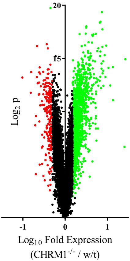
Levels of RNA for 15,501 annotated genes and noncoding RNA Table 3) especially as some of these protein classes have well-
were detectable in the cortex from Chrm1−/− and w/t mice of defined roles in CNS function.
which, levels of 1,467 RNAs were higher and 229 RNAs were lower We also completed a manual curation of the functional role of
in the Chrm1−/− mice (Fig. 1; Supplementary Table 1). Notably, the genes with altered levels of expression in the cortex of Chrm1−/−
signal for mRNA from the Chrm1 gene was essentially equal to the mice to identify groupings of genes that could be clustered within
background, giving significant face validity to our analyses. specific cellular functions. This analysis showed that genes
involved in mitochondrial function, protein degradation, mem-
brane transport, neurotransmission, receptor signalling, ribosomal
function, calcium biochemistry, phosphorylation, heat shock,
Redox reactions, tubulin biochemistry, inflammation, energy, and
metabolism, as well as lipid metabolism had altered levels of
expression in the cortex of the Chrm1−/− mouse (Fig. 3 and
Supplementary Table 4).
1234567890():,;
Fig. 2 Protein classes affected by changes in gene expression in
the Chrm1−/− mouse. Protein classes containing enriched or
depleted levels of genes with altered levels of expression in the
cortex of Chrm1−/− mouse.
Fig. 1 Levels of gene expression in the cortex of the Chrm1−/−
compared to that in wild type mice. Volcano plot showing the Fig. 3 Cellular functions affected by changes in cortical gene
levels of expression of 15,501 genes in the cortex of Chrm1−/− expression in the Chrm1−/− mouse. The number of coding and
compared to that in wild type (w/t) mice. Green dots = increased noncoding RNA that are known to be associated with specific
gene expression, red dots = decreased gene expression, black dots cellular functions that had either higher (green columns) or lower
unchanged gene expression. (red columns) levels on the cortex of Chrm1−/− mouse.
npj Schizophrenia (2021) 44 Published in partnership with the Schizophrenia International Research SocietyB. Dean and E. Scarr
3
Changes in cortical gene expression in Chrm1−/− mice and in cell cultures isolated from newborn Chrm1−/− mice24. Interest-
patients with schizophrenia ingly, our data shows the expression of two MAPKs (Mapk8 and
One of the strengths of our study design is we measured levels Mapk10) were higher in the cortex of Chrm1−/− mice which could
of gene expression in the mouse cortex with a GeneChip™ be a compensatory response to decrease signalling through the
Mouse Exon 1.0 ST Array, which is comparable to the GeneChip™ Chrm1. In addition, Chrm1 agonist-stimulated phosphatidyl
Human Exon 1.0 ST Array used in our study comparing gene inositol (PI) hydrolysis is reduced by >60% in primary cortical cell
expression in three cortical regions from patients with schizo- cultures from the Chrm1−/− mouse24. Our study showed there was
phrenia to that in age-matched controls18. These two studies a higher expression of four enzymes critical to the functioning of
showed there were 47 genes with changed levels of expression the PI hydrolysis (Pi4k2a, Pip5k1a, Pik3r3, and Ip6k1). It has also
in both the cortex of Chrm1−/− mice and Brodmann’s (BA) 10 been shown that pan-CHRM agonist-stimulated [35S]-GTPγS
from patients with schizophrenia18 (Supplementary Table 5). By binding could not be detected in the cortex of the Chrm1-/-
contrast, there were no genes that had changed levels of mice25. Our data argue signalling via G-proteins would be further
expression in the cortex of Chrm1−/− mice and BA 9 or BA 33 affected because of changed cortical expression of guanine
from patients with schizophrenia. nucleotide-binding proteins (Higher: Gnai3, Gnb4; Lower: Gng4,
Gnao1) and regulators of G-protein signalling (Higher: Rgs4, Rgs14,
Rgs17; Lower: Rgs2) in the Chrm1−/− mouse. Our data therefore
Changes in cortical gene expression in Chrm1−/− mice: significantly increase understanding of the molecular mechanisms
relevance to the human cognition GWAS causing changes in Chrm1 signalling previously shown to be
As the Chrm1−/− mice had some aberrations in the cognitive present in the cortex of Chrm1−/− mice.
function we postulated some changes in cortical gene expression Primary cortical neuron cultures from the Chrm1−/− mouse
could be associated with cognition that could be relevant to have increased production of the amyloid precursor protein
humans. It was therefore notable that there were changes in the (APP)26. It is therefore significant that our study has shown higher
levels of expression of 69 genes reported as being associated with expression of proteins related to APP (Apbb1, Appbp2, Apbb3) in
variation in human cognition in the cortex of the Chrm1−/− mouse the cortex of the Chrm1−/− mouse. Apbb1 (amyloid beta (A4)
(Supplementary Table 6). As cognitive deficits are a symptom of precursor protein-binding, family B, member 1) is a member of the
schizophrenia we also compared changes in gene expression in Fe65 protein family, which interacts with APP to modulate its
BA 10 from patients with schizophrenia to genes associated activity27. Appbp2 (amyloid-beta precursor protein (cytoplasmic
with variation in human cognitive ability and found 12 genes with tail) binding protein 2) has been shown to regulate APP protein
altered levels of expression in BA 10 from patients with transport and /or processing28. Apbb3 (amyloid beta (A4)
schizophrenia that have been associated with varying cognitive precursor protein-binding, family B, member 3) is a protein in
ability in humans (Supplementary Table 7). In addition, there were the cytosol that binds to the intracellular domain of APP to
two genes (AKT interacting protein (Aktip) and nuclear receptor modulate the internalisation of that protein29. Thus, our data add
subfamily 1, group D, member 2 (Nrld2) which had changed levels to data suggesting Chrm1 signalling impacts on APP function,
of expression in the cortex of Chrm1−/− mice and BA 10 from which has long been thought to be important in the pathophy-
people with schizophrenia that have also been shown to be siological processes underlying Alzheimer’s disease30.
associated with varying cognitive ability in humans. By using homologous technologies to measure cortical gene
expression, we were able to compare changes in cortical gene
expression in the Chrm1−/− mouse and BA 9, 10, and 33 from
DISCUSSION patients with schizophrenia which showed that 47 genes had
Here we show Chrm1 signalling in mouse CNS is associated with altered levels of expression in the Chrm1−/− mouse and in BA 10,
changes in cortical gene expression. Changes in gene expression but not BA 9 or 33, from patients with schizophrenia. It could be
are mostly higher in the Chrm1−/− mouse, thus our data argues argued the comparable changes in cortical gene expression in the
Chrm1 signalling predominantly acts to suppress cortical gene Chrm1−/− and BA 10 from patients with schizophrenia were due
expression. Changes in cortical gene expression in the Chrm1−/− to chance. However, the changes in gene expression in BA 9, 10,
mouse include the expression of enzymes, SNARE proteins, and 33 from patients with schizophrenia do not occur in the same
chaperone proteins, as well as proteins involved in receptor three cortical regions from patients with bipolar disorder or major
signalling, gene transcription and translation, metabolism, and depressive disorders31 who do not have changes in their cortical
immunity. Such changes in cortical gene expression would impact CHRM1 levels32,33. These data argue that the diagnostically
on mitochondrial function, protein translation and degradation, selective changes in gene expression present in the Chrm1−/−
membrane transport, neurotransmission, receptor signalling, and and schizophrenia are likely associated with lower levels of
calcium biochemistry, which are critical to maintaining CNS CHRM1. Moreover, existing data implicates 26 of these genes with
function. One limitation of transcriptomic studies is that changes changed levels of expression in Chrm1−/− mouse to the
in coding RNA (i.e., gene expression) can be directly, inversely, or pathophysiology of schizophrenia (Supplementary Table 8),
not related to changes in levels of protein encoded by messenger further arguing the changes in cortical gene expression in the
RNA21 whilst changes in non-coding RNA can activate or inhibit Chrm1−/− mouse are not purely due to chance.
gene expression22. Hence, understanding the impact of changes It was notable that the direction of change of 44 of the 47 genes
in the cortical transcriptome of the Chrm1−/− mouse on the with changed levels of expression in BA 10 from patients with
cortical proteome will be required to better understand the schizophrenia was in the opposite direction to what occurred in
changes in the activity of affected biochemical pathways. the cortex of the Chrm1−/− mice. Our premise for studying the
Behaviourally, the Chrm1−/− mouse does not have seizures Chrm1−/− mouse was that it was a good model in which to
after exposure to pilocarpine but does have increased locomotor identify changes in cortical gene expression that would be
activity, mild and selective cognitive impairments, and a number expected in the absence of signalling by that receptor. Following
of electrophysiological deficits23. Importantly, the Chrm1−/− that hypothesis, we would expect to have found comparable
mouse has impairments in nonmatching-to-sample working changes in the expression of some genes in the cortex of patients
memory and consolidation, a behaviour requiring cortical inter- with schizophrenia where we have shown lower levels of CHRM18
actions with the hippocampus14. At the biochemical level, Chrm1 and no changes in levels of CHRM2, 39, or 48 compared to that in
agonist-stimulated activation of the mitogen-activated protein controls. However, our study in the Chrm1−/− has revealed
kinase (MAPK) pathway is essentially abolished in primary cortical opposing changes in levels of most of the genes that are also
Published in partnership with the Schizophrenia International Research Society npj Schizophrenia (2021) 44B. Dean and E. Scarr
4
changed in BA 10 from patients with schizophrenia. Notably, it has phosphorylation, redox state, tubulin, inflammation, as well as
been suggested that derangements of biochemical pathways energy and lipid metabolism. Thus, the absence of
from their “normal” physiological responsiveness are associated Chrm1 signalling, at least at the level of gene expression, would
with the molecular pathology of disorders of the CNS34. Hence, be expected to have significant effects on cortical function. It is
one explanation for the opposing changes in expression of the 44 therefore significant that the Chrm1−/− mouse did not show
genes in the Chrm1−/− mouse and BA 10 from patients with profound changes at the level of behaviour23. That been said,
schizophrenia is that those genes are an important component of changed mitochondrial function, protein degradation, membrane
the pathology of the disorder. transport, neurotransmission, receptor signalling, ribosomal func-
Our gene expression studies in the cortex of the Chrm1−/− mice tion, calcium biochemistry, protein phosphorylation, redox state,
and in the cortex of patients with schizophrenia have been carried tubulin, inflammation as well as energy and lipid metabolism have
out using RNA from tissue homogenates. We15, and others35, have all been implicated in the pathophysiology of schizophrenia18,44–
shown there are high levels of CHRM1 on pyramidal neurons15 but 50
and in cognitive functioning51–55. This suggests the Chrm1−/−
the CHRM1 is also present on inhibitory neurons35, astrocytes35, mouse may be more useful in studying the molecular, rather than
and CNS microvascular endothelial cells36. Thus, at this time it is behavioral, changes regulated by Chrm1 signalling especially as
not possible to determine which cells are affected by the changes some of these molecular changes appear to be relevant to
in gene expression that we have described in the Chrm1−/− schizophrenia6 and cognition56.
mouse and in BA 10 from patients with schizophrenia18. There are limitations using the Chrm1−/− gene knockout model
A confound faced by all studies into the molecular pathology of in studying biological systems and pathways. First, the loss of
schizophrenia is that the diagnosis encompasses a syndrome of Chrm1 signalling is universal across all tissue, which may not be an
disorders, which has led to the argument that the pathophysiol- issue in patients with schizophrenia as both neuroimaging57 and
ogy of schizophrenia cannot be fully elucidated until it is possible postmortem39 studies suggest lower levels of CHRM1 are wide-
to study definable sub-groups within schizophrenia37. We have spread in the CNS of those with the disorder. In addition, the use
identified a sub-group of patients (~25%) within the syndrome of of the gene knockout mouse can now be more refined as there
schizophrenia that can be characterised because they have are techniques to manipulate gene expression that allow
markedly lower (~75%) levels of cortical CHRM138,39. It would increasing control of which cells over or under-express a gene
therefore be reasonable to postulate that there may be more and when the onset of either over or under expression is
similarities in changes in cortical gene expression in the Chrm1−/− triggered58. This gives the opportunity to study the impact of
mouse in BA 10 from the sub-group of patients within changes in Chrm1 signalling within specific cell types and/or
schizophrenia and identifying such comparable changes may specific CNS regions and to control for when the changed level
significantly contribute to understanding their pathophysiology. of gene expression is triggered rather than the absence of
Unfortunately, our data on gene expression in BA 10 from patients Chrm1 signalling being present from conception. This level of
with schizophrenia were from cohorts that were predominantly control could be critical in better understanding the contribution
(80%) compose of patients with schizophrenia that do not have a of decreased signalling through the CHRM1 to the pathophysiol-
marked loss of cortical CHRM138. ogy in disorders such as schizophrenia which has a peak age of
Cognitive deficits are a core feature of both schizophrenia40 and onset in late adolescence to early adulthood59. However, even
Alzheimer’s disease41 and the CHRM1 is known to play an without such refinements, our study is significant because it
important role in regulating cognitive ability7. It is therefore of shows that the modulation of gene expression is an important
interest that 69 genes with changed levels of expression in the mechanism of action of the Chrm1 that will be important in
cortex of the Chrm1−/− mouse have been identified by the GWAS controlling cortical function.
on human cognition to be associated with varying cognitive
ability20. Relevant to the involvement of the cortical CHRM1 and
cognition, we have reported cortical CHRM1 expression varies METHODS
with catechol-O-methyltransferase (COMT) genotypes that are Chrm1−/− mice
associated with varying levels of cognitive ability but do not vary Homozygous, inbred, specific pathogen-free breeding colonies of Chrm1−/−
with COMT genotypes not associated with varying cognitive mice and C57Bl/6NTac wild-type (WT) mice with the same genetic
ability19. More recently, we have shown that individuals with the background were obtained from Taconic (Cambridge City, IN). Progeny were
COMT genotypes that vary with cognitive ability have varying bred from these mice and homozygous Chrm1−/− offspring confirmed by
levels of soluble (sCOMT), but not membrane-bound, COMT in the genotyping with male F3 progeny being used for these studies; we used male
human prefrontal cortex42. Importantly, sCOMT regulates the mice as in our postmortem studies nearly all cases are males. As the onset of
schizophrenia in males is mainly in adolescence60 we used two groups
levels of catecholestrogens that, in the presence of low levels of (Chrm1−/− and w/t) of 10 × 8 week old mice as they would have all reached
oestrogen, bind oestrogen receptors causing them to translocate breeding age. All mice were housed in 12 h light/dark conditions with ad
to the nucleus where they can affect gene expression by binding libitum food and water. After cervical dislocation, grey matter, which had been
to oestrogen receptor elements. Pursuing that hypothesis, we excised from the frontal cortex (1−3 mm anterior with respect to bregma),
have confirmed that cortical gene expression does vary between was rapidly frozen by immersion in –40 °C isopentane prior to being stored at
individuals with the different COMT genotypes that are associated − 80 °C until required.
with varying cognitive ability43. Relevant to our Chrm1−/− mouse
study, one of those genes (Cadm4) had a higher level of Ethical approval
expression in the cortex of the Chrm1−/− mouse. Whilst this All studies reported had prior approval from the Florey Institute for
suggests there is not a large overlap between CHRM1-mediated Neuroscience and Mental Health Animal Ethics Committee.
changes at the level of gene expression and changes in gene
expression associated with COMT genotypes it will be important
RNA extraction and quantification
to determine if Cadm4 may be an important bridge between the
two mechanisms. To measure cortical gene expression in Chrm1−/− and w/t, total RNA was
isolated from ∼ 50 mg of frozen grey matter using 0.5 ml TRIzol reagent
In more general terms, our study suggests that the changes in (Life Technologies, Scoresby, VIC, Australia). After homogenisation and
gene expression in the absence of signalling through the Chrm1 phase separation according to the manufacturer’s instructions, an equal
would have profound effects on mitochondrial function, protein volume of 70% ethanol was added to the aqueous phase and RNA was
degradation, membrane transport, neurotransmission, receptor then isolated using RNeasy mini kits (Qiagen, Chadstone Centre, VIC,
signalling, ribosomal function, calcium biochemistry, protein Australia). All samples were treated with DNase using on-column digestion;
npj Schizophrenia (2021) 44 Published in partnership with the Schizophrenia International Research SocietyB. Dean and E. Scarr
5
the absence of DNA contamination being proven using PCR and primers DATA AVAILABILITY
specific for genomic DNA. RNA quantity and quality were analysed by The funding to obtain the core array data (.CEL files) from which the data in this
spectrophotometry (NanoDrop; Thermo Fisher Scientific Australia, Scor- publication have been extracted was from the CRC for Mental Health under a
esby, VIC, Australia) and by obtaining RNA integrity numbers (RINs) using contract that required these data to keep confidential. However, following a review of
an Agilent 2100 bioanalyser (Agilent Technologies, Santa Clara, CA, USA). the core data it was decided that the data on annotated RNA could be made
All samples used for the microarray study had RINs of ⩾9.0 to ensure an available for publication as these data were not adding to the intellectual property
RNA quality suitable for microarray hybridisation. portfolio relating to research translation being undertaken by the CRC for Mental
Levels of RNA in the mouse cortex were measured at the Australian Health. With regards to core data, restrictions currently apply to the availability of the
Genome Research Facility (AGRF; Melbourne, Australia). At the AGRF, .cel files because some of the data they contain are contributing to the
ribosomal RNA was eliminated prior to generating cRNA that was end commercialisation IP portfolio of CRC for Mental Health and other stakeholders.
labelled with biotin using the Affymetrix synthesis and labelling kit with Thus, for the next 6 months, access to the core array data may be allowed for bone
fide researchers who should initially approach the Corresponding Authors who will
random priming. Samples passing the quality checkpoints were prepared
seek to establish an agreement with the CRC for Mental Health and other
for hybridisation using a standard probe cocktail. Each sample was loaded
stakeholders for release the data under agreed conditions. It is expected all array
onto an Affymetrix Mouse Exon 1.0 ST Array (Affymetrix, Santa Clara, CA, data will be freely available after 6 months at which time it will be lodged in a
USA) and hybridised overnight. Following post-hybridisation washes, the publicly accessible database.
chips were scanned and the fluorescent signals converted into a DAT file.
After visual confirmation of the scans and quality control analysis, these
files were used to generate cell intensity (CEL) and chip (CHP) files for Received: 3 March 2021; Accepted: 14 July 2021;
analysis by the authors. Data files are held by the CRC for Mental Health
and will be made available to bona fide researchers upon request.
Data analyses and statistics REFERENCES
All CEL files were imported into JMP Genomics 9.0 (SAS, Cary, NC, USA) at 1. American Psychiatric Association. Diagnostic and Statistical Manual of Mental
the gene level, collapsing the exon-level data onto known transcripts. To Disorders 5th edn (American Psychiatric Association, 2013).
control for array-to-array variation, the data were normalised using the 2. Wible, C. G. et al. Prefrontal cortex, negative symptoms, and schizophrenia: an
Robust Multichip Average (RMA) algorithm. The data were then log2 MRI study. Psychiatry Res. 108, 65–78 (2001).
transformed and, because they were to be analysed across multiple 3. Smigielski, L., Jagannath, V., Rössler, W., Walitza, S. & Grünblatt, E. Epigenetic
groups, normalised for between-group comparisons using the ‘Least mechanisms in schizophrenia and other psychotic disorders: a systematic review
Square means’ method (see: http://support.sas.com/onlinedoc/913/ of empirical human findings. Mol. Psychiatry 25, 1718–1748 (2020).
getDoc/en/statug.hlp/glm_sect34.htm) and then imported using metap- 4. Emilsson, V. et al. Genetics of gene expression and its effect on disease. Nature
452, 423–428 (2008).
robe set and probe set list files.
5. Horvath, S. & Mirnics, K. Schizophrenia as a disorder of molecular pathways. Biol.
Being aware of problems in analysing transcriptomic data61, and having
Psychiatry 77, 22–28 (2015).
compared three recognised criteria for such analyses (Supplementary
6. Dean, B. & Scarr, E. Muscarinic M1 and M4 receptors: hypothesis driven drug
Material 1), we used the criteria suggested to be appropriate for studies of
development for schizophrenia. Psychiatry Res. 288, 112989 (2020).
gene expression in the mouse62, which uses a significance of p < 0.01 and a 7. Bakker, G. et al. Relationship between muscarinic M1 receptor binding and
fold change in gene expression of ≥ ±0.5 to define a changed level of gene cognition in medication-free subjects with psychosis. Neuroimage Clin. 18,
expression between experimental groups. Then, to better understand 713–719 (2018).
the potential biological relevance of changes in cortical gene expression in 8. Dean, B., McLeod, M., Keriakous, D., McKenzie, J. & Scarr, E. Decreased muscarinic
the Chrm1−/− mouse, genes with altered levels of expression in the cortex (1) receptors in the dorsolateral prefrontal cortex of subjects with schizophrenia.
of that mouse were included in analyses using the Panther Gene Ontology Mol. Psychiatry 7, 1083–1091 (2002).
Classification System63, which identifies pathways and functions that 9. Scarr, E., Keriakous, D., Crossland, N. & Dean, B. No change in cortical muscarinic
includes genes with a change level of expression at a statistically M2, M3 receptors or [35S]GTPgammaS binding in schizophrenia. Life Sci. 78,
significant over or under-representation. Finally, we manually reviewed 1231–1237 (2006).
the changes in gene expression in the cortex of Chrm1−/− mice to 10. von der Kammer, H. et al. Regulation of gene expression by muscarinic acet-
determine if there were clusters of genes that would be expected to ylcholine receptors. Biochem. Soc. Symp. 67, 131–140 (2001).
impact on specific cellular functions. 11. Brannan, S. K. et al. Muscarinic cholinergic receptor agonist and peripheral
In our study design, we were aware it had been standard practice to antagonist for schizophrenia. N. Engl. J. Med. 384, 717–726 (2021).
12. Wess, J. et al. M1-M5 muscarinic receptor knockout mice as novel tools to study
validate data from transcriptomic technologies using an alternative
the physiological roles of the muscarinic cholinergic system. Recept. Channels 9,
technology such as in situ hybridisation64 or quantitative polymerase
279–290 (2003).
chain reaction (qPCR)65. We used this approach in a number of our
13. Bymaster, F. P., McKinzie, D. L., Felder, C. C. & Wess, J. Use of M1-M5 muscarinic
transcriptomic studies and showed that changes in levels of gene receptor knockout mice as novel tools to delineate the physiological roles of the
expression could be repeatedly validated using qPCR16,17,46,66,67. As we muscarinic cholinergic system. Neurochem. Res. 28, 437–442 (2003).
had repeatedly shown qPCR does validate results from the Affymetrix 14. Anagnostaras, S. G. et al. Selective cognitive dysfunction in acetylcholine M1
gene expression arrays we have more recently accepted the data from muscarinic receptor mutant mice. Nat. Neurosci. 6, 51–58 (2003).
these arrays without further validation18,31,43,68, which is the design we 15. Scarr, E. et al. Low levels of muscarinic M1 receptor positive neurons in cortical
adopted for this study. layers III and V in Brodmann’s areas 9 and 17 from individuals with schizophrenia.
J. Psychiatry Neurosci. 43, 338–346 (2018).
16. Narayan, S. et al. Molecular profiles of schizophrenia in the CNS at different stages
Data analyses: cross species comparison of illness. Brain Res. 1239, 235–248 (2008).
To determine if changes in gene expression in the cortex of Chrm1−/− had 17. Scarr, E., Udawela, M., Thomas, E. A. & Dean, B. Changed gene expression in
similarities to those in patients with schizophrenia, we compared changes subjects with schizophrenia and low cortical muscarinic M1 receptors predicts
in gene expression in the cortex of that mouse to those in the cortex from disrupted upstream pathways interacting with that receptor. Mol. Psychiatry 23,
patients with schizophrenia18. In addition, because of the role of the 295–303 (2018).
Chrm1 in cognition19, we compared the genes with the changed level of 18. Scarr, E., Udawela, M. & Dean, B. Changed frontal pole gene expression suggest
expression in the Chrm1−/− mouse to genes associated with changing altered interplay between neurotransmitter, developmental, and inflammatory
levels of cortical ability in humans as identified by GWAS20. pathways in schizophrenia. npj Schizophrenia 4, 4 (2018).
19. Dean, B. & Scarr, E. COMT genotype is associated with differential expression of
muscarinic M1 receptors in human cortex. Am. J. Med. Genet. B: Neuropsychiatr.
Reporting summary Genet. 171, 784–789 (2016).
Further information on research design is available in the Nature Research 20. Davies, G. et al. Study of 300,486 individuals identifies 148 independent genetic
Reporting Summary linked to this article. loci influencing general cognitive function. Nat. Commun. 9, 2098 (2018).
Published in partnership with the Schizophrenia International Research Society npj Schizophrenia (2021) 44B. Dean and E. Scarr
6
21. Liu, Y., Beyer, A. & Aebersold, R. On the dependency of cellular protein Levels on 47. Dean, B. Signal transmission, rather than reception, is the underlying neuro-
mRNA abundance. Cell 165, 535–550 (2016). chemical abnormality in schizophrenia. Aust. Nz. J. Psychiatry 34, 560–569 (2000).
22. Palazzo, A. F. & Lee, E. S. Non-coding RNA: what is functional and what is junk? 48. Porokhovnik, L. N. et al. Active ribosomal genes, translational homeostasis and
Front. Genet. 6, 2–2 (2015). oxidative stress in the pathogenesis of schizophrenia and autism. Psychiatr.
23. Wess, J. Muscarinic acetylcholine receptor knockout mice: novel phenotypes and Genet. 25, 79–87 (2015).
clinical implications. Annu. Rev. Pharmacol. Toxicol. 44, 423–450 (2004). 49. Dean, B. Understanding the role of inflammatory-related pathways in the
24. Hamilton, S. E. & Nathanson, N. M. The M1 receptor is required for muscarinic pathophysiology and treatment of psychiatric disorders: evidence from human
activation of mitogen-activated protein (MAP) kinase in murine cerebral cortical peripheral studies and CNS studies. Int. J. Neuropsychopharmacol. 14, 997–1012
neurons. J. Biol. Chem. 276, 15850–15853 (2001). (2011).
25. Berkeley, J. L. et al. M1 muscarinic acetylcholine receptors activate extracellular 50. Narayan, S., Head, S. R., Gilmartin, T. J., Dean, B. & Thomas, E. A. Evidence for
signal-regulated kinase in CA1 pyramidal neurons in mouse hippocampal slices. disruption of sphingolipid metabolism in schizophrenia. J. Neurosci. Res. 87,
Mol. Cell Neurosci. 18, 512–524 (2001). 278–288 (2009).
26. Davis, A. A., Fritz, J. J., Wess, J., Lah, J. J. & Levey, A. I. Deletion of M1 muscarinic 51. Moore, H. L., Blain, A. P., Turnbull, D. M. & Gorman, G. S. Systematic review of
acetylcholine receptors increases amyloid pathology. Vitr. Vivo. J. Neurosci. 30, cognitive deficits in adult mitochondrial disease. Eur. J. Neurol. 27, 3–17 (2020).
4190–4196 (2010). 52. Bibb, J. A., Mayford, M. R., Tsien, J. Z. & Alberini, C. M. Cognition enhancement
27. Guénette, S. Y., Chen, J., Jondro, P. D. & Tanzi, R. E. Association of a novel human strategies. J. Neurosci.: Off. J. Soc. Neurosci. 30, 14987–14992 (2010).
FE65-like protein with the cytoplasmic domain of the beta-amyloid precursor 53. Chamberlain, S. R. et al. Neurochemical modulation of response inhibition and
protein. Proc. Natl Acad. Sci. USA 93, 10832–10837 (1996). probabilistic learning in humans. Science 2006, 861–863 (2006).
28. Zheng, P., Eastman, J., Vande Pol, S. & Pimplikar, S. W. PAT1, a microtubule- 54. Coppens, V., Morrens, M., Destoop, M. & Dom, G. The interplay of inflammatory
interacting protein, recognizes the basolateral sorting signal of amyloid precursor processes and cognition in alcohol use disorders—a systematic review. Front.
protein. Proc. Natl Acad. Sci. USA 95, 14745–14750 (1998). Psychiatry 10, 632 (2019).
29. Duilio, A., Faraonio, R., Minopoli, G., Zambrano, N. & Russo, T. Fe65L2: a new 55. Morley, J. E. & Banks, W. A. Lipids and cognition. J. Alzheimers Dis. 20, 737–747
member of the Fe65 protein family interacting with the intracellular domain of (2010).
the Alzheimer’s beta-amyloid precursor protein. Biochemical J. 330, 513–519 56. Erskine, D. et al. Cholinergic muscarinic M1 and M4 receptors as therapeutic
(1998). targets for cognitive, behavioural, and psychological symptoms in psychiatric and
30. Isacson, O., Seo, H., Lin, L., Albeck, D. & Granholm, A.-C. Alzheimer’s disease and neurological disorders. Drug Discov. Today 24, 2307–2314 (2019).
Down’s syndrome: roles of APP, trophic factors and ACh. Trends Neurosci. 25, 57. Raedler, T. J. et al. In vivo determination of muscarinic acetylcholine receptor
79–84 (2002). availability in schizophrenia. Am. J. Psychiatry 160, 118–127 (2003).
31. Scarr, E., Udawela, M. & Dean, B. Changed cortical risk gene expression in major 58. Komor, A. C., Kim, Y. B., Packer, M. S., Zuris, J. A. & Liu, D. R. Programmable editing
depression and shared changes in cortical gene expression between major of a target base in genomic DNA without double-stranded DNA cleavage. Nature
depression and bipolar disorders. Aust. Nz. J. Psychiatry 53, 1189–1198 (2019). 533, 420–424 (2016).
32. Gibbons, A. S., Scarr, E., McLean, C., Sundram, S. & Dean, B. Decreased muscarinic 59. McCutcheon, R. A., Reis Marques, T. & Howes, O. D. Schizophrenia—an overview.
receptor binding in the frontal cortex of bipolar disorder and major depressive JAMA Psychiatry 77, 201–210 (2020).
disorder subjects. J. Affect. Disord. 116, 184–191 (2009). 60. Gogtay, N., Vyas, N. S., Testa, R., Wood, S. J. & Pantelis, C. Age of onset of schi-
33. Gibbons, A. S., Jeon, W. J., Scarr, E. & Dean, B. Changes in muscarinic M2 receptor zophrenia: perspectives from structural neuroimaging studies. Schizophr. Bull. 37,
levels in the cortex of subjects with bipolar disorder and major depressive dis- 504–513 (2011).
order and in rats after treatment with mood stabilisers and antidepressants. Int. J. 61. Benjamini, Y. & Hochberg, Y. Controlling the false discovery rate: a practical and
Neuropsychopharmacol. 19, pyv118 (2016). powerful approach to multiple testing. J. R. Stat. Soc. Ser. B 57, 289–300 (1995).
34. Schwarcz, R., Bruno, J. P., Muchowski, P. J. & Wu, H. Q. Kynurenines in the 62. Chiu Isaac, M. et al. A neurodegeneration-specific gene-expression signature of
mammalian brain: when physiology meets pathology. Nat. Rev.: Neurosci. 13, acutely isolated microglia from an amyotrophic lateral sclerosis mouse model.
465–477 (2012). Cell Rep. 4, 385–401 (2013).
35. Oda, S. et al. Immunolocalization of muscarinic M1 receptor in the rat medial 63. Mi, H., Muruganujan, A., Ebert, D., Huang, X. & Thomas, P. D. PANTHER version 14:
prefrontal cortex. J. Comp. Neurol. 526, 1329–1350 (2018). more genomes, a new PANTHER GO-slim and improvements in enrichment
36. Radu, B. M. et al. All muscarinic acetylcholine receptors (M1-M5) are expressed in analysis tools. Nucleic Acids Res. 47, D419–D426 (2018).
murine brain microvascular endothelium. Sci. Rep. 7, 5083 (2017). 64. Mirnics, K., Middleton, F. A., Stanwood, G. D., Lewis, D. A. & Levitt, P. Disease-
37. Tamminga, C. A. et al. Strategies for advancing disease definition using bio- specific changes in regulator of G-protein signaling 4 (RGS4) expression in
markers and genetics: the bipolar and schizophrenia network for intermediate schizophrenia. Mol. Psychiatry 6, 293–301 (2001).
phenotypes. Biol. Psychiatry.: Cogn. Neurosci. Neuroimaging 2, 20–27 (2017). 65. Arion, D., Unger, T., Lewis, D. A., Levitt, P. & Mirnics, K. Molecular evidence for
38. Scarr, E. et al. Decreased cortical muscarinic receptors define a subgroup of increased expression of genes related to immune and chaperone function in the
subjects with schizophrenia. Mol. Psychiatry 14, 1017–1023 (2009). prefrontal cortex in schizophrenia. Biol. Psychiatry 62, 711–721 (2007).
39. Gibbons, A. et al. Widespread decreases in cortical muscarinic receptors in a 66. Dean, B., Keriakous, D., Scarr, E. & Thomas, E. A. Gene expression profiling in
subset of people with schizophrenia. Int. J. Neuropsychopharmacol. 16, 37–46 Brodmann’s area 46 from subjects with schizophrenia. Aust. Nz. J. Psychiatry 41,
(2013). 308–320 (2007).
40. Carruthers, S. P., Van Rheenen, T. E., Gurvich, C., Sumner, P. J. & Rossell, S. L. 67. Udawela, M. et al. SELENBP1 expression in the prefrontal cortex of subjects with
Characterising the structure of cognitive heterogeneity in schizophrenia spec- schizophrenia. Trans. Psychiatry 5, e615 (2015).
trum disorders. A systematic review and narrative synthesis. Neurosci. Biobehav. 68. Dean, B. & Gogos, A. The impact of ovariectomy and chronic estrogen treatment
Rev. 107, 252–278 (2019). on gene expression in the rat cortex: implications for psychiatric disorders. Psy-
41. Perry, R. J. & Hodges, J. R. Attention and executive deficits in Alzheimer’s disease. choneuroendocrinology 127, 105192 (2021).
A critical review. Brain 122, 383–404 (1999).
42. Parkin, G. M., Udawela, M., Gibbons, A., Scarr, E. & Dean, B. Catechol-O-
methyltransferase (COMT) genotypes are associated with varying soluble, but not
membrane-bound COMT protein in the human prefrontal cortex. J. Hum. Genet. ACKNOWLEDGEMENTS
63, 1251–1258 (2018). The authors gratefully acknowledge Geoff Pavey and Madhara Udawela for their
43. Dean, B., Parkin, G. M. & Gibbons, A. S. Associations between catechol-O-
technical assistance. This study was supported in part by the National Health and
methyltransferase (COMT) genotypes at rs4818 and rs4680 and gene expression
Medical Research Council Project Grant GNT01045619 and the CRC for Mental Health.
in human dorsolateral prefrontal cortex. Exp. Brain Res. 238, 477–486 (2020).
44. Rajasekaran, A., Venkatasubramanian, G., Berk, M. & Debnath, M. Mitochondrial
dysfunction in schizophrenia: pathways, mechanisms, and implications. Neurosci.
Biobehav. Rev. 48, 10–21 (2015).
45. Luza, S. et al. The ubiquitin proteasome system and schizophrenia. Lancet Psy- AUTHOR CONTRIBUTIONS
chiatry 7, 528–537 (2020). All authors had full access to all the data in the study and take responsibility for the
46. Scarr, E. et al. Altered expression of the zinc transporter SLC39A12 suggests a integrity of the data and the accuracy of the data analysis. Conceptualisation, B.D. and
breakdown in zinc cortical homeostasis as part of the pathophysiology of schi- E.S.; methodology, B.D. and E.S.; formal analysis, B.D.; writing—original draft, B.D.;
zophrenia. NPJ Schizophrenia 2, 16002 (2016). writing—review and editing, B.D. and E.S.; funding acquisition, B.D. and E.S.
npj Schizophrenia (2021) 44 Published in partnership with the Schizophrenia International Research SocietyB. Dean and E. Scarr
7
COMPETING INTERESTS Open Access This article is licensed under a Creative Commons
The authors declare no competing interests. Attribution 4.0 International License, which permits use, sharing,
adaptation, distribution and reproduction in any medium or format, as long as you give
appropriate credit to the original author(s) and the source, provide a link to the Creative
Commons license, and indicate if changes were made. The images or other third party
ADDITIONAL INFORMATION material in this article are included in the article’s Creative Commons license, unless
Supplementary information The online version contains supplementary material indicated otherwise in a credit line to the material. If material is not included in the
available at https://doi.org/10.1038/s41537-021-00174-z. article’s Creative Commons license and your intended use is not permitted by statutory
regulation or exceeds the permitted use, you will need to obtain permission directly
Correspondence and requests for materials should be addressed to Brian Dean. from the copyright holder. To view a copy of this license, visit http://creativecommons.
org/licenses/by/4.0/.
Reprints and permission information is available at http://www.nature.com/
reprints
© The Author(s) 2021
Publisher’s note Springer Nature remains neutral with regard to jurisdictional claims
in published maps and institutional affiliations.
Published in partnership with the Schizophrenia International Research Society npj Schizophrenia (2021) 44You can also read