Electrotransfer of IL-15/IL-15Rα Complex for the Treatment of Established Melanoma - MDPI
←
→
Page content transcription
If your browser does not render page correctly, please read the page content below
cancers
Article
Electrotransfer of IL-15/IL-15Rα Complex
for the Treatment of Established Melanoma
Shawna A. Shirley 1 , Cathryn G. Lundberg 1 and Richard Heller 1,2, *
 1    Frank Reidy Research Center for Bioelectrics, Old Dominion University, Norfolk, VA 23508, USA;
      sashirle2@gmail.com (S.A.S.); clundber@odu.edu (C.G.L.)
 2    Department of Medical Engineering, University of South Florida, Tampa, FL 33512, USA
 *    Correspondence: rheller@usf.edu; Tel.: +01-813-974-1221

 Received: 10 September 2020; Accepted: 19 October 2020; Published: 21 October 2020

 Simple Summary: The stimulation of the immune system through the administration of immunomodulatory
 agents such as cytokines has the potential to be an effective anti-cancer therapy. Obtaining the correct dose
 is an important aspect with respect to minimizing toxicity and obtaining the desired effect. A method to
 decrease the toxicity of this type of treatment is to replace the high-dose recombinant protein injections
 by using DNA expressing genes for one or more of these anti-cancer agents. In this current study, we
 have evaluated the delivery of interleukin-15 and its receptor in the form of plasmid DNA in a mouse
 melanoma model. We utilize a delivery approach that can deliver plasmid DNA in a manner that results
 in the desired level of expression being produced and induces a potent anti-tumor response as well as
 an immune memory response.

 Abstract: Gene electrotransfer (GET) is a safe, reliable, and effective method of delivering plasmid
 DNA (pDNA) to solid tumors. GET has been previously used to deliver interleukin-15 (IL-15) to
 mouse melanoma, resulting in long-term tumor regression and the survival of a percentage of treated
 animals after challenge. To enhance this effect, we evaluated modulating the expression levels of
 IL-15 and co-expressing its receptor, IL-15Rα. GET was used to deliver plasmids encoding IL-15
 and IL-15Rα to established B16.F10 tumors on days 0, 4, and 7. Two delivery protocols that yielded
 different expression profiles were utilized. Mice that were tumor-free for 50 days were then challenged
 with B16.F10 cells on the opposite flank and monitored for an additional 50 days. The amount of
 IL-15 expressed and the presence or absence of IL-15Rα in the treated tumors did not significantly
 affect the tumor regression and long-term survival. Upon challenge, however, low levels of IL-15
 were more protective and resulted in a greater production of anti-tumor cytokines such as IFN-γ
 and MIP-1β and a greater amount of CD11b+ and CD3e+ cells infiltrating tumors. While mice with
 high levels of IL-15 showed CD11b+ and CD3e+ cell infiltrate, there was a substantial presence of NK
 cells that was absent in other treated groups. We can conclude that the level of IL-15 expressed in
 tumors after GET is an important determinant of the therapeutic outcome, a finding that will help us
 finetune this type of therapy.

 Keywords: IL-15; IL15Rα; gene therapy; immunotherapy; melanoma; cytokines

1. Introduction
     Interleukin-15 (IL-15) is an important mediator of immune function. It is a pleiotropic cytokine
related to IL-2 that regulates the function of a variety of cells by signaling through the β and γ chain of
the IL-2 receptor [1]. IL-15 is stabilized by binding to its high-affinity alpha receptor (Rα), which is
structurally related to IL-2Rα [2]. The ability of IL-15 to regulate and stimulate both innate and adaptive
immune cells makes it an attractive anti-cancer agent [3–6]. Specifically, its ability to recruit, maintain,

Cancers 2020, 12, 3072; doi:10.3390/cancers12103072                              www.mdpi.com/journal/cancers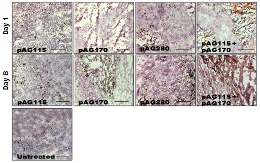
Cancers 2020, 12, 3072                                                                                   2 of 15

and stimulate CD8+ T cells, NK cells, and NKT cells within the tumor itself would be a significant
advantage to overcoming immune evasion.
     IL-15 is highly regulated and its expression is tightly controlled [7]. Binding to its high-affinity alpha
receptor stabilizes the protein, prevents degradation, and increases bioactivity [8–13]. The IL-15/IL-15Rα
complex formation is important for trans-presentation to target cells and subsequent signaling though
the βγ receptor [14,15]. The delivery of genes encoding a soluble receptor α along with IL-15
could improve the efficacy of this anti-cancer immune therapy. Previously, we demonstrated that
the electrotransfer of IL-15 directly to tumors leads to the regression and long-term survival of
mice with melanoma [16,17]. Rowley et al. [18] explored the use of IL-15 together with Il-15Rα by
transducing tumor cells with a retrovirus encoding IL-15 linked to IL-15Rα. Following the injection of
the transduced tumor cells, a slowing of tumor growth as well as an enhanced immune response was
observed. An additional study transfecting unstimulated CD8+ T-cells with RNA encoding IL-15Rα
using nucleofection (electroporation) followed by exposure to IL-15 resulted in increased viability
and proliferation. This was also observed when transfecting unstimulated T-cells with IL-15 linked to
IL-15Rα which enhanced this effect further, as well as after cells were injected into mice [19]. While this
previous study successfully demonstrated the potential of combining IL-15/IL-15Rα, here we seek to
improve on this therapy by delivering both IL-15 and IL-15Rα using electrotransfer protocols and to test
the concept further by treating established tumors and evaluating whether the regression of existing
tumors could be achieved.
     Gene electrotransfer (GET) is an efficient, safe, and effective method used to physically deliver
DNA to tissues [20,21]. GET is mediated by the generation of electric fields in tissue through
the administration of electric pulses via electrodes placed around the tumor. The level of gene expression
generated can be controlled based on the selection of electrode and pulse parameters [22–27]. We
previously demonstrated that achieving the appropriate level of cytokine expression locally is important
in generating the most effective immune response, resulting in tumor regression and long-term
survival [23].
     In the current study, in order to augment the anti-tumor effects previously obtained several
aspects of this immunotherapy approach were examined. A major emphasis of the current study was
to evaluate the delivery of IL-15Rα together with IL-15 either on separate plasmids or together on
one plasmid. Three plasmids were utilized to test this approach. One plasmid encoded only IL-15
(pAG170), a second plasmid encoded only IL-15Rα (pAG115), and one plasmid encoded both IL-15
and IL-15Rα using a dual promoter (pAG208). One aspect that was explored in this current study was
the impact of expression levels on efficacy. Therefore, two GET delivery protocols were utilized in order
to achieve different levels of IL-15 following the delivery of the different plasmids. The evaluation of
these protocols included level of response against the treated tumor and the ability to induce protection
following challenge with a second injection of tumor cells.

2. Results
     Previous studies from our lab showed that IL-15 GET delivered to established tumors in
a mouse melanoma model resulted in complete tumor regression [16,17]. Systemic immune effects of
the treatment, evidenced by protection from challenge, were present in 45% of the treated animals.
In this study, we evaluated the delivery of IL-15 alone or in combination with its high-affinity receptor
IL-15Rα to potentially improve the therapeutic outcome. The two GET protocols used were EP1
and EP2. EP1 (six rotating pulses of 1300 V/cm, 100 µs duration, delivered using a circular six-needle
array) has been shown to generate relatively low levels of gene expression, while EP2 (10 unidirectional
pulses of 600 V/cm, 5 ms in duration, delivered using parallel plate applicator) has been shown
to generate high levels of gene expression. Both protocols are widely used for GET in preclinical
and clinical studies. Experiments were designed to determine the optimal electrotransfer parameters
that would deliver the most appropriate IL-15 levels to improve the therapeutic outcome. Work was
Cancers 2020, 12, 3072                                                                             3 of 15

performed in a mouse melanoma model. B16.F10 cells were injected into the flank of C57Bl/6 mice.
Once the tumors reached approximately 4–5 mm in diameter (6–7 days), treatments were initiated.

2.1. IL-15 and IL-15/IL-15Rα Complex Expression Following Intratumor Delivery
     The initial experimental approach was designed to determine the expression levels that could be
obtained following direct delivery to tumors. Three plasmids and two GET protocols were evaluated.
The three plasmids utilized were a dual-promoter plasmid encoding IL-15 and IL-15Rα (pAG208),
along with plasmids encoding IL-15 (pAG170) and IL-15Rα (pAG115). The two GET protocols utilized
were EP1 and EP2, along with two electrode configurations. The needle electrode applicator (N)
consisted of six needles in a circular array with an outer diameter of 1 cm. Only four of the six needles
were active for each pulse, and the field was rotated 60◦ after each pulse so that at the end of the six
pulses a full 360◦ circle was completed [27]. The gap between the active electrodes was 0.92 cm.
The caliper electrode (C) consisted of two flat stainless-steel plates (5 mm × 5 mm) mounted on the end
of a Vernier caliper. The two plates were placed on either side of the tumor prior to administering
pulses, the gap was measured, and the applied voltage was set according to the gap.
     The plasmids were injected directly into the tumor at a volume of 50 µL and a concentration
of 1 mg/mL. The three plasmids were evaluated individually and pAG115 and pAG170 were also
delivered together using equal volumes of both. The first evaluation was to test the expression
levels of IL-15 following delivery at several time points from 6 h to 8 days. Delivery was performed
on days 0, 4, and 7. The highest expression was observed when pAG170 was delivered with EP2.
Elevated expression was also seen when pAG170 was delivered with EP1. In both cases, the expression
dropped significantly after day 1 (Figure 1A). Subsequent deliveries did not restore the expression
levels, which may have been due to the regression of the tumors. With respect to pAG208, the dual
plasmid expression was elevated with EP2, though not to the extent as that obtained with pAG170,
and although there was a relative decrease from the initial levels, the expression was maintained longer
with this plasmid. The delivery of pAG208 with EP1 had only a slight elevation of the expression
levels. The delivery of pAG170 together with pAG115 resulted in expression levels similar to those of
pAG170 alone for each of the two GET conditions; however, the expression was for a longer period,
even though there was also tumor regression. The results observed with pAG208 and pAG170 together
with pAG115 are interesting, as they support the notion that the half-life of IL-15 can be extended
when complexed with IL-15Rα.
     The levels of the IL-15/IL-15Rα complex following delivery were also explored. The same experimental
setup as described above was performed. Not surprisingly, the presence of the complex was only seen
after the delivery of the dual plasmid or when both pAG170 and pAG115 were delivered together.
The highest and most sustained levels were seen when pAG170 and pAG115 were delivered together
using EP2 (Figure 1B). High levels of the complex were observed with EP1, but they dropped off after
day 1. There was a low to moderate presence of the complex after the delivery of pAG208 with EP2, which
was maintained for 5 days, and the levels were even lower when the delivery was performed with EP1.

2.2. Tumor Regression and Long-Term Survival after IL-15 GET
     Plasmids were delivered to established tumors using either EP1 or EP2 as a series of three
treatments on days 0, 4, and 7. The tumor volume was measured over the course of nine weeks, as
shown in Figure 2. The injection of plasmid with no electrotransfer pulses was used as a control.
All the animals that received this treatment showed no tumor regression. The delivery of pAG208
using EP1 resulted in complete regression in only 60% of the treated animals. This is less than when
EP1 was used to deliver both pAG115 and pAG170 and pAG170 alone, which showed complete
regression in 100% and 90% of the treated animals, respectively (Figure 2). Once the tumors regressed,
the animals remained tumor-free for the duration of the 9-week observation period. The delivery of
pAG115 with EP1 showed no regression in any of the treated animals. The delivery of pAG208 using
EP2 resulted in complete regression in 90% of the animals compared to 60% and 70% of animals treated
Cancers 2020, 12, 3072 4 of 15
with both pAG115 and pAG170 or pAG170 alone, respectively (Figure 2). From these results, it appears
that the selection of electrotransfer protocol plays a role in the tumor regression and overall survival
(Figure 3). For example, the delivery of pAG208 with EP2 resulted in higher levels of expression and a
higher percentage of complete tumor regressions than delivery with EP1. In contrast, the delivery of
pAG170 or the combination of pAG170 and pAG115 with EP2 again resulted in a higher expression;
however, a better tumor response was obtained with EP1. Overall, pAG208 resulted in a lower
expression than the delivery of the other plasmids, but high levels of tumor regression were observed
when delivered with EP2. It is possible that the high levels of expression achieved with EP2 when
delivering pAG170 or pAG115 together with pAG170 may not have induced a sufficient anti-tumor
immune response. With respect to IL-15Rα, these data do not suggest a clear advantage for selecting to
deliver IL-15 with IL-15Rα rather than IL-15 alone.
Figure 1. IL-15 and IL-15/IL-15Rα complex expression after GET. B16.F10 tumors were established on
the left flank of C57Bl/6 mice. Once tumors reached 3–5 mm in diameter, the intratumor injection of
the plasmid was performed as described in methods. Following injection, electrotransfer was performed
using either EP1 or EP2. EP1 = 1300 V/cm, 100 us, 6 pulses of EP1 delivered using a 6 penetrating
needle array. EP2 = 600 V/cm, 5 ms, 10 pulses of EP2 delivered using caliper electrodes. Level of
transgene expression within the B16.F10 tumors was measured by ELISA using tumor homogenate
collected at the indicated time points. n = 4 for each group. (A) levels of Il-15 after delivery; (B) levels
of IL-15/IL-15Rα after delivery. IO = injection only.
Figure 2. IL-15 GET induces tumor regression. Animals were given a series of three deliveries of
intratumoral plasmid DNA using GET on days 0, 4, and 7. (A) EP1 delivered using a 6 penetrating
needle array and (B) EP2 delivered using caliper electrodes. Tumor volume was measured in mm3
and monitored weekly over a nine-week period. Data are presented as the mean tumor volume and error
bars represent the standard deviation. n = 10 for each group. IO = injection only; EP = electrotransfer.Cancers 2020, 12, 3072 5 of 15
Figure 3. IL-15 GET induces the long-term survival of mice. The survival of mice monitored above is
represented as the percent survival of the experimental group over time. Electrotransfer protocols were
used as indicated. (A) EP1 = 1300 V/cm, 100 us, 6 pulses of EP1 delivered using a 6 penetrating needle
array. (B) EP2 = 600 V/cm, 5 ms, 10 pulses of EP2 delivered using caliper electrodes. n = 10 for each
group. IO = injection only; EP = electrotransfer.
2.3. Delivery of IL-15/IL-15Rα Protects a Greater Percentage of Mice from Challenge
The animals that were tumor-free after the treatment described in Section 2.2 were challenged
subcutaneously with B16.F10 cells on the opposite flank and closely monitored for tumor development.
Animals that developed tumors post-challenge were termed non-resistant and those that did not
develop tumors were termed resistant. Table 1 shows the number of animals that were resistant to
challenge after 50 days, as well as the overall percentage of the original number that were tumor-free
after treatment for the duration of the entire experiment (primary and challenge). Resistance to
challenge indicates the generation of a systemic immune memory response.
The delivery of IL-15/IL15Rα (pAG208) protected more animals from developing a second tumor
than the delivery of IL-15 alone (pAG170) or the delivery of both genes on separate plasmids (pAG115
and pAG170). Using EP1 to deliver the plasmids showed only a slight advantage in using IL-15/IL-15Rα
over IL-15 alone by protecting 40% of the animals from challenge compared to 30%, respectively. When
both genes were delivered on separate plasmids, only 20% of the animals were protected. Using EP2 to
deliver the plasmids had a more definitive outcome. In this case, delivering pAG208 was able to protect
70% of the animals from developing a second tumor, the highest level of protection for all groups
tested. The delivery of IL-15 alone or both genes on separate plasmids using EP2 was not able to confer
protection to any of the treated animals. From these results, it appears that different combinations of
plasmid and electrotransfer parameters may be inducing different cellular responses within the tumor.
It is also important to note that the delivery of pAG208 with EP2 did not result in high levels of IL-15
or IL-15/IL-15Rα complex. However, this did result in the highest level of protection from challenge.
2.4. Cytokines Generated in the Tumor after GET
In order to explore how immune activation may be involved in the observed tumor regression
and long-term systemic response, a panel of cytokines was measured using a magnetic bead multiplex
assay. The cytokines included in the multiplex were: IFNγ, IL-1α, IL-6, IL-10, IL-12 p40, IL-12 p70,
IL-15, IP-10, MCP-1, MIP-1β, and TNFα. Tumors were collected at various time points over the course
of treatment and assayed for cytokine levels. Other than IL-15, only IFNγ, IL-1α, IL-6, and MIP-1β
showed variation between the treatment groups. The results for these four cytokines are shown
in Figure 4. The groups chosen for analysis were based on the percentage of overall survival after
treatment and challenge. Since the delivery of both genes on separate plasmids did not induce a robustCancers 2020, 12, 3072 6 of 15
systemic response, they were not included in this analysis. Direct comparisons are made for groups that
received both genes on a single plasmid and groups that received IL-15 only using both electrotransfer
protocols. Animals treated with IL-15Rα only using EP2 and pAG208 injection only were included as
controls for the purposes of this experiment.
Table 1. Long term survival of mice that received gene electrotransfer.
GET Challenged %
Plasmid Original Resistant
Protocol (Disease Free at 50 Days) Resistant
n n n
Injection
pAG115 (IL-15Rα) 10 0 - -
Only
Injection
pAG208 (IL-15/IL-15Rα) 10 0 - -
Only
Injection
pAG170 (IL-15) 10 0 - -
Only
Injection
pAG170 + pAG115) 10 0 - -
Only
pAG115 (IL-15Rα) EP1 10 0 - -
pAG208 (IL-15/IL-15Rα) EP1 10 6 4 40
pAG170 (IL-15) EP1 10 9 3 30
pAG170 + pAG115 EP1 10 10 2 20
pAG115 (IL-15Rα) EP2 10 2 0 0
pAG208 (IL-15/IL-15Rα) EP2 10 9 7 70
pAG170 (IL-15) EP2 10 7 0 0
pAG170 + pAG115 EP2 10 6 0 0
Overall, the choice of EP1 or EP2 to deliver each plasmid did not result in a significant difference in
the level of cytokines induced. The expression of IL-15 from pAG208 and pAG170 is driven by different
promoters and, as such, the expression levels detected in the tumor are different. IL-15 appears to be
generated at higher quantities from pAG170 than pAG208 (Figure 1A). At the early time point, after
the first treatment with pAG170 slightly higher levels of IL-1α and IL-6 compared to pAG208 were
observed (Figure 4A,B). After the second treatment, the groups treated with pAG208 showed higher
levels of IFN-γ and MIP-1β compared to pAG170 (Figure 4C,D). This indicates there may be a more
cellular, non-specific response to the delivery of pAG170, while the delivery of pAG208 stimulates
a more specific response.
2.5. GET Immunotherapy Generates Local Lymphocytic Infiltrate
In order to determine what effects the therapy had on the local tumor environment, tumors
were collected shortly after the first and last treatments (day 1 and day 9) and examined by
immunohistochemistry. Only one electrotransfer condition, EP2, was selected for this analysis, as
it showed protection from challenge in the largest percentage of animals as well as the greatest disparity
in protection based on the selection of plasmid.Cancers 2020, 12, 3072 7 of 15
Figure 4. IL-15 GET induces cytokine in the tumor microenvironment. Cytokine levels were measured
using a multiplex assay from tumor homogenate collected at various time points during the treatment
protocol. Cytokines included in the multiplex were IFNγ, IL-1α, IL-6, IL-10, IL-12 p40, IL-12 p70, IL-15,
IP-10, MCP-1, MIP-1β, and TNFα. Results are shown for cytokines that were observed to have variation
between groups. pDNA was delivered on days 0, 4, and 7. Data are represented as the mean expression
for each group. (A) levels of IL-1α; (B) levels of IL-6; (C) levels of IFNγ; (D) levels of MIP-1β. Error
bars represent the standard deviation. n = 4 for each group.
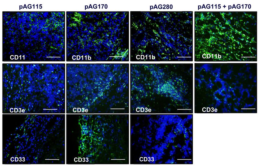
The presence of an increased lymphocytic infiltrate in the treated tumors could be an indication
that tumor regression is immune cell-mediated and not due to strong electric fields generated by
the electrotransfer pulses. The histology of tumors collected during the course of treatment showed
increased tumor damage when IL-15 was locally expressed compared to the controls (Figure 5).
CD11b, CD3e, and CD335 antibodies were used to determine the general populations of monocytes
and antigen-presenting cells, T lymphocytes, and natural killer (NK) cells, respectively, present in
the tumors after treatment (Figure 6). Compared to non-treated tumors, the plasmid electrotransfer
increased the presence of immune cells in the tumor. pAG208 induced more CD11b+ and CD3e+ cells
than AG170, but the opposite is true for CD335+ cells. This indicates that, while pAG170 induces
cellular infiltrate, the immune response driving tumor regression may be non-specific. The increased
presence of CD335 + NK cells and the amount of early tumor damage compared to the other groups
are indicators of this. The delivery of pAG208 induces early CD11b+ cell infiltrate that facilitates
antigen presentation and late CD3+ cell infiltrate, which suggests the generation of a specific immune
response to tumor antigen. The lack of increase in NK cell infiltrate seen with pAG170 also lends itself
to the theory of the generation of a tumor-specific response.Cancers 2020, 12, 3072 8 of 15
Figure 5. Histology of tumors after electrotransfer. Hematoxylin and eosin stained sections of tumor
collected on day 1 (24 h after the first treatment) and on day 8 (24 h after the third treatment) after
the electrotransfer of pAG115, pAG170, and pAC208 using EP2. Images are representative sections of
n = 2 tumors. Scale bar = 500 µm.
Figure 6. IL-15 GET induces immune cell infiltrate into the tumor microenvironment. Haematoxylin
and eosin stain. Anti-CD3e (green-T cells), anti-CD11b (green-APCs), and anti-CD335 (green—natural
killer cells) staining (green) reveal the presence of immune cells within the tumors of mice treated using
EP2 to deliver pAG115, p,G170, and pAG208 on days 0, 4, and 7. Tissue was collected on day 8 (24 h
after the third treatment). Images are the best representative sample of n = 2 for each group. Sections
were counterstained with a nuclear dye 40 ,6-diamidino-2-phenylindole (DAPI) (Blue). Mag = 20×.
Scale bar = 500 µm.Cancers 2020, 12, 3072 9 of 15
2.6. Get Immunotherapy Generates a Systemic Response
The GET delivery of pAG208 with EP2 generated the highest level of protection following
challenge with an injection of fresh B16.F10 cells. This protocol also resulted in the highest level of
local immune response with regards to the infiltrate of T-cells and a cytokine profile indicating a more
specific response. Being able to sustain a long-term disease-free state and the generation of local
immune response is important in controlling tumor spread; it is also important to determine if a robust
systemic response could be generated that could prevent and/or eliminate distant tumors in visceral
organs. To test this, we utilized a lung colonization model.
In addition to pAG208 delivered with GET EP2, the groups included pAG170 GET EP1 (best
response with that plasmid) and pAG115 delivered with GET EP2 as a control. Tumors were established
on the left flank of mice and treated on days 0, 4, and 7. On day 0, the mice received an intravenous
injection of B16.F10 cells that had been stably transfected with luciferase. The mice were imaged with
an IVIS in vivo imager to detect growth within the lungs. Mice that were treated with pIL-15/IL-15Rα
complex had a greater level of protection from secondary tumor formation using a lung colonization
model (Figure 7). However, to a lesser extent, treatment using GET EP1 to deliver pAG170 slowed
the tumor growth within the lungs.
Figure 7. IL-15/IL-15Rα GET protects mice from secondary tumor formation in a lung colonization
model. B16.F10 tumors were induced on the flanks of C57BL/6 Albino mice and treated with GET using
the EP2 protocol on days 0, 4, and 7. On the day of first treatment, B16.F10-Luc G5 cells were injected
intravenously. Tumor metastasis was monitored using an IVIS spectrum and reported as luminescence
(photons/sec). n = 9 for each group.
3. Discussion
The administration of cytokines to stimulate an immune-mediated anti-tumor response has been
explored for several decades [28–30]. Interferon-alpha (IFN-α) was the first cytokine to be approved
by the U.S. Food and Drug Administration (FDA), initially for the treatment of hairy cell leukemia
in 1986 and then for metastatic melanoma in 1995 [28,29]. Interleukin-2 (IL-2) was also evaluated
as a potent stimulator of T-cell activity. High-dose IL-2 received FDA approval for metastatic renal
cell cancer in 1992 and advanced melanoma in 1998 [28,29]. Since then, several other cytokines
have been tested in a variety of indications, including interferon-gamma, granulocyte macrophage
colony-stimulating factor, IL-12, IL-15, and IL-21 [28–30]. These cytokines were initially evaluated as
single agents. In these trials, while there was some low level of responses, there was also significant
toxicity. One drawback of these approaches is the toxicity associated with the systemic administration
of these agents. This may have also contributed to the low response rates related to low levels of
the cytokines within the tumor environment.
A typical issue with these agents is their short half-life. One approach to potentially overcome this
is to administer cytokine therapies at high doses which can result in adverse reactions. To potentiallyCancers 2020, 12, 3072 10 of 15
get around this issue, as well as to prolong the presence of the therapeutic agent, a gene-based approach
can be used. One example of this approach is the delivery of IL-12 in the form of plasmid DNA (pIL-12).
To better control the expression levels and kinetics, it was delivered using gene electrotransfer [23].
Preclinical studies demonstrated the efficacy and safety of this approach [31,32]. In clinical trials
testing the approach in melanoma patients, this approach was shown to be effective with no systemic
treatment-related adverse effects [33]. In addition, in both Phase I and Phase II trials responses were
seen in both treated and untreated lesions, with approximately 15% of the patients achieving durable
complete response of all lesions [34]. This approach is being tested with other tumor types, including
Merkel cell carcinoma [35].
IL-15 has shared functions with IL-2 and was thought to be an attractive alternative and, unlike
IL-2, does not attract T-regulatory cells nor induces activation-induced cell death. However, IL-15
as a monotherapy did not result in a sustained anti-tumor response [28,29]. It was noted that, for
IL-15 to be effective, it was necessary for it to be associated with IL-15 Rα [28,36]. Several studies have
been conducted utilizing IL-15/IL-15Rα (superagonist). In clinical trials with the IL-15 superagonist,
there were higher levels of T-cytotoxic cells in the circulation for a longer period. There was also
a higher level of NK cells and T-effector cells. Although it is well established that IL-15 or IL-15/IL-15Rα
complex can be a potent stimulator of T cytotoxic and NK cells, high levels of IL-15 or the complex can
actually result in reduced proliferation, exhaustion, and reduced anti-tumor activity [37]. A correlation
between high IL-15 serum levels, poor prognosis in melanoma patients, and reduced efficacy in
treatment with ipilimumab was observed [38]. High-dose IL-15 has also been associated with severe
adverse effects. including systemic inflammation [37]. These effects are related to an extended half-life
of the IL-15 complex. There are also additional studies being conducted evaluating the potential
of engineering cells to express the IL-15/IL-15Rα complex or to deliver via plasmid DNA or viral
vectors [39]. These approaches are designed to extend the time that IL-15 is present and to potentially
augment the anti-tumor response.
In the study reported here, we set out to answer two questions: (1) Does the delivery of IL-15 along
with its soluble receptor IL-15Rα improve therapeutic outcome? (2) Can the selection of electrotransfer
parameters enhance this outcome by modulating the level of IL-15 expression? Using GET to deliver
IL-15 and the soluble IL-15Rα on a single plasmid (pAG208), we were able to demonstrate an effective
local anti-tumor response similar to that obtained using IL-15 only (pAG170). The systemic response
generated by pAG208 exceeded that of delivering only pAG170. By selecting appropriate delivery
parameters, we were able to enhance the long-term protective effects of this therapy and show protection
from challenge in 70% of treated animals, which is an improvement on what we have described
previously [16,17]. Interestingly, this was similar to results obtained in preclinical studies in the same
model with pIL-12 [22], although the GET parameters to obtain this response were different.
The electrotransfer protocols were selected because they generate different levels of gene
expression [23,39]. We sought to determine if increased local IL-15 expression, with or without
the presence of IL-15Rα would result in greater anti-tumor response and protection. Due to gene
expression being driven by different promoters, pAG208 produced less measurable IL-15 than pAG170
even when EP2 was used for delivery. To account for this discrepancy and examine the effects of
the presence of the receptor, IL-15 and IL-15Rα on separate plasmids (driven by the same promoter)
were delivered simultaneously. Though there was not an apparent advantage in using IL-15/IL-15Rα
over IL-15 alone, or high levels of IL-15 over low levels of IL-15 to induce tumor regression of the treated
tumor and long-term survival, it became clear upon challenge that high levels of IL-15 do not protect
from challenge and a lower expression induces specific anti-tumor responses. The delivery of both
genes on separate plasmids showed similar levels of protection to the delivery of IL-15 alone.
The expression of lower levels of IL-15 in the tumors generated higher levels of the anti-tumor
cytokines IFN-γ and MIP-1β than what was generated when higher levels of IL-15 were obtained.
IFN-γ and MIP-1β peaked five days into the treatment regimen, creating favorable conditions for
the generation of a specific rather than a non-specific immune response that would result in immuneCancers 2020, 12, 3072 11 of 15
memory. The histological examination of treated tumors revealed that high levels of IL-15 induced
a robust innate response that appears to be mainly driven by NK cells that resulted in a great amount
of tissue damage. The lower levels of IL-15 induced far less NK cell infiltrate but more CD3+ cells.
This high level of IL-15 in the tumor resulted in tissue destruction that occurred rapidly, and though
antigen-presenting cells may have been present, the low number of T cells in the tumor did not allow
for the generation of an adequate memory response.
The results presented in this study as well as by others have shown a clear advantage of utilizing
IL-15/IL-15Rα over the monotherapy. In clinical trials evaluating IL-15, a low level of response was
observed. To potentially overcome these poor responses, studies are being conducted to determine
the potential of combining IL-15 or IL-15/IL-15Rα with other immune modifiers, including checkpoint
inhibitors [40–42]. Interestingly, in the pIL-12 clinical trials, patients who had a poor response were
observed to have elevated tumor levels of PD-1 and PD-L1 [34,43]. A phase II clinical trial combining
pIL-12 and anti-PD-1 in immunologically quiescent melanomas resulted in a higher than expected
response rate [44], with a complete response rate (all tumors responding) of 36% [44]. Determining
the appropriate delivery conditions for a plasmid encoding IL-15/IL-15Rα and combining with
a checkpoint inhibitor could result in similar or higher response levels.
4. Materials and Methods
4.1. Plasmids, Cells, Mice
The plasmids used in this study were gifted to us by Drs. B. Felber and G. Pavlakis (NCI, Bethesda
MD, USA) and commercially prepared by Aldevron, (Fargo, ND, USA), with endotoxin levelsCancers 2020, 12, 3072 12 of 15
The protocol EP2 consisted of 10 unidirectional 600 V/cm, 5 ms pulses delivered using adjustable
parallel plate caliper electrodes. The mice received a series of three plasmid deliveries on days 0, 4,
and 7.
4.4. Cytokine Measurements
Tumors were collected from animals in separate experiments at 6 and 24 h after a single
delivery, 24 h after two deliveries, and 24 h after all three deliveries, and then homogenized
in cold phosphate buffered saline (Mediatech, Manassas, VA, USA) containing protease inhibitor
(Roche, Indianapolis, IN, USA). The homogenate was centrifuged and the supernatant collected
and assayed for cytokine expression using a Milliplex MAP kit mouse cytokine magnetic bead panel
multiplex (Millipore, Billercia, MA, USA) according to the manufacturer’s instructions.
4.5. Immunohistochemistry
Tumors were collected 24 h after a single delivery, two deliveries, and three deliveries of pDNA
in separate experiments; embedded in OCT; frozen on dry ice; and stored at −80 ◦ C. Standard
protocols were used. Briefly, 7 µm cryosections were prepared and fixed using a 75% acetone and 25%
ethanol mix. Sections were incubated in 5–10% animal serum blocking buffer for at least one hour
at room temperature. An avidin/biotin blocking kit (Vector Labs, Burlingame, CA, USA) was used
to block the endogenous biotin present in the samples prior to the addition of the primary antibody.
Antibodies to the markers CD11b (14-0112, 1:200; eBioscience, San Diego, CA, USA), CD3e (14-0031,
1:100; eBioscience), and CD335 (NKp46) (137615, 1:50; BioLegend, San Diego, CA, USA) were used.
Biotinylated secondary antibodies anti-rat IgG (13-4813, 1:500; eBioscience) and anti-armenian hamster
IgG (13-4113, 1:300; eBioscience) were used, except for the samples stained for CD335, as the primary
antibody is biotinylated. Signal was detected using FITC conjugated streptavidin (11-4317, 1:500;
eBioscience). DAPI (Sigma, St. Louis, MO, USA) was used as a counterstain to visualize nuclei.
Cover slips were mounted using VectaSheild Reagent (Vector Labs) and the images recorded using
a DP70 camera attached to a BX-51 fluorescent microscope (Olympus, Center Valley, PA, USA).
4.6. Lung Colonization Model
B16.F10 (B16.F10-Luc) cells that are stably transfected to express luciferase were used. This enabled
the cells to be tracked noninvasively. Mice with B16.F10 tumors already established on the left flank
as described above were injected intravenously into the tail vein with 1 × 10⁵ B16.F10-Luc cells in
a volume of 50 µL of sterile PBS on the first day of treatment. Only the subcutaneous tumor was
treated and the treatment was as described above. Mice were imaged on days 6, 8, and 11 using
an IVIS Spectrum whole body imaging system (Perkin Elmer, Waltham, MA, USA). To obtain an image,
luciferin is administered via an intraperitoneal injection at a concentration of 15 mg/mL and a dose of
150 mg/kg weight. For a 20 g mouse, 200 µL is injected. All the animals are imaged within 15 min after
the injection of luciferin. The IVIS Spectrum takes an image of the animal and can measure the amount
of luminescence being produced at specific sites.
4.7. Statistical Analysis
Data were log transformed to achieve normal distribution. Multiple groups were compared using
a one-way ANOVA with the Tukey–Kramer Multiple Comparison post-test. Pair-wise comparisons
were conducted using a paired t-test. The critical alpha for all the comparisons was 0.05.
5. Conclusions
From the results obtained in this study, it appears that the level of IL-15 expressed in the tumor has
a greater impact on the overall survival and protection from challenge than the presence or absence of
the soluble receptor. At higher expression levels, an innate immune response was induced, and whileCancers 2020, 12, 3072 13 of 15
it was sufficient to induce the complete regression of the treated tumor, a robust memory response
was not induced, resulting in a reduced protection following challenge and less protection from lung
colonization. In addition, though the presence of IL-15Rα in tumors is thought to stabilize the protein
and allow for more efficient trans-presentation and signaling on the target cells, from these results we
cannot at this time fully discern its role in the responses seen. Further studies need to be done at low
levels of expression in the presence and absence of the receptor to tease out its role, if any, in this GET
therapy. Additional studies should also be conducted to determine how the presence of checkpoints
may influence the response and to test the combination of IL-15/IL-15Rα with checkpoint inhibitors.
Author Contributions: Conceptualization, S.A.S. and R.H.; methodology, S.A.S., C.G.L., and R.H.; formal analysis,
S.A.S. and R.H.; investigation, S.A.S., C.G.L., and R.H.; resources, R.H.; writing—original draft preparation, S.A.S.;
writing—review and editing, R.H.; supervision, S.A.S. and R.H.; project administration, R.H.; funding acquisition,
R.H. All authors have read and agreed to the published version of the manuscript.
Funding: This research was funded by U.S. National Institutes of Health, R01 CA122518.
Acknowledgments: We would like to thank G. Pavlakis (National Cancer Institute) for the gift of the plasmids;
Niculina Burcus for processing the samples for histology; and Fanying Li for assaying the samples for
cytokine expression.
Conflicts of Interest: R. Heller is an inventor on patents which cover the technology that was used in the work
reported in this manuscript. In addition, R. Heller owns stock in Inovio Pharmaceuticals, Inc., and stock and stock
options in OncoSec, Inc.
References
1. Giri, J.; Ahdieh, M.; Eisenman, J.; Shanebeck, K.; Grabstein, K.; Kumaki, S.; Namen, A.; Park, L.; Cosman, D.;
Anderson, D. Utilization of the Beta and Gamma Chains of the IL-2 Receptor by the Novel Cytokine IL-15.
EMBO J. 1994, 13, 2822–2830. [CrossRef] [PubMed]
2. Giri, J.G.; Kumaki, S.; Ahdieh, M.; Friend, D.J.; Loomis, A.; Shanebeck, K.; DuBose, R.; Cosman, D.; Park, L.S.;
Anderson, D.M. Identification and Cloning of a Novel IL-15 Binding Protein That Is Structurally Related to
the Alpha Chain of the IL-2 Receptor. EMBO J. 1995, 14, 3654–3663. [CrossRef] [PubMed]
3. Lodolce, J.P.; Boone, D.L.; Chai, S.; Swain, R.E.; Dassopoulos, T.; Trettin, S.; Ma, A. IL-15 Receptor Maintains
Lymphoid Homeostasis by Supporting Lymphocyte Homing and Proliferation. Immunity 1998, 9, 669–676.
[CrossRef]
4. Mortier, E.; Advincula, R.; Kim, L.; Chmura, S.; Barrera, J.; Reizis, B.; Malynn, B.A.; Ma, A. Macrophage-
and Dendritic-Cell-Derived Interleukin-15 Receptor Alpha Supports Homeostasis of Distinct CD8+ T Cell
Subsets. Immunity 2009, 31, 811–822. [CrossRef]
5. Ohteki, T.; Suzue, K.; Maki, C.; Ota, T.; Koyasu, S. Critical Role of IL-15–IL-15R for Antigen-Presenting Cell
Functions in the Innate Immune Response. Nat. Immunol. 2001, 2, 1138–1143. [CrossRef]
6. Basak, G.W.; Zapala, L.; Wysocki, P.J.; Mackiewicz, A.; Jakóbisiak, M.; Lasek, W. Interleukin 15 Augments
Antitumor Activity of Cytokine Gene-Modified Melanoma Cell Vaccines in a Murine Model. Oncol. Rep.
2008, 19, 1173–1179. [CrossRef]
7. Waldmann, T.A.; Tagaya, Y. The Multifaceted Regulation of Interleukin-15 Expression and the Role of This
Cytokine in Nk Cell Differentiation and Host Response to Intracellular Pathogens. Annu. Rev. Immunol.
1999, 17, 19–49. [CrossRef]
8. Bergamaschi, C.; Rosati, M.; Jalah, R.; Valentin, A.; Kulkarni, V.; Alicea, C.; Zhang, G.-M.; Patel, V.; Felber, B.K.;
Pavlakis, G.N. Intracellular Interaction of Interleukin-15 with Its Receptor α During Production Leads to
Mutual Stabilization and Increased Bioactivity. J. Biol. Chem. 2007, 283, 4189–4199. [CrossRef]
9. Bergamaschi, C.; Bear, J.; Rosati, M.; Beach, R.K.; Alicea, C.; Sowder, R.; Chertova, E.; Rosenberg, S.A.;
Felber, B.K.; Pavlakis, G.N. Circulating IL-15 Exists as Heterodimeric Complex with Soluble IL-15Rα in
Human and Mouse Serum. Blood 2012, 120, e1–e8. [CrossRef]
10. Stoklasek, T.A.; Schluns, K.S.; Lefrancois, L. Combined IL-15/IL-15Rα Immunotherapy Maximizes IL-15
Activity in Vivo. J. Immunol. 2006, 177, 6072–6080. [CrossRef]
11. Rubinstein, M.P.; Kovar, M.; Purton, J.F.; Cho, J.H.; Boyman, O.; Surh, C.D.; Sprent, J. Converting IL-15 to
a Superagonist by Binding to Soluble IL-15Rα. Proc. Natl. Acad. Sci. USA 2006, 103, 9166–9171. [CrossRef]
[PubMed]Cancers 2020, 12, 3072 14 of 15
12. Bergamaschi, C.; Jalah, R.; Kulkarni, V.; Rosati, M.; Zhang, G.-M.; Alicea, C.; Zolotukhin, A.S.; Felber, B.K.;
Pavlakis, G.N. Secretion and biological activity of short signal peptide IL-15 is chaperoned by IL-15 receptor
alpha in vivo. J. Immunol. 2009, 183, 3064–3072. [CrossRef] [PubMed]
13. Jalah, R.; Rosati, M.; Kulkarni, V.; Patel, V.; Bergamaschi, C.; Valentin, A.; Zhang, G.-M.; Sidhu, M.K.;
Eldridge, J.H.; Weiner, D.B.; et al. Efficient Systemic Expression of Bioactive IL-15 in Mice Upon Delivery of
Optimized DNA Expression Plasmids. DNA Cell Biol. 2007, 26, 827–840. [CrossRef] [PubMed]
14. Dubois, S.; Mariner, J.; Waldmann, T.A.; Tagaya, Y. IL-15Rα Recycles and Presents IL-15 in Trans to
Neighboring Cells. Immunity 2002, 17, 537–547. [CrossRef]
15. Burkett, P.R.; Koka, R.; Chien, M.; Chai, S.; Boone, D.L.; Marcia, C. Coordinate Expression and Trans
Presentation of Interleukin (IL)-15Rα and IL-15 Supports Natural Killer Cell and Memory CD8+ T Cell
Homeostasis. J. Exp. Med. 2004, 200, 825–834. [CrossRef]
16. Ugen, K.E.; Kutzler, M.A.; Marrero, B.; Westover, J.; Coppola, D.; Weiner, D.B.; Heller, R. Regression of
Subcutaneous B16 Melanoma Tumors After Intratumoral Delivery of an IL-15-Expressing Plasmid Followed
by in Vivo Electroporation. Cancer Gene Ther. 2006, 13, 969–974. [CrossRef]
17. Marrero, B.; Shirley, S.; Heller, R. Delivery of Interleukin-15 to B16 Melanoma by Electroporation Leads to
Tumor Regression and Long-Term Survival. Technol. Cancer Res. Treat. 2013, 13, 551–560. [CrossRef]
18. Rowley, J.; Monie, A.; Hung, C.-F.; Wu, T.-C. Inhibition of Tumor Growth by NK1.1+ Cells and CD8+ T Cells
Activated by IL-15 through Receptor β/Common γ Signaling in Trans1. J. Immunol. 2008, 181, 8237–8247.
[CrossRef]
19. Rowley, J.; Monie, A.; Hung, C.-F.; Wu, T.C. In Expression of IL-15RA or an IL-15/IL-15RA Fusion on CD8+ T
Cells Modifies Adoptively Trasnferred T-Cell Function in Cis. Eur. J. Immunol. 2009, 39, 491–506. [CrossRef]
20. Heller, L.C.; Heller, R. In Vivo Electroporation for Gene Therapy. Hum. Gene Ther. 2006, 17, 890–897.
[CrossRef]
21. Heller, R.; Heller, L.C. Gene Electrotransfer Clinical Trials. Adv. Genet. 2015, 89, 235–262. [CrossRef]
22. Efavard, C.; Dean, D.S.; Rols, M.-P. Electrotransfer as a Non Viral Method of Gene Delivery. Curr. Gene Ther.
2007, 7, 67–77. [CrossRef]
23. Shirley, S.A.; Lundberg, C.G.; Li, F.; Burcus, N.; Heller, R. Controlled Gene Delivery Can Enhance Herapeutic
Outcome for Cancer Immune Therapy for Melanoma. Curr. Gene Ther. 2015, 15, 32–43. [CrossRef]
24. Gehl, J.; Mir, L.M. Determination of Optimal Parameters for in Vivo Gene Transfer by Electroporation,
Using a Rapid in Vivo Test for Cell Permeabilization. Biochem. Biophys. Res. Commun. 1999, 261, 377–380.
[CrossRef] [PubMed]
25. Wolf, H.; Rols, M.-P.; Boldt, E.; Neumann, E.; Teissie, J. Control by Pulse Parameters of Electric Field-Mediated
Gene Transfer in Mammalian Cells. Biophys. J. 1994, 66, 524–531. [CrossRef]
26. Cemazar, M.; Sersa, G.; Wilson, J.; Tozer, G.M.; Hart, S.L.; Grosel, A.; Dachs, G.U. Effective Gene
Transfer to Solid Tumors Using Different Nonviral Gene Delivery Techniques: Electroporation, Liposomes,
and Integrin-Targeted Vector. Cancer Gene Ther. 2002, 9, 399–406. [CrossRef]
27. Gilbert, R.A.; Jaroszeski, M.J.; Heller, R. Novel Electrode Designs for Electrochemotherapy. Biochim. Biophys.
Acta BBA Gen. Subj. 1997, 1334, 9–14. [CrossRef]
28. Conlon, K.C.; Miljkovic, M.D.; Waldmann, T.A. Cytokines in the Treatment of Cancer. J. Interf. Cytokine Res.
2019, 39, 6–21. [CrossRef] [PubMed]
29. Waldmann, T.A. Cytokines in Cancer Immunotherapy. Cold Spring Harb. Perspect. Biol. 2017, 10, a028472.
[CrossRef]
30. Flores, T.; Tarhini, A.A. Anticancer Cytokines: Biology and Clinical Effects of IFN-α2, IL-2, IL-15, IL-21
and IL-12. Semin. Oncol. 2015, 42, 539–548. [CrossRef]
31. Lucas, M.; Heller, L.; Coppola, D.; Heller, R. IL-12 Plasmid Delivery by in Vivo Electroporation for
the Successful Treatment of Established Subcutaneous B16.F10 Melanoma. Mol. Ther. 2002, 5, 668–675.
[CrossRef]
32. Shi, G.; Edelblute, C.; Arpag, S.; Lundberg, C.; Heller, R. IL-12 Gene Electrotransfer Triggers a Change in
Immune Response within Mouse Tumors. Cancers 2018, 10, 498. [CrossRef]
33. Daud, A.I.; DeConti, R.C.; Andrews, S.; Urbas, P.; Riker, A.I.; Sondak, V.K.; Munster, P.N.; Sullivan, D.M.;
Ugen, K.E.; Messina, J.L.; et al. Phase I Trial of Interleukin-12 Plasmid Electroporation in Patients with
Metastatic Melanoma. J. Clin. Oncol. 2008, 26, 5896–5903. [CrossRef]Cancers 2020, 12, 3072 15 of 15
34. Greaney, S.K.; Algazi, A.P.; Tsai, K.K.; Takamura, K.T.; Chen, L.; Twitty, C.G.; Zhang, L.; Paciorek, A.;
Pierce, R.H.; Le, M.H.; et al. Intratumoral Plasmid IL12 Electroporation Therapy in Patients with Advanced
Melanoma Induces Systemic and Intratumoral T-Cell Responses. Cancer Immunol. Res. 2019, 8, 246–254.
[CrossRef]
35. Bhatia, S.; Longino, N.V.; Miller, N.J.; Kulikauskas, R.; Iyer, J.G.; Ibrani, D.; Blom, A.; Byrd, D.R.;
Parvathaneni, U.; Twitty, C.G.; et al. Intratumoral Delivery of Plasmid IL12 Via Electroporation Leads to
Regression of Injected and Noninjected Tumors in Merkel Cell Carcinoma. Clin. Cancer Res. 2020, 26, 598–607.
[CrossRef]
36. Robinson, T.O.; Schluns, K.S. The Potential and Promise of IL-15 in Immuno-Oncogenic Therapies. Immunol.
Lett. 2017, 190, 159–168. [CrossRef]
37. Cristiani, C.M.; Garofalo, C.; Passacatini, L.C.; Carbone, E. New Avenues for Melanoma Immunotherapy:
Natural Killer Cells? Scand. J. Immunol. 2020, 91, e12861. [CrossRef]
38. Tallerico, R.; Cristiani, C.M.; Staaf, E.; Garofalo, C.; Sottile, R.; Capone, M.; De Coaña, Y.P.; Madonna, G.;
Palella, E.; Wolodarski, M.; et al. IL-15, TIM-3 and NK Cells Subsets Predict Responsiveness to Anti-CTLA-4
Treatment in Melanoma Patients. OncoImmunology 2016, 6, e1261242. [CrossRef]
39. Guo, Y.; Luan, L.; Patil, N.K.; Sherwood, E.R. Immunobiology of the IL-15/IL-15Rα Complex as an Antitumor
and Antiviral Agent. Cytokine Growth Factor Rev. 2017, 38, 10–21. [CrossRef]
40. Cemazar, M.; Golzio, M.; Sersa, G.; Hojman, P.; Kranjc, S.; Mesojednik, S.; Rols, M.-P.; Teissie, J. Control by
Pulse Parameters of DNA Electrotransfer into Solid Tumors in Mice. Gene Ther. 2009, 16, 635–644. [CrossRef]
41. Waldmann, T.A.; Dubois, S.; Miljkovic, M.D.; Conlon, K.C. IL-15 in the Combination Immunotherapy of
Cancer. Front. Immunol. 2020, 11, 868. [CrossRef] [PubMed]
42. Barroso-Sousa, R.; Ott, P.A. Transformation of Old Concepts for a New Era of Cancer Immunotherapy:
Cytokine Therapy and Cancer Vaccines as Combination Partners of PD1/PD-L1 Inhibitors. Curr. Oncol. Rep.
2018, 20, 1. [CrossRef] [PubMed]
43. Algazi, A.; Bhatia, S.; Agarwala, S.; Molina, M.; Lewis, K.; Faries, M.; Fong, L.; Levine, L.; Franco, M.;
Oglesby, A.; et al. Intratumoral Delivery of Tavokinogene Telseplasmid Yields Systemic Immune Responses
in Metastatic Melanoma Patients. Ann. Oncol. 2020, 31, 532–540. [CrossRef] [PubMed]
44. Algazi, A.P.; Twitty, C.G.; Tsai, K.K.; Le, M.; Pierce, R.; Browning, E.; Hermiz, R.; Canton, D.A.; Bannavong, D.;
Oglesby, A.; et al. Phase II Trial of IL-12 Plasmid Transfection and PD-1 Blockade in Immunologically
Quiescent Melanoma. Clin. Cancer Res. 2020, 26, 2827–2837. [CrossRef]
45. Tomayko, M.M.; Reynolds, C.P. Determination of Subcutaneous Tumor Size in Athymic (nude) Mice. Cancer
Chemother. Pharm. 1989, 24, 148–154. [CrossRef]
Publisher’s Note: MDPI stays neutral with regard to jurisdictional claims in published maps and institutional
affiliations.
© 2020 by the authors. Licensee MDPI, Basel, Switzerland. This article is an open access
article distributed under the terms and conditions of the Creative Commons Attribution
(CC BY) license (http://creativecommons.org/licenses/by/4.0/).You can also read