Wharton's jelly-derived mesenchymal stem cells in the treatment of four patients with alopecia areata
←
→
Page content transcription
If your browser does not render page correctly, please read the page content below
Original papers

Wharton’s jelly-derived mesenchymal stem cells
in the treatment of four patients with alopecia areata
Anna Czarnecka1,2,A–D,F, Agnieszka Odziomek1,B–D, Magdalena Murzyn3,B,E,
Joanna Dubis1,C,E, Marta Bagłaj-Oleszczuk4,B,C, Anita Hryncewicz-Gwóźdź4,A,D,F
1
  Regional Specialist Hospital, Research and Development Centre, Wrocław, Poland
2
  Faculty of Physiotherapy, University School of Physical Education, Wrocław, Poland
3
  The Polish Stem Cells Bank, Warszawa, Poland
4
  Department of Dermatology, Venereology and Allergology, Wroclaw Medical University, Poland

A – research concept and design; B – collection and/or assembly of data; C – data analysis and interpretation;
D – writing the article; E – critical revision of the article; F – final approval of the article

    Advances in Clinical and Experimental Medicine, ISSN 1899–5276 (print), ISSN 2451–2680 (online)                                        Adv Clin Exp Med. 2021;30(2):211–218

Address for correspondence
Anita Hryncewicz-Gwóźdź
                                                                 Abstract
E-mail: anhryn@gmail.com                                         Background. Alopecia areata (AA) is the second most common cause of non-scarring alopecia. Little
                                                                 is known on the etiopathogenesis of AA. It is considered an autoimmune disease, with T lymphocytes and
Funding sources
None declared
                                                                 antibodies directed against hair follicle structures. Topical and systemic therapies are used for the treatment
                                                                 of AA, but none of the therapies used to date have a permanent therapeutic effect.
Conflict of interest                                             Objectives. To evaluate the efficacy and safety of AA treatment through a single intradermal injection
None declared
                                                                 of a suspension of allogeneic MSCs extracted from Wharton’s jelly (WJ-MSCs) into the alopecia foci.
                                                                 Materials and methods. The study involved 4 AA patients who underwent experimental therapy with
Received on December 18, 2020                                    a suspension of WJ-MSCs. The AA intensity was measured using the SALT score. This measure was performed
Reviewed on December 22, 2020
Accepted on December 31, 2020
                                                                 3 times during treatment: 1st measure (SALT0) prior to treatment; 2nd measure (SALT12) 12 weeks after
                                                                 the treatment; and 3rd measure (SALT24) 24 weeks after the treatment. Furthermore, during each follow-up
Published online on February 26, 2021                            visit (6, 12, 18, and 24 weeks after the administration of WJ-MSCs) the patient’s general condition (physical
                                                                 examination) and local condition were assessed, their mood was evaluated, and a photo of the scalp was taken.
                                                                 Results. Hair regrowth was observed in all patients by an average of 67% at the sites where the cell suspen-
                                                                 sion was administered. In all cases, we observed greater dynamics of hair regrowth in the first 3 months after
                                                                 the treatment, with an average increase of 52.2%, compared to the following 3 months, with an average
                                                                 of 32%.
                                                                 Conclusions. The results of the applied intradermal injections of an allogeneic WJ-MSC suspension were posi-
                                                                 tive with hair growth observed in all participants and the therapy was found to be safe, with no side effects.
Cite as                                                          Key words: alopecia areata, Wharton’s jelly, mesenchymal stem cells, MSC, WJ-MSC
Czarnecka A, Odziomek A, Murzyn M, Dubis J, Bagłaj-
Oleszczuk M, Hryncewicz-Gwóźdź A. Wharton’s jelly-derived
mesenchymal stem cells in the treatment of four patients
with alopecia areata. Adv Clin Exp Med. 2021;30(2):211–218.
doi:10.17219/acem/132069

DOI
10.17219/acem/132069

Copyright
© 2021 by Wroclaw Medical University
This is an article distributed under the terms of the
Creative Commons Attribution 3.0 Unported (CC BY 3.0)
(https://creativecommons.org/licenses/by/3.0/)
212 A. Czarnecka et al. Stem cells in treatment of alopecia areata
Background glucocorticoids, methotrexate, cyclosporine, azathioprine,
and sulfasalazine).1,6,14 Moreover, treatment also includes
Alopecia areata (AA) is the 2nd most common cause UV phototherapy in the form of excimer laser and pso-
of non-scarring alopecia after androgenic alopecia (AGA).1 ralen and ultraviolet A (PUVA) therapy,1 as well as super-
The prevalence is estimated at 2% and has increased ten- ficial cryotherapy.15,16 However, none of the therapies used
fold since the 1970s when it affected 0.1–0.2% of the pop- to date have a permanent therapeutic effect.
ulation.2 It equally affects both genders.1 The severity The risk of side effects of the commonly used therapies
of the disease varies widely, from a single focus of a few significantly reduces their use. New therapeutic alter-
centimeters in diameter to extensive hair loss in all natives are therefore constantly being sought. Recently,
or some regions of the body, including the scalp, eyebrows the efficacy of Janus kinase inhibitors (applied topically
and eyelashes, and the rest of the body. In some patients, and systematically), prostaglandin analogues, statins,
nail dystrophy has also been observed. The disease leads platelet-rich plasma, and stem cells has been reported.
to a significant deterioration of patients’ quality of life.3 The medical literature contains limited descriptions of ex-
There are several clinical forms of the disease: focal hair perimental therapies using autologous mesenchymal stem
loss (the most common), diffuse alopecia, ophiasis, total cells (MSCs) extracted from the patient’s adipose tissue for
alopecia (total loss of scalp hair), and alopecia universalis the treatment of AA.9,17
(loss of all body hair).4 Alopecia focal lesions are not usu- Stem cells can be divided into 3 different types: em-
ally accompanied by subjective complaints, although 14% bryonic stem cells (ESCs), adult stem cells (ASCs) and
of patients report itching or burning. The course of the dis- induced pluripotent stem cells (IPSCs). The use of ESCs
ease is unpredictable, with 90% of patients experiencing raises ethical controversies that do not apply to tissue-
recurrences after the 1st episode in the first 5 years, and derived stem cells, e.g., MSCs.17 In cell therapies, MSCs are
some showing spontaneous remissions.5 used most commonly. The MSCs can be extracted from
Little is known on the AA etiopathogenesis. It is consid- various tissues: bone marrow, adipose tissue, umbilical
ered an autoimmune disease, with T lymphocytes and an- cord blood, Wharton’s jelly (allogeneic MSCs extracted
tibodies directed against hair follicle structures.6 Indeed, from Wharton’s jelly – WJ-MSCs), and the amniotic mem-
histopathological examination of the skin reveals abundant brane.18 Cells extracted from bone marrow, adipose tis-
perifollicular lymphocytic infiltrates,7,8 and this infiltra- sue and Wharton’s jelly (WJ) are of most practical impor-
tion mainly affects follicles in the anagen phase.6 tance.17,19,20 It is worth noting that the extraction of MSCs
The autoantigenic epitopes are thought to include mela- from WJ is noninvasive.21 In 2006, the International So-
nin and melanin-related proteins, and keratinocyte anti- ciety for Cellular Therapy (ISCT) defined the minimum
gens. In the pathogenesis of the disease, the loss of immune criteria required for a cell to qualify as an MSC: (1) adhe-
privilege is also important. Healthy hair follicles are clas- sion to plastic; (2) expression of CD73, CD90 and CD105
sified as tissues with immune privilege, i.e., they do not surface antigens in the absence of hematopoietic antigens
induce an aggressive immune response when transplanted CD34, CD45, CD14 or CD11b, CD79α or CD19, and hu-
using an allogeneic regimen. The immunological privilege man leukocyte antigen – DR isotype (HLA DR) surface
in healthy hair follicles is attributed to the reduced expres- antigens; and (3) the ability to differentiate into osteoblasts,
sion of class I major histocompatibility complex (MHC I) adipocytes or chondrocytes in vitro.22
antigens on the keratinocytes, a lack of expression of these Mesenchymal stem cells possess immunomodulatory
antigens on melanocytes and a lack of antigen-presenting properties.18 It has been demonstrated that MSCs reduce
cells in the lower part of the hair follicle, together with the proliferative properties of T and B lymphocytes and NK
a small number of immunocompetent cells and the pres- cells, secrete numerous paracrine factors including anti-
ence of immunosuppressive factors.9,10 inflammatory cytokines, and have the ability to migrate
Genome-wide association studies (GWAS) have identi- toward the site of damage. The MSCs change the secre-
fied various genes associated with the pathogenesis of AA tion profile of immune cells towards anti-inflammatory
(e.g., IL2/IL21, IL2RA, CTLA4, ULBP3, and STX17).11 cytokines and contribute to an increase in the regulatory
Genetic factors are also involved in the pathogenesis T lymphocyte population.9,22 The most important cytokine
of AA, as the disease is also seen in first-degree relatives secreted by MSCs that modulate T lymphocytes is inter-
and monozygotic twins.12,13 In 16% of patients, other auto- leukin 6 (IL-6).18
immune diseases are also present, most often vitiligo and Due to the immunological properties of MSCs harnessed
autoimmune thyroid diseases.1,2,12 from adult and fetal tissues they can be administered
Topical and systemic therapies are used for the treat- through an allogeneic regimen without the need to test
ment of AA. In topical therapy, the following medications the recipient and donor’s HLA systems and initiate immu-
are used: glucocorticoids in the form of external prepa- nosuppressive therapy in the recipient. Unlike embryonic
rations and injections into the affected skin, minoxidil, cells, MSCs have no tumorigenic potential.23
contact immunotherapy (e.g., diphenylcyclopropenone In the European Union, cell therapy products that
and cygnolin), and drugs administered systematically (e.g., include MSCs have been considered medications since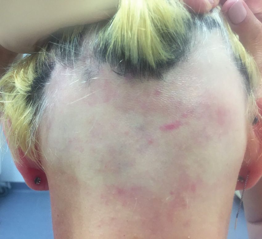
Adv Clin Exp Med. 2021;30(2):211–218                                                                                            213

2003.24 They are used to treat shin ulcers25 and hemato-              all of the patients had undergone the following therapies:
logical, orthopedic, urological, and gastroenterological              systemic and topical glucocorticoids, topical minoxidil,
disorders.26                                                          cryotherapy, and phototherapy (UVB 311 nm). One patient
  Previous attempts to use autologous MSCs for the treat-             was treated with contact immunotherapy (diphenylcyclo-
ment of alopecia have demonstrated the effectiveness and              propenone). None of these methods resulted in complete
safety of this form of treatment.9 So far, allogeneic trans-          hair regrowth.
plantation of WJ-MSCs has not been used in AA treat-                    The patients did not suffer from any skin diseases or sys-
ment. Due to the limited efficacy of the currently applied            temic diseases other than alopecia. For 6 weeks prior
methods for the treatment of AA, it is important to search            to the study, the patients did not take any medications.
for new forms of therapy.                                             In all patients with AA, additional tests were performed
                                                                      prior to the initiation of therapy, which revealed C-reac-
                                                                      tive protein (CRP) values ≤5 mg/L, negative antinuclear
Objectives                                                            antibodies (ANA) panel results, and no hepatitis C virus
                                                                      (HCV), hepatitis B virus (HBV) and human immunodefi-
  The aim of this study was to evaluate the efficacy and              ciency virus (HIV) infections. On the day of administra-
safety of AA treatment through a single intradermal in-               tion of the WJ-MSC suspension, no clinical signs of other
jection of a WJ-MSC suspension into the alopecia foci.                active infections were found.
The primary endpoint was the percentage change in the Se-
verity of Alopecia Tool (SALT) score during treatment.                Preparation of the Wharton’s jelly
                                                                      mesenchymal stem cell suspension
Materials and methods                                                    All umbilical cord (UC) collections were performed af-
                                                                      ter obtaining informed consent of the parents. The UCs
  This experimental study was conducted with patients                 samples were collected after natural delivery or caesarean
undergoing treatment at the Regional Specialist Hospital              sections. The fragment of the UC as long as 20–30 cm was
in Wrocław, Poland, and was performed in collaboration                placed in a sterile container into with 0.9% natrium chlora-
with the Polish Stem Cell Bank (Polski Bank Komórek                   tum solution (Fresenius Kabi, Bad Homburg vor der Höhe,
Macierzystych – PBKM). The study protocol was approved                Germany) and transported in protective boxes to the labo-
by the local bioethics committee of the Research and                  ratory. The transport conditions were monitored and UC
Development Centre of the Regional Specialist Hospital                tissue was processed within 72 h after delivery. Qualifica-
in Wroclaw (approval No. KB 01/2019) and all procedures               tion of UC tissue requires providing complete responses
performed in the study were carried out in accordance                 to a medical questionnaire and submitting by donor-
with the ethical standards of the Helsinki Declaration.               mother a peripheral blood sample for infectious agents
All patients were informed in detail about the purpose                testing for HBV, HCV, HIV, cytomegalovirus (CMV), and
and methods of the study, as well as the potential benefits           syphilis. All steps of manufacturing were performed in ac-
and risks of therapy. All participants provided written in-           cordance the principles of Good Manufacturing Practice
formed consent to participate in the study.                           (GMP) and Good Laboratory Practice (GLP). Umbilical
                                                                      cord fragment was removed from transportation container
Patients                                                              and placed into a new container with 0.9% natrium chlo-
                                                                      ridum solution (Fresenius Kabi) supplemented with 1%
   The study involved 4 AA patients who underwent experi-             Antibiotic Antimycotic (Thermo Fisher Scientific/Gibco,
mental therapy with a suspension of WJ-MSCs. The pa-                  Waltham, USA). After washing, the fragment was dis-
tient population included 3 men aged 36, 43 and 49 years              sected into 2 cm fragments, put on 90 mm Petri dish (Med-
and 1 woman aged 57 years. The duration of the disease                lab Products Sp. z o.o., Raszyn, Poland) and cut along with
ranged from 2 to 9 years (mean of 5 years). Three or more             surgical blade; then, the arteries and the vein were removed
AA foci were observed in each patient (Table 1). In the past,         with tweezers. After all blood vessels were removed, WJ

Table 1. Characteristics of the patients
                                                                                   Duration of the disease     Number of foci
        Patient/patients’ initials         Gender               Age [years]
                                                                                           [years]              of alopecia
 1/BR                                      female                   57                       9                       3
 2/PT                                       male                    43                       6                       4
 3/DH                                       male                    36                       2                       4
 4/PI                                       male                    49                       3                       9
214 A. Czarnecka et al. Stem cells in treatment of alopecia areata
tissue was minced into 1–2 mm3 scraps and placed in xeno- of the WJ-MSC suspension in the 12th and 24th weeks,
free, serum-free media into culture flasks for primary assuming that the condition before treatment commence-
explants cultures development. Flasks were incubated ment was 100%,28 according to the following formula:
in optimal conditions. After 14 days in culture, the tissues
(A − B/A) × 100% = I or D,
were removed from culture and the adherent cells were
trypsinized using TrypLE™ Select 1× (Life Technologies, where A indicates the percentage of baldness before treat-
Carlsbad, USA) and passed into new flasks for further ex- ment, B is the percentage of baldness after treatment and
pansion. The criteria for defining multipotent MSCs were I (improvement) indicates the amount of hair regrowth.
established in 2006 by ISCT.27 They are based on cells ad- If increased hair loss was observed after treatment,
herence to plastic, presence of specific antigens like CD90, the condition was indicated by D (deterioration). Regrowth
CD73 and CD105, together with the absence of hemato- observed 12 weeks following treatment was marked as I12
poietic and immune system markers (CD45, CD34, CD14 and regrowth after 24 weeks was marked as I24 (in both
or CD11b, CD79a, CD19) and HLA-DR surface antigens, cases taking the pretreatment condition as baseline). Addi-
as well as conferring stem cell identity to differentiate into tionally, in order to examine the dynamics of hair regrowth
osteoblasts, chondrocytes and adipocytes. The pharma- in relation to the time that had passed since treatment,
ceutical form of the product is a frozen preparation of cells the I24:12 regrowth was determined 24 weeks after the treat-
in a cryoprotective liquid. The administration and dosage ment, taking the 12-week condition as baseline.
form of the treatment is a thawed suspension of WJ-MSCs The efficacy of therapy was assessed for the whole sur-
with the phenotype CD73(+), CD90(+), CD105(+). face of the scalp and for the following individual areas:
vertex, right side, left side, and posterior (Table 2). Hair
Procedure regrowth or loss in individual areas was determined ac-
cording to the formula:
For each patient, a single WJ-MSC suspension was
(I or D)12vertex.
administered to several alopecia foci with a total area
of about 15 cm2 in the form of intradermal injections. For Follow-up visits
1 treatment, 2 mL of the suspension was used, which con-
tained 5 × 10⁶ WJ-MSCs of phenotype CD73(+), CD90(+), Follow-up visits took place on an outpatient basis 6,
CD105(+) suspended in a saline solution. Injections were 12, 18, and 24 weeks after the WJ-MSCs administration.
performed into every 0.5 cm of skin surface. For each in- During each follow-up visit, the patient’s general condi-
jection site, 0.01 mL of the suspension was administered. tion (physical examination), mental wellbeing and topical
A needle (32 G, 0.23 × 6 mm) was used to administer conditions were assessed, and a photo of the scalp was
the treatment. The scalp was anesthetized locally with taken. When assessing the condition of the topical local
EMLA cream (5% lidocaine) (Aspen Pharmacare, Durban, site, particular attention was paid to symptoms that could
South Africa) 1 h before the treatment. indicate adverse effects of intradermal cell administration,
On the day of WJ-MSCs administration, patients were such as redness or swelling. The SALT assessments were
hospitalized for 1 day. Before the procedure, the severity performed 12 weeks (SALT12) and 24 weeks SALT24) fol-
of alopecia was assessed using the SALT score (SALT0), lowing treatment. Hair regrowth or loss were calculated
the patient’s general condition (physical examination) and and expressed as (I or D)12, (I or D)24 and (I or D)24:12.
well-being were assessed, and photographs of scalp skin
were taken. Patients’ general and local condition and well-
being were reassessed 24 h after the procedure. Results
Evaluation of disease severity On the day of the administration of the WJ-MSC sus-
and effect of therapy pension, the SALT0 score for the 4 patients (Table 2) was
26.4% for patient 1 (BR), 23.4% for patient 2 (PT), 19.4%
The AA intensity was measured with the SALT score.28 for patient 3 (DH), and 40% for patient 4 (PI). The average
For this purpose, the scalp was divided into 4 areas where SALT0 score for all patients was 27.3% of hair loss.
hair loss was assessed (40% vertex, 18% right side, 18% In all patients, hair regrowth was observed at the sites
left side, 24% posterior); then, the percentage of hair of cell suspension administration 12 weeks after the pro-
loss over the entire scalp was calculated.28 This measure cedure, together with a decrease in the SALT12 value.
was performed 3 times during treatment: SALT0 prior The SALT12 values for patients 1, 2, 3, and 4 were 17.6%,
to treatment; SALT12 12 weeks after the treatment; and 7.2%, 12.2%, and 22.5%, respectively, with an average
SALT24 24 weeks after the treatment; then, the observa- SALT12 score of 14.9%.
tions concluded. Further improvement was observed in the 24th week.
To evaluate the effects of therapy, we calculated the dif- Hair loss after 24 weeks, measured using the SALT24 score,
ference in alopecia surface area before and after application was lower than that observed after 12 weeks (SALT12), andAdv Clin Exp Med. 2021;30(2):211–218 215
Table 2. The AA intensity measured using the SALT score in the 4 regions (vertex, right side, left side, posterior) and in the total scalp. Improvement
or deterioration in the 4 regions (vertex, right side, left side, posterior) and in the total scalp
Parameter Patient 1 (BR) Patient 2 (PT) Patient 3 (DH) Patient 4 (PI)
posterior, left side, posterior, vertex, posterior, vertex, posterior,
Injected sites of the scalp
right side right side right side left side, right side
week week week week week week week week week week week week
Follow-up visit
0 12 24 0 12 24 0 12 24 0 12 24
Vertex SALT 0% 8% 12% 0% 0% 0% 12% 8% 6% 28% 16% 14%
Vertex improvement I12, I24 – 0 0 – – – – 33% 50% – 42.8% 50%
Vertex deterioration D12, D24 – 8% 12% – – – – 0 0 – 0 0
Vertex improvement D24:12 – – – – – – – – 25% – – 12.5%
Vertex deterioration D24:12 – – 33% – – – – – 0 – – 0
Left side SALT 1.8% 0% 0% 0% 0% 0% 0% 0% 0% 1.8% 0.9% 0.9%
Left side improvement I12, I24 – 100% 100% – – – – – – – 50% 50%
Left side improvement I24:12 – – – – – – – – – – – 0
Right side SALT 1.8% 0% 0% 0.4% 0% 0% 3.6% 1.8% 1.8% 1.8% 0.9% 0.9%
Right side improvement I12, I24 – 100% 100% – 100% 100% – 50% 50% – 50% 50%
Right side improvement I24:12 – – 0 – – 0 – – 0 – – 0
Posterior SALT 22.8% 9.6% 4.8% 18% 7.2% 4.8% 4.8% 2.4% 1.2% 8.4% 3.6% 2.4%
Posterior improvement I12, I24 – 57% 79% – 60% 73% – 50% 75% – 57% 71%
Posterior improvement I24:12 – – 50% – – 33% – – 50% – – 33%
Total scalp SALT 24.6% 17.6% 16.8% 23.4% 7.2% 4.8% 19.4% 12.2% 9% 40% 22.5% 18.2%
Improvement in injection site I12, I24 – 60% 80% – 69% 79% – 37% 53% – 43% 54%
Improvement in injection site I24:12 – – 50% – – 33% – – 26% – – 19%
SALT – Patients’ Severity of Alopecia Tool; Improvement I12, I24/Deterioration D12, D24 – difference between the alopecia surface area before the treatment
and after the treatment in the 12th and 24th week, assuming that the condition before treatment was 100%; Improvement I24:12/Deterioration D24:12
– difference between the alopecia surface area after the 12th week and after the 24th week, assuming that the condition in the 12th week was 100%.
was 16.8% for patient 1, 4.8% for patient 2, 9% for patient of observation (D24vertex). The rate of hair loss increased
3, and 18.2% for patient 4. The average SALT24 score was and the D24:12vertex score was 33% (Table 2). No hair loss
12.2% (Table 2). was found in the remaining area of the scalp, while hair
Regrowth (I24) of the scalp where the procedure was regrowth was observed in the injected sites (Table 2).
carried out was between 53% and 80%, with values of 80%, In patients 2, 3 and 4, no new foci of alopecia were found
79%, 53%, and 54% for patients 1, 2, 3, and 4, respectively during the 24-week observation period. Improvement was
(Table 2). None of the observed cases achieved 100% hair observed in all treated areas: vertex, posterior, left side,
regrowth. The mean hair regrowth in the injected sites and right side. Detailed results are presented in Table 2.
(I24) was 67%. During the 24-week observation period, patients did not
All the patients were found to present with more hair present any abnormalities in the physical examination. No
regrowth after the first 12 weeks (I12) than in the following local side effects (rash or swelling) at the site of intradermal
12 weeks (I24:12) at the site where the intradermal WJ-MSC injection of the WJ-MSC suspension were found in any
suspension was administered (Table 2). For patient 1, the I12 patients. All patients reported good general health state
was 60% for the first 12 weeks and the I24:12 was 50% for and did not report any subjective symptoms.
the following 12 weeks; patient 2 had an I12 of 69% and I24:12 Figures 1,2,3 present the condition before the treatment
of 33%; patient 3 had values of 37% and 26%, respectively; and results of the therapy in patient 1.
and patient 4 had an I12 of 43% and I24:12 of 19% (Table 2).
The mean I12 and I24:12 were 52.2% and 32%, respectively.
A noteworthy case was patient 1 (BR), who received pos- Discussion
terior, left- and right-side injections, and by the 12th week
was found to have hair loss in the vertex area which was The introduction of stem cell-based therapies to repair
not treated with injections of the WJ-MSC suspension. and regenerate various tissues and organs offers innovative
This deterioration was expressed by the symbol (Dvertex), therapeutic solutions. Mesenchymal stem cells play an im-
and in the 12th week of therapy, the deterioration (D12vertex) portant role in the production of active agents for tissue
was 8% and increased to 12% after the following 12 weeks regeneration, affecting the proliferation and migration216 A. Czarnecka et al. Stem cells in treatment of alopecia areata
of endothelial cells, fibroblasts and skin cells.29,30 Studies
also show that, in addition to proangiogenic, chemoattrac-
tive and anti-inflammatory potential, MSCs may modu-
late the activity of the immune system.31 Attempts have
recently been made to use MSCs to reactivate hair follicle
stem cells and thus prevent hair loss.32
There are only a few reports in the medical literature
on the use of MSCs in the treatment of alopecia. The most
commonly used MSCs in AA therapy are those derived
from bone marrow or adipose tissue.33–35 In a therapeutic
experiment, autologous MSCs derived from bone marrow
were used on a group of 40 people with hair loss, including
20 people with AA and 20 people with AGA. Six months
after a single injection of stem cells into the scalp, the au-
thors observed a significant improvement, confirmed with
digital dermoscopy. There was no significant difference
in the effectiveness of treatment between the 2 types of alo-
pecia. No serious adverse events were reported.36
Fig. 1. Patient No. 1, posterior region of the scalp, before treatment Another interesting study reported the use of human
autologous adipose-derived adult cells of stromal vascular
fraction (ADSVC) for the treatment of 20 AA patients. Pa-
tients were given a single injection of autologous ADSVC
cells extracted by lipoaspiration from adipose tissue into
the alopecia foci at concentrations of 4–4.7 × 10 6 cells.
The growth and thickness of the hair improved signifi-
cantly within the first 6 months after treatment. A de-
crease in the intensity of the hair pull test was also ob-
served. No side effects of ADSVC treatment were observed,
and patients assessed the therapy as satisfactory.34
Mesenchymal stem cells can also be extracted from WJ
in the umbilical cord. It is an ideal source of stem cells
due to its availability, noninvasive and painless extraction,
weak immunogenic potential, no risk of adverse effects for
the donor or recipient, and no ethical restrictions.22,32,35
Medical literature suggests the potential effectiveness
of WJ-MSCs in hair follicle regeneration and hair re-
growth.33,34 An additional advantage of using material ex-
Fig. 2. Patient No. 1, posterior region of the scalp, 12 weeks after treatment tracted from WJ is the possibility of obtaining its decellu-
larized fraction (DWJM), which is considered an excellent
natural biocompatible 3D scaffold. The DWJM, as a 3D
scaffold, can be used as a regenerative drug to promote
stem cell adhesion, penetration, growth, and multiplication
both in vitro and in vivo.37
In our study, a single suspension of allogeneic WJ-MSCs
was injected into the selected AA foci at a concentra-
tion of 5 × 10⁶ cells, followed by a six-month observation
of the treatment effects. All patients who participated
in this experimental study had previously been treated
with standard procedures, but without significant long-
term clinical improvements. All patients showed a reduc-
tion in the area of baldness by an average of 67%. None
of the patients experienced complete hair regrowth. No-
tably, we observed improved hair regrowth dynamics
in all cases in the first 3 months after the procedure, with
an increase of 52.2% on average compared to the following
Fig. 3. Patient No. 1, posterior region of the scalp, 24 weeks after treatment 3 months, when the increase was 32% on average.Adv Clin Exp Med. 2021;30(2):211–218 217
The case of patient 1 (BR) is particularly interesting, 4. Burgdorf WHC, Plewing G, Wolff HH, Landthaler M. Dermatologia.
Vol. 1. Lublin, Poland: Czelej; 2017.
because in the 12th week of observation, the subject was
5. Strazzulla LC, Wang EHC, Avila L, et al. Alopecia areata: An apprais-
diagnosed with hair loss in the frontal area where no intra- al of new treatment approaches and overview of current therapies.
dermal injection of WJ-MSC suspension was applied. No J Am Acad Dermatol. 2018;78(1):15–24. doi:10.1016/j.jaad.2017.04.1142
hair loss was detected in the remaining area of the scalp, 6. Simakou T, Butcher JP, Reid S, Henriquez FL. Alopecia areata: A multi
factorial autoimmune condition. J Autoimmun. 2019;98:74–85.
while in the injected sites (i.e., the parietal area, right and 7. Gilhar A, Etzioni A, Paus R. Alopecia areata. N Engl J Med. 2012;366(16):
left side of the scalp), hair regrowth did occur. This case in- 1515–1525. doi:10.1056/NEJMra1103442
dicates that the applied therapy is effective only at the site 8. Guo H, Cheng Y, Shapiro J, McElwee K. The role of lymphocytes in the
development and treatment of alopecia areata. Expert Rev Clin Immunol.
of cell administration itself. 2015;11(12):1335–1351. doi:10.1586/1744666X.2015.1085306
There are very few reports of allogeneic therapies with 9. Li Y, Yan B, Wang H, et al. Hair regrowth in alopecia areata patients fol-
WJ-MSCs in the medical literature to date. The authors lowing Stem Cell Educator therapy. BMC Med. 2015;13:87. doi:10.1186/
s12916-015-0331-6
of these papers stress the safety of this type of therapy, 10. Sudnik W. Rola selektyn E, L, P w patomechanizmie łysienia placko-
including the lack of tumorigenic potential.22,32,38 watego [PhD thesis]. Poznań, Poland: Poznan University of Medical
Our experiment also evaluated the safety of the proce- Sciences; 2012.
dures used. No side effects were observed during the pro- 11. Hordinsky MK. Treatment of alopecia areata: What is new on the hori-
zon? Dermatol Ther. 2011;24(3):364–368. doi:10.1111/j.1529-8019.2011.
cedure or after application of the WJ-MSC suspension 01421.x
in the allogeneic system. 12. Biran R, Zlotogorski A, Ramot Y. The genetics of alopecia areata: New
approaches, new findings, new treatments. J Dermatol Sci. 2015;78(1):
11–20. doi:10.1016/j.jdermsci.2015.01.004
13. Hordinsky MK. Overview of alopecia areata. J Investig Dermatol Symp
Limitations Proc. 2013;16(1):S13–S15. doi:10.1038/jidsymp.2013.4
14. Łuczak M, Łuczak T, Ciesińska C, Czajkowski R. Leczenie ogólne łysie-
nia plackowatego. Przegl Dermatol. 2013;100:53–58.
The limitation of the study was the small number of pa- 15. Nowicka D, Maj J, Jankowska-Konsur A, Hryncewicz-Gwóźdź A. Effi-
tients enrolled. cacy of diphenylcyclopropenone in alopecia areata: A comparison
of two treatment regimens. Postepy Dermatol Allergol. 2018;35(6):
577–581. doi:10.5114/ada.2018.77608
Conclusions 16. Dainichi T, Kabashima K. Alopecia areata: What’s new in epidemiolo-
gy, pathogenesis, diagnosis, and therapeutic options? J Dermatol Sci.
2017;86(1):3–12. doi:10.1016/j.jdermsci.2016.10.004
The presented report supports the effectiveness and 17. Szabłowska-Gadomska I, Bużańska L, Małecki M. Właściwości komó-
rek macierzystych, regulacje prawne oraz zastosowanie w medycynie.
safety of the applied therapy – intradermal injections Postepy Hig Med Dosw (Online). 2017;71:1216–1230.
of an allogeneic WJ-MSC suspension. To our knowledge, 18. Marino L, Castaldi MA, Rosamilio R, et al. Mesenchymal stem cells
this is the first clinical study to describe the application from the Wharton’s jelly of the human umbilical cord: Biological prop-
erties and therapeutic potential. Int J Stem Cells. 2019;12(2):218–226.
of an allogeneic MSC transplant in patients with AA. doi:10.15283/ijsc18034
The results of treatment were positive, with hair growth ob- 19. Bajek A, Olkowska J, Drewa T. Mezenchymalne komórki macierzy-
served in all participants, and the therapy was found to be ste narzędziem terapeutycznym w regeneracji tkanek i narządów.
Postepy Hig Med Dosw. 2011;65:124–132.
safe, with no side effects. The question remains as to how 20. Wang HS, Hung SC, Peng ST, et al. Mesenchymal stem cells in the
many cells should be given to the patient to achieve full Wharton’s jelly of the human umbilical cord. Stem Cells. 2004;22(7):
hair regrowth and how often the treatments should be 1330–1337. doi:10.1634/stemcells.2004-0013
21. Davies JE, Walker JT, Keating A. Concise review: Wharton’s jelly:
repeated to achieve 100% therapy efficacy with no relapse.
The rich, but enigmatic, source of mesenchymal stromal cells.
We emphasize the need to conduct further studies with Stem Cells Transl Med. 2017;6(7):1620–1630. doi:10.1002/sctm.16-0492
a randomized control group. 22. Dominici M, Le Blanc K, Mueller I, et al. Minimal criteria for defin-
ing multipotent mesenchymal stromal cells: The International Soci-
ety for Cellular Therapy position statement. Cytotherapy. 2006;8(4):
ORCID iDs 315–317. doi:10.1080/14653240600855905
Anna Czarnecka https://orcid.org/0000-0002-6621-9537 23. Pojda Z, Machaj E, Kurzyk E, et al. Mezenchymalne komórki macie-
Agnieszka Odziomek https://orcid.org/0000-0002-3574-171X rzyste. Adv Biochem. 2013;59(2):187–197.
Magdalena Murzyn https://orcid.org/0000-0001-8115-4287 24. Martín PG, Martinez AR, Lara VG, Naveros BC. Regulatory consider-
Joanna Dubis https://orcid.org/0000-0002-6814-580X ations in production of a cell therapy medicinal product in Europe to
Marta Bagłaj-Oleszczuk https://orcid.org/0000-0002-4554-7603 clinical research. Clin Exp Med. 2014;14(1):25–33. doi:10.1007/s10238-
Anita Hryncewicz-Gwóźdź https://orcid.org/0000-0002-1601-471X 012-0213-6
25. Masłowski L, Paprocka M, Czyżewska-Buczyńska A, et al. Autotrans-
plantation of the adipose tissue-derived mesenchymal stromal cells
References in therapy of venous stasis ulcers. Arch Immunol Ther Exp (Warsz).
1. Pratt CH, King LE Jr, Messenger AG, Christiano AM, Sundberg JP. 2020;68(1):5. doi:10.1007/s00005-020-00571-9
Alopecia areata. Nat Rev Dis Primers. 2017;3:17011. doi:10.1038/nrdp. 26. Szydlak R. Produkty lecznicze zaawansowanej terapii medycznej
2017.11 oparte na mezenchymalnych komórkach macierzystych. Farm Pol.
2. Safavi K. Prevalence of alopecia areata in the First National Health 2018;74(3):178–183.
and Nutrition Examination Survey. Arch Dermatol. 1992;128(5):702. 27. Dominici M, Le Blanc K, Mueller I, et al. Minimal criteria for defining
doi:10.1001/archderm.1992.01680150136027 multipotent mesenchymal stromal cells: The International Society for
3. Abedini R, Hallaji Z, Lajevardi V, et al. Quality of life in mild and severe Cellular Therapy position statement. Cytotherapy. 2006;8(4):315–317.
alopecia areata patients. Int J Womens Dermatol. 2017;4(2):91–94. doi:10.1080/14653240600855905
doi:10.1016/j.ijwd.2017.07.001218 A. Czarnecka et al. Stem cells in treatment of alopecia areata
28. Olsen EA, Hordinsky MK, Price VH, et al; National Alopecia Areata 34. Anderi R, Makdissy N, Azar A, Rizk F, Hamade A. Cellular therapy
Foundation. Alopecia areata investigational assessment guidelines. with human autologous adipose-derived adult cells of stromal vas-
Part II. National Alopecia Areata Foundation. J Am Acad Dermatol. cular fraction for alopecia areata. Stem Cell Res Ther. 2018;9(1):141.
2004;51(3):440–447. doi:10.1186/s13287-018-0889-y
29. Maxson S, Lopez EA, Yoo D, Danilkovitch-Miagkova A, Leroux MA. 35. Sabapathy B, Sundaram SVM, Mankuzhy P, Kumar S. Human Whar-
Concise review: Role of mesenchymal stem cells in wound repair. ton’s jelly mesenchymal stem cells plasticity augments scar-free
Stem Cells Transl Med. 2012;1(2):142–149. doi:10.1016/j.jaad.2003.09.032 skin wound healing with hair growth. PLoS One. 2014;9 (4):e93726.
30. López JF, Sarkanen JR, Huttala O, Kaartinen IS, Kuokkanen HO, Ylikomi T. doi:10.1371/journal.pone.0093726
Adipose tissue extract shows potential for wound healing: In vitro 36. Ibrahim ZA, Elmaadawi IH, Mohamed BM, et al. Stem cell therapy
proliferation and migration of cell types contributing to wound heal- as a novel therapeutic intervention for resistant cases of alopecia area-
ing in the presence of adipose tissue preparation and platelet rich ta and androgenetic alopecia. J Dermatol Treat. 2018;29(5):431–440.
plasma. Cytotechnology. 2018;70(4):1193–1204. doi:10.1007/s10616- doi:10.1080/09546634.2016.1227419
018-0211-y 37. Jadalannagari S, Converse G, McFall C, et al. Decellularized Wharton’s
31. Otero-Viñas M, Falanga V. Mesenchymal stem cells in chronic wounds: jelly from human umbilical cord as a novel 3D scaffolding material
The spectrum from basic to advanced therapy. Adv Wound Care for tissue engineering applications. PLoS One. 2017;12(2):e0172098.
(New Rochelle). 2016;5(4):149–163. doi:10.1089/wound.2015.0627 doi:10.1371/journal.pone.0172098
32. Owczarczyk-Saczonek A, Krajewska-Włodarczyk M, Kruszewska, A, et al. 38. Gentile P, Garcovich S. Advances in regenerative stem cell therapy
Therapeutic potential of stem cells in follicle regeneration. Stem Cells Int. in androgenic alopecia and hair loss: Wnt pathway, growth-factor,
2018;2018:1049641. doi:10.1155/2018/1049641 and mesenchymal stem cell signaling impact analysis on cell growth
33. Egger A, Tomic-Canic M, Tosti A. Advances in stem cell-based thera- and hair follicle development. Cells. 2019;8(5):466. doi:10.3390/cells
py for hair loss. CellR4 Repair Replace Regen Reprogram. 2020;8:e2894. 8050466You can also read