Prospective Trial of Intense Pulsed Light for the Treatment of Meibomian Gland Dysfunction - Drycom
←
→
Page content transcription
If your browser does not render page correctly, please read the page content below
Clinical Trials

             Prospective Trial of Intense Pulsed Light for the Treatment
             of Meibomian Gland Dysfunction
             Jennifer P. Craig, Yen-Heng Chen, and Philip R. K. Turnbull
             Ocular Surface Laboratory, Department of Ophthalmology, New Zealand National Eye Centre, University of Auckland, Auckland,
             New Zealand

             Correspondence: Jennifer P. Craig,          PURPOSE. To evaluate the effect of intense pulsed light (IPL) applied to the periocular area for
             Department of Ophthalmology, Uni-           meibomian gland dysfunction (MGD) in a prospective, double-masked, placebo-controlled,
             versity of Auckland, Private Bag            paired-eye study.
             92019, Auckland 1142, New Zea-
             land;                                       METHODS. Twenty-eight participants underwent IPL treatment, with homogeneously
             jp.craig@auckland.ac.nz.                    sequenced light pulses delivered to one eye and placebo treatment to the partner control
             Submitted: September 29, 2014               eye at 1, 15, and 45 days following baseline (BL) evaluation. Lipid layer grade (LLG),
             Accepted: February 4, 2015                  noninvasive tear break-up time (NIBUT), tear evaporation rate (TER), tear meniscus height
                                                         (TMH), and subjective symptom score using visual analogue scales (VAS) were compared with
             Citation: Craig JP, Chen Y-H, Turnbull
                                                         BL and control values at each visit.
             PRK. Prospective trial of intense
             pulsed light for the treatment of           RESULTS. Lipid layer grade improved significantly from BL to Day (D) 45 in the treated eye (P <
             meibomian gland dysfunction. Invest         0.001), but not the control eye (P ¼ 0.714), with 82% of treated eyes improving by at least
             Ophthalmol Vis Sci. 2015;56:1965–           one LLG. Noninvasive tear break-up time also improved significantly from BL to D45 in the
             1970. DOI:10.1167/iovs.14-15764             treated (P < 0.001) but not in the control eye (P ¼ 0.056) and was significantly longer than in
                                                         the treated eye at D45 (14.1 6 9.8 seconds versus 8.6 6 8.2 seconds, P < 0.001). The tear
                                                         evaporation rate was not different in the treated eye compared with the control eye at any
                                                         visit. Tear meniscus height did not change from BL in either eye (P > 0.05). Visual analog scale
                                                         symptom scores improved from BL in the treated (P ¼ 0.015), but not the control eye (P ¼
                                                         0.245), with 86% of participants noting reduced symptoms in the treated eye by D45.
                                                         CONCLUSIONS. Intense pulsed light with multiple sculpted pulses shows therapeutic potential
                                                         for MGD, improving tear film quality and reducing symptoms of dry eye. (https://www.anzctr.
                                                         org.au number, ACTRN12614000162617.)
                                                         Keywords: intense pulsed light, meibomian gland dysfunction, dry eyes

                   eibomian gland dysfunction (MGD) is a common cause of                     aqueous layer, in turn leading to symptoms of dry eye and
             M     evaporative dry eye, affecting almost 70% of the popula-
             tion in some parts of the world.1 It manifests with symptoms of
                                                                                             ocular surface inflammation.12
                                                                                                 Current management paradigms range from self-adminis-
             ocular surface burning and irritation, fluctuating visual acuity,               tered or practitioner-administered treatments with artificial
             and red, often watery, eyes.2 These symptoms, combined with                     tears, heat application, and manual gland expression13,14 (often
             frequently ineffective treatment options, can severely affect                   providing only transient relief) to therapies that aim to restore
             quality of life.3,4 In MGD, the glands can become narrowed, the                 the natural balance of lipids within the meibum. Such therapies
             acini atrophy and hyperkeratinise,5 and the meibum increases                    include omega-3 supplementation,15 topical antibiotics to
             in viscosity.6 This reduces meibum outflow, encouraging                         lessen the local bacterial load, oral tetracyclines to reduce the
             proliferation of commensal bacteria.7 These bacteria secrete                    level of pro-inflammatory cytokines,16 corticosteroids, or
             lipases that can change the composition of lipids in the                        topical cyclosporine.3 Despite the broad range of therapies
                                                                                             available, MGD management is commonly considered unsatis-
             meibum, increasing the level of esterified cholesterol (and its
                                                                                             factory by both clinicians and affected patients, and alternative
             melting point), which further reduces MG output.6,8
                                                                                             options for management are continually sought.
                 Biomicroscopic signs of MGD can be unremarkable in the                          Intense pulsed light (IPL) therapy is widely used in the
             case of nonobvious obstructive MGD9 but can include plugged                     cosmetic industry as well as therapeutically for the removal of
             or capped meibomian gland (MG) orifices, along with lid                         hypertrichosis, benign cavernous hemangiomas, benign venous
             margin thickening, irregularity, telangiectasia, and hyperemia.10               malformations, telangiectasia, port-wine stains, and pigmented
             Comprehensive examination can further reveal MG dropout                         lesions.17 Systematic review demonstrates that IPL is an effective,
             and solidified toothpaste-like secretions on gland expression in                well-tolerated treatment option for a range of dermatologic
             more severe cases.8,11 Tear break-up time is most often                         conditions, resulting in a reduction in telangiectasia and severity
             reduced, and the tear film is frequently contaminated by                        of facial erythema.18 Concurrent ocular surface health improve-
             endogenous debris and foam.11 Ultimately, the meibomian                         ments have been observed serendipitously in patients undergo-
             glands fail to secrete a suitable or sufficient oil layer for the tear          ing IPL for the dermatologic manifestations of rosacea, leading to
             film, which allows a higher evaporation rate of the underlying                  interest in evaluating IPL as a potential therapy for MGD.

             Copyright 2015 The Association for Research in Vision and Ophthalmology, Inc.
             www.iovs.org j ISSN: 1552-5783                                                                                                               1965

Downloaded From: http://iovs.arvojournals.org/pdfaccess.ashx?url=/data/Journals/IOVS/933681/ on 10/13/2016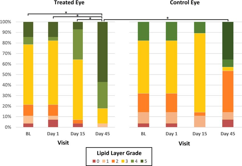
Prospective Trial of IPL for Treatment of MGD IOVS j March 2015 j Vol. 56 j No. 3 j 1966
TABLE. Intense Pulsed Light Treatment Intensity (J/cm2) With E>Eye
IPL Device Derived From Fitzpatrick Skin Type Grading
E>Eye
Fitzpatrick Treatment Fluence,
Skin Type Skin Appearance Level J/cm2
I Pale white 6 13.0
II White 5 12.2
III Light brown 4 11.4
IV Medium brown 3 10.6
V Dark brown 2 9.8
VI Very dark brown/black Unsuitable for E>Eye IPL
Experimental Design
The prospective, double-masked, paired-eye, placebo-con-
trolled study was conducted over a period of 45 days, with
IPL treatment administered to the skin area immediately below
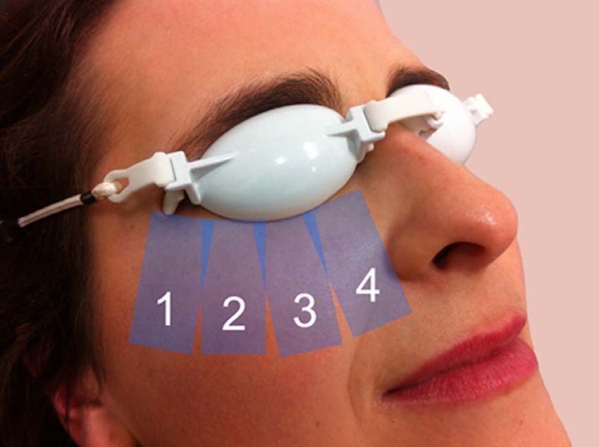
FIGURE 1. IPL treatment was applied to four periocular zones inferior the lower eyelid during three separate treatment sessions on
to the eye, while the eyes were protected by opaque goggles. Both eyes
received treatment, with a light-blocking filter applied to the tip of the
Day (D) 1, D15, and D45 as per manufacturer recommenda-
E>Eye in the control eye. tions. Four pulses were applied as shown in Figure 1 at a pulse
intensity that ranged from 9 to 13 J/cm2 and was inversely
related to the individual skin phototype level as determined by
the Fitzpatrick grading scale (Table).21
Intense pulsed light devices contain high-intensity light One eye was selected for treatment according to a
sources, which emit polychromatic light extending from the computer-generated randomization program, with the other
visible (515 nm) to the infrared spectrum (1200 nm). The light eye assigned to serve as a mock-treated control. The researcher
is directed to the skin tissue and is then absorbed by the collecting the clinical data was masked as to which eye was
targeted structure, resulting in the production of heat (>808C), treated, and participant masking was employed with a white-
which destroys the pigmented skin lesions. Appropriate blocking filter applied over the tip of the IPL probe during
wavelengths can be selected for different targets depending application to the nontreated eye only.
on the absorption behavior and the penetration depth of the The study was conducted in accordance with the tenets of
light emitted, and specific filters can be chosen to limit the the Declaration of Helsinki, and the protocol was approved by
the local ethics review committee (UAHPEC 9531). All partici-
delivery of wavelengths to the treatment area resulting in
pants provided written informed consent before participating.
selective thermal delivery.17,19
A third-generation IPL device designed specifically for
periocular application with multiple homogenously sculpted Clinical Assessments
light pulses (E>Eye; E-SWIN, Paris, France) has recently become Prior to, and at least 5 minutes following, IPL, a battery of tests
commercially available and is currently the only medically was conducted on each eye in the same order, from least to
certified IPL device for treating MGD. Delivering multiple most invasive. Parameters assessed included best spectacle
homogenously sculpted light pulses with a spectral range of corrected visual acuity (logMAR), bulbar conjunctival injection
580 to 1200 nm, according to a proprietary algorithm, the E>Eye graded on a visual analog scale (VAS), noninvasive tear break-
has recently become commercially available. This study sought to up time (NIBUT), and fluorescein and lissamine green corneal
investigate the potential for IPL applied to the inferior periocular and conjunctival staining. Further tear film assessment
skin, to alter tear film characteristics and symptoms in subjects included assignment of the lipid layer grade (LLG) through
suffering from MGD. We report the results of what we believe is tear film interferometry20 (Tearscope Plus; Keeler, Berkshire,
the first prospective, randomized, double-masked, placebo- UK), tear meniscus height (TMH), tear osmolality (Tearlab
controlled clinical trial evaluating IPL as a therapy for MGD. Osmolarity System; Tearlab, San Diego, CA, USA), and tear
evaporation rate ([TER] VapoMeter; Delfin, Kuopio, Finland).
Symptoms were also compared prior to each treatment using
METHODS the Standard Patient Evaluation of Eye Dryness (SPEED)22
validated questionnaire, and each subject summarized their
Patient Selection perceived severity of dry eye symptoms before and after
treatment for each eye, on a VAS anchored at each end with
A total of 28 participants (20 female) with mild to moderate ‘‘No symptoms’’ and ‘‘Constant symptoms’’ as descriptors.
clinical signs of MGD,20 and a mean age of 45 6 15 years
(range, 22–73 years) were enrolled in the prospective study.
Prior to enrollment, general health, as well as current and Statistical Testing
recent medication use, was screened to exclude individuals for Post-IPL scores at D1, D15, and D45 were compared with pre-
whom light therapy was contraindicated. Participants who had IPL scores at baseline (BL). Repeated measures analysis enabled
received clinical skin treatments within the prior 2 months, or comparison of data across the various time points, and paired
implants beneath the treatment area, were also excluded from analyses allowed comparison of pre- and post-IPL data at
the study, as were those with tattoos, semipermanent makeup, individual time points. Variables were tested for normality with
or pigmented lesions in the treatment area. Contact lens the Kolmogorov-Smirnoff test. Ordinal variables (e.g., LLG) or
wearing within 48 hours of commencing the study, or during those with nonnormal distributions were analyzed with
the study, also resulted in exclusion. Friedman 2-way analysis of variance (ANOVA), with pairwise
Downloaded From: http://iovs.arvojournals.org/pdfaccess.ashx?url=/data/Journals/IOVS/933681/ on 10/13/2016
Prospective Trial of IPL for Treatment of MGD                                                         IOVS j March 2015 j Vol. 56 j No. 3 j 1967

             FIGURE 2. Lipid layer grade (0 ¼ worst) frequencies in the treated (left) and control eye (right) at BL, D1, D15, and D45. At D45, LLG in the treated
             eye was higher than at BL (P < 0.001), D1 (P < 0.001), and D15 (P ¼ 0.002). Day 45 LLG was better in the treated eye compared with the control
             eye (P ¼ 0.002). The control eye was not different from BL at any visit (P ¼ 0.802). *Significant difference in distribution of LLG at P < 0.005.

             Wilcoxon (paired) or Mann-Whitney (nonpaired) post hoc                      control eye showed no improvement from BL over the three
             testing as required, and are reported as medians with                       visits (Friedman, P ¼ 0.802).
             interquartile ranges (IQRs). Normally distributed continuous                    While there was no difference between the treated and
             data are reported as mean 6 SD and were assessed with a                     control eye lipid grade on D1 (Wilcoxon, P ¼ 0.932) or D15 (P
             repeated measures 2-way ANOVA with Tukey honest significant                 ¼ 0.101); by D45, lipid grade in the treated eye was better than
             difference (HSD) post hoc testing. Differences between treated              that of the control eye (P ¼ 0.002).
             and nontreated eye data were compared with the paired                           Noninvasive tear break-up time increased from BL to D45 in
             samples t-test and the Wilcoxon signed rank test for parametric             the treated eye (ANOVA, 5.28 6 1.42 seconds to 14.11 6 9.75
             and nonparametric data, respectively. Correlations between                  seconds; P < 0.001; Fig. 3), but not the control eye (5.29 6
             parametric and nonparametric data were assessed with                        1.42 seconds to 7.31 6 1.50 seconds; P ¼ 0.56). Noninvasive
             Pearson product-moment correlation or Spearman rank order                   tear break-up time was not different between the treated and
             correlation, respectively. Outcomes were considered signifi-                control eye at D1 (P ¼ 0.991) or D15 (P ¼ 0.055) but was higher
             cant if P < 0.05.                                                           in the treated eye than the control eye at D45 (P < 0.001).
                                                                                             Tear evaporation rate showed high variability between
                                                                                         visits, and there were differences in TER between visits in both
             RESULTS                                                                     the treated (Friedman, P ¼ 0.003) and control eye (P ¼ 0.012);
             The full cohort of 28 enrolled participants completed measure-              however, there was no overall trend, and the TERs were highly
             ments across all three appointments and were included in the                correlated between the eyes at each visit (P < 0.001). Further,
             analysis, with all reporting at least some symptoms of MGD and              there was no difference between the control and treated eye at
             89% reporting significant symptoms (Ocular Surface Disease                  any visit.
             Index [OSDI] > 12) (see Supplementary Table S1) prior to                        Tear osmolarity did not change over the four visits in either
             treatment. At BL, there was no significant difference between the           the control (ANOVA, P ¼ 0.741) or the treated eye (P ¼ 0.308).
             treated and control eyes in any outcome variable (P > 0.05 in all           There was also no difference in bulbar conjunctival hyperemia
             cases).                                                                     in either the control (Friedman, P ¼ 0.414) or the treated eye
                                                                                         (P ¼ 0.348) nor was there a difference in TMH from BL in either
             Clinical Assessment                                                         the control (ANOVA, P ¼ 0.559) or treated eye (P ¼ 0.348).

             Lipid layer grade in the treated eye improved from BL                        Questionnaires
             (Friedman, P < 0.001; Fig. 2), with median improvements
             between BL and D45 (Wilcoxon, P < 0.001), D1 and D45 (P <                    Subjective self-reported rating of dry eye symptoms on a
             0.001), and between D15 and D45 (P ¼ 0.002). The LLG of the                  single VAS (0–100 mm) showed an improvement over time in

Downloaded From: http://iovs.arvojournals.org/pdfaccess.ashx?url=/data/Journals/IOVS/933681/ on 10/13/2016
Prospective Trial of IPL for Treatment of MGD IOVS j March 2015 j Vol. 56 j No. 3 j 1968
5.28 to 14.11 seconds represents a meaningful clinical
improvement, as previous research has shown that NIBUT
values less than 10 seconds are associated with significantly
higher TERs.27
The self-reported dry eye symptoms on a VAS improved
from BL in the treated eye at D45, but not in the control eye,
which suggests at least some of this improvement seen in LLG
and NIBUT translates into subjective improvements. However,
while SPEED scores were lower at D45 compared with BL in
both eyes, this questionnaire did not reveal a difference in
symptoms between the eyes. This difference may be a result of
the VAS being interpreted more openly, while the SPEED
questionnaire specifies exact symptoms over different time
periods, which may miss or dilute any changes caused by IPL.
FIGURE 3. Noninvasive tear break-up time of the treated and control Nevertheless, the fact that SPEED symptoms in both eyes
eye at each of the four visits. Noninvasive tear break-up time was improved from BL, with only one eye treated, bodes well for
higher at D45 relative to BL in the treated eye (P < 0.001), but not in bilateral treatment.
the control eye (P ¼ 0.56). Additionally, NIBUT was significantly higher The absence of a significant difference in TER following
in the treated eye compared with the control eye at D45 (P < 0.001). treatment is perhaps not surprising given the study sample.
*Significant difference between groups at P < 0.005. Error bars denote An increased TER occurs in the presence of a nonvisible or
SEM. incomplete lipid layer, but where even a thin, continuous
lipid layer exists, tear evaporation is inhibited.27 Thus, unless
the treated eye (Friedman, P ¼ 0.015), but not in the control blinking is actively prohibited during evaporation rate
eye (P ¼ 0.245). Post hoc testing showed a decrease in the measurement, a finding of increased tear evaporation might
VAS score in the treated eye at D45 (median, 30.5; IQR, 7.0– not be anticipated owing to the presence of a continuous
60.1) compared with BL (median, 35; IQR, 12.3–64.8; P ¼ lipid layer, irrespective of thickness. While blinking was
0.015). discouraged during evaporation measurement in the current
Median SPEED scores decreased (indicating reduced symp- study, it was not prohibited for the sake of patient comfort
toms) over the time period between D1 and D45 in both the and potential effect on reflex tearing. The infrequency of
treated eye (Friedman, 14.0–8.5; P < 0.001) and control eye noncontinuous lipid layers at BL (only 4% exhibited an LLG
(Friedman, 11.0–8.5; P < 0.001). The scores between the eyes of zero) likely contributes to the lack of a significant
were highly correlated at each visit (Spearman’s r, D1 ¼ 0.922, difference in tear evaporation observed post treatment in
P < 0.001; D15 ¼ 0.874, P < 0.001; D45 ¼ 0.871, P < 0.001), the presence of a thickened lipid layer. This finding is further
and there were no significant differences between the treated reflected in the tear osmolarity results, which were also
and control eye SPEED scores at any visit. observed not to change significantly with treatment. More-
over, while it is possible to demonstrate between-eye
differences in LLG and NIBUT parameters, the bilateral
DISCUSSION nature of the lacrimal gland response would likely diminish
any effect in osmolarity observed as a result of changes in a
The management of MGD in clinical practice remains single eye.28 Nevertheless, the increases observed in LLG and
challenging, as patient compliance with physician-recommend- NIBUT support the concept of having created a more robust
ed self-administered therapies is notoriously poor.3 Our results tear film lipid layer, and the corresponding reduction in
suggest therapeutic potential for sculpted pulse IPL therapy SPEED symptoms suggests that the clinical signs possibly
with the E>Eye for the management of MGD, on the basis of translate into a tear film better able to withstand adverse
significant improvements in LLG, tear film stability, and conditions.
reduced symptoms. The serendipitous discovery of ocular
benefits following facial rosacea treatment has led to clinical Mechanism
centers offering IPL as a treatment for MGD on the basis of
reports of reduced fluorescein staining and severity of MGD, as The mechanism by which MGD signs and symptoms improved
well as improvements in visual function and comfort, with following treatment remains incompletely understood. Pro-
some suggesting an apparent cumulative effect.23–25 However, posed mechanisms include heat transfer, which softens the
evidence of the success of this treatment modality to date has meibum and aids expression.24 However such a mechanism
been largely anecdotal, arising from retrospective, open-label would be anticipated to induce only short-term effects.13 A
evaluations, and no randomized controlled, investigator- preliminary safety evaluation by the authors using infrared (IR)
masked studies are yet available (Vegunta S, et al. IOVS thermography noted only minimal skin surface temperature
2014;55:ARVO E-Abstract 2018). changes (Prospective Trial of IPL for Treatment of MGD IOVS j March 2015 j Vol. 56 j No. 3 j 1969
load on the lid margin and ocular adnexa could be directly Acknowledgments
affected by the IPL.29 There is a further possibility that IPL has
Supported by a summer studentship grant from the New Zealand
the potential to modify the mitochondrial output of reactive
Association of Optometrists (YHC) and consumables funding from
oxidative species,30 which have been implicated in dry eye France Medical.
disease.31 The wide spectrum light source of E>Eye contains
near-IR wavelengths used in low-level laser therapy, a Disclosure: J.P. Craig, France Medical (F); Y.-H. Chen, None;
controversial technique,32 which purportedly acts on mito- P.R.K. Turnbull, None
chondrial cytochrome c.33
References
Limitations 1. Schaumberg DA, Nichols JJ, Papas EB, Tong L, Uchino M,
A number of factors suggest that the current study provides a Nichols KK. The International Workshop on Meibomian Gland
conservative estimate of the potential of IPL to manage MGD. Dysfunction: report of the subcommittee on the epidemiology
In order for the effects on the tear film to be isolated to the of, and associated risk factors for, MGD. Invest Ophthalmol Vis
IPL treatment, attempts were made within the study design to Sci. 2011;52:1994–2005.
limit confounding variables. It may be that more substantial 2. Shimazaki J, Sakala M, Tsubota K. Ocular surface changes and
effects would be observed in clinical practice, where a discomfort in patients with meibomian gland dysfunction.
combination of therapeutic approaches, such as pretreatment Arch Ophthalmol. 1995;113:1266–1270.
lid margin debridement or posttreatment gland expression 3. Geerling G, Tauber J, Baudouin C, et al. The International
might be employed alongside IPL therapy. An additional Workshop on Meibomian Gland Dysfunction: report of the
limiting factor was the time frame of 45 days, especially as the subcommittee on management and treatment of meibomian
results appeared to be cumulative. It cannot be deduced from gland dysfunction. Invest Ophthalmol Vis Sci. 2011;52:2050–
this study whether the results at D45 represent the maximal 2064.
effect, or whether further benefit would be realized from 4. Gipson IK, Argüeso P, Beuerman R, et al. Research in dry eye:
measurement at a later time point or, indeed, subsequent to report of the research subcommittee of the international Dry
further treatments. This time-frame limitation is particularly Eye WorkShop (2007). Ocul Surf. 2007;5:179–193.
applicable for the symptomatic evaluation that was carried 5. Obata H. Anatomy and histopathology of human meibomian
out before IPL treatment at each visit, meaning that SPEED gland. Cornea. 2002;21:S70–S74.
scores evaluated on D45 actually relate to the symptoms 6. Borchman D, Foulks GN, Yappert MC, Milliner SE. Differences
experienced by the participants after having received only 2 in human meibum lipid composition with meibomian gland
treatments (on D1 and D15). A greater effect may have been dysfunction using NMR and principal component analysis.
observed if symptoms had been evaluated beyond D45, as this Invest Ophthalmol Vis Sci. 2012;53:337–347.
would account for any perceived improvement in symptoms 7. Graham JE, Moore JE, Jiru X, et al. Ocular pathogen or
arising from the third treatment. On the basis of the authors’ commensal: a PCR-based study of surface bacterial flora in
continued clinical experience with IPL, it is suspected that normal and dry eyes. Invest Ophthalmol Vis Sci. 2007;48:
further treatments are beneficial in many patients, but further 5616–5623.
controlled trials are required to establish the optimal 8. Mathers WD, Shields WJ, Sachdev MS, Petroll WM, Jester JV.
treatment regime and the potential of IPL therapy. Meibomian gland dysfunction in chronic blepharitis. Cornea.
Placebo control was considered critical in the study design 1991;10:277–285.
to reduce risk of bias from patient knowledge of which eye had 9. Blackie CA, Korb DR, Knop E, Bedi R, Knop N, Holland EJ.
been treated; however, the logistics of conducting a placebo Nonobvious obstructive meibomian gland dysfunction. Cor-
treatment on one eye in each individual presented challenges. nea. 2010;29:1333–1345.
Ultimately, mock treatments were performed in an identical 10. Knop E, Knop N, Millar T, Obata H, Sullivan DA. The
fashion to actual treatments but with a white-blocking filter in International Workshop on Meibomian Gland Dysfunction:
place. While this had the desired effect with respect to patient report of the subcommittee on anatomy, physiology, and
masking, a greater relative effect of the therapy might have pathophysiology of the meibomian gland. Invest Ophthalmol
been observed in the treated eye if the control had been Vis Sci. 2011;52:1938–1978.
exposed to no treatment at all. The effect of the light escaping 11. Smith RE, Flowers CW Jr. Chronic blepharitis: a review. CLAO
from around the blocking filter cannot be accurately quanti- J. 1995;21:200–207.
fied, and it is possible that positive differences in lipid layer 12. Arciniega JC, Wojtowicz JC, Mohamed EM, McCulley JP.
thickness and stability in the control eye might be partially Changes in the evaporation rate of tear film after digital
attributable to an incomplete control. Additionally, the tightly expression of meibomian glands in patients with and without
fitting goggles worn during the IPL procedure (Fig. 1) may have dry eye. Cornea. 2011;30:843–847.
contributed to inadvertent MG expression of both eyes during 13. Bilkhu PS, Naroo SA, Wolffsohn JS. Effect of a commercially
treatment from pressure on the skin overlying the meibomian available warm compress on eyelid temperature and tear film
glands. Despite these factors, which may have served to reduce in healthy eyes. Optom Vis Sci. 2014;91:163–170.
the apparent effect of treatment, significant differences 14. Romero JM, Biser SA, Perry HD, et al. Conservative treatment
between the control and treated eyes were seen on D45, of meibomian gland dysfunction. Eye Contact Lens. 2004;30:
suggesting a true and possibly underestimated effect of IPL 14–19.
therapy. 15. Oleñik A, Mahillo-Fernández I, Alejandre-Alba N, et al. Benefits
The results presented here show an improvement in both of omega-3 fatty acid dietary supplementation on health-
clinical signs and symptoms of MGD following a course of related quality of life in patients with meibomian gland
three E>Eye IPL treatments over a 45-day period. Further dysfunction. Clin Ophthalmol. 2014;8:831–836.
evaluation of IPL for MGD is required to determine the 16. Sobolewska B, Doycheva D, Deuter C, Pfeffer I, Schaller M,
optimal treatment regime, and to better understand its Zierhut M. Treatment of ocular rosacea with once-daily low-
mechanism of action, which individuals have the greatest dose doxycycline. Cornea. 2014;33:257–260.
potential to benefit, and the duration of the effect of IPL as a 17. Raulin C, Greve B, Grema H. IPL technology: a review. Lasers
therapy for MGD. Surg Med. 2003;32:78–87.
Downloaded From: http://iovs.arvojournals.org/pdfaccess.ashx?url=/data/Journals/IOVS/933681/ on 10/13/2016Prospective Trial of IPL for Treatment of MGD IOVS j March 2015 j Vol. 56 j No. 3 j 1970
18. Wat H, Wu DC, Rao J, Goldman MP. Application of intense 26. Craig JP, Tomlinson A. Importance of the lipid layer in human
pulsed light in the treatment of dermatologic disease: a tear film stability and evaporation. Optom Vis Sci. 1997;74:8–
systematic review. Dermatol Surg. 2014;40:359–377. 13.
19. Schroeter CA. Haaf-Von Below S, Neumann HAM. Effective 27. Pflugfelder SC, Tseng SCG, Sanabria O, et al. Evaluation of
treatment of rosacea using intense pulsed light systems. subjective assessments and objective diagnostic tests for
Dermatol Surg. 2005;31:1285–1289. diagnosing tear-film disorders known to cause ocular irrita-
20. Tomlinson A, Bron AJ, Korb DR, et al. The International tion. Cornea. 1998;17:38–56.
Workshop on Meibomian Gland Dysfunction: report of the 28. Pepose JS, Sullivan BD, Foulks GN, Lemp MA. The value of tear
diagnosis subcommittee. Invest Ophthalmol Vis Sci. 2011;52: osmolarity as a metric in evaluating the response to dry eye
2006–2049. therapy in the clinic and in clinical trials. Am J Ophthalmol.
21. Roberts WE. Skin type classification systems old and new. 2014;157:4–6.e1.
Dermatol Clin. 2009;27:529–533. 29. Farrell HP, Garvey M, Cormican M, Laffey JG, Rowan NJ.
22. Ngo W, Situ P, Keir N, Korb D, Blackie C, Simpson T. Investigation of critical inter-related factors affecting the
Psychometric properties and validation of the standard patient efficacy of pulsed light for inactivating clinically relevant
evaluation of eye dryness questionnaire. Cornea. 2013;32: bacterial pathogens. J Appl Microbiol. 2010;108:1494–1508.
1204–1210.
30. Chung H, Dai T, Sharma S, Huang Y-Y, Carroll J, Hamblin M.
23. Gupta PK, Vora GK, Stinnett SS. Outcomes of intense pulsed The nuts and bolts of low-level laser (light) therapy. Ann
light therapy for treatment of evaporative dry-eye disease. In: Biomed Eng. 2012;40:516–533.
ASCRS-ASOA Symposium & Congress. Boston, MA: American
Society of Cataract and Refractive Surgery; 2014. Abstract 31. Wakamatsu TH, Dogru M, Matsumoto Y, et al. Evaluation of
6388. lipid oxidative stress status in Sjögren syndrome patients.
Invest Ophthalmol Vis Sci. 2013;54:201–210.
24. Toyos R. Intense, pulsed light for dry eye syndrome. Cataract
& Refractive Surgery Today. http://crstoday.com/2009/04/ 32. Hamblin MR, Huang YY, Sharma SK, Carroll J. Biphasic dose
CRST0409_14.php. Published April 2009. Accessed July 31, response in low level light therapy—an update. Dose
2013. Response. 2011;9:602–618.
25. Toyos R, Buffa CM, Youngerman SM. Case report: dry-eye 33. Hamblin MR, Demidova TN. Mechanisms of low level light
symptoms improve with intense pulsed light treatment. therapy. Photobiological Sciences Online. Published August
EyeWorld. http://www.eyeworld.org/article.php?sid¼2698. 14, 2008. Available at: http://www.photobiology.info/Hamblin.
Published September 2005. Accessed July 31, 2013. html. Accessed July 21, 2014.
Downloaded From: http://iovs.arvojournals.org/pdfaccess.ashx?url=/data/Journals/IOVS/933681/ on 10/13/2016You can also read