The Fate of Partially Thrombosed Intracranial Aneurysms Treated with Endovascular Intervention
←
→
Page content transcription
If your browser does not render page correctly, please read the page content below
Clinical Article
J Korean Neurosurg Soc 2021 Feb 26. [Epub ahead of Print]
https://doi.org/10.3340/jkns.2020.0195                                                                                      pISSN 2005-3711 eISSN 1598-7876

The Fate of Partially Thrombosed Intracranial Aneurysms
Treated with Endovascular Intervention
Jeongjun Lee,1 Won-Sang Cho,2 Roh Eul Yoo,3 Dong Hyun Yoo,3 Young Dae Cho,3 Hyun-Seung Kang,2 Jeong Eun Kim2
Department of Neurosurgery,1 Dongguk University Ilsan Hospital, Goyang, Korea
Department of Neurosurgery,2 Seoul National University Hospital, Seoul, Korea
Department of Radiology, 3 Seoul National University Hospital, Seoul, Korea

Objective : The fate of partially thrombosed intracranial aneurysms (PTIAs) is not well known after endovascular treatment. The
authors aimed to analyze the treatment outcomes of PTIAs.
Methods : We retrospectively reviewed the medical records of 27 PTIAs treated with endovascular intervention between January
1999 and March 2018. Twenty-one aneurysms were treated with intraluminal embolization (ILE), and six were treated with parent
artery occlusion (PAO) with or without bypass surgery. Radiological results, clinical outcomes and risk factors for major recurrence
were assessed.
Results : The initial clinical status was similar in both groups; however, the last status was better in the ILE group than in the PAO
group (p=0.049). Neurological deterioration resulted from mass effect in one case and rupture in one after ILE, and mass effect
in two and perforator infarction in one after PAO. Twenty cases (94.2%) in the ILE group initially achieved complete occlusion
or residual neck status. However, 13 cases (61.9%) showed major recurrence, the major causes of which included coil migration
or compaction. Seven cases (33.3%) ultimately achieved residual sac status after repeat treatment. In the PAO group, all initially
showed complete occlusion or a residual neck, and just one case ultimately had a residual sac. Two cases showed major recurrence,
the cause of which was incomplete PAO. Aneurysm wall calcification was the only significantly protective factor against major
recurrence (odds ratio, 36.12; 95% confidence interval, 1.85 to 705.18; p=0.018).
Conclusion : Complete PAO of PTIAs is the best option if treatment-related complications can be minimized. Simple fluoroscopy is
a useful imaging modality because of the recurrence pattern.

Key Words : Intracranial aneurysm · Endovascular procedures · Treatment outcome · Risk factors.

INTRODUCTION                                                                          PTIAs have a different natural history and are very challeng-
                                                                                      ing to treat, compared to typical saccular aneurysms with no
  Partially thrombosed intracranial aneurysms (PTIAs), char-                          intraluminal thrombus. Physicians are easily tempted to per-
acterized by organized intraluminal thrombi, are rare, and most                       form a simple coil embolization within a small intraluminal
of them are known to be large or giant sized6,8,9,11,12,13,20,26).                    space. However, the natural course after the endovascular in-

• Received : July 6, 2020 • Revised : August 19, 2020 • Accepted : September 19, 2020
•A ddress for reprints : Won-Sang Cho
  Department of Neurosurgery, Seoul National University Hospital, 101 Daehak-ro, Jongno-gu, Seoul 03080, Korea
  Tel : +82-2-2072-2824, Fax : +82-2-744-8459, E-mail : nsdrcho@gmail.com, ORCID : https://orcid.org/0000-0002-3345-8718

T his is an Open Access article distributed under the terms of the Creative Commons Attribution Non-Commercial License (http://creativecommons.org/licenses/by-nc/4.0)
which permits unrestricted non-commercial use, distribution, and reproduction in any medium, provided the original work is properly cited.

                                                                                               Copyright © 2021 The Korean Neurosurgical Society                     1
J Korean Neurosurg Soc | 2021 Feb 26. [Epub ahead of Print]

tervention is little known, and there are potential risks of an-    WI, USA). PTIAs were treated with intraluminal emboliza-
eurysm recurrence, growth and subsequent complications.             tion (ILE) or parent artery occlusion (PAO) with or without
The authors aimed to assess the radiological results and clini-     bypass surgery. ILE entails coil embolization within the rem-
cal outcomes of PTIAs treated with endovascular intervention        nant aneurysmal sac surrounded by the thrombus, and PAO
and to analyze the risk factors for major recurrence.               entails coil embolization at both the aneurysmal sac and the
                                                                    parent artery harboring the aneurysm. Various types of coils,
                                                                    such as bare platinum or modified detachable coils, and
MATERIALS AND METHODS
                                                                    Table 1. Basal characteristics
  This study was approved by the Institutional Review Board
                                                                                                                         Value
of Seoul National University Hospital (IRB No. 1910-133-107).
                                                                    No. of aneurysms/patients                            27/27
                                                                    Female : male                                        11 : 16
Patient selection
                                                                    Age (years)                                    54.3±12.7 (11–73)
   A retrospective analysis was conducted, reviewing 34 PTIAs
                                                                    Presenting symptom
in 34 patients who were treated with endovascular interven-
                                                                      Mass effect                                         16 (59.3)
tion at our institute between November 1999 and March 2018
                                                                      Headache or dizziness                               10 (37.0)
under the approval of our institutional review board. Patients
                                                                      Asymptomatic finding                                 6 (22.2)
followed less than 6 months after treatment (n=3), those who
                                                                      Seizure                                              3 (11.1)
were treated initially at other institutes (n=2), those with poor
                                                                      Rupture                                              6 (22.2)
medical records (n=1), and those in which the intraluminal
                                                                    Maximal diameter of aneurysm including        26.0±12.7 (11.0–65.9)
thrombus spontaneously resolved just before the intervention
                                                                     thrombus (mm)
(n=1) were excluded. Finally, 27 patients harboring 27 PTIAs
                                                                      Small-sized (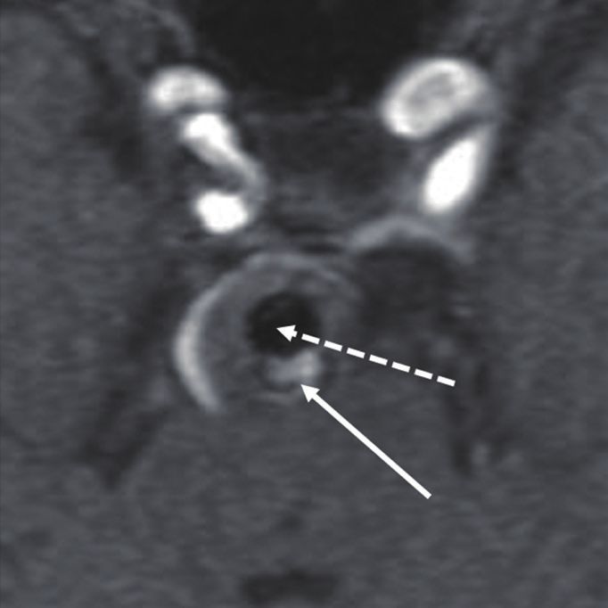
EVT for Thrombosed Cerebral Aneurysms | Lee J, et al.

stents, such as Neuroform (Stryker Neurovascular, Fremont,          variables, respectively. A univariate analysis of parameters im-
CA, USA) and Enterprise (Codman & Shurtleff, Raynham,               pacting major recanalization was conducted via binary logis-
MA, USA) stents, were used according to the physicians’ pref-       tic regression, including age, sex, rupture, aneurysm size, lu-
erence.                                                             men to whole aneurysm ratio, aneurysm location (post vs.
   Antiplatelet premedication and concurrent monitoring with        anterior circulation), treatment method (ILE vs. PAO), initial
the VerifyNow P2Y12 assay were used for patients with un-           occlusion result, the use of a stent, thrombus signal intensity
ruptured aneurysms. If patients were poor responders based          on T1 or time-of-flight MR imaging (high vs. iso- or low), fol-
on the VerifyNow P2Y12 assay, additional antiplatelet medica-       low-up duration and wall calcification. Variables with p-values
tion was administered5). A bolus of heparin (3000 IU), injected
J Korean Neurosurg Soc | 2021 Feb 26. [Epub ahead of Print]

  There was no mortality case related to the aneurysms. Two                    tary Table 1). Ultimately, seven cases (33.3%) still showed re-
patients (7.4%) transiently experienced periprocedural throm-                  sidual sac (mean follow-up duration of 62.8±50.6 months
boembolic events. One patient (3.7%) had a permanent neuro-                    [range, 6.4–207.3]).
logical deficit due to the occlusion of the lenticulostriate artery               Imaging patterns of major recurrence after ILE in 13 cases
after the trapping of the middle cerebral artery (MCA) har-                    included coil migration or dispersion into the thrombus in 10
boring the aneurysm.                                                           (76.9%), coil compaction in seven (53.8%), a decrease in
                                                                               thrombus volume in four (30.8%) and aneurysm growth in
Radiological results                                                           two (15.4%) (Fig. 1). Only two cases showed major recurrence
   A total of 27 cases were treated with ILE for 21 and PAO for                without coil mass change. Interestingly, in the other eight cas-
six. Two of 6 PAO cases underwent simultaneous bypass sur-                     es without major recurrence during the follow-up, there was
gery for flow restoration in the territory of the occluded parent              no evidence of coil compaction or migration, and seven of
artery. The angiographic results are summarized in Table 2.                    eight cases were accompanied by calcification along the aneu-
Twenty-six aneurysms (96.3%) achieved complete occlusion                       rysm wall (Supplementary Fig. 2).
or residual neck states immediately after initial treatment. De-                  Of the six aneurysms initially treated with PAO, five cases
spite repeat treatment in 12 cases, eight (29.6%) ultimately re-               were completely occluded at first (Fig. 2). Two aneurysms
mained with a status of residual sac at the last follow-up, with               (33.3%) showed major recurrence during the follow-up : the
a mean follow-up duration of 53.5±48.7 months (range, 2.9–                     intraluminal space of the one case just distal to the MCA bi-
207.3).                                                                        furcation had been completely occluded, and the inferior divi-
   Of 21 aneurysms initially treated with ILE, 94.2% (n=20) of                 sion of the MCA had been occluded, with the superior divi-
the cases initially showed complete occlusion or residual neck                 sion of the MCA remaining unoccluded; and the other case
states. However, 13 aneurysms (61.9%) resulted in major re-                    located at the vertebral artery had been incompletely occluded,
currence in a mean duration of 30.7±48.7 months (range, 4.6–                   with the proximal parent artery trapped, but the distal parent
182.4) after the initial treatment, among which nine cases had                 artery remained untrapped after the initial treatment. The
achieved a complete occlusion, three had a residual neck, and                  cause of major recurrence was the incomplete trapping of par-
the last one had a residual sac at first. Three cases (each one                ent arteries, and coil compaction on fluoroscopy was identi-
from each status of complete occlusion, residual neck and sac)                 fied in all cases. All patients were repeatedly treated with by-
were conservatively observed because of the patients’ refusal,                 pass surgery and endovascular or surgical trapping of the
and the other 10 underwent repeat embolization. Repeat ILE                     parent arteries. Only one case (16.7%), which was one of two
was performed once in seven cases, twice in two cases and                      cases of major recurrence, ultimately achieved residual sac sta-
three times in one case. Even after the repeat treatment in 10                 tus because of the retrograde filling from the distal vertebral
cases, 40% remained with a status of residual sac (Supplemen-                  artery, and the other five cases ultimately showed states of

Table 2. Angiographic results

                                                                           Total patient (n=27)               ILE (n=21)      PAO (n=6)
Immediately after initial treatment
    Complete occlusion                                                            15 (55.6)                    10 (47.6)        5 (83.3)
    Residual neck                                                                 11 (40.7)                    10 (47.6)        1 (16.7)
    Residual sac                                                                   1 (3.7)                      1 (4.8)         0 (0.0)
At the last follow-up period
    Complete occlusion                                                            11 (40.7)                     7 (33.3)        4 (66.7)
    Residual neck                                                                  8 (29.6)                     7 (33.3)        1 (16.7)
    Residual sac                                                                   8 (29.6)                     7 (33.3)        1 (16.7)
Values are presented as number of cases (%). ILE : intraluminal embolization, PAO : parent artery occlusion

4         https://doi.org/10.3340/jkns.2020.0195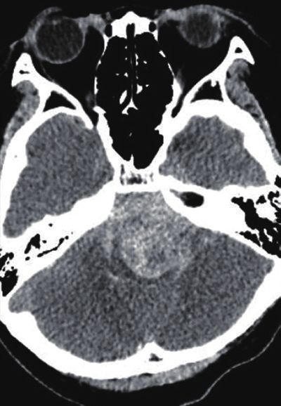
EVT for Thrombosed Cerebral Aneurysms | Lee J, et al.
A B C
D E F
Fig. 1. An approximately 20-mm-sized thrombosed aneurysm arising from the basilar artery on cerebral angiography (A) and time-of-flight magnetic
resonance angiography (B; arrow : basilar artery, dotted arrow : thrombus). After the initial stent-assisted intraluminal coil embolization, the coil mass
and stent (dotted lines) were identified on fluoroscopy (C) and magnetic resonance imaging (D; arrow : basilar artery, dotted arrow : coil mass). During
the follow-up, however, aneurysm recurrence was identified at 24 months, and aneurysm growth (approximately 28 mm) and the migration of the coil
mass were definitely demonstrated at 30 months on fluoroscopy (E; dotted lines: stent, black arrows: coil migration from the stent) and magnetic
resonance imaging (F; arrow : basilar artery, thick arrow : recurred sac, dotted line : migrating coil mass).
A B C D E
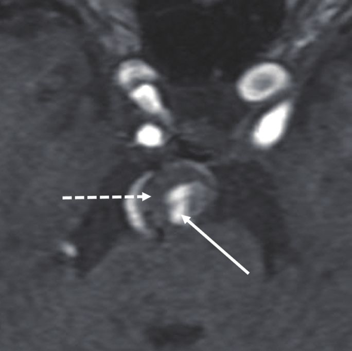
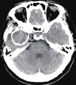
Fig. 2. An approximately 18-mm-sized thrombosed aneurysm at the left vertebral artery presenting with subarachnoid hemorrhage on cerebral
angiography (A) and computed tomography (B). Parent artery occlusion (the trapping of the proximal vertebral artery and intraluminal embolization)
was performed (C). Coil mass (white arrow) and intra-aneurysmal thrombus (dotted black arrow) were identified on the 1-month follow-up time-of-
flight magnetic resonance angiography (D). On the 14-month follow-up imaging, the thrombus was completely resolved, and a coil mass (white arrow)
was observed (E).
J Korean Neurosurg Soc 2021 Feb 26. [Epub ahead of Print] 5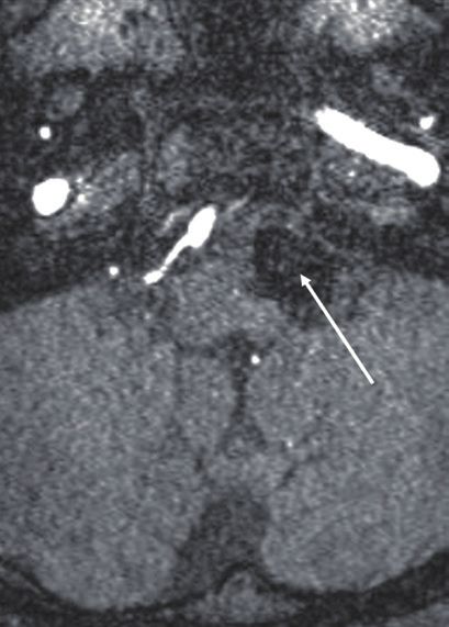
J Korean Neurosurg Soc | 2021 Feb 26. [Epub ahead of Print]

complete occlusion or residual neck during the mean follow-                  3 and 4).
up period of 21.1±21.8 months (range, 2.9–63.0) (Supplemen-
tary Fig. 3).
   According to the last follow-up angiography, a decrease in                DISCUSSION
aneurysm size was more frequently observed in completely
occluded lesions, while an increase in size was more com-                      PTIAs are generally considered different from typical sac-
monly observed in lesions with residual sac status after the                 cular aneurysms because of their morphology, symptoms
treatment. Complete occlusion was achieved in 11 cases : an-                 such as mass effect or thromboembolism rather than rupture,
eurysm size was stable in six cases and decreased in five cases              and difficulty in treatment with usual strategies8,10-12). There
(45.5%). Residual neck status was shown in eight cases, seven                are some explanations about the pathophysiology of PTIAs.
(87.5%) of which were stable in size and one showed a decrease               Basically, aneurysmal dilatation is thought to originate from
in size. Residual sac status was observed in eight cases, in                 repeated subintimal dissection by intrinsic and extrinsic fac-
which three cases (37.5%) showed an increase in aneurysm                     tors14). Thereafter, an intra-aneurysmal thrombus may be
size, three were stable in size and the other two were unable to             formed by several possible mechanisms. First, the vasa vaso-
be measured because of incomplete imaging data (Supple-                      rum within the aneurysm wall may induce repeated intramu-
mentary Table 2).                                                            ral hemorrhage and progressive thrombus formation22). This
                                                                             hypothesis was based on imaging findings such as the rim
Risk factor analysis for major recurrence                                    enhancement of the aneurysm wall and fresher clot near the
  In univariate and multivariate analyses, aneurysm wall                     periphery of the thrombus12,22,23). Second, intrathrombotic
without calcification was found to be significantly associated               capillary formation may cause repeat intramural and intrath-
with major recurrence in all cases (odds ratio [OR], 36.12; 95%              rombotic hemorrhage and progressive thrombosis based on
confidence interval [CI], 1.85 to 705.18; p=0.018) and in the                histological examination16). The third possible mechanism is
ILE group (OR, 19.02; 95% CI, 1.55 to 233.16; p=0.021) (Tables               the hemodynamic effect that blood stagnation and clot for-

Table 3. Risk factor analysis of major recurrence after endovascular         Table 4. Risk factor analysis of major recurrence after intraluminal
treatment in total cases                                                     embolization in 21 cases

Variable                                    p-value      OR (95% CI)         Variable                               p-value          OR (95% CI)
Univariate analysis                                                          Univariate analysis
    Aneurysm size                            0.569    0.64 (0.14–2.94)         Aneurysm size                          0.965       1.04 (0.17–6.40)
    Lumen/whole size ratio                   0.236    15.85 (0.16–1536.93)     Lumen/whole size ratio                 0.568       4.28 (0.03–630.62)
    Location                                 0.080     4.44 (0.84–23.58)       Location                               0.999
    Ruptured aneurysm                        0.313     0.38 (0.06–2.52)        Ruptured aneurysm                      0.855       1.27 (0.10–16.81)
    Treatment method                         0.077     8.13 (0.80–82.73)       Initial occlusion result               0.115       0.21 (0.03–1.47)
    Initial occlusion result                 0.346     0.48 (0.10–2.23)        Use of stent                           0.335       0.32 (0.03–3.56)
    Sex                                      0.816     1.20 (0.26–5.59)        Thrombus signal on MR                  0.920        1.11 (0.14–8.68)
    Age                                      0.218     1.05 (0.97–1.13)        Sex                                    0.625       0.63 (0.11–3.71)
    Follow-up period                         0.874     1.00 (0.98–1.02)        Age                                    0.813        1.01 (0.92–1.11)
    Wall without calcification               0.017     8.25 (1.45–46.86)       Follow-up period                       0.766       1.00 (0.98–1.02)
Multivariate analysis                                                          Wall without calcification             0.012      23.33 (1.99–273.29)
    Location                                 0.999                           Multivariate analysis
    Treatment method                         0.999                             Initial occlusion result               0.373       0.34 (0.03–3.65)
    Wall without calcification               0.018    36.12 (1.85–705.18)      Wall without calcification             0.021      19.02 (1.55–233.16)
OR : odds ratio, CI : confidence interval                                    OR : odds ratio, CI : confidence interval, MR : magnetic resonance

6          https://doi.org/10.3340/jkns.2020.0195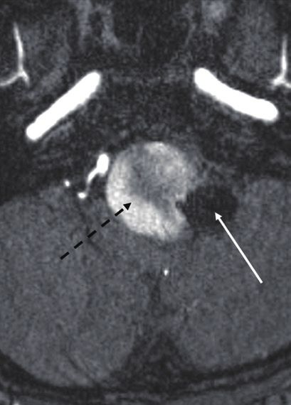
EVT for Thrombosed Cerebral Aneurysms | Lee J, et al.

mation may occur in large or giant aneurysms2). One of our              rysms4,15). In the PTIAs, however, evidence is limited that an-
patients excluded from this study showed complete resolution            giographic results were better in surgical treatment, and clini-
of their thrombus just before the treatment.                            cal outcomes remained inconclusive9,13,21,24,26). Two recent
   Endovascular treatment is one of the standard modalities             systemic reviews reported a higher rate of complete exclusion
for cerebral aneurysms4,15). Despite its low morbidity and mor-         in surgery than in neurointervention (approximately 94% vs.
tality, the major drawback of endovascular treatment is a high          48–65%)9,21). Lawton et al.13) demonstrated that surgery
risk of recurrence and retreatment compared to surgical treat-          achieved complete exclusion in 97% and that clinical out-
ment15,19). The recurrence and retreatment rates in typical             comes were satisfactory (improved or unchanged in 87% and
nonthrombosed aneurysms range from 10% to 33.6% and                     independent in 79%). A systemic review showed better angio-
from 4.7% to 12.3%, respectively19). A higher recurrence rate is        graphic results in surgery and similar clinical outcomes; how-
reported in PTIAs. Kim and Choi11) demonstrated that the re-            ever, all of them were located in the MCA21). A certain surgical
canalization rate in PTIAs was up to fivefold higher than that          group reported 50% of the disability even after complete ex-
in nonthrombosed aneurysms. A systemic review revealed a                clusion24). Because of the high risk of treatment and the rarity
higher recurrence in neurointervention than surgical treat-             of incidence in PTIAs, such controversies would exist for a
ment (62% vs. 2%)9). In this study, 29.6% finally showed major          while. Thus, an interdisciplinary treatment strategy would be
recurrence. If not treated, the recurrence of PTIAs can lead to         the best option based on the factors of aneurysm, patient and
progressive neurological deterioration due to growth, subse-            institution. Recently, f low diverters have been actively at-
quent mass effect or rupture, and embolic infarction originat-          tempted in the field of neurointervention, and promising re-
ing from the intra-aneurysmal thrombus. Therefore, treat-               sults have been reported. Flow diverters could be a good alter-
ment strategies for the PTIAs should be different from those            native in the near future. There are also reports about very
of typical aneurysms.                                                   rare cases that should be surgically removed even after the
   Endovascular treatment for PTIAs consists of ILE and PAO             complete occlusion of the aneurysms from the parent artery
with or without bypass surgery. According to the previous re-           because of the progressive growth by the vasa vasorum10). Phy-
ports, complete PAO seems better than ILE and incomplete                sicians should also be aware of such a rare situation in treating
PAO in terms of angiographic outcomes8,9,11,20,21,26). Ferns et al.8)   PTIAs.
reported that there was no additional treatment required after             As recurrence is very common in PTIAs after the endovas-
PAO, while 75% of PTIAs after ILE showed recurrence. Yang               cular treatment of intracranial aneurysms, imaging follow-up
et al.26) demonstrated that no PTIAs required retreatment af-           is very important. Although DSA, MR and computed tomog-
ter PAO. In the current study, 33.3% of the cases treated with          raphy are effective as imaging tools, they have some invasive-
ILE ultimately showed major recurrence even after repeated              ness and limitations. Fortunately, simple fluoroscopy can be
treatment, and there were no recurrences in four initial and            effective because of the recurrence patterns of PTIAs. In this
one repeat complete PAO. In addition, size reduction was                study as well as in previous reports, the dominant patterns of
more frequently observed in completely occluded lesions,                recurrence are coil migration into the thrombus and coil com-
while aneurysm growth was observed only in cases with ma-               paction8,11). Thus, a simple X-ray would be effective, economic
jor recurrence. Ferns et al.8) also demonstrated that reduction         and safe as a follow-up imaging modality. Although there is a
in aneurysm size after PAO occurred more often than after               lack of evidence for the follow-up protocol of simple fluoros-
ILE (17/18 vs. 2/28). On the other hand, clinical outcomes              copy, we recommend that it is performed every 6 or 12 months
seem controversial. Some reports showed better clinical out-            after the endovascular treatment. If PTIA shows stable occlu-
comes in the complete PAO group, and vice versa in others,              sion during the follow-up period, it could be performed every
including this study6,8,11,20). Therefore, treatment techniques         12 or 24 months.
should be determined considering the recurrence rate and                   Until now, there has been no study on the risk factors for re-
procedural risks.                                                       currence in PTIAs after neurointervention. Generally known
   It is generally accepted that angiographic results are superior      risk factors for recurrence after the endovascular treatment of
and clinical outcomes are inferior in surgery in typical aneu-          typical cerebral aneurysms are large size, wide neck, rupture,

                                                                        J Korean Neurosurg Soc 2021 Feb 26. [Epub ahead of Print]      7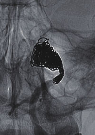
J Korean Neurosurg Soc | 2021 Feb 26. [Epub ahead of Print]

location, incomplete occlusion, follow-up duration and                tion would cause an inflammatory reaction within the throm-
stent3,17,18). Wall calcification was the only significant factor     bus, resulting in a change in thrombus composition. Addi-
protecting for major recanalization in this study. Vascular cal-      tionally, inflammatory cell invasion into the thrombus may
cification is a marker of atherosclerosis, occurring in the inti-     develop via the vasa vasorum or intrathrombotic capillary
mal layer of the arterial wall, commonly found in the aorta           channels. For such reasons, there may be a limitation in evalu-
and the coronary, carotid and renal arteries. Intimal calcifica-      ating the MR signal intensity as a candidate risk factor for re-
tion represents advanced atherosclerosis formation and tissue         currence.
degeneration1,7). Cho et al.6) suggested that mural calcification        Our study has a few limitations. It was a retrospective, ob-
within aneurysmal thrombi indicated an organized thrombus             servational, and single-center study with a small sample size.
for a substantial period. It is thought that our result may sup-      This study focused on the endovascular treatment of PTIAs,
port a vasa vasorum theory about the pathophysiology of               although the surgical role is also important.
PTIAs12,22). PTIAs continue to grow by repeated intramural
hemorrhage and thrombus formation via the vasa vasorum.
When the intimal calcification starts to form, however, the           CONCLUSION
vasa vasorum would degenerate, and thrombus formation
would stop. Finally, previous thrombi would become hard and              Durability was not satisfactory in the treatment of PTIAs
organized. Then, recurrence would rarely occur because it is          with endovascular intervention. Comparing the two technical
difficult for coils to migrate into the hard, organized throm-        options of PAO and ILE, the angiographic results were better
bus. In addition, repeat ILE may cure the aneurysm even               in the PAO group, and the clinical outcomes were better in the
when coil migration occurs because wall calcification would           ILE group. ILE and incomplete PAO seem to be a major cause
prevent aneurysm growth. Therefore, ILE can be a good alter-          of angiographic recurrence. Neurological deterioration was
native to PAO in some PTIAs with calcified walls.                     caused by rupture or the mass effect of recurred aneurysms
   The correlation between recurrence and MR signal intensity         and perforator infarction after PAO. Aneurysm wall calcifica-
of the thrombus has not been studied. Most imaging studies            tion was a protective factor against major recurrence. In treat-
have examined the pathophysiology of PTIAs in which rim               ing PTIAs, therefore, complete PAO with or without surgical
enhancement represented a vascularized wall and an intra-             bypass can be recommended as the first treatment option if
aneurysmal thrombus looked like an onion shell with fresh             the risk of treatment-related complications can be minimized.
hemorrhage at the periphery of the thrombus near the wall             ILE should be considered as an alternative if other options are
and older clot near the central lumen12,23). The authors tried to     not available or for PTIAs with a calcified wall. However, the
elucidate the relationship between recurrence and thrombus            selection of treatment modalities should consider patients’
hardness on the assumption that recurrence might happen               condition, the characteristics of the aneurysm and adjacent
more frequently in lesions with a softer thrombus. However,           vessels, and institutional situations. Most cases with major re-
such an assumption failed to be proven because the authors            currence showed coil migration into the thrombus or coil
used the thrombus signal around the central lumen on the              compaction. Therefore, fluoroscopy can be a useful imaging
initial MR imaging, considering it as a fixed status of the           modality for follow-up.
whole thrombus. The thrombus at a certain period had a het-
erogeneous signal intensity with multiple layers. In addition,
the signal intensity of the thrombus at a certain area was dy-        CONFLICTS OF INTEREST
namic, changing over time. The thrombus signal intensity
merely had a tendency : high near the central lumen and isog-           No potential conflict of interest relevant to this article was
enous or low at the periphery in approximately 60% of the             reported.
cases, which is contrary to the findings of previous reports12,23).
It may be speculated that blood inflow from the parent artery
to the lumen after recurrence by coil migration and compac-

8       https://doi.org/10.3340/jkns.2020.0195
EVT for Thrombosed Cerebral Aneurysms | Lee J, et al.

INFORMED CONSENT                                                  References

    This type of study does not require informed consent.          1. Albanese I, Khan K, Barratt B, Al-Kindi H, Schwertani A : Atherosclerotic
                                                                      calcification: Wnt is the hint. J Am Heart Assoc 7 : e007356, 2018
                                                                   2. Black SP, German WJ : Observations on the relationship between the
                                                                      volume and the size of the orifice of experimental aneurysms. J Neuro-
AUTHOR CONTRIBUTIONS                                                  surg 17 : 984-990, 1960
                                                                   3. Chalouhi N, Jabbour P, Singhal S, Drueding R, Starke RM, Dalyai RT,
    Conceptualization : WSC                                           et al. : Stent-assisted coiling of intracranial aneurysms: predictors of
    Data curation : JL, REY, WSC                                      complications, recanalization, and outcome in 508 cases. Stroke 44 :
                                                                      1348-1353, 2013
    Formal analysis : WSC
                                                                   4. Cho WS, Kim JE, Park SQ, Ko JK, Kim DW, Park JC, et al. : Korean clinical
    Funding acquisition : JL, WSC
                                                                      practice guidelines for aneurysmal subarachnoid hemorrhage. J Korean
    Methodology : WSC, YDC, HSK                                       Neurosurg Soc 61 : 127-166, 2018
    Project administration : JL, WSC                               5. Cho WS, Lee J, Ha EJ, Kim KH, Lee J, Cho YD, et al. : Low-dose prasugrel
    Visualization : JL, WSC                                           vs clopidogrel-based tailored premedication for endovascular treatment
    Writing - original draft : JL, REY, WSC                           of cerebral aneurysms. Neurosurgery 85 : E52-E59, 2019
    Writing - review & editing : WSC, REY, DHY, YDC, HSK,          6. Cho YD, Park JC, Kwon BJ, Hee Han M : Endovascular treatment of
                                                                      largely thrombosed saccular aneurysms: follow-up results in ten pa-
                                JEK
                                                                      tients. Neuroradiology 52 : 751-758, 2010
                                                                   7. Doherty TM, Asotra K, Fitzpatrick LA, Qiao JH, Wilkin DJ, Detrano RC, et
                                                                      al. : Calcification in atherosclerosis: bone biology and chronic inflamma-
ORCID                                                                 tion at the arterial crossroads. Proc Natl Acad Sci USA 100 : 11201-
                                                                      11206, 2003
    Jeongjun Lee       https://orcid.org/0000-0001-6847-1130       8. Ferns SP, van Rooij WJ, Sluzewski M, van den Berg R, Majoie CB : Par-
                                                                      tially thrombosed intracranial aneurysms presenting with mass effect:
    Won-Sang Cho       https://orcid.org/0000-0002-3345-8718
                                                                      long-term clinical and imaging follow-up after endovascular treatment.
    Roh Eul Yoo        https://orcid.org/0000-0002-5625-5921
                                                                      AJNR Am J Neuroradiol 31 : 1197-1205, 2010
    Dong Hyun Yoo      https://orcid.org/0000-0003-1658-5341       9. Güresir E, Wispel C, Borger V, Hadjiathanasiou A, Vatter H, Schuss P :
    Young Dae Cho      https://orcid.org/0000-0002-5293-2761          Treatment of partially thrombosed intracranial aneurysms: single-center
    Hyun-Seung Kang    https://orcid.org/0000-0002-6957-1907          series and systematic review. World Neurosurg 118 : e834-e841,
    Jeong Eun Kim      https://orcid.org/0000-0002-6927-3109          2018
                                                                  10. Iihara K, Murao K, Sakai N, Soeda A, Ishibashi-Ueda H, Yutani C, et al. :
                                                                      Continued growth of and increased symptoms from a thrombosed giant
●
    Acknowledgements                                                  aneurysm of the vertebral artery after complete endovascular occlusion
                                                                      and trapping: the role of vasa vasorum. Case report. J Neurosurg 98 :
  This research was supported by grant of the Korea Health            407-413, 2003
Technology R&D Project through the Korea Health Industry          11. Kim SJ, Choi IS : Midterm outcome of partially thrombosed intracranial
Development Institute (KHIDI) funded by the Ministry of               aneurysms treated with guglielmi detachable coils. Interv Neurora-
                                                                      diol 6 : 13-25, 2000
Health & Welfare, Republic of Korea (grant number :
                                                                  12. Krings T, Alvarez H, Reinacher P, Ozanne A, Baccin CE, Gandolfo C, et
HI17C1561).
                                                                      al. : Growth and rupture mechanism of partially thrombosed aneurysms.
                                                                      Interv Neuroradiol 13 : 117-126, 2007
●
    Supplementary materials                                       13. Lawton MT, Quiñones-Hinojosa A, Chang EF, Yu T : Thrombotic intracra-
                                                                      nial aneurysms: classification scheme and management strategies in 68
   The online-only data supplement is available with this arti-       patients. Neurosurgery 56 : 441-454, 2005
                                                                  14. Mizutani T, Miki Y, Kojima H, Suzuki H : Proposed classification of non-
cle at https://doi.org/10.3340/jkns.2020.0195.
                                                                      atherosclerotic cerebral fusiform and dissecting aneurysms. Neurosur-
                                                                      gery 45 : 253-259, 1999
                                                                  15. Molyneux A, Kerr R, Stratton I, Sandercock P, Clarke M, Shrimpton J,
                                                                      et al. : International subarachnoid aneurysm trial (ISAT) of neurosurgi-

                                                                  J Korean Neurosurg Soc 2021 Feb 26. [Epub ahead of Print]                   9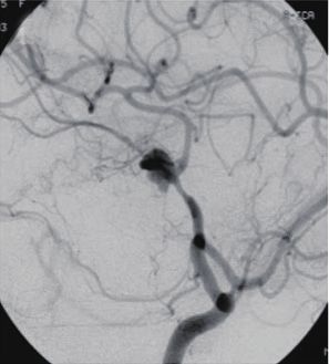
J Korean Neurosurg Soc | 2021 Feb 26. [Epub ahead of Print]

      cal clipping versus endovascular coiling in 2143 patients with ruptured       21. Scerrati A, Sabatino G, Della Pepa GM, Albanese A, Marchese E, Puca A,
      intracranial aneurysms: a randomised trial. Lancet 360 : 1267-1274,               et al. : Treatment and outcome of thrombosed aneurysms of the middle
      2002                                                                              cerebral artery: institutional experience and a systematic review. Neu-
16.   Nagahiro S, Takada A, Goto S, Kai Y, Ushio Y : Thrombosed growing gi-             rosurg Rev 42 : 649-661, 2019
      ant aneurysms of the vertebral artery: growth mechanism and manage-           22. Schubiger O, Valavanis A, Wichmann W : Growth-mechanism of giant
      ment. J Neurosurg 82 : 796-801, 1995                                              intracranial aneurysms; demonstration by CT and MR imaging. Neuro-
17.   Nishido H, Piotin M, Bartolini B, Pistocchi S, Redjem H, Blanc R : Analysis       radiology 29 : 266-271, 1987
      of complications and recurrences of aneurysm coiling with special em-         23. Teng MM, Nasir Qadri SM, Luo CB, Lirng JF, Chen SS, Chang CY : MR
      phasis on the stent-assisted technique. AJNR Am J Neuroradiol 35 :                imaging of giant intracranial aneurysm. J Clin Neurosci 10 : 460-464,
      339-344, 2014                                                                     2003
18.   Raymond J, Guilbert F, Weill A, Georganos SA, Juravsky L, Lambert A,          24. Uede T, Ohtaki M, Tanabe S, Hashi K : Direct surgical management of
      et al. : Long-term angiographic recurrences after selective endovascular          giant and large intracerebral aneurysms, associated with intraluminal
      treatment of aneurysms with detachable coils. Stroke 34 : 1398-1403,              thrombus and/or atherosclerotic thickening of aneurysmal neck. No
      2003                                                                              Shinkei Geka 25 : 1007-1015, 1997
19.   Ries T, Siemonsen S, Thomalla G, Grzyska U, Zeumer H, Fiehler J : Long-       25. Uyttenboogaart M, Stewart RE, Vroomen PC, De Keyser J, Luijckx GJ :
      term follow-up of cerebral aneurysms after endovascular therapy predic-           Optimizing cutoff scores for the Barthel index and the modified Rankin
      tion and outcome of retreatment. AJNR Am J Neuroradiol 28 : 1755-                 scale for defining outcome in acute stroke trials. Stroke 36 : 1984-
      1761, 2007                                                                        1987, 2005
20.   Roccatagliata L, Guédin P, Condette-Auliac S, Gaillard S, Colas F, Boulin     26. Yang K, Park JC, Ahn JS, Kwon DH, Kwun BD, Kim CJ : Characteristics
      A, et al. : Partially thrombosed intracranial aneurysms: symptoms, evolu-         and outcomes of varied treatment modalities for partially thrombosed
      tion, and therapeutic management. Acta Neurochir (Wien) 152 :                     intracranial aneurysms: a review of 35 cases. Acta Neurochir (Wien)
      2133-2142, 2010                                                                   156 : 1669-1675, 2014

10        https://doi.org/10.3340/jkns.2020.0195
EVT for Thrombosed Cerebral Aneurysms | Lee J, et al.
0 0
1 1
2 2
3 3
4 4
5 5
6 6
At first At last At first At last
A B
Supplementary Fig. 1. Comparison of the modified Rankin Scale (mRS) in intraluminal embolization (A) and parent artery occlusion (B) groups at the
first period just before treatment and the last period. Vertical axes means mRS and horizontal axes means periods when the mRS was checked.
J Korean Neurosurg Soc 2021 Feb 26. [Epub ahead of Print] 11J Korean Neurosurg Soc | 2021 Feb 26. [Epub ahead of Print] A B C D E F Supplementary Fig. 2. A patient presented with seizure. Brain computed tomography (A) and T2-weighted magnetic resonance imaging (B) showed a 34 mm-sized calcified and thrombosed round lesion in the right temporal pole. Right internal carotid angiograms (C and D) showed an aneurysm at the right middle cerebral artery bifurcation. Intraluminal embolization was performed (E). On the 11-year follow-up time-of-flight magnetic resonance imaging (F), just minor recurrence (white arrow) was identified without other changes. 12 https://doi.org/10.3340/jkns.2020.0195
EVT for Thrombosed Cerebral Aneurysms | Lee J, et al.

 A                                                  B                                                 C

 D                                                  E                                                 F

 G                                                  H

Supplementary Fig. 3. A patient presented with symptoms related to brainstem compression. An about 27 mm-sized thrombosed aneurysm was
identified at the left vertebral artery (VA) just distal to posterior inferior cerebellar artery (PICA) on magnetic resonance imaging (A) and cerebral
angiography (B). Intraluminal sac was completely embolized, however, the PICA orifice and parent artery harboring aneurysm neck were remained (C).
The aneurysm enlarged on the 18-month follow-up magnetic resonance imaging (D) and major recurrence by coil compaction and aneurysm growth
(white arrow) was identified on cerebral angiography (E). VA just proximal to PICA was trapped after the surgical bypass between left occipital artery
and PICA (F). In 44 months after the second treatment, the aneurysm more enlarged (G) because of the recurrence (black arrow) by retrograde filling
from the distal VA via the remaining parent artery harboring aneurysm neck (H).

                                                                              J Korean Neurosurg Soc 2021 Feb 26. [Epub ahead of Print]            13
J Korean Neurosurg Soc | 2021 Feb 26. [Epub ahead of Print]
Supplementary Table 1. Angiographic results in 10 cases repeatedly treated after the initial intraluminal embolization
At the initial treatment During the follow-up At last
Complete occlusion 6 (60.0) 0 (0.0) 4 (40.0)
Residual neck 3 (30.0) 0 (0.0) 2 (20.0)
Residual sac 1 (10.0) 10 (100.0) 4 (40.0)
Values are presented as number of cases (%)
14 https://doi.org/10.3340/jkns.2020.0195EVT for Thrombosed Cerebral Aneurysms | Lee J, et al.
Supplementary Table 2. Size change of all the aneurysms according to the angiographic results at the last follow-up
Increase Stationary Decrease Not available
Complete occlusion 0 6 5 0
Residual neck 0 7 1 0
Residual sac 3 3 0 2
J Korean Neurosurg Soc 2021 Feb 26. [Epub ahead of Print] 15You can also read