Calcification patterns in femoral and carotid atheromatous plaques: A comparative morphometric study
←
→
Page content transcription
If your browser does not render page correctly, please read the page content below
EXPERIMENTAL AND THERAPEUTIC MEDICINE 22: 865, 2021

           Calcification patterns in femoral and carotid atheromatous
                   plaques: A comparative morphometric study
                            MIRCEA CATALIN COSARCA1*, EMŐKE HORVÁTH2*, CALIN MOLNAR3,
                          GYOPÁR‑BEÁTA MOLNÁR4, ELIZA RUSSU5 and VASILE ADRIAN MUREȘAN5

       1
        Doctoral School and Departments of 2Pathology and 3General Surgery, ‘George Emil Palade’ University of Medicine,
                    Pharmacy, Science and Technology, 540142 Târgu‑Mureș; 4Pathological Anatomy Service,
                  Târgu‑Mureș County Emergency Clinical Hospital (SCJU Târgu‑Mureș), 540136 Târgu‑Mureș;
                         5
                          Department of Vascular Surgery, ‘George Emil Palade’ University of Medicine,
                              Pharmacy, Science and Technology, 540142 Târgu Mureș, Romania

                                            Received April 5, 2021; Accepted May 5, 2021

                                                    DOI: 10.3892/etm.2021.10297

Abstract. This comparative study was designed to focus on           (P= 0.0007 and P= 0.0009). pCSs calculated in plaques with
the mineral patterns in human atherosclerotic plaques based         extensive calcification pattern showed a lower value in the
on quantitative measurements of calcium deposits through the        CA vs. the pSFA plaques (P= 0.004). A less pronounced, but
morphometric method. A total of 101 atherosclerotic plaques         significant difference was observed between the pCS of pSFA
were harvested by conventional transluminal angioplasty             and dSFA plaques (P= 0.017). Femoral and carotid plaques
from the carotid artery (CA) and different segments of the          exhibited different morphology and tendency for calcification.
femoral‑popliteal axis (FPA), fixed in formalin and sent for        In parallel with the narrowing of the vascular lumen diameter,
histological processing. The histological grade of the athero‑      the mineralized surface increased at the level of different
sclerotic plaque and the calcification pattern were evaluated,      FPA segments. These results suggest that the mechanism is
followed by a morphometric analysis of the mineral deposits.        site‑specific, and wall structure‑dependent.
Regarding the localization, the advanced plaques (VII and
VIII types) developed predominantly at the level of the super‑      Introduction
ficial femoral artery (SFA) compared to the CA (P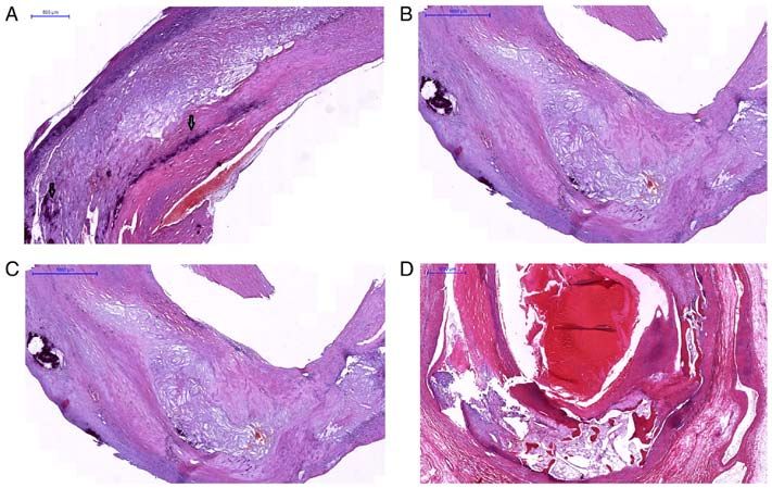
2                COSARCA et al: CALCIFICATION PATTERNS IN FEMORAL AND CAROTID ATHEROMATOUS PLAQUES

ulceration are associated with intra‑plaque hemorrhage, and they      A total of 101 cases were selected for the clinic pathological
may be a substitute for higher risk lesions (10,11). Some studies     study, based on strict criteria which included patients with
have investigated the role of size and location of calcification      complete clinical documentation and the written consent
regarding the restenosis rate after surgical treatment. Compared      of enrolment in the study and an appropriate quantitative
with the low percentage of restenosis after coronary drug‑eluted      and structural specimen for histological examination. Prior
stent replacement (10‑15%) (12), restenosis after lower limb          to surgery, for the clinical assessment of the severity of the
artery stenting reaches 40‑50% (13,14), while the variable rate       CA and the FPA stenosis, a CT angiography and a Doppler
of this complication is between 1 and 36% after intervention at       ultrasonography were performed. The subjects were first
the carotid level (15).                                               divided into two groups: Group 1 (n=21) included patients with
    Numerous factors such as demography, comorbidity, sex,            symptomatic CA stenosis (either transient ischemic attack
cardiovascular risk factors, high‑degree stenosis, metabolic          or stroke on the ipsilateral side of the carotid stenosis); and
and hemodynamic factors, biochemical parameters, the lesion           group 2 (n=80), patients with stenosis in different segments of
length, the underlying site‑specific wall structure and the nature    the FPA presenting claudication or critical limb ischemia. The
(including calcification) of the atheromatous plaque alter the        atherosclerotic plaques from the FPA were collected from the
restenosis rate after endarterectomy or stent implantation, of        following levels: The proximal third of the superficial femoral
which the crucial role is attributed to the appearance and extent     artery (pSFA), adjacent to the inguinal ligament (Poupart's
of calcification (16,17). Atherosclerotic plaque calcification        ligament); the distal segment of the superficial femoral artery
varies according to each arterial bed; femoral plaques exhibit a      (dSFA) at the level of the Hunter's canal and the proximal
significantly different type of calcification pattern and osteoid     segment of the popliteal artery (PA), respectively. All samples
metaplasia than carotid plaques (18), which suggests the role of      from the carotid artery (CA) and FPA were immediately fixed
a site‑dependent mechanism in plaque calcification. Clinically,       in 10% neutral buffered formalin and sent for histological
two types of plaque calcification have distinct implications in the   processing. The study was conducted according to the prin‑
progression and regression of atherosclerosis: Macrocalcification,    ciples of the Helsinki Declaration and was approved by the
which leads to plaque stability, and microcalcification with          Ethics Committee of the ‘George Emil Palade’ University of
a pivotal role in plaque rupture and consequent complica‑             Medicine, Pharmacy, Science and Technology of Târgu‑Mureș,
tions (19,20). Recent research has demonstrated that the spatial      Romania (no. 884 and 11420/30.04.2020). All patients signed
distribution of mineral mass, calcification size and location are     an informed consent for inclusion in the study.
important determinants of the plaque rupture risk (21,22).
    The effect of calcification is considered biphasic, from          Histological processing. The specimens were processed using
pro‑inflammatory properties of ‘microcalcification’ to                a standard method to produce paraffin sections for staining
anti‑inflammatory properties of ‘macrocalcification’. Plaque          with hematoxylin and eosin (H&E), and the calcified speci‑
rupture has been shown to be positively correlated with micro‑        mens were treated with an ethylenediaminetetraacetic acid
calcifications, and conversely with extensive calcifications.         (EDTA) solution (pH 7.0).
Macroscopic calcification is easily detected and quantified
(calcium scores as predictive value for cardiovascular incidence)     Establishment of the histological grade of the atherosclerotic
using the CT scan method. In contrast, microcalcification,            plaques. The histological grade of the atherosclerotic plaque
the early stage of plaque calcification, is observed only with        was evaluated on 4‑µm thick and H&E-stained sections
positron emission tomography (PET)/CT imaging and optical             according to the modified American Heart Association (AHA)
coherence tomography (diagnostic methods that are not used            classification based on 9, well‑defined categories, of which
in daily practice) (9).                                               6 types correspond to atheromatous plaque (23).
    However, the CT analysis of calcification patterns is limited         Briefly, type IV is considered to be the first advanced stage
by the resolution and blooming artefacts. In this context, the        of the disease with confluent extracellular lipid core. An ather‑
histopathological examination of the endarterectomy speci‑            omatous plaque with fibrous cap associated with prominent
mens provides useful information to the clinician to develop          fibrosis is included in type V. All atheromatous plaques with
a treatment strategy.                                                 surface damage (ulceration and thrombosis) must be classified
    This present study is a detailed cross‑sectional morpho‑          as type VI. A fibrocalcified plaque with extensive calcifica‑
logical comparative characterization of the intra‑plaque              tion is categorized as type VII. In type VIII, fibrous changes
calcification of advanced atherosclerotic lesions based on            predominate. Complete occlusion (type IX) in our study was
morphometric methods involving the most important segments            not taken into account.
of the FPA and CA.
                                                                      Determination of the degree (type) of calcification of the
Materials and methods                                                 plaques. Focusing on the calcification pattern, the plaques
                                                                      were included in four categories depending on the calcified
Patients and tissue fragments. In this prospective comparative        patch distribution, size and shape: a) Sheet‑like calcification
study, tissue fragments harvested by conventional translu‑            (defined as numerous micronodules/scattered small mineral
minal angioplasty from patients diagnosed with symptomatic            foci forming a calcification front within fibrosis (Fig. 1A);
PAD and CA atherosclerosis were included. The material                b) nodular calcification (single/multiple stratified mineral
was collected from different patients between January 2017            deposits with a nodular aspect (Fig. 1B); c) extensive (confluent)
and December 2018, at the Vascular Surgery Clinic, within             calcification (conglomerate of mineral material with irregular
the Mureș County Emergency Clinical Hospital (Romania).               edges (Fig. 1C); and d) osteoid metaplasia (mature bone with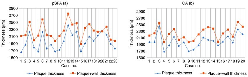
EXPERIMENTAL AND THERAPEUTIC MEDICINE 22: 865, 2021                                                                   3

Figure 1. Microscopic aspects of human atherosclerotic plaque calcification types; H&E staining. (A) Sheet‑like calcification (defined as numerous micronod‑
ules/scattered small mineral foci forming a calcification front within fibrosis. (B) Nodular calcification (single/multiple stratified mineral deposits with nodular
aspect. (C) Extensive (confluent) calcification (conglomerate of mineral material with irregular edges and disruption of internal elastic lamina and (D) osteoid
metaplasia represented by mature bone with lamellar structure and bone marrow. H&E, hematoxylin and eosin.

lamellar structure and bone marrow (Fig. 1D). Although there                        Mann‑Whitney U test. A correlation analysis was performed
is no conventional standard of size, there is a general consensus                   according to Spearman. The level of statistical significance
that categorizes microcalcifications and macrocalcifications                        was set at P
4 COSARCA et al: CALCIFICATION PATTERNS IN FEMORAL AND CAROTID ATHEROMATOUS PLAQUES
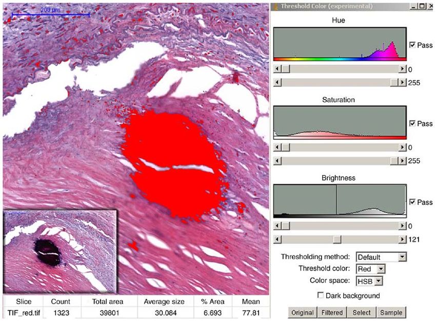
Figure 2. Morphometric analysis of mineral deposits for the quantification of the pCS (% area) in the atheromatous plaques. Representative foci with calcifica‑
tion (H&E stain, inset) were captured with x20 magnification, followed by processing of the obtained image and HSB (hue, saturation, brightness) color
filtering of the NIH's Image J 1.46 software. H&E, hematoxylin and eosin.
Regarding the localization, we found that the plaques Comparative morphometric analysis of mineral deposits in
included in AHA VIII type (45 cases) developed predomi‑ the femoral and carotid artery plaques. In the present study,
nantly at the level of the superficial femoral artery [proximal we included only calcified plaques (from 81 cases), that showed
third and the distal segment (62.5%) compared to the CA a higher frequency among men (PEXPERIMENTAL AND THERAPEUTIC MEDICINE 22: 865, 2021 5
Figure 3. Distribution of plaques according to the calcification type in different arterial segments. Statistical analysis shows a significantly higher presence of
nodular calcification (type II) in the femoral‑popliteal axis (FPA), than in the carotid arterial (CA) bed. dSFA, distal segment of the superficial femoral artery;
pSFA, proximal third of the superficial femoral artery; PA, popliteal artery.
Figure 4. The average thickness of the plaques and the values of the ratio plaque thickness/total thickness at the level of the pSFA (a) compared to CA (b) did
not show a significant difference. pSFA, proximal third of the superficial femoral artery; CA, carotid artery.
at which calcification occupied significantly larger areas According to our knowledge, this comparative study
(P=0.0007 and P=0.0009). At the level of pSFA segments, the between the carotid artery (CA) and the femoral‑popliteal axis
nodular calcification showed significant differences in favor (FPA) is the first to follow the extent of calcification depending
of the PA (P= 0.007). Extensive calcification surface area did on the variation of the vascular caliber.
not differ between the CA and pSFA plaques (P= 0.83), but Our patients undergoing carotid and femoral endarterec‑
the pCS of the CA plaques was lower than those measured in tomy represented a select risk group with high grade artery
dSFA (P=0.004). A less pronounced, but significant difference stenosis; a large part of the atherosclerotic plaques being clas‑
was observed between the pCS of the pSFA and dSFA plaques sified based on morphological aspects of type VII and VIII,
(P=0.017) (Table I and Fig. 5). over three quarters of them with intra‑plaque calcification.
Preliminary histological studies demonstrated considerable
Discussion differences between plaques with identical degrees of stenosis,
respectively. Certain plaque features are associated with an
The majority of studies focusing on calcification are based on increased risk of ischemic event (25). At the same time, it has
calcium score (CS) determination by imaging; few of them use been shown that carotid plaque thickness or plaque volume
morphometric analysis of mineral content on the histopatho‑ is more strongly associated with ischemic events than the
logical sections of the endarterectomy samples involving one degree of stenosis (26). Although vascular calcification in
type of artery (21) or two types (18). the lower extremities is commonly a hallmark of peripheral6 COSARCA et al: CALCIFICATION PATTERNS IN FEMORAL AND CAROTID ATHEROMATOUS PLAQUES
Table I. Comparison of total calcified area in different arterial segments.
Arterial segment
‑‑‑‑‑‑‑‑‑‑‑‑‑‑‑‑‑‑‑‑‑‑‑‑‑‑‑‑‑‑‑‑‑‑‑‑‑‑‑‑‑‑‑‑‑‑‑‑‑‑‑‑‑‑‑‑‑‑‑‑‑‑‑‑‑‑‑‑‑‑‑‑‑‑‑‑‑‑‑‑‑‑‑‑‑‑‑‑‑‑‑‑‑‑‑‑‑‑‑‑‑‑‑‑‑‑‑‑‑‑‑‑‑‑‑‑‑‑‑‑‑‑‑‑‑‑‑‑‑‑‑‑‑‑‑‑‑‑‑‑‑‑‑‑‑‑‑‑‑‑‑‑‑‑‑‑‑‑‑‑‑‑‑‑‑‑‑‑‑‑‑‑‑‑‑‑‑‑‑‑‑
Total calcified area (pCS) CA plaque (n=19) pSFA plaque (n=18) dSFA plaque (n=35) PA plaque (n=9)
I. Sheet‑like calcification 2.09±0.47 6.34±0.85 8.27±0.75 8.07±1.25
II. Nodular calcification 7.5±1.06e,f 11.27±0.88d,g 26.09±1.47e,g 27.89±2.90d,f
III. Extensive calcification 26.28±2.00c 27.33±2.46b 35.73±1.59b,c 38.52±0.00
Average calcification 14.88±2.51a 13.54±2.26b 21.63±1.93a,b 22.47±4.09
Values are expressed as the mean ± standard error. pCS was defined as the percentage (%) of positive calcified surface from the total examined
area. P‑values were calculated by the Mann‑Whitney U test. a,bPEXPERIMENTAL AND THERAPEUTIC MEDICINE 22: 865, 2021 7
the surrounding environments around the calcification within from the interface between the fibrous cap and lipid core to
plaques. Based on the findings in the literature according to the interface between the fibrous cap and vessel lumen (38).
which superficial calcifications are independently associated Some authors claim that although extensive calcifications are
with plaque vulnerability, and the type of calcification extension associated with plaque stability, the role of the calcification
(dispersed or compacted), dimensions, shapes and positions may size in plaque vulnerability may be biphasic. They explain this
play different roles in plaque evolution (11), we continued our phenomenon by the interaction of mechanical forces with the
study by determining the total calcified surface area (as pCS) of mass of the plaque. Normally, mechanical stress is expected
remote plaques by morphometry and we focused on the extent to be concentrated at the interfaces between materials with
of calcification in terms of spectrum between the sheet‑like different stiffness, within a plaque at the interfaces between
nodular‑extensive calcification. In the present study we showed calcium deposits and the rest of the plaque elements. Calcium
that the calcified area varied depending on the arterial segment stiffness is at least four times higher than that of other plaque
affected by atherosclerosis; the total calcified area was higher components (39), as a result, if the degree of calcification
in femoral plaques than in carotid plaques, but without a increases, the calcium deposits merge and the soft surface area
significant difference between the plaques of the two arteries progressively decreases (11).
with the same structure (pSFA and CA). At the same time, we We are aware of the limitations of our study. The relatively
noted a significant increase in mineralized surface at the level small sample number of a single center material characterizes
of the different FPA segments in parallel with a decrease in the the CA and FPA plaques at a well‑determined moment. The
vascular lumen diameter. Differences in the total calcification morphological aspects of the plaques of the two arteries from
of plaques depending on the involved arterial segments were different patients compromise the comparison due to their
consistent with the histological grade, according to which most development in different pathological conditions. At the same
of the femoral plaques were classified as fibrocalcific (VII and time, most of the lesions came from advanced atherosclerotic
VIII AHA types), whereas carotid plaques were classified as plaques, with extensive calcification associated with a rela‑
fibrous cap atheroma (IV and V types) (18). tively small number of sheet‑like calcifications. Regarding the
At the same time, we found that, even when the three femoral morphometric analysis, we determined the total surface and not
segment plaques and carotid plaques explain different calcifica‑ the total amount of mineral salts of the plaques, because different
tion pattern, the total mineralized area did not depend on the patterns were combined in a single plaque, but we agree that the
shape and pattern of the plaques, but rather on vascular caliber; nodular type of macrocalcifications has a large volume but a
in parallel to the narrowing of the lumen, the calcified area of small surface area (40,41). A technical disadvantage of the study
the plaque increased. This finding was also supported by the was the loss of calcification (during cutting) in some sections
results obtained in the case of an extensive calcification pattern, and morphology distortion. In some cases, we tried to overcome
in the sense that the mineralized area did not differ between this by following the perimeter of the calcified cavity, a method
the CA and pSFA plaques, but in contrast, the pCS of the CA that is possible only in nodular calcifications.
plaques was lower than those measured in distal (d)SFA. In conclusion, despite being exposed to similar risk
Although several molecular aspects of this mechanism have factors, peripheral arteries develop heterogeneous lesions.
been elucidated regarding the two types of arterial calcification Femoral and carotid plaques show different morphologies
in the different vessel types (large elastic vs. smaller muscular and tendencies for calcification. In the present study, we
arteries) and parts (proximal vs. distal) (32,33), few morpho‑ demonstrated that a population with similar demographic and
logic studies have focused on the plaque calcification in the biological data develops at the level of smaller caliber arteries
different segments of the FPA in comparison with the CA. The (femoral‑popliteal/popliteal artery) larger areas of calcifica‑
most significant results are in the study of Herisson et al (18), tion than at the level of larger caliber arteries (femoral/carotid).
who focused on the calcification pattern, complemented with These results suggest that the mechanism is site‑specific,
quantitative measurements of calcium and lipids within the and wall structure‑dependent. In advanced carotid and FPA
plaque. This complex approach of calcification underscored the atherosclerosis, calcification has a heterogeneous pattern with
high prevalence of sheet‑like and nodular calcification parallel a simultaneous presence of large calcified area and numerous
with significantly higher amounts of calcium at the level of FA small calcific patches. Due to the heterogeneous composi‑
plaques in comparison with CA plaques, and did not lead to the tion of the calcified plaques, extensive calcification remains
staging of calcification according to different segments of AFP. controversial in terms of plaque stability.
In studies that included the measurement of calcium scores
and occlusion grade, it was reported that patients with PAD Acknowledgements
presenting increased occlusion and calcification scores were
strongly associated with more severe stages of ischemia (34), Not applicable.
and independently predicted amputation and mortality (35‑37).
However, the exact mechanism of atherosclerotic calcification, Funding
including the territorial distribution and the size of calcified
regions within atherosclerotic lesions and its role in plaque No funding was received.
vulnerability, remains incompletely understood (21).
Microcalcifications (0.5‑50 mm) represent an early stage Availability of data and materials
in the spectrum of the vascular calcification cascade. They
are considered predictors of cardiovascular events, leading The data and materials that support the results of this study are
to rupture of the plaque through the transfer of the stress available from the corresponding author (EH).8 COSARCA et al: CALCIFICATION PATTERNS IN FEMORAL AND CAROTID ATHEROMATOUS PLAQUES
Authors' contributions 11. Shi X, Gao J, Lv Q, Cai H, Wang F, Ye R and Liu X: Calcification
in atherosclerotic plaque vulnerability: Friend or Foe? Front
Physiol 11: 56, 2020.
MCC conceived the study, coordinated the surgical team, 12. Tang L, Cui QW, Liu DP and Fu YY: The number of stents was
performed the surgery, analyzed and interpreted the patient an independent risk of stent restenosis in patients undergoing
percutaneous coronary intervention. Medicine (Baltimore) 98:
data. MCC, ER and CM selected the patients and performed e18312, 2019.
the surgery. EH designed the study, performed histopatho‑ 13. Laird JR and Yeo KK: The treatment of femoropopliteal in‑stent
logical and morphometric analyses of the biopsies, contributed restenosis: Back to the future. J Am Coll Cardiol 59: 24‑25, 2012.
14. Gerardi D, Alfani A, Tesorio T, Cioppa A, Esposito G and
to data analysis, performed figure design and provided critical Stabile E: Drug‑coated balloon in superficial femoral artery
review of the manuscript and was responsible for the final in‑stent restenosis. Postepy Kardiol Interwencyjnej 14: 9‑14, 2018.
edited version. GBM performed the histopathological and 15. Gaudry M, Bartoli JM, Bal L, Giorgi R, De Masi M, Magnan PE
and Piquet P: Anatomical and technical factors influence the rate
morphometric analyses of the biopsies, contributed to data of in‑stent restenosis following carotid artery stenting for the
analysis and was responsible for the acquisition of the data. treatment of post‑carotid endarterectomy stenosis. PLoS One 11:
VAM acquired the data and revised the manuscript critically e0161716, 2016.
16. Katano H, Nishikawa Y, Yamada H and Mase M: Calcification in
for important intellectual content and coordinated the research. original plaque and restenosis following carotid artery stenting.
All authors contributed to data interpretation and editing the Surg Neurol Int 8: 279, 2017.
manuscript and read and approved the final version. 17. Katano H, Mase M, Nishikawa Y, Yamada H and Yamada K:
Analysis of recurrent stenosis after carotid endarterectomy
featuring primary plaque calcification. Neurosurgery 80: 863‑70,
Ethics approval and consent to participate 2017.
18. Herisson F, Heymann MF, Chetiveaux M, Charrier C, Battaglia S,
Pilet P, Rouillon T, Krempf M, Lemarchand P, Heymann D and
The study was approved by the Ethics Committee of ‘George Gouëffic Y: Carotid and femoral atherosclerotic plaques show
Emil Palade’ University of Medicine, Pharmacy, Science and different morphology. Atherosclerosis 216: 348‑354, 2011.
Technology of Târgu‑Mureș (no. 884 and 11420/30.04). All 19. Kelly‑Arnold A, Maldonado N, Laudier D, Aikawa E, Cardoso L
and Weinbaum S: Revised microcalcification hypothesis for
patients provided informed consent for inclusion in the study. fibrous cap rupture in human coronary arteries. PNAS 110:
10741‑1046, 2013.
Patient consent for publication 20. Jinnouchi H, Sato Y, Sakamoto A, Cornelissen A, Mori M,
Kawakami R, Gadhoke NV, Kolodgie FD, Virmani R and
Finn AV: Calcium deposition within coronary atherosclerotic
This manuscript does not contain particular cases, personal lesion: Implications for plaque stability. Atherosclerosis 306:
information or images which would require patient personal 85‑95, 2020.
21. Han RI, Wheeler TM, Lumsden AB, Reardon MJ, Lawrie GM,
consent. Grande‑Allen KJ, Morrisett JD and Brunner G: Morphometric
analysis of calcification and fibrous layer thickness in carotid
Competing interests endarterectomy tissues. Comput Biol Med 70: 210‑219, 2016.
22. Tavakoli S and Sadeghi MM: 18F‑NaF PET and plaque calcifica‑
tion: How complicated can it be? Circ Cardiovasc Imaging 12:
The authors declare that they have no competing interests. e008712, 2019.
23. Stary HC: Natural history and histological classification of
atherosclerotic lesion: An update. Arterioscler Thromb Vasc
References Biol 20: 1177‑1178, 2000.
24. Ferreira T and Rasband W: ImageJ User Guide. Image
1. Eurostat:YourkeytoEuropeanstatistics:https://ec.europa.eu/eurostat/ Processing and Analysis in Java. National Institutes of Health,
statistics‑xplained/index.php/Causes_of_death_statistics_‑people_ 2012. http://rsb.info.nih.gov/ij.
over_65https://ec.europa.eu/eurostat/statistics‑explained/index.php/ 25. Redgrave JNE, Lovett JK, Gallagher PJ and Rothwell P:
Cardiovascular_diseases_statistics. Histological assessment of 526 symptomatic carotid plaques in
2. Singh RB, Mengi SA, Xu YJ, Arneja AS and Dhalla NS: relation to the nature and timing of ischemic symptoms: The
Pathogenesis of atherosclerosis: A multifactorial process. Exp Oxford plaque study. Circulation 113: 2320‑2328, 2006.
Clin Cardiol 7: 40‑53, 2002. 26. Zhu G, Hom J, Li Y, Jiang B, Rodriguez F, Fleischmann D,
3. Albanese I, Khan K, Barratt B, Al‑Kindi H and Schwertani A: Saloner D, Porcu M, Zhang Y, Saba L and Wintermark M:
Atherosclerotic calcification: Wnt is the hint. J Am Heart Carotid plaque imaging and the risk of atherosclerotic cardio‑
Assoc 7: e007356, 2018. vascular disease. Cardiovasc Diagn Ther 10: 1048‑1067, 2020.
4. Peace A, van Mil A, Jones H and Thijssen DHJ: Similarities and 27. Reneman RS, Arts T and Hoeks AP: Wall shear stress‑an impor‑
differences between carotid artery and coronary artery function. tant determinant of endothelial cell function and structure‑in the
Curr Cardiol Rev 14: 254‑263, 2018. arterial system in vivo discrepancies with theory. J Vasc Res 43:
5. Schiano V, Sirico G, Giugliano G, Laurenzano E, Brevetti L, 251‑269, 2006.
Perrino C, Brevetti G and Esposito G: Femoral plaque echo‑ 28. VanderLaan PA, Reardon CA and Getz GS: Site specificity of
genicity and cardiovascular risk in claudicants. JACC Cardiovasc atherosclerosis: Site selective responses to atherosclerotic modu‑
Imaging 5: 348‑357, 2012. lators. Arterioscler Thromb Vasc Biol 24: 12‑22, 2004.
6. Golomb BA, Dang TT and Criqui MH: Peripheral arterial
disease: Morbidity and mortality implications. Circulation 114: 29. Otsuka F, Sakakura K, Yahagi K, Joner M and Virmani R: Has our
688‑699, 2006. understanding of calcification in human coronary atherosclerosis
7. Maleckis K, Anttila E, Aylward P, Poulson W, Desyatova A, progressed? Arterioscler Thromb Vasc Biol 34: 724‑736, 2014.
MacTaggart J and Kamenskiy A: Nitinol Stents in the femoro‑ 30. Chistiakov DA, Myasoedova VA, Melnichenko AA, Grechko AV
popliteal artery: A mechanical perspective on material, design, and Orekhov AN: Calcifying matrix vesicles and atherosclerosis.
and performance. Ann Biomed Eng 46: 684‑704, 2018. Biomed Res Int 2017: 7463590, 2017.
8. Kwee RM: Systematic review on the association between calcifi‑ 31. Janzen J: The microscopic transitional zone between elastic and
cation in carotid plaques and clinical ischemic symptoms. J Vasc muscular arteries. Arch Mal Coeur Vaiss 97: 909‑914, 2004.
Surg 51: 1015‑1025, 2010. 32. Amann K: Media calcification and intima calcification are
9. Shioi A and Ikari Y: Plaque Calcification during atherosclerosis distinct entities in chronic kidney disease. Clin J Am Soc
progression and regression. J Atheroscler Thromb 25: 294‑303, 2018. Nephrol 3: 1599‑1605, 2008.
10. Yang J, Pan X, Zhang B, Yan Y, Huang Y, Woolf AK, Gillard JH, 33. Allison MA, His S, Wassel CL, Morgan C, Ix JH, Wright CM
Teng Z and Hui P: Superficial and multiple calcifications and and Criqui MH: Calcified atherosclerosis in different vascular
ulceration associate with intraplaque hemorrhage in the carotid beds and the risk of mortality. Arterioscler Thromb Vasc Biol 32:
atherosclerotic plaque. Eur Radiol 28: 4968‑4977, 2018. 140‑146, 2012.EXPERIMENTAL AND THERAPEUTIC MEDICINE 22: 865, 2021 9
34. Zettervall SL, Marshall AP, Fleser P and Guzman RJ: Association 39. Lee RT, Grodzinsky AJ, Frank EH, Kamm RD and Schoen FJ:
of arterial calcification with chronic limb ischemia in patients Structure‑dependent dynamic mechanical behavior of fibrous
with peripheral artery disease. J Vasc Surg 67: 507‑513, 2018. caps from human atherosclerotic plaques. Circulation 83:
35. Huang CL, Wu IH, Wu YW, Hwang JJ, Wang SS, Chen WJ, 1764‑1770, 1993.
Lee WJ and Yang WS: Association of lower extremity arterial 40. Wang Y, Osborne MT, Tung B, Li M and Li Y: Imaging cardio‑
calcification with amputation and mortality in patients with symp‑ vascular calcification. J Am Heart Assoc 7: e008564, 2018.
tomatic peripheral artery disease. PLoS One 9: e90201, 2014. 41. Dweck MR, Aikawa E, Newby DE, Tarkin JM, Rudd JH,
36. Blacher J, Guerin AP, Pannier B, Marchais SJ and London GM: Narula J and Fayad ZA: Noninvasive molecular imaging of
Arterial calcifications, arterial stiffness, and cardiovascular risk disease activity in atherosclerosis. Circ Res 119: 330‑340, 2016.
in end‑stage renal disease. Hypertension 38: 938‑942, 2001.
37. Guzman RJ, Brinkley DM, Schumacher PM, Donahue RMJ,
Beavers H and Qin X: Tibial artery calcification as a marker of
amputation risk in patients with peripheral arterial disease. J Am This work is licensed under a Creative Commons
Coll Cardiol 51: 1967‑1974, 2008. Attribution-NonCommercial-NoDerivatives 4.0
38. Rambhia SH, Liang X, Xenos M, Alemu Y, Maldonado N, International (CC BY-NC-ND 4.0) License.
Kelly A, Chakraborti S, Weinbaum S, Cardoso L, Einav S and
Bluestein D: Microcalcifications increase coronary vulnerable
plaque rupturepotential: A patient‑based micro‑CT fluid‑struc‑
ture interaction study. Ann Biomed Eng 40: 1443‑1454, 2012.You can also read