Increased sensitivity to TNF α promotes keloid fibroblast
←
→
Page content transcription
If your browser does not render page correctly, please read the page content below
EXPERIMENTAL AND THERAPEUTIC MEDICINE 21: 502, 2021

            Increased sensitivity to TNF‑α promotes keloid fibroblast
                  hyperproliferation by activating the NF‑κB,
                         JNK and p38 MAPK pathways
                              QIJIE LI1*, FENGRUI CHENG2*, KAI ZHOU2, LU FANG2, JUNLIANG WU2,
                                    QINGJIE XIA3, YING CEN2, JUNJIE CHEN2 and YONG QING2

                          1
                         Laboratory of Anesthesia and Critical Care Medicine, Department of Anesthesiology,
            Translational Neuroscience Center; 2Department of Plastic and Burn Surgery, West China School of Medicine;
               3
                 Department of Anesthesiology, Institute of Neurological Diseases, Translational Neuroscience Center,
                           West China Hospital, Sichuan University, Chengdu, Sichuan 610041, P.R. China

                                         Received August 19, 2020; Accepted January 18, 2021

                                                       DOI: 10.3892/etm.2021.9933

Abstract. Hyperproliferation of fibroblasts is the main cause            In conclusion, KFs exhibited increased expression of sTNFR1,
of keloid formation. However, the pathogenesis of keloids has            which may contribute to the increased sensitivity to TNF‑α,
yet to be fully elucidated. Tumor necrosis factor (TNF)‑ α               resulting in low concentrations of TNF‑ α activating the
may play an important role in the formation and proliferation            NF‑κ B, JNK and p38 MAPK pathways, thereby promoting the
of keloids, as it is implicated in the pathogenesis of various           sustained and excessive proliferation of KFs.
fibrous disorders. In the present study, the expression level
of TNF‑α and its receptors, soluble TNF receptor (sTNFR)1                Introduction
and sTNFR2, in the peripheral blood and skin tissues was
detected by ELISA, reverse transcription‑quantitative PCR or             Keloid is a type of proliferative fibroma that may form after
immunohistochemistry. There was no statistically significant             trauma, and is characterized by abnormal wound healing,
difference in the expression of TNF‑ α and sTNFR2 in the                 fibroblast proliferation and extracellular matrix deposition.
peripheral blood and skin tissues between patients with keloids          Keloid is considered as a benign human tumor without malig‑
and healthy participants (P>0.05), while the sTNFR1 mRNA                 nant potential, which invades the adjacent normal skin tissue
level in fibroblasts cultured in vitro and its protein level in          beyond the initial boundaries of the trauma (1,2). Fibroblasts are
keloid skin samples were significantly higher compared with              considered to play a key role in the formation and development
those in normal skin (P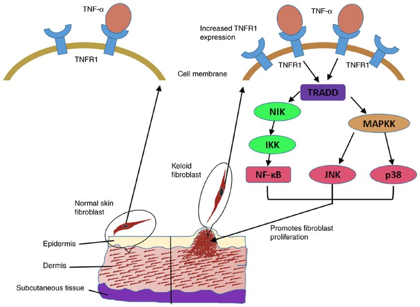
2                           LI et al: TNF-α PROMOTES HYPERPROLIFERATION OF KELOID FIBROBLASTS

action of TNF‑α in the pathogenesis of keloids are unclear.         Table I. Demographics of participants included in the present
In tumor research, TNF‑α was found to promote cell prolif‑          study.
eration through activating the nuclear factor (NF)‑κ B, c‑Jun
N‑terminal kinase (JNK) and p38 mitogen‑activated protein           Characteristics    Keloid group (n=20) Control group (n=18)
kinase (MAPK) pathways, which were regulated by the
concentration of TNF‑α (11). However, further experimental          Age, years              28.00±6.58              31.47±10.06
research is required to determine whether the function and          Sex, male/female           9/11                     6/12
mechanism of action of TNF‑ α in tumors is involved in
                                                                    Data are presented as the mean ± SD or count.
keloid pathogenesis.
    Based on the keloid characteristics and the role of TNF‑α
in fibrotic and neoplastic diseases, it was hypothesized
that TNF‑α may be involved in the pathogenesis of keloids.
Therefore, the aim of the present study was to explore the effect       The exclusion criteria of healthy control patients were
and mechanism of action of TNF‑α in keloid formation. First,        as follows: i) Exclusion criteria for patients with keloids as
the expression of TNF‑α and its main receptors, sTNFR1 and          aforementioned; patients ii) diagnosed with inflammatory or
sTNFR2, was detected in the peripheral blood of patients with       immune responses; and iii) diagnosed with a skin disorder or
keloids and healthy participants, whereas keloid and normal         skin abnormality.
skin tissues and fibroblast culture supernatants were cultured          The demographics of the patients are summarized in
in vitro. Subsequently, keloid and normal skin fibroblasts          Table I. There was no statistically significant difference in age
cultured in vitro were stimulated using recombinant human           and sex distribution between the two groups.
TNF‑α protein, and the effects of different TNF‑α concentra‑            The protocol of the present study was reviewed and
tions on the cell survival of the two types of fibroblasts and      approved by the Ethics Committee of West China Hospital of
the underlying mechanisms were investigated, in the hope            Sichuan University (approval no. 2014‑65) on April 29, 2014
of elucidating the role of TNF‑α in keloid pathogenesis and         and written consent was obtained from all the participants.
providing theoretical support and an experimental basis for the         Keloid samples (n=20) were obtained from the chest,
diagnosis and treatment of keloids.                                 shoulders or abdomen. The areas were marked with a
                                                                    surgical pen and collected using a scalpel following local
Materials and methods                                               anesthesia with 2% lidocaine. Normal skin samples (n=18)
                                                                    were obtained from the abdomen or chest via an incision
Surgical specimens and peripheral blood samples. Patients           of a 2‑4‑mm rim of normal skin peripheral border without
with keloids (n=20) and healthy control patients (n=18) who         lesions. A tension‑free primary closure was ensured.
underwent surgical treatment between February 2016 and              Following excision and removal of the epidermis, connec‑
March 2018 at the Department of Plastic and Burn Surgery            tive tissue and subcutaneous fat, each sample was divided
of West China Hospital, Sichuan University (Chengdu, China)         into three parts: One part was immediately stored in liquid
were enrolled in the present study. All keloids included in the     nitrogen (‑196˚C) for RNA extraction, one was immersed in
study shared certain characteristics and were confirmed by          4% formaldehyde at room temperature for 24 h to be used
pathological examination. A brief description is as follows (12):   for immunohistochemistry, and the remaining sample was
Exhibits continuous growth, generally exceeding 2 years, and        used for primary culture of fibroblasts.
does not subside spontaneously. It appears as a hard, mildly            Fasting peripheral blood samples (1 ml) were collected
tender, raised tumor with a shiny surface and, occasionally,        from 10 patients with keloids and 9 healthy individuals, who
with telangiectasia. The surface epithelium becomes thinner,        were randomly selected from the participants used for skin
ranging in color from pink to purple, and may be accompanied        sample collection, using heparin as an anticoagulant. Within
by hyperpigmentation. The boundary is clearly delineated, but       30 min of collection, the samples were centrifuged at 3,000 x g
the outline is irregular. The patient may experience itching and    for 15 min at room temperature, and the supernatants were
pain in the affected area.                                          preserved at ‑80˚C.
    The inclusion criteria patients with keloids were as follows:
i) Keloid diagnosed by a plastic surgeon; ii) no keloid treat‑      Antibodies and reagents. Human TNF‑α recombinant protein
ments, such as radiotherapy and injection therapy, during the       (cat. no. 300‑01A) was purchased from PeproTech, Inc.; inflix‑
previous 5 years; and iii) no ulceration or infection in keloid     imab (Remicade) was from Janssen Biotech, Inc.; Johnson &
tissue or surrounding skin.                                         Johnson; the Cell Counting Kit‑8 (CCK‑8; cat. no. CK04) was
    The exclusion criteria patients with keloids were as follows:   from Dojindo Molecular Technologies, Inc.; human TNF‑ α
Patients i) diagnosed with malignant tumors; ii) diagnosed          (cat. no. ZC‑35733), sTNFR1 (cat. no. ZC‑54321) and sTNFR2
with psychological diseases; iii) diagnosed with metabolic          (cat. no. ZC‑54322) ELISA kits were purchased from ZCI
diseases; iv) diagnosed with rheumatic immune diseases; and         Bio, China (http://www.zcibio.com/chanpinzhongxin_2073.
v) receiving radiotherapy, injection therapy and other keloid       html); the cDNA reverse transcription kit (PrimeScript™
treatments over the previous 5 years.                               RT Reagent Kit; cat. no. RR037A) was obtained from Takara
    The inclusion criteria of healthy control patients were as      Biotechnology Co., Ltd. and 2X SYBR® Green premix was from
follows: i) Non‑keloid as diagnosed by a plastic surgeon; ii) the   Bio‑Rad Laboratories, Inc.; propidium iodide (cat. no. ST511)
skin had not undergone radiation or chemotherapy; and iii) no       was from Beyotime Institute of Biotechnology; rabbit mono‑
skin disease.                                                       clonal antibodies against TNF‑α (cat. no. ab6671), TNFR1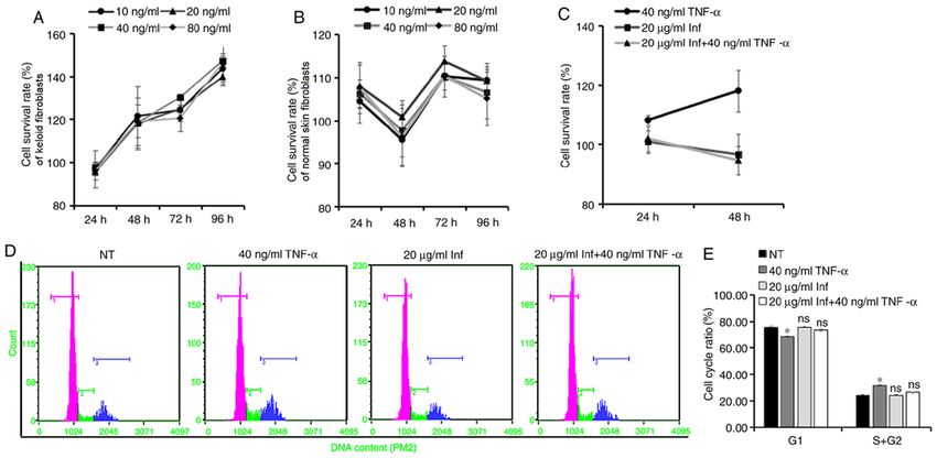
EXPERIMENTAL AND THERAPEUTIC MEDICINE 21: 502, 2021 3
(cat. no. ab19139) and TNFR2 (cat. no. ab109322) were all as follows: β‑actin forward, 5'‑CGAGGCCCAGAGCAAGAG
purchased from Abcam; rabbit polyclonal antibodies against AG‑3' and reverse, 5'‑CGGTTGGCCTTAGGGTTCAG‑3'; TNF‑α
inhibitor of NF‑κ B (Iκ B‑α; cat. no. 9242s), phosphorylated forward, 5'‑GGCAGTCAGATCATCTTCTCGA‑3' and reverse,
(p)‑SAPK/JNK (cat. no. 4668s), p38 MAPK (cat. no. 9212), 5'‑CGGTTCAGCCACTGGAGCT‑3'; TNFR1 forward, 5'‑CAA
p‑p38 MAPK (cat. no. 4511s), p‑Iκ B‑α (cat. no. 9246s) and GTGCCACAAAGGAACCTAC‑3' and reverse, 5'‑CAGCTG
SAPK/JNK (cat. no. 9252s) were all obtained from Cell AGGCAGTGTCTGA‑3'; TNFR2 forward, 5'‑CTATGACCAGAC
Signaling Technology, Inc.; mouse polyclonal antibody against AGCTCAGATG‑3' and reverse, 5'‑CAGTTCCAGAGCTGGGTG
β‑actin (cat. no. 60008‑1‑lg) was purchased from PeproTech, TAT‑3'; IL‑6 forward, 5'‑ATGCAATAACCACCCCTGAC‑3' and
Inc.; horseradish peroxidase (HRP)‑labeled goat anti‑mouse reverse, 5'‑CTGCGCAGAATGAGATGAGT‑3'. The SYBR-Green
IgG (H+L) (cat. no. ZB‑2305) and HRP‑labeled goat anti‑rabbit qPCR amplification conditions were as follows: Initial denaturation
IgG (H+L) (cat. no. ZB‑2301) were obtained from OriGene for 1 min at 95˚C; 40 cycles of 10 sec at 95˚C and 30 sec at 58˚C.
Technologies, Inc. Sequence detection software, version 1.2.3 (Applied Biosystems;
Thermo Fisher Scientific, Inc.) was used to analyze the Cq value of
Primary culture of fibroblasts. On a clean bench, tissue each amplification reaction in the qPCR, and the 2‑ΔΔCq method was
samples were washed with PBS solution containing used for relative quantitative analysis (13).
100 U/ml penicillin/streptomycin three times, cut into small
pieces (0.5‑1 mm3) with eye scissors, and then seeded every Immunohistochemistry. Fixed tissue was embedded in paraffin
1 cm into a T25 culture bottle (Thermo Fisher Scientific, Inc.). and cut into 4‑µm serial sections. After dewaxing sections to
The culture bottle was placed upside down for 2 h at 37˚C, water, the sections were treated with 3% hydrogen peroxide in
5% CO2 and 95% saturated humidity, after which time the methanol for 30 min, then washed with 0.01 M PBS for 5 min
cells were supplemented with DMEM (Gibco; Thermo Fisher and the process was repeated three times. After blocking
Scientific, Inc.) containing 10% FBS (Gibco; Thermo Fisher with 5% bovine serum albumin (Biosharp Life Sciences) in
Scientific, Inc.) and 100 U/ml penicillin/streptomycin. Culture PBS for 20 min at 37˚C, the sections were incubated with
was continued in the incubator until the fibroblasts grew out of diluted primary antibodies against TNF‑ α (1:100), TNFR1
the tissue block and covered the bottom of the culture bottle. (1:50) or TNFR2 (1:50) overnight at 4˚C, then washed with
Cells used for experiments were between passages 3 and 5. 0.01M PBS for 5 min and the process was repeated three
Keloid and normal skin fibroblasts were respectively treated times. Next, the sections were incubated with secondary
with 20, 40, 80 ng/ml TNF‑α for 0, 24, 48, 72 and 96 h to antibodies (1:200; cat. no. ZB‑2301; OriGene Technologies,
detect the cell survival rate using CCK‑8 assay. Keloid fibro‑ Inc.) labelled with HRP for 1 h at 37˚C. After staining with
blasts were treated with 40 ng/ml TNF‑α, 20 µg/ml infliximab 3,3'‑diaminobenzidine for 10 min and hematoxylin solution
or 20 µg/ml infliximab + 40 ng/ml TNF‑α for 0, 24 and 48 h to for 20 sec at room temperature, the expression and distribu‑
detect the cell survival rate using CCK‑8 assay and for 48 h to tion of the target proteins were observed under an inverted
analyze the cell cycle by flow cytometry, and were also treated phase contrast microscope (magnification, x400; Olympus
with 40 ng/ml TNF‑α for 3 h to detect the phosphorylation of Corporation). The results were analyzed using Image-Pro Plus
JNK, p38 and Iκ Bα by western blot. software 6.0 (Media Cybernetics, Inc.) and scored according
to Crambert et al (14).
ELISA. A total of 50 µl diluted sample (100X diluted serum, or
10X diluted cell culture supernatant) were added into each well CCK‑8 assay. Cells were seeded in a 96‑well plate at a density
and incubated for 1 h at 37˚C, followed by the addition of 100 µl of 5x10³ cells/well. When observed at exponential growth,
biotin antibody to each well and further incubation for 1 h cells were treated as aforementioned in the ‘Primary culture
at 37˚C. Each well was aspirated and washed, and the process of fibroblasts’ section and incubated for 24, 48, 72 and 96 h.
was repeated twice for a total of three washes. Subsequently, Next, 10 µl CCK‑8 solution was added to each well and
100 µl HRP‑avidin was added into each well and incubated for incubated for 4 h at 37˚C. Subsequently, the absorbance was
1 h at 37˚C. After washing, 100 µl 3,3', 5,5'‑tetramethylbenzi‑ measured on a microplate reader (Thermo Fisher Scientific,
dine substrate solution was added to each well, followed by an Inc.) at 450 nm. Cell viability was calculated as follows: Cell
incubation for 30 min at 37˚C. Stop solution (50 µl) was then viability (%)=[treated (OD)‑blank (OD)/control (OD)‑treated
added, and the optical density of each well was determined (OD)] x100%.
within 15 min, using a microplate reader (Multiskan™ FC;
Thermo Fisher Scientific, Inc.) set to 450 nm. Flow cytometry. The cells were harvested, fixed in 70% cold
ethanol for 16 h overnight at 4˚C, and centrifuged at 300 x g
Total RNA extraction and quantitative PCR (qPCR) analysis. for 5 min at 4˚C. The supernatants were discarded. Then, the
The keloid and normal skin dermis stored in liquid nitrogen cell pellet was stained with 0.4 ml propidium iodide staining
were ground in a mortar filled with liquid nitrogen and then total solution in a bath at 37˚C for 30 min in the dark. Cell cycle
RNA was extracted according to the manufacturer's protocol of distribution was analyzed using FACSCalibur flow cytometer
the RNA extraction kit (Aidlab Biotechnologies Co., Ltd.). Total (BD Biosciences), and the percentage of cells in each cell cycle
RNA was quantified using a microspectrophotometer (NanoDrop phase was evaluated (FACSCalibur software version 1.3; BD
2000; NanoDrop Technologies; Thermo Fisher Scientific, Inc.), Biosciences).
and reverse transcribed into cDNA using the aforementioned
reagent (1 h at 42˚C and 10 min at 70˚C). The qPCR primers were Western blot analysis. The cells were collected and lysed in
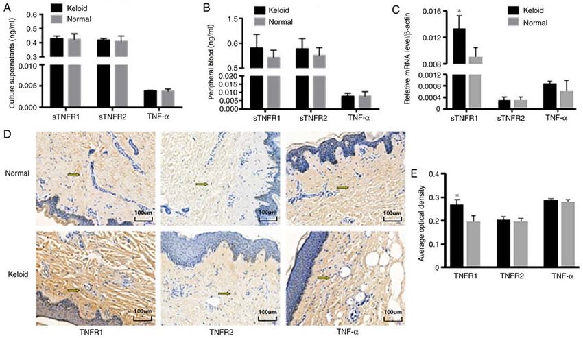
synthesized by Sangon Biotech Co., Ltd., and their sequences are RIPA buffer (Beyotime Institute of Biotechnology), then total4 LI et al: TNF-α PROMOTES HYPERPROLIFERATION OF KELOID FIBROBLASTS Figure 1. Expression of TNF‑α, sTNFR1 and sTNFR2 in keloid tissue and normal skin. The content of TNF‑α, sTNFR1 and sTNFR2 was detected by ELISA (A) in the culture supernatants of (n=9) of primary fibroblasts isolated from keloid or normal skin of patients, and (B) in peripheral blood samples (n=5) from patients with keloids or healthy participants. (C) mRNA levels of TNF‑α, sTNFR1 and sTNFR2 in keloid or normal fibroblasts were detected by quantitative PCR analysis and β‑actin served as the reference gene (n=6). (D) Dermis from patients with keloids or healthy participants was embedded in paraffin and used to detect the protein levels of TNF‑α, sTNFR1 and sTNFR2 by immunohistochemistry (magnification, x400). (E) Results were quantitated and the mean optical density was plotted as a histogram to compare the protein levels (healthy n=9; keloid n=10). *P0.05; Fig. 1B). Surgical specimens of SPSS 16.0 (SPSS Inc.). For normally distributed data, indepen‑ keloids and normal skin were used to study the expression dent samples t‑test was applied to analyze differences between of the aforementioned molecules at the mRNA and protein
EXPERIMENTAL AND THERAPEUTIC MEDICINE 21: 502, 2021                                                                  5

Figure 2. TNF‑α regulates the proliferation of keloid fibroblasts. (A) Keloid or (B) normal skin fibroblasts were stimulated with different concentrations (0, 10,
20, 40 and 80 ng/ml) of TNF‑α recombinant protein, and the CCK‑8 assay results were used to evaluate the effect of the treatment on cell proliferation (n=6).
The untreated group at each time point was set to 100% and compared with the cell survival rates of the corresponding treated groups. Keloid fibroblasts were
pretreated with Inf, and were subsequently stimulated with or without TNF‑α recombinant protein (40 ng/ml). (C) CCK‑8 assay results were used to evaluate
the effect of the treatment on cell proliferation (n=6). (D) Cell cycle distribution was analyzed after 48 h treated with TNF‑α and infliximab by flow cytometry
(rose color indicates the G1 cell count; green indicates the S phase cell count; and blue indicates the G2 phase cell count), and (E) cell cycle ratio was plotted
as a histogram to compare the number of cells in different phases of the cell cycle (n=3). *P0.05),                          phase cells compared with the NT group, while pretreatment
while the sTNFR1 mRNA level in KFs was significantly                               with infliximab eliminated this increased proportion of S and
higher compared with that in normal skin (P0.05), while the sTNFR1 protein level in keloid                      TNF‑ α regulates the NF‑κ B, JNK and p38 MAPK pathways
tissue was significantly higher compared with that in normal                       in KFs. The aforementioned results demonstrated that TNF‑α
skin (P
6                                LI et al: TNF-α PROMOTES HYPERPROLIFERATION OF KELOID FIBROBLASTS

Figure 3. TNF‑ α regulates NF‑κ B, JNK and p38 MAPK pathway activation in keloid fibroblasts. (A) Keloid fibroblasts were stimulated with or without
40 ng/ml TNF‑ α recombinant protein for 24 h, and the IL‑6 mRNA level in keloid fibroblasts was detected by quantitative PCR analysis, with ACTB
serving as the reference gene (n=6). Keloid fibroblasts were stimulated with or without 40 ng/ml TNF‑α recombinant protein for 30 min, and the protein
levels of (B) p‑JNK, JNK and β‑actin, (C) Iκ B‑α, p‑Iκ B‑α and β‑actin, and (D) p‑p38, p38 and β‑actin were detected by western blotting (n=3). *P
EXPERIMENTAL AND THERAPEUTIC MEDICINE 21: 502, 2021                                                               7

Figure 4. TNF‑α promotes KF hyperproliferation by activating the NF‑κ B, JNK and p38 MAPK pathways. Increased expression of TNFR1 in KFs compared
with that in normal fibroblasts may contribute to their enhanced sensitivity to TNF‑α, and then the persistent stimulation by a low concentration of TNF‑α may
lead to repeated activation of the NF‑κ B, JNK and p38 pathways, thereby promoting the sustained and excessive proliferation of KFs. TNF, tumor necrosis
factor; TNFR, TNF receptor; NF‑κ B, nuclear factor‑κ B; JNK, c‑Jun N‑terminal kinase; MAPK, p38 mitogen‑activated protein kinase; KF, keloid fibroblast;
NIK, NF‑κ B inducing kinase; TRADD, tumor necrosis factor receptor type 1‑associated DEATH domain protein.

membrane bound TNF‑α. TNFR1 mediates almost all known                            its biological effects, its higher expression may lead to more
biological effects of TNF‑ α. The activation of TNFR1 can                        potent sensitization of KFs to the stimulatory action of TNF‑α.
further activate a variety of signal transduction pathways                           Hyperproliferation of fibroblasts is the main cause
by recruiting a series of intracellular adaptor proteins (7,11).                 underlying keloid formation and progression (3). Previous
Since sTNFR1, the main receptor of TNF‑α, has been found                         studies have demonstrated that TNF‑α is involved in various
to be more highly expressed in keloid tissue compared with                       fibrotic diseases. Miyazaki et al (17) found that transgenic
normal skin, it was hypothesized that KFs exhibit increased                      mice expressing high levels of TNF‑α in the lung were more
sensitivity to TNF‑α compared that in normal skin fibroblasts.                   susceptible to fibroalveolitis compared with those with low
The expression of the TNF‑α receptors sTNFR1 and sTNFR2,                         levels of TNF‑α. Guo et al (18) reported that the renal intersti‑
was also detected in the current study. There was no signifi‑                    tial volume of TNFR1‑/‑/TNFR2‑/‑ double knockout mice was
cant difference in the expression of TNFR2 between keloid                        significantly lower compared with that of the control group.
and normal skin, but the expression of TNFR1 in keloid tissues                   Gurevitch et al (19) found that cardiac fibroblasts were able
and fibroblasts cultured in vitro was higher compared with that                  to proliferate and transform into myofibroblasts under TNF‑α
in normal skin. It was previously reported that the expression                   stimulation. Weiner et al (20) demonstrated that TNF‑ α
of TNFR1 in normal skin was significantly higher compared                        directly promoted the proliferation of Ito cells and fibroblasts
with that in keloid tissue (15), but this was contradictory to                   in the liver, and Elias et al (21) reported that TNF‑α promoted
the hypothesis that the upregulation of TNFR1 inhibits apop‑                     the proliferation of lung fibroblasts. All these previous findings
tosis and promotes the proliferation of fibroblasts. In addition,                indicate that TNF‑α can promote fibroblast proliferation. In
Peruccio et al (16) observed that the TNFR1 mRNA level in                        the present study, the regulatory effect of TNF‑α on the prolif‑
hypertrophic scars after burn injury was lower compared with                     eration of KFs was investigated. The results demonstrated that
that in keloids. This conclusion was different from the findings                 40 ng/ml TNF‑α could promote the proliferation of KFs, but
of the present study, as hypertrophic scars after burn injury                    not that of normal fibroblasts. As an increased expression of
were not examined in the present study, and it may be hypoth‑                    TNFR1 potentially increases the sensitivity of KFs to TNF‑α,
esized that this is due to the different scar types and testing                  it may be hypothesized that, during the process of keloid
sites of surgical specimens, as previous studies either focused                  formation, KFs with a higher sensitivity to TNF‑α are under
on hypertrophic scars or did not explicitly mention the testing                  constant stimulation. Consequently, the intracellular signaling
sites of specimens (15,16). Based on the results of the present                  pathways are repeatedly activated, eventually leading to
study, as TNFR1 is the main receptor of TNF‑α mediating                          sustained and excessive fibroblast proliferation.
8                           LI et al: TNF-α PROMOTES HYPERPROLIFERATION OF KELOID FIBROBLASTS

    The present study further investigated the mechanism           University written consent was obtained from all the partici‑
underlying the role of TNF‑α in the regulation of KF prolifera‑    pants.
tion. After binding to TNFR1, TNF‑α may activate NF‑κ B,
JNK, p38 MAPK, or other intracellular signaling pathways.          Patient consent for publication
Since the activation of these signaling pathways may differ
among different cell types, the specific pathways activated        Not applicable.
in KFs have not yet been fully elucidated. NF‑κ B is the most
important signaling molecule for TNF‑α to exert its promoting      Competing interests
effect on cell proliferation and inflammation (10,22), whereas
the JNK and p38 MAPK pathways may also promote cell                The authors declare that they have no competing interests.
survival and proliferation (11,22,23). In the present study, it
was observed that TNF‑α simultaneously activated the NF‑κ B,       References
JNK and p38 MAPK pathways in KFs, which suggested that
the proliferation of KFs induced by TNF‑α stimulation was           1. Ikeda K, Torigoe T, Matsumoto Y, Fujita T, Sato N and
closely associated with the activation of these pathways. This         Yotsuyanagi T: Resveratrol inhibits fibrogenesis and induces
                                                                       apoptosis in keloid fibroblasts. Wound Repair Regen 21: 616‑623,
was consistent with previous findings reporting the promotion          2013.
of cell proliferation by these pathways (7,11,22,23). Therefore,    2. Hunasgi S, Koneru A, Vanishree M and Shamala R: Keloid:
it was inferred that the persistent activation of the NF‑κ B,          A case report and review of pathophysiology and differences
                                                                       between keloid and hypertrophic scars. J Oral Maxillofac
JNK and p38 pathways may be the mechanism underlying                   Pathol 17: 116‑120, 2013.
TNF‑α‑induced KF proliferation (Fig. 4).                            3. Bettinger DA, Yager DR, Diegelmann RF and Cohen IK: The
    In conclusion, compared with normal fibroblasts, increased         effect of TGF‑beta on keloid fibroblast proliferation and collagen
                                                                       synthesis. Plastic Reconst Surg 98: 827‑833, 1996.
expression of TNFR1 in KFs may confer a unique sensitivity          4. Harty M, Neff AW, King MW and Mescher AL: Regeneration or
to TNF‑α, and persistent stimulation by a low concentration of         scarring: An immunologic perspective. Dev Dyn 226: 268‑279,
TNF‑α may lead to repeated activation of the NF‑κ B, JNK and           2003.
                                                                    5. Dong XL, Mao SL and Wen H: Upregulation of proinflamma‑
p38 pathways, thereby promoting the sustained and excessive            tory genes in skin lesions may be the cause of keloid formation
proliferation of KFs.                                                  (Review). Biomed Rep 1: 833‑836, 2013.
                                                                    6. Frances B: TNF‑alpha in promotion and progression of cancer.
                                                                       Cancer Metastasis Rev 25: 409‑416, 2006.
Acknowledgements                                                    7. Bahcecioglu IH, Koca SS, Poyrazoglu OK, Yalniz M, Ozercan
                                                                       IH, Ustundag B, Sahin K, Dagli AF and Isik A: Hepatoprotective
Not applicable.                                                        effect of infliximab, an anti-TNF-α agent, on carbon tetrachlo‑
                                                                       ride-induced hepatic fibrosis. Inflammation 31: 215-221, 2008.
                                                                    8. McCauley RL, Chopra V, Li YY, Herndon DN and Robson MC:
Funding                                                                Altered cytokine production in black patients with keloids. J Clin
                                                                       Immunol 12: 300‑308, 1992.
                                                                    9. Messadi DV, Doung HS, Zhang Q, Kelly AP, Tuan TL,
The present study was supported by the Sichuan Basic Applied           Reichenberger E and Le AD: Activation of NFkappaB signal
Research Project (grant no. 2017FZ0055), the Post‑Doctor               pathways in keloid fibroblasts. Arch Dermatol Res 296: 125‑133,
                                                                       2004.
Research Project, West China Hospital, Sichuan University          10. Zhu GY, Cai JL, Zhang J, Zhao YR and Xu B: Abnormal nuclear
(grant no. 2018HXBH082) and the China Postdoctoral Science             factor (NF)‑kappaB signal pathway and aspirin inhibits tumor
Foundation (grant no. 2019M653413).                                    necrosis factor α‑induced NF‑kappaB activation in keloid fibro‑
                                                                       blasts. Dermatol Surg 33: 697‑708, 2007.
                                                                   11. Wajant H, Pfizenmaier K and Scheurich P: Tumor necrosis factor
Availability of data and materials                                     signaling. Cell Death Differ 10: 45‑65, 2003.
                                                                   12. Gauglitz GG, Korting HC, Pavicic T, Ruzicka T and Jeschke MG:
                                                                       Hypertrophic scarring and keloids: Pathomechanisms and
The datasets used and/or analyzed during the current study are         current and emerging treatment strategies. Mol Med 17: 113‑125,
available from the corresponding author on reasonable request.         2011.
                                                                   13. Livak KJ and Schmittgen TD: Analysis of relative gene expres‑
                                                                       sion data using real‑time quantitative PCR and the 2(‑Delta Delta
Authors' contributions                                                 C (T)) method. Methods 25: 402‑408, 2001.
                                                                   14. Crambert G, Li C, Claeys D and Geering K: FXYD3 (Mat‑8),
QL and FC participated in the design of the experiments,               a new regulator of Na, K‑ATPase. Mol Biol Cell 16: 2363‑2371,
                                                                       2005.
optimization of experimental conditions, conduction of the         15. Liu LP, Chen FC, Guo LL and Chen YT: Expression and
experiments and writing of the manuscript. KZ, LF and JW               significance of apoptosis‑related TNFR1 and Bcl‑2 in fibroblasts
participated in the recruitment of participants and the collec‑        derived from pathologic scar. Chin J Prac Aesthetic and Plastic
                                                                       Surg 15: 272‑274, 2004.
tion of clinical specimens. QX and YC participated in the          16. Peruccio D, Castagnoli C, Stella M, D'Alfonso S, Momigliano PR,
experimental design and data analysis. JC and YQ participated          Magliacani G and Alasia ST: Altered biosynthesis of tumour
in designing the present study and revising the manuscript and         necrosis factor (TNF) alpha is involved in postburn hypertrophic
                                                                       scars. Burns 20: 118‑121, 1994.
confirmed the authenticity of all the raw data. All authors read   17. Miyazaki Y, Araki K, Vesin C, Garcia I, Kapanci Y, Whitsett JA,
and approved the final manuscript.                                     Piguet PF and Vassalli P: Expression of a tumor necrosis
                                                                       factor‑alpha transgene in murine lung causes lymphocytic and
                                                                       fibrosing alveolitis. A mouse model of progressive pulmonary
Ethics approval and consent to participate                             fibrosis. J Clin Invest 96: 250‑259, 1995.
                                                                   18. Guo G, Morrissey J, McCracken R, Tolley T and Klahr S: Role
                                                                       of TNFR1 and TNFR2 receptors in tubulointerstitial fibrosis
The protocol of the present study was reviewed and approved            of obstructive nephropathy. Am J Physiol 277: F766‑F772,
by the Ethics Committee of West China Hospital of Sichuan              1999.
EXPERIMENTAL AND THERAPEUTIC MEDICINE 21: 502, 2021 9
19. Gurevitch J, Frolkis I, Yuhas Y, Lifschitz‑Mercer B, Berger E, 22. Lamb JA, Ventura JJ, Hess P, Flavell RA and Davis RJ: JunD
Paz Y, Matsa M, Kramer A and Mohr R: Anti‑tumor necrosis mediates survival signaling by the JNK signal transduction
factor‑alpha improves myocardial recovery after ischemia and pathway. Mol Cell 11: 1479‑1489, 2003.
reperfusion. J Am Coll Cardiol 30: 1554‑1561, 1997. 23. Ventura JJ, Hübner A, Zhang C, Flavell RA, Shokat KM and
20. Weiner FR, Giambrone MA, Czaja MJ, Shah A, Annoni G, Davis RJ: Chemical genetic analysis of the time course of signal
Takahashi S, Eghbali M and Zern MA: Ito‑cell gene expression transduction by JNK. Mol Cell 21: 701‑710, 2006.
and collagen regulation. Hepatology 11: 111‑117, 1990.
21. Elias JA, Freundlich B, Kern JA and Rosenbloom J: Cytokine This work is licensed under a Creative Commons
networks in the regulation of inflammation and fibrosis in the Attribution-NonCommercial-NoDerivatives 4.0
lung. Chest 97: 1439‑1445, 1990. International (CC BY-NC-ND 4.0) License.You can also read