Osteocalcin, osteopontin and RUNX2 expression in patients with arteriosclerosis
←
→
Page content transcription
If your browser does not render page correctly, please read the page content below
WORLD ACADEMY OF SCIENCES JOURNAL 3: 32, 2021

                    Osteocalcin, osteopontin and RUNX2 expression
                            in patients with arteriosclerosis
                   JÖRG UKKAT, CUONG HOANG-VU, BOGUSZ TROJANOWICZ and ARTUR REBELO

                             Department of Visceral, Vascular and Endocrine Surgery, Martin Luther
                                  University Halle-Wittenberg, 06120 Halle (Saale), Germany

                                     Received February 10, 2021; Accepted April 20, 2021

                                                   DOI: 10.3892/wasj.2021.103

Abstract. Knowledge
            �������������������������������������������������
                         of the role of the skeleton as an endo�   Introduction
crine organ has increase over the past years. The role of
calcification in arteriosclerotic disease has also been a matter   The understanding of the role of the skeleton as an endocrine
of investigation. Since there is a clear association between the   organ has increased over the past years (1). Calcification is a
levels of osteocalcin (OC), osteopontin (OPN) and Runt‑related     highly relevant process in terms of the development of cardio�
transcription factor 2 (RUNX2) and the development of arte�        vascular diseases (CVDs). Arteriosclerosis, Mönckeberg
riosclerotic vascular disease, the present study examined the      medial calcific sclerosis and arteriolosclerosis are the three
expression of OC, OPN and RUNX2 in vascular tissue and their       main lesions derived from classification (2,3). In patients with
possible employment as diagnostic markers for cardiovascular       end‑stage chronic kidney disease (CKD), arterial calcification
disease. For this purpose, 13 vessel tissues, including tissues    is an independently predictive factor of CVD and mortality (4).
from six patients without arteriosclerotic disease (PAD‑)          The prevention of CVD is currently a highly relevant topic.
and 7 patients with arteriosclerotic disease (PAD+) were           CVDs are a group of disorders affecting the heart and blood
obtained by surgical resection. Immunohistochemistry and           vessels. It has been demonstrated that 17.9 million individuals
reverse transcription‑quantitative polymerase chain reaction       worldwide succumb to CVDs annually, representing 31% of
(RT‑qPCR) analysis were performed. The results revealed            all deaths worldwide (5). Thus, establishing novel and reli�
that tissues representing PAD+ exhibited a noticeably stronger     able predictive biomarkers for the identification of patients
protein expression of OC, OPN and RUNX2 as compared                with related calcification is a highly relevant topic, of utmost
with the corresponding PAD‑ tissues. The transcript levels of      urgency. Vascular calcification is a potential predictor of future
OPN and RUNX2 were associated with protein expression and          cardiovascular events in asymptomatic patients (6).
were significantly elevated in PAD+ (OPN, P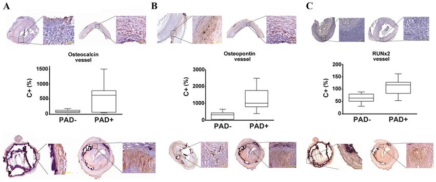
2                         UKKAT et al: OSTEOCALCIN, OSTEOPONTIN AND RUNX2 IN ARTERIOSCLEROSIS

in blood, the presence of OC‑positive cells, or histological        transcribed using the Superscript II kit (Gibco; Thermo
staining for OC were compared to the extent of calcification        Fisher Scientific, Inc.) at 42˚C for 30 min followed by enzyme
or atherosclerosis. A total of 26 positive, 17 negative and         inactivation at 95˚C for 5 min. The qPCR reactions for OC
29 neutral associations were reported (11).                         were performed using the Rotor‑Gene System (Qiagen GmbH)
     Osteopontin (OPN) is also a protein with a major role in       and qPCRBIO Probe Mix (Nippon Genetics). Samples were
mineralization and calcification; however, it also has multifunc�   amplified as double replicates by the employment of TaqMan
tional roles in physiological and pathophysiological processes.     assays specific for OC (Hs01587814_g1 BGLAP) and ACTB
Its role in inflammatory processes has been studied in over the     (Hs99999903‑m1 ACTB). The thermal cycling conditions for
years (12). It has been demonstrated that low levels of OPN         TaqMan were as follows: Holding for 10 min at 95˚C, 40 cycles
may play a protective role concerning vascular tissue injury        of 10 sec/95˚C and 30 sec/60˚C. OPN and RUNX2 were
and high levels may have potentially damaging effects (13).         amplified with 5X HOT FIREPol® EvaGreen® qPCR Mix
Runt‑related transcription factor 2 (RUNX2), is also a bone         (Solis Biodyne) using specific primers (Table II) and under
regulator and has been detected in calcified atherosclerotic        following conditions: Holding for 10 min at 95˚C, followed
lesions (14,15). Furthermore, the role of RUNX2 in vascular         by 40 cycles of 15 sec at 95˚C, 30 sec at 60˚C and 30 sec at
calcification has been studied in in recent years (16,17).          72˚C. Normalization was performed with primers specific
     Since there is a clear association between OC, OPN and         for ACTB, GAPDH, 18S and ACTB TaqMan Probes in the
RUNX2, and the development of arteriosclerotic vascular             case of OC (Table II). Data evaluation was performed using
disease, the present study investigated the expression of           Rotor‑Gene 2.0.2 software and the comparative quantifica�
OC, OPN and RUNX2 in vascular tissue, and their possible            tion method (2‑ΔΔCq) (18), which calculated the efficiencies of
employment as diagnostic markers for CVD.                           each gene for each individual PCR reaction (is based on the
                                                                    second differential maximum method (19) to calculate single
Patients and methods                                                reaction efficiencies. Briefly, reported concentrations of each
                                                                    PCR‑product, including housekeeping genes, were compared
Patients and tissue preparation. A total of 13 vessel tissues,      to the positive control (C+; cDNA from follicular thyroid
including tissues from six patients without arteriosclerotic        cancer cells, FTC‑133; cat. no. 94060901‑1VL, Sigma‑Aldrich;
disease (PAD‑) and seven patients with arteriosclerotic             Merck KGaA) set as 1. Thereafter, all reported concentrations
disease (PAD+) were collected at the Department of Surgery          were transformed to percentage values (C+ as 100%) and
of Martin‑Luther‑University Halle, Halle (Saale), Germany           target transcripts were normalized to the housekeeping genes.
by surgical resection (Table I). The patients in the control        For the whole study, the same C+, the same negative controls
group (without arteriosclerotic disease) were mostly patients       and the same housekeeping genes were employed.
that underwent arterial resections in oncologic surgery. The
pathological diagnosis of the tissue sections was confirmed         H&E staining. H&E staining and morphological investiga�
by hematoxylin and eosin (H&E) staining. All tissues were           tions of selected pathologically changed and not changed
snap frozen in liquid nitrogen and stored at ‑80˚C for further      vessels were performed. As demonstrated in Fig. 1, freshly cut
proceedings. Total RNA was extracted from all samples using         cryo‑embedded serial 6‑µm‑thick sections of selected vessel
TRIzol® reagent (Thermo Fisher Scientific, Inc.) according to       tissues were air‑dried at room temperature, shortly washed
the manufacturer's instructions.                                    with cold Acetone (Sigma‑Aldrich; Merck KGaA) and dried
    Additionally, selected tissues from all groups were             again. Thereafter, the sections were shortly washed with
investigated in immunohistochemical assays with antibodies          distilled water and incubated for 1 min at room temperature
specific for OC, OPN and RUNX2, as described below. The             in Hämalaun (Merck) solution followed by washing twice with
Ethics Committee of the Martin Luther University, Faculty of        tap water. The sections were then incubated for 1 min in eosin
Medicine, approved the study and all patients provided written      (Merck KGaA) solution, washed with tap water, shortly washed
informed consent.                                                   twice with 96% ethanol and finally shortly dipped in xylol (Carl
                                                                    Roth GmbH Chemikalien). The sections were then covered
RNA isolation. Briefly, the vessels were snap‑frozen in liquid      with Entellan® (Merck KGaA) and coverslips and examined
nitrogen and homogenized in Teflon‑Tubes at 2,500 rpm for           under a under a light microscope (Axiovert 200; Zeiss GmbH).
45 sec by employing Mikro‑Dismembrator S (Braun Biotech             The staining of BMP4 was performed on cryosections fixed
International GmbH). The resulting powder tissue (ca. 100 mg)       with methanol/H2O2 and BMP4 antiserum (Abcam ab155033)
was mixed with 1 ml TRIzol® reagent, vortexed for 30 sec            diluted 1:100 in TBS. Tissues were lightly counterstained with
and incubated at room temperature for 5 min. Following the          Mayer's hematoxylin (Merck KGaA) and photographed under
addition of 0.2 ml chloroform, the samples were centrifuged         a light microscope (Biozero‑3000; Keyence).
15 min by 14,000 x g at 4˚C and RNA‑containing upper phase
was collected and mixed with 0.5 ml isopropanol. Following          Immunohistochemistry. Freshly cut cryo‑embedded serial
centrifugation (10 min by 14,000 x g at 4˚C) the pelleted RNA       6‑µm‑thick sections of selected vessel tissues were washed
was washed twice with 75% ethanol, air‑dried and dissolved in       with PBS and fixed in a 1:4 mixture of 3% H 2O 2 (Carl
RNase‑free water. Finally, the dissolved RNA was incubated          Roth GmbH Chemikalien) in ice‑cold 90% methanol
for 10 min at 60˚C and stored at ‑80˚C until use.                   (Sigma‑Aldrich; Merck KGaA) for 20 min. After washing
                                                                    twice with PBS, the slides were blocked with Envision Flex
Reverse transcription‑quantitative polymerase chain reac‑           Antibody Diluent (Dako; Agilent Technologies, Inc.) for 1 h
tion (RT‑qPCR). A total of 1 µg of total RNA was reverse            at room temperature and then incubated overnight at 4˚C with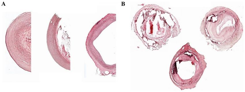
WORLD ACADEMY OF SCIENCES JOURNAL 3: 32, 2021                                                        3

Table I. Clinical data of the patients included in the present study.

                  Peripheral
			                 artery			                      Diabetes     Anti-
Patient Age,		 disease stage Vessel		              mellitus  vitamin K      Vitamin D-	    OC OPN RUNX2
no.     years Sex (Fontaine) location Dyslipidemia type II anticoagulants supplementation score score score

 1         53     M          No          Thyroid   No  Yes No No                                                          1       2         1
 2         78     M          No          Aorta     No		    No No                                                          2       2         0
 3         62     M          No          Aorta	-	-	-	-	                                                                   1       1         1
 4         78     W          No          Femoral   No  No  No No                                                          3       2         2
 5         59     W          No          Neck      No  No  No No                                                          2       1         1
 6         65     M          No          Thyroid   No  No  No No                                                          1       1         1
 7         84     M          III         Popliteal No  No  No No                                                          3       4         4
 8         56     M          III         Femoral   No  No  No No                                                          3       6         6
 9         66     M          IV          Femoral   Yes Yes No Yes                                                         4       4         9
10         77     W          IV          Femoral   No  Yes No No                                                          4       4         8
11         97     W          IV          Femoral   Yes Yes No No                                                          6       6         4
12         74     M          IV          Femoral   No  No  No No                                                          4       4         6
13         83     W          IV          Femoral   Yes No  No No                                                          4       6         6

OC, OPN and RUNX2 scores determined by multiplying two factors: Overall staining intensity of the slide and specific staining. M, male; F, female.

Table II. Primer pairs for the amplifications of target and housekeeping (HKG) gene transcripts.

Gene                                   Primer sequence (5'-3')                             Size (bp)                       Supplier

RUNX2                  Forward: CCCTGAACTCTGCACCAAGT                                          120              Metabion
                       Reverse: GGCTCAGGTAGGAGGGGTAA
Osteopontin            Forward: GCCGAGGTGATAGTGTGGTT                                          149              Metabion
                       Reverse: AACGGGGATGGCCTTGTATG
HKG
 β-actin               Forward: AGG CAC CAG GGC GTG AT                                         51              Metabion
                       Reverse: GCC CAC ATA GGA ATC CTT CTG AC
 GAPDH                 Forward: ACC CAG AAG ACT GTG GAT GG                                    233              Metabion
                       Reverse: TTC TAG ACG GCA GGT CAG GT
 18s                   Forward: GTT GGT GGA GCG ATT TGT CTG G                                 151              Metabion
                       Reverse: AGG GCA GGG ACT TAA TCA ACG C
Osteocalcin            HS01587814_G1 BGLAP                                                    138              Thermo Fisher Scientific, Inc.
HKG
 β-actin               HS99999903-M1 ACTB		                                                                    Thermo Fisher Scientific, Inc.

the rabbit polyclonal antibody against OC (2.5 µg/ml; ab93876,            immunostaining was visualized with diaminobenzidine chro�
Abcam), mouse monoclonal antibody against OPN (1:300;                     mogenic solution (1:50; Dako; Agilent Technologies, Inc.).
ab166709; Abcam) and rabbit polyclonal antibody against                   Finally, the cells were lightly counterstained with Mayer's
RUNX2 (1:1,000; ab23981; Abcam). Negative control sections                hematoxylin (Merck KGaA) and photographed under a light
were exposed to the secondary antibody only and processed                 microscope (Axiovert 200; Zeiss GmbH) (Fig. 1).
as described below. All the following steps were performed at                 Two independent reviewers, semi‑quantitatively using
room temperature. After 3x10 min washing in PBS, the slides               an Axioplan light microscope (Zeiss GmbH), examined all
were incubated for 30 min at room temperature with a 1:10,00              immunostained tissue sections. The OC, OPN and RUNX2
dilution of biotinylated goat anti‑rabbit secondary antibody              scores was determined by multiplying two factors: The oerall
(sc‑2004; Santa Cruz Biotechnology, Inc.) followed by incuba�             staining intensity of the slide and specific staining. In overall
tion with an avidin‑biotin‑peroxidase complex (Dako; Agilent              staining, the slides were evaluated according to a 0‑3 points
Technologies, Inc.). After 3x10  min washing in PBS, specific             scale, where 0 indicates negative staining and 3 the strongest
4 UKKAT et al: OSTEOCALCIN, OSTEOPONTIN AND RUNX2 IN ARTERIOSCLEROSIS
Figure 1. Representative H&E staining of (A) healthy and (B) diseased vessels obtained from PAD‑ and PAD+ femoral arteries. (A) Normal‑appearing femoral
artery with no evidence of intimal thickening or medial calcification and (B) adaptive intimal thickening with focal calcification of the internal elastic lamina.
Magnification: Healthy vessels, x5; diseased vessels, x2.5. There are three representative images of PAD‑ and PAD+ shown for each image. H&E, hematoxylin
and eosin; PAD‑, patients without arteriosclerotic disease; PAD+ patients with arteriosclerotic disease.
staining. The points for specific staining were calculated as PAD+ tissues compared with the PAD‑ group (Fig. 2B). The
follows: 0, 0‑10% (negative); 1, 10‑30% (low expression); median percentage transcript expression for OPN in PAD+ was
2, 30‑50% (moderate expression); 3, 50‑80% (high expres� 1,015% and 322.5% for PAD‑ (P= 0.0085). Specific staining
sion); 4, ≥80% (very high expression). with anti‑OPN antibody also demonstrated the same tendency
as the transcript analyses (Fig. 2B).
Statistical analysis. Each experiment was performed in
quadruplicate. The distribution of the quantitative variables Expression and immunostaining of RUNX2. A significantly
within qPCR data of all investigated transcripts was tested elevated mRNA expression of RUNX2 was observed in the
with Kolmogorov‑Smirnov normality tests. Since the expres� PAD+ group compared with the PAD‑ group (Fig. 2C). The
sion of OC, OPN and RUNX2 passed the normality tests, a median percentage transcript expression for RUNX2 in PAD+
parametric (differences between paired values are consistent) was 117% and 64% in PAD‑ (P=0.0134). As shown in Fig. 2C,
two‑sided t‑test was used. Data are presented as medians with a more intense OC expression was also detected in PAD+ as
inter quartile ranges. For comparisons within vascular tissues, compared with PAD‑.
the Bonferroni correction was applied and P‑values PWORLD ACADEMY OF SCIENCES JOURNAL 3: 32, 2021 5
Figure 2. Analysis of the mRNA expression of (A) OC, (B) OPN and (C) RUNX2 in vascular tissue from PAD+ (top panels) and PAD‑ (bottom panels). An
elevated mRNA expression of OC, OPN and RUNX2 was found in the PAD+ group (middle panels). The median percentage transcript expression for OC in
PAD+ was 634% and 88.5% for PAD‑ (P= 0.067). The median percentage transcript expression for OPN in PAD+ was 1015% and 322.5% for PAD‑ (P= 0.0085).
The median percentage transcript expression for RUNX2 in PAD+ was 117% and for 64% PAD‑ (P= 0.0134). The expression of all transcripts was compared to
the positive control (C+) set as 100% and presented as % values as compared to C+. (A‑C) There are two images of PAD‑ tissues (top panels) and two images
of PAD+ tissues shown (bottom panels) for each marker. The images in all panels are presented at magnifications of x2.5 and zoomed in at x10, respectively.
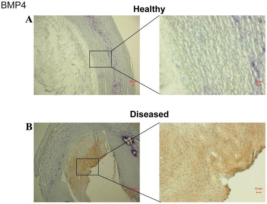
Figure 3. Representative staining of BMP4 in (A) healthy and (B) diseased vessels. The staining was performed on cryosections fixed with methanol/H2O2 and
BMP4 antiserum Note that vessels obtained from arteriosclerotic patients demonstrated much more intense staining for BMP4 than healthy vessels.
In the present study, a clear association was found between the protein kinase Cδ (PKCδ)����������������������������������
/���������������������������������
Toll like receptor 4 (TLR4)������
/�����
reac�
the levels of OC expression and symptomatic arteriosclerotic tive oxygen species/cyclooxygenase‑2 signaling cascade (24).
disease. In a previous study, the same association was made. On the other hand, in another study, no significant associa�
Yuen et al (24) investigated the molecular mechanisms under� tion between vascular calcification and OC (25) was found in
lying the promoting effects of OC on vascular pathogenesis patients undergoing peritoneal dialysis.
and remodeling. The main finding of that study was that OC
transforms adventitial fibroblasts into myofibroblasts by stimu� OPN. In a recent study from Italy, OPN plasma levels were
lating angiotensin II release and the subsequent activation of found to be associated with type II diabetes and obesity (26).6 UKKAT et al: OSTEOCALCIN, OSTEOPONTIN AND RUNX2 IN ARTERIOSCLEROSIS
OPN is also referred to as a marker of the outcome of Funding
patients with vascular diseases, including peripheral artery
disease (13,27). In mice, OPN has been found to be an induc� No funding was received.
ible inhibitor of vascular calcification (28). In adidtion, the
plasma levels of OPN have been shown to be associated with Availability of data and materials
cardiovascular pathologies, such as abdominal aortic aneu�
rysm (29,30). Furthermore, elevated plasma levels of OPN The datasets used and/or analyzed during the current study are
have been discussed as an independent predictor of coronary available from the corresponding author on reasonable request.
calcification in patients with diabetes and asymptomatic coro�
nary disease (31). In the present study, OPN was found to be Authors' contributions
strongly associated with symptomatic arteriosclerotic disease.
The findings presented herein are consistent with previous JU and BT were involved in the conceptualization of the study.
findings that OPN may be a marker of the disease (32). BT was involved in the study methodology and in the provi�
RUNX2. Recently, RUNX2 activity and arteriosclerotic sion of software. CHV was involved in data validation. BT
vascular calcification were found to be strongly associated and CHV were involved in formal analysis. JU and AR were
in patients with diabetes mellitus (33). In another study, involved in the investigative aspects of the study, as well as
Tanaka et al (34) stated that RUNX2 played a major role in in data curation, study supervision and project administration.
osteogenic conversion in arteriosclerotic lesions. In mouse AR was involved in the provision of resources, in the writing
models, RUNX2 has been shown to be strongly associated of the original draft, in the reviewing and editing of the manu�
with the vessel calcification process (35,36). These findings script. All authors confirm the authenticity of all the raw data
support the results of the present study and identify RUNX2 and all authors have read and approved the final manuscript.
as a novel potential drug target in the treatment of patients
with severe calcification due to arteriosclerosis. Ethics approval and consent to participate
It was hypothesized that the diseased vessel wall may act
as chemoattractant for immune cells, which in turn produce The Ethics Committee of the Martin Luther University, Faculty
increased amounts of cytokines and affect endothelial and of Medicine, approved the study and all patients provided
muscle cells. These in turn react with increased or modulated written informed consent.
expression of OC, OPN and RUNX2. In addition, the immune
cells infiltrate vessel wall and induce signaling pathways Patient consent for publication
affecting OC, OPN and RUNX2. Finally, the increased
calcium and/or phosphate levels lead to the dysfunction of Not applicable.
immune cells and/or endothelial cells/muscle cells. As a result,
the levels of OC, OPN and RUNX2 are increased. Competing interests
The present study has some limitations. The present study
lacks functional data, particularly as regards the influence of OC, The authors declare that they have no competing interests.
OPN and RUNX2‑modifying medication on the progression of
arteriosclerotic disease. In addition, whether circulating blood References
levels of OC, OPN and RUNX2 in serum/plasma by means of a
non‑invasive assay may support the development of the disease. 1. Ferron M and Lacombe J: Regulation of energy metabolism by the
Furthermore, the differences in the investigated vessel types skeleton: Osteocalcin and beyond. Arch Biochem Biophys 561:
137‑146, 2014.
for PAD‑ compared with PAD+ (all femoral) may represent a 2. Fishbein GA and Fishbein MC: Arteriosclerosis: Rethinking the
limitation of the present study. The present pilot study was also current classification. Arch Pathol Lab Med 133: 1309‑1316, 2009.
conducted on a small patient collective. However, on the whole, 3. Rennenberg RJ, Kessels AG, Schurgers LJ, van Engelshoven JM,
de Leeuw PW and Kroon AA: Vascular calcifications as a marker
the promising results presented herein justify future investiga� of increased cardiovascular risk: A meta‑analysis. Vasc Health
tions using larger cohorts and make possible the more effective Risk Manag 5: 185‑197, 2009.
diagnosis and follow‑up of these patients. 4. Shobeiri N, Adams MA and Holden RM: Vascular calcification
in animal models of CKD: A review. Am J Nephrol 31: 471‑481,
In conclusion, in the present study, the expression of OC, 2010.
OPN and RUNX2 in arterial tissue was found to be strongly 5. World Health Organization: Cardiovascular Diseases. https://
associated with symptomatic arteriosclerotic disease. However, www.who.int/health‑topics/cardiovascular‑diseases/#tab=tab_1.
Accessed December 2, 2020.
the role that these three markers play in the development of CVD 6. Wexler L, Brundage B, Crouse J, Detrano R, Fuster V, Maddahi J,
remains unclear. Further investigations are required to evaluate Rumberger J, Stanford W, White R and Taubert K: Coronary
the roles of OC, OPN and RUNX2 as potential biomarkers in artery calcification: Pathophysiology, epidemiology, imaging
methods, and clinical applications. Circulation 94: 1175‑1192,
the diagnosis and even the follow‑up of these patients. 1996.
7. Zoch ML, Clemens TL and Riddle RC: New insights into the
Acknowledgements biology of osteocalcin. Bone 82: 42‑49, 2016.
8. Lee NK, Sowa H, Hinoi E, Ferron M, Ahn JD, Confavreux C,
Dacquin R, Mee PJ, McKee MD, Jung DY, et al: Endocrine regu�
The authors would like to thank Ms. Kathrin Hammje from lation of energy metabolism by the skeleton. Cell 130: 456‑469,
Martin Luther University Halle‑Wittenberg for providing 2007.
9. Sheng L, Cao W, Cha B, Chen Z, Wang F and Liu J: Serum osteo�
excellent technical assistance with the creation of this manu� calcin level and its association with carotid atherosclerosis in
script. patients with type 2 diabetes. Cardiovasc Diabetol 12: 22, 2013.WORLD ACADEMY OF SCIENCES JOURNAL 3: 32, 2021 7
10. Tacey A, Qaradakhi T, Brennan‑Speranza T, Hayes A, Zulli A 26. Schinzari F, Tesauro M, Bertoli A, Valentini A, Veneziani A,
and Levinger I: Potential role for osteocalcin in the development Campia U and Cardillo C: Calcification biomarkers and vascular
of atherosclerosis and blood vessel disease. Nutrients 10: E1426, dysfunction in obesity and type 2 diabetes: Influence of oral hypo�
2018. glycemic agents. Am J Physiol Endocrinol Metab 317: E658‑E666,
11. Millar SA, Patel H, Anderson SI, England TJ and O'Sullivan SE: 2019.
Osteocalcin, vascular calcification, and atherosclerosis: A systematic 27. Icer MA and Gezmen‑Karadag M: The multiple functions and
review and meta‑analysis. Front Endocrinol (Lausanne) 8: 183, mechanisms of osteopontin. Clin Biochem 59: 17‑24, 2018.
2017. 28. Speer MY, McKee MD, Guldberg RE, Liaw L, Yang HY,
12. Lund SA, Giachelli CM and Scatena M: The role of osteopontin Tung E, Karsenty G and Giachelli CM: Inactivation of the
in inflammatory processes. J Cell Commun Signal 3: 311‑322, osteopontin gene enhances vascular calcification of matrix Gla
2009. protein‑deficient mice: Evidence for osteopontin as an inducible
13. Lok ZSY and Lyle AN: Osteopontin in vascular disease. inhibitor of vascular calcification in vivo. J Exp Med 196:
Arterioscler Thromb Vasc Biol 39: 613‑622, 2019. 1047‑1055, 2002.
14. Moe SM, Duan D, Doehle BP, O'Neill KD and Chen NX: Uremia 29. Scatena M, Liaw L and Giachelli CM: Osteopontin: A multifunc�
induces the osteoblast differentiation factor Cbfa1 in human tional molecule regulating chronic inflammation and vascular
blood vessels. Kidney Int 63: 1003‑1011, 2003. disease. Arterioscler Thromb Vasc Biol 27: 2302‑2309, 2007.
15. Tyson KL, Reynolds JL, McNair R, Zhang Q, Weissberg PL 30. Golledge J, Muller J, Shephard N, Clancy P, Smallwood L,
and Shanahan CM: Osteo/chondrocytic transcription factors Moran C, Dear AE, Palmer LJ and Norman PE: Association
and their target genes exhibit distinct patterns of expression in between osteopontin and human abdominal aortic aneurysm.
human arterial calcification. Arterioscler Thromb Vasc Biol 23: Arterioscler Thromb Vasc Biol 27: 655‑660, 2007.
489‑494, 2003. 31. Berezin AE and Kremzer AA: Circulating osteopontin as a
16. Li Y, Wang W, Chao Y, Zhang F and Wang C: CTRP13 attenuates marker of early coronary vascular calcification in type two
vascular calcification by regulating Runx2. FASEB J 33: 9627‑9637, diabetes mellitus patients with known asymptomatic coronary
2019. artery disease. Atherosclerosis 229: 475‑481, 2013.
17. Zhu L, Zhang N, Yan R, Yang W, Cong G, Yan N, Ma W, Hou J, 32. Cho HJ, Cho HJ and Kim HS: Osteopontin: A multifunctional
Yang L and Jia S: Hyperhomocysteinemia induces vascular protein at the crossroads of inflammation, atherosclerosis, and
calcification by activating the transcription factor RUNX2 via vascular calcification. Curr Atheroscler Rep 11: 206‑213, 2009.
Krüppel‑like factor 4 up‑regulation in mice. J Biol Chem 294: 33. Lino M, Wan MH, Rocca AS, Ngai D, Shobeiri N, Hou G, Ge C,
19465‑19474, 2019. Franceschi RT and Bendeck MP: Diabetic vascular calcification
18. Livak KJ and Schmittgen TD: Analysis of relative gene expression mediated by the collagen receptor discoidin domain receptor 1
data using real‑time quantitative PCR and the 2(‑ΔΔC(T)) method. via the phosphoinositide 3‑kinase/Akt/Runt‑related transcription
Methods 25: 402‑408, 2001. factor 2 signaling axis. Arterioscler Thromb Vasc Biol 38:
19. Rasmussen R: Quantification on the Light Cycler. In: Rapid Cycle 1878‑1889, 2018.
Real‑Time PCR. Meuer S, Wittwer C and Nakagawara KI (eds). 34. Tanaka T, Sato H, Doi H, Yoshida CA, Shimizu T, Matsui H,
Springer, Heidelberg, pp21-34 2001. Yamazaki M, Akiyama H, Kawai‑Kowase K, Iso T, et al: Runx2
20. Mizokami A, Kawakubo‑Yasukochi T and Hirata M: Osteocalcin represses myocardin‑mediated differentiation and facilitates
and its endocrine functions. Biochem Pharmacol 132: 1‑8, 2017. osteogenic conversion of vascular smooth muscle cells. Mol Cell
21. Li J, Zhang H, Yang C, Li Y and Dai Z: An overview of osteo� Biol 28: 1147‑1160, 2008.
calcin progress. J Bone Miner Metab 34: 367‑379, 2016. 35. Al‑Huseini I, Ashida N and Kimura T: Deletion of Iκ B‑Kinase β
22. Kord‑Varkaneh H, Djafarian K, Khorshidi M and Shab‑Bidar S: in smooth muscle cells induces vascular calcification through
Association between serum osteocalcin and body mass index: A β ‑catenin‑Runt‑related transcription factor 2 signaling. J Am
systematic review and meta‑analysis. Endocrine 58: 24‑32, 2017. Heart Assoc 7: e007405, 2018.
23. Pal SN, Rush C, Parr A, Van Campenhout A and Golledge J: 36. Deng L, Huang L, Sun Y, Heath JM, Wu H and Chen Y: Inhibition
Osteocalcin positive mononuclear cells are associated with the of FOXO1/3 promotes vascular calcification. Arterioscler
severity of aortic calcification. Atherosclerosis 210: 88‑93, 2010. Thromb Vasc Biol 35: 175‑183, 2015.
24. Yuen CY, Wong SL, Lau CW, Tsang SY, Xu A, Zhu Z, Ng CF,
Yao X, Kong SK, Lee HK, et al: From skeleton to cytoskeleton:
Osteocalcin transforms vascular fibroblasts to myofibroblasts via This work is licensed under a Creative Commons
angiotensin II and Toll‑like receptor 4. Circ Res 111: e55‑e66, 2012. Attribution-NonCommercial-NoDerivatives 4.0
25. Ramirez‑Sandoval JC, Casanova I, Villar A, Gomez FE, Cruz C International (CC BY-NC-ND 4.0) License.
and Correa‑Rotter R: Biomarkers associated with vascular calci�
fication in peritoneal dialysis. Perit Dial Int 36: 262‑268, 2016.You can also read