The role of optical coherence tomography guidance in scaffold versus stent optimization
←
→
Page content transcription
If your browser does not render page correctly, please read the page content below
Al Nooryani et al. The Egyptian Heart Journal
https://doi.org/10.1186/s43044-020-00110-z
                                                          (2020) 72:77
                                                                                                                                        The Egyptian Heart
                                                                                                                                                   Journal

 RESEARCH                                                                                                                                            Open Access

The role of optical coherence tomography
guidance in scaffold versus stent
optimization
Arif A. Al Nooryani1, Nagwa A. Abdelrahman1,2*, Hatem A. Helmy2, Yehia T. Kishk2 and Ayman K. M. Hassan2

  Abstract
  Background: Optical coherence tomography showed a great ability to identify adverse features during
  percutaneous coronary intervention with drug-eluting stents and resulted in better clinical outcomes. The study
  aimed to assess the impact of optical coherence tomography on intraoperative decision-making during
  implantation of Absorb bioresorbable scaffolds versus everolimus drug-eluting stents.
  Results: We performed an observational study that included 223 consecutive patients post optical coherence
  tomography-guided implantation of either Absorb bioresorbable scaffolds (162 patients) or everolimus drug-eluting
  stents (61 patients). We studied the influence of optical coherence tomography on intraoperative decision-making
  during implantation of bioresorbable scaffolds versus drug-eluting stents by analyzing the total rate of optical
  coherence tomography-dependent modifications in each device.
  After satisfactory angiographic results, the total rate of required intervention for optical coherence tomography
  detected complications was significantly higher in the bioresorbable scaffolds arm compared to drug-eluting stents
  arm (47.8% versus 32.9%, respectively; p = 0.019). The additional modifications encompassed further optimization in
  the case of device underexpansion or struts malapposition, and even stenting in the case of strut fractures, or
  significant edge dissection.
  Conclusions: Compared to drug-eluting stents, Absord scaffold was associated with a significantly higher rate of
  optical coherence tomography-identified intraprocedural complications necessitating further modifications. The
  study provides some hints on the reasons of scaffolds failure in current PCI practice; it offers a new insight for the
  enhancement of BRS safety and presents and adds to the growing literature for successful BRS utilization.
  Keywords: Bioresorbable scaffold, Optical coherence tomography, Malapposition, Strut fracture

Background                                                                              addition to vasomotion impairment and physiological
Despite significant advances in drug-eluting stents (DES)                               blood flow disturbance [9, 11].
technology, metallic stents are still associated with some                                Bioresorbable vascular scaffold (BRS) development was
drawbacks, as prolonged contact with either the metallic                                a key step in interventional cardiology due to the added
alloy or the polymer accelerates neoatherogenesis with                                  advantage of complete scaffold biodegradation, allowing
increased hazard of thrombosis and revascularization in                                 the recovery of vascular pulsatility, vasomotion, and
                                                                                        endothelial function compared to DES. The most fre-
                                                                                        quently studied and clinically used scaffold is the Absorb
                                                                                        BRS (Abbott Vascular, Santa Clara, California) [8].
* Correspondence: Nagwaabdelrahman@aun.edu.eg
1
 Cardiovascular Department, Al Qassimi Hospital, Sharjah, United Arab                     Recently, interventional cardiology is at critical cross-
Emirates
2
                                                                                        roads due to the dissatisfactions with the Absorb scaf-
 Cardiovascular Department, Faculty of Medicine, Assiut University, Asyut,
                                                                                        fold. The 3–year follow-up of the ABSORB III trial
Egypt

                                        © The Author(s). 2020 Open Access This article is licensed under a Creative Commons Attribution 4.0 International License,
                                        which permits use, sharing, adaptation, distribution and reproduction in any medium or format, as long as you give
                                        appropriate credit to the original author(s) and the source, provide a link to the Creative Commons licence, and indicate if
                                        changes were made. The images or other third party material in this article are included in the article's Creative Commons
                                        licence, unless indicated otherwise in a credit line to the material. If material is not included in the article's Creative Commons
                                        licence and your intended use is not permitted by statutory regulation or exceeds the permitted use, you will need to obtain
                                        permission directly from the copyright holder. To view a copy of this licence, visit http://creativecommons.org/licenses/by/4.0/.
Al Nooryani et al. The Egyptian Heart Journal (2020) 72:77 Page 2 of 8
revealed a higher risk of scaffold thrombosis (ScT). A the case of significant edge dissection or residual lesion
subsequent systematic review and meta-analysis with > post-scaffold deployment.
10,000 patients displayed doubling the risk of ScT [5, 6].
Owing to the inherent differences in recoil characteris- Data collection schedule
tics between BRS and DES and the thicker BRS struts, An identification number was generated for each patient
scaffold underexpansion and malapposition are more in an electronic worksheet. Patient’s data (demographics,
frequent and are possibly incriminated for the increased biological parameters) and coronary angiography review
risk of scaffold thrombosis [2]. including procedure and lesion description according to
Optical coherence tomography (OCT) showed a great the ACC/AHA classification were recorded.
ability to identify adverse features during percutaneous Each stent/scaffold was reviewed to detect if additional
coronary intervention (PCI) with DES, resulting in better intervention was required based on the OCT analysis
clinical outcomes [13]. after what was supposed to be an angiographically suc-
Recently, the 2018 ESC/EACTS Guidelines on myocar- cessful implantation.
dial revascularization upgraded the OCT role due to its
accuracy in detecting intraprocedural complications OCT performance and analysis
such as malapposition, underexpansion, and edge dissec- OCT acquisition was done using either the ILUMIEN
tion [12]. OPTIS PCI Optimization System with the Dragonfly-
In the present study, we aimed to assess the influence Duo imaging catheter (both St. Jude Medical, MN, USA)
of OCT on intraprocedural decision-making during im- or the LUNAWAVE OCT System with the Fastview im-
plantation of Absord BRS versus everolimus-DES. aging catheter (both Terumo, Japan). A non-occlusive
technique with injection of iso-osmolar iodixanol (Visi-
Methods paque) was used for acquisition to limit blood artifacts.
Study design and population Underexpansion was defined as a minimum device
We studied retrospectively consecutive patients (223 pa- area < 80% of the mean proximal and distal reference
tients) who had OCT-guided implantation of either Ab- lumen area [2]. Edge dissection was defined as luminal
sorb BRS or everolimus-DES from March 2013 to surface disruption at the stent edge resulting in a flap
October 2017 at Al Qassimi Hospital, Sharjah, UAE. (Fig. 1a). Residual disease was considered in the case of
All patients (age ≥ 18 years) received one or more of ≥ 50% narrowing of the mean luminal area in the OCT
either Absorb BRS or everolimus-DES, either the Xience images within 5 mm proximal or distal to the stent/scaf-
everolimus-eluting cobalt–chromium stent (Abbott Vas- fold edges. Stent fracture was assumed in the case of
cular, Santa Clara, USA) or the Promus Everolimus- overriding contiguous struts, disconnection from the ex-
Eluting Platinum-Chromium Stent (Boston Scientific, pected device circularity, and isolated struts lying unap-
Massachusetts, USA). Patients who had OCT images posed in the lumen (Fig. 1c, e). BRS strut malapposition
with considerable artifacts were excluded from the was defined as the presence of struts separated from the
study. adjacent vessel wall (Fig. 1g, h). In the case of DES indu-
The influence of OCT guidance on intraoperative cing posterior dropout, malapposition was considered
decision-making during implantation of BRS versus DES when the axial distance between the strut’s surface and
was reported by analyzing the total rate of OCT-based the luminal surface exceeds the strut thickness (90 um)
modification in each device. (Fig. 1i, j) [10].
BRS implantation in 2013–2017 Study endpoint
All patients signed informed consent for scaffold deploy- The study endpoint was the total rate of required modi-
ment and OCT use before starting the procedure. fications based on OCT findings, including further post-
All patients were pretreated with aspirin 300 mg and dilation for underexpansion and malapposition, and add-
either clopidogrel 600 mg or ticagrelor 180 mg as itional BRS/DES implantation in the case of significant
required. edge dissection or strut fracture.
Lesion predilatation was performed upon operators’
discretion in 231 (94.3%) scaffolds at baseline. Scaffolds Statistical analysis
were implanted according to common practice with pro- Data analysis was done using SPSS version 19 (Statistical
gressive inflation. Post-dilatation with non-compliant Package for Social Science). Data were presented as
(NC) balloon was performed in 235 (96%) scaffolds. number, percentage, mean, median, and standard devi-
Planned overlapping-BRS strategy was performed ation. Chi-square test and Fisher’s exact test were used
when the lesion length exceeded the maximum scaffold to compare qualitative variables. Mann-Whitney test was
length (28 mm), while unplanned overlap was done in used to compare quantitative variables in case of non-Al Nooryani et al. The Egyptian Heart Journal (2020) 72:77 Page 3 of 8
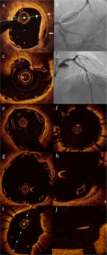
Fig. 1 OCT detected BRS and DES complications. a OCT image of
intimal dissection at the edge of Absorb scaffold (arrow), b PA
caudal view of the same scaffold implanted in proximal LAD (oval
shape) in which edge dissection could not be identified, c OCT
image of calcium spike inducing scaffold fracture with loss of the
expected scaffold circularity, d PA cranial view of the same scaffold
implanted in mid LAD (oval shape) without evidence of fracture, e
OCT image of scaffold fracture in the form of overriding contiguous
struts post culotte bifurcation stenting, f post successful stenting of
the same scaffold using DES, g OCT showing malapposed scaffold
struts, h magnified picture showing scaffold struts clearly separated
from the vessel wall, i OCT picture showing metallic struts
malapposition with posterior dropout, and j magnified picture
showing that the axial distance between the strut’s surface and the
luminal surface exceeds the strut thickness in the malapposing struts
parametric data. A univariate regression analysis was
used to identify the independent predictors of the re-
quirement for OCT-based modifications in each arm
and to estimate odds ratio (OR) and 95% confidence in-
tervals (CIs). A multivariate logistic analysis was used to
identify the independent predictors of OCT-induced PCI
modifications in all studied population. First, the follow-
ing covariates were screened in univariate models: (1)
clinical presentation, (2) angiographic variables (target
vessel, lesion complexity), and (3) procedural variables
(stent type, scaffold/stent diameter). Second, a multivari-
able analysis of predictors at p < 0.20 by univariate ana-
lysis was performed to identify independent predictors
of OCT-induced PCI modifications and to estimate ad-
justed odds ratios and 95% confidence intervals (CIs). p
value < 0.05.was considered statistically significant.
Results
Patients and procedures
Consecutive patients (n:242) post OCT-guided implant-
ation of either Absorb BRS or everolimus-DES from
March 2013 to October 2017 were assessed for eligibil-
ity. Nineteen patients were excluded because of artifacts
rendering proper OCT images analysis unfeasible. We
included 223 patients, with either Absorb BRS (162 pa-
tients; 75% males; mean age 53.5 ± 26 years) or
everolimus-DES (61 patients; 77% males; mean age 51 ±
25 years).
The Absorb arm had 179 lesions (245 scaffolds), ver-
sus 65 lesions (82 stents) in the DES arm.
Patients’ baseline characteristics and PCI indications
were almost similar in both groups (Table 1). Lesion and
procedural characteristics are summarized in Table 2.
OCT-detected intraoperative complications
After satisfactory angiographic results, the total rate of
required intervention for OCT-detected complications
was significantly higher in the BRS arm compared to theAl Nooryani et al. The Egyptian Heart Journal (2020) 72:77 Page 4 of 8
Table 1 Baseline demographics and clinical characteristics
Characteristic Absorb scaffold (N = 162) Everolimus-DES (N = 61) p value
No. (%) No. (%)
Male 122(75.3%) 47(77.0%) 0.787
Age/years 54.36 ± 10.02 54.13 ± 10.89 0.805
Body mass index 28.93 ± 4.17 28.97 ± 5.32 0.361
Hypertension 104(64.2%) 35(57.4%) 0.349
Diabetes mellitus 93(57.4%) 41(67.2%) 0.183
Insulin-treated 29(17.9%) 15(24.6%) 0.263
Dyslipidemia 120(74.1%) 46(75.4%) 0.838
Current smoker 57(36.5%) 23(37.7%) 0.873
Previous myocardial infarction 25(15.4%) 10(16.4%) 0.860
Post-CABG 2(1.2%) 3(4.9%) 0.127
Clinical presentation:
Silent ischemia 19(11.7%) 7(11.5%) 0.958
Stable angina 50(30.9%) 21(34.4%) 0.611
Unstable angina/NSTEMI 63(38.9%) 28(45.9%) 0.928
Primary PCI 20(12.3%) 4(6.6%) 0.214
Late or lysed STEMI 10(6.2%) 1(1.6%) 0.297
Plus–minus values are means ± SD
BMI body mass index is the weight in kilograms divided by the square of the height in meters, CABG coronary artery bypass grafting, DES drug-eluting stents, PCI
percutaneous coronary intervention, STEMI ST-elevation myocardial infarction
DES arm (47.8% versus 32.9%, respectively; p = 0.019), underexpansion, as well as OCT-detected struts fracture
(Table 3). in 4 scaffolds. All 6 lesions were treated successfully with
BRS bioresorbable scaffolds, DES drug-eluting stent DES (Fig. 1c–f). There were no adverse events associated
Stenting for OCT-detected dissection was required in with OCT imaging in the studied population.
6.5% of BRS and 1.2% of DES arms (p = 0.06) (Fig. 1a, NSTEMI presentation, type B2/C lesion, moderate/se-
b), (video 1 and 2 in the supplemental material). Under- vere calcification, and overlapping stents, all were found
expansion necessitating stenting was observed in 2 scaf- to have a significant positive relationship with the need
folds (0.8%) that showed significant underexpansion for intraoperative modification post-OCT review in the
despite multiple attempts of postdilatation with NC bal- BRS arm. Bifurcation stenting was the only variable that
loon at high pressure, so upgrade of the NC balloon size has a positive relation in the DES arm (Table 4).
and pressure beyond the manufacturer limit was re- By performing a multivariate logistic regression ana-
quired, and thus stents were implanted for fear of struts lysis of all studied population, BRS scaffold, type B2/C
fracture. Stenting due to fractured struts was required in lesion, moderate/severe calcification, and primary PCI
4 scaffolds (1.6%), but not in the DES arm (Fig. 1c–f). presentation, all were found to have a significant rela-
Thrombus aspiration post BRS implantation was tionship with the OCT-detected complications post de-
attempted by the operators in a case where thrombus vice implantation (Table 5).
was extending to more than 3 frames (6 mm), appearing
as a thrombus protruding between struts in one frame Discussion
(8 × 6 mm), and as small thrombi moving freely inside The invention of bioresorbable vascular scaffolds was
the lumen in other frames. Thrombus stenting was done considered a revolution in the field of interventional car-
in a case of a large thrombus (4 × 3 mm) protruding out diology due to the potential advantages of scaffold
of the distal scaffold edge with a free tail in the vessel bioresorption.
lumen, which could be a substrate for scaffold throm- To our knowledge, this is the first study to compare
bosis, or at least microvascular occlusion and no-reflow. the influence of OCT on the operators’ decisions during
The total rate of bailout devices used was significantly deployment of Absorb BRS versus everolimus-eluted
higher in the BRS arm (n:25; 10.2%) compared to DES metallic stents.
arm (n:1; 1.2%) (p = 0.009). Optimal scaffold placement entails a balance between
Among 6 cases with acute scaffold failure, two were perfect struts embedment and absence of edge dissec-
due to heavily calcified lesions with subsequent tion. Uncorrected residual stenosis, struts malapposition,Al Nooryani et al. The Egyptian Heart Journal (2020) 72:77 Page 5 of 8
Table 2 Lesions and procedural characteristics
Variable Lesion based
Absorb scaffold (N = 179) Everolimus-DES (N = 65) p value
Target vessel:
LM 4 (2.2%) 15 (23.1%) < 0.001
LAD 100 (55.9%) 40 (61.5%) 0.428
LCX 35 (19.6%) 12 (18.4%) 0.863
RCA 43 (24.0%) 10 (15.4%) 0.148
SVG 1 (0.6%) 1 (1.5%) 0.463
ACC/AHA lesion class B2/C 116 (64.8%) 46 (70.8%) 0.383
Scaffold/stent based
Absorb scaffold (n = 245) Everolimus-DES (n = 82) p value
Bifurcation stenting 18 (7.3%) 17 (20.7%) < 0.001
Moderate/severe calcification 86 (35.1%) 38 (46.3%) 0.069
Thrombus 24 (9.8%) 5 (6.1%) 0.308
Overlap 141 (57.6%) 59 (72.0%) 0.020
In-stent implantation 15 (6.1%) 28 (34.1%) < 0.001
Chronic total occlusion 11 (4.5%) 3 (3.7%) 0.74
Pre-dilatation 231 (94.3%) 71 (86.6%) 0.023
Pre-dilatation NC-balloon 161 (65.7%) 37 (45.1%) 0.001
Pre-dilatation cutting balloon 24 (9.8%) 19 (23.2%) 0.002
Pre-dilatation balloon/device diameter-ratio 0.96 ± 0.11 0.90 ± 0.13 < 0.001
Pre-dilatation inflation pressure/atm 14.38 ± 3.92 13.79 ± 3.64 0.464
Stent inflation pressure/atm 11.95 ± 2.67 12.70 ± 2.87 0.099
Stent inflation time/sec 76.66 ± 18.00 16.50 ± 5.57 < 0.001
Post-dilatation NC-balloon 235(95.9%) 81 (98.8%) 0.303
Post-dilatation balloon/device diameter ratio 1.10 ± 0.09 1.08 ± 0.12 0.025
Post-dilatation balloon-device diameter/mm 0.34 ± 0.25 0.29 ± 0.34 0.04
Post-dilatation inflation pressure/atm 17.89 ± 4.04 16.84 ± 4.03 0.028
Post-dilatation inflation time/sec 15.88 ± 6.22 13.70 ± 3.44 0.004
Device success 239 (97.6%) 81 (98.8%) 0.685
Use of bailout device 25 (10.2%) 1 (1.2%) 0.009
Plus–minus values are means ± SD
ACC/AHA American College of Cardiology/American Heart Association, DES drug-eluting stents, LM left main, LAD left anterior descending, LCX left circumflex, NC
non-compliant, RCA right coronary artery, SVG saphenous vein graft
Table 3 Comparison of OCT-guided modification in BRS vs. DES
Modifications BRS (n = 245) DES (n = 82) p value
No. (%) No. (%)
Total modifications 117 (47.6) 27 (32.9) 0.019
Post-dilatation due to underexpansion or malapposition 90 (36.7) 26 (31.7) 0.410
Dissection stenting 16 (6.5) 1 (1.2) 0.060
Stenting due to underexpansion 2 (0.8) 0 (0.0) 0.411
Fracture stenting 4 (1.6) 0 (0.0) 0.576
Thrombus aspiration 1 (0.4) 0 (0.0) 0.562
Thrombus stenting 1 (0.4) 0 (0.0) 0.562
Residual lesion stenting 3 (1.2) 0 (0.0) 0.576Al Nooryani et al. The Egyptian Heart Journal (2020) 72:77 Page 6 of 8
Table 4 Univariate logistic regression analysis of OCT based modification in BRS and DES
BRS DES
Odds ratio (95% CI) p value Odds ratio (95% CI) p value
Clinical presentation:
CSA 1.77 (0.9–3.48) 0.095 1.02 (0.34–3.04) 0.964
UA 1.34 (0.54–3.31) 0.519 0.99 (0.21–4.59) 0.99
NSTEMI 2.165 (1.03–4.52) 0.04* 0.42 (0.13–1.39) 0.158
Late/lysed MI 0.7 (1.19–2.59) 0.59 – –
Primary PCI 2.2 (0.82–5.84) 0.113 1.71 (0.22–13.08) 0.603
Silent ischemia 0.45 (0.16–1.26) 0.131 5.0 (0.88–28.33) 0.069
Target vessel:
LAD 1.03 (0.57–1.87) 0.911 2.18 (0.7–6.72) 0.175
LCX 0.56 (0.25–1.21) 0.144 0.76 (0.172–3.38) 0.721
RCA 1.62 (0.815–3.24) 0.168 0.63 (0.14–2.72) 0.539
ACC/AHA B2/C 1.82 (1.016–3.28) 0.044* 1.96 (0.63–6.07) 0.239
Bifurcation stenting 1.5 (0.57–3.95) 0.407 2.93 (0.981–8.79) 0.05*
Moderate/severe calcification 2.41 (1.407–4.15) 0.001* 1.30 (0.55–3.5) 0.484
Overlap 1.84 (1.09–3.08) 0.021* 1.56 (0.53–4.57) 0.413
In-stent restenosis 1.81 (0.62–5.27) 0.272 1.9 (0.74–5.07) 0.171
CTO 0.65 (0.18–2.29) 0.509 1.01 (0.08–11.76) 0.988
Ostial 0.65 (0.32–1.31) 0.234 1.39 (0.55–3.52) 0.484
ACC/AHA American College of Cardiology/American Heart Association, BRS bioresorbable scaffold, CSA chronic stable angina, CI confidence interval, CTO chronic
total occlusion, DES drug-eluting stents, LAD left anterior descending, LCX left circumflex, NSTEMI non-ST elevation myocardial infarction, OCT optical coherence
tomography, PCI percutaneous coronary intervention, RCA right coronary artery, UA unstable angina
and edge dissections increase the risk of future scaffold intraoperative modification, thus highlighting the value
restenosis and thrombosis [2]. of OCT guidance for BRS optimization.
In the present study, there was a significant difference Even if not identified on angiography, the OCT-
in the impact of OCT on intraoperative decision-making detected complications may have a critical impact on pa-
during implantation of BRS versus DES. Despite angio- tients’ outcome if left untreated [13]. It is well known
graphic success, we reported a significantly higher rate that the recent Absorb setback was mainly due to a
of OCT detected device-related complications in the higher tendency of very late scaffold thrombosis in the
BRS arm (47.8%) compared to the DES arm (32.9%). ABSORB trials [5, 15].
The detected complications necessitated further Increased rates of subacute scaffold thrombosis in AB-
modifications such as further optimization in case of SORB III trial were explained by the high rate of residual
device underexpansion or struts malapposition, and stenosis, while the high very late scaffold thrombosis rate in
even additional stenting in case of strut fractures, or ABSORB II was attributed to underexpansion and incom-
significant edge dissection reaching up to 10.2% of plete coverage due to malapposition, as detected with intra-
bailout device use in the BRS arm versus 1.2% only vascular ultrasound (IVUS) [3, 4]. In the ABSORB Japan
in the DES arm. trial, OCT showed incomplete coverage due to overhanging
In ABSORB cohort B substudy, OCT detected subopti- struts in very late scaffold thrombosis cases as malapposing
mal deployment in more than 25% of patients with struts prevent scaffold bioresorption and delay endotheliali-
mainly type A lesions, and this number might have the- zation, thus increasing the risk of thrombosis [7].
oretically increased with the expanded BRS use into The difference in the rate of required interventions be-
complex lesions [1, 14]. This theory was verified by our tween both devices can be explained by the intrinsic bio-
findings, where type B2/C lesion, moderate/severe lesion mechanical differences due to the eccentric expansion
calcification, and overlapping stents had a positive rela- pattern of the Absorb scaffold that results into a higher
tionship with the need for OCT-guided additional inter- rate of underexpansion and struts malapposition [2].
ventions, emphasizing the importance of lesion selection Moreover, the radiolucency of the scaffold to the con-
before BRS implantation. BRS scaffold was found to have ventional angiography renders underexpansion and frac-
a significant positive relationship with the OCT-based tures less likely to be spotted by this modality.Al Nooryani et al. The Egyptian Heart Journal (2020) 72:77 Page 7 of 8
Table 5 Multivariate logistic regression analysis of OCT-based modification in the studied population
Univariate Multivariate
Odds ratio (95% CI) p value Odds ratio (95% CI) p value
Stent type (BRS) 1.74 (1.03–2.94) 0.038* 2.35 (1.28–4.31) 0.006*
Clinical presentation:
CSA 0.76 (0.48–1.22) 0.271 – –
UA 0.79 (0.42–1.47) 0.459 – –
NSTEMI 2.02 (1.19–3.42) 0.009* 1.71 (0.97–3.01) 0.062
Late/Lysed MI 1.8 (0.67–4.82) 0.238 – –
Primary PCI 2.51 (1.15–5.46) 0.02* 2.55 (1.1–5.9) 0.028*
Silent ischemia 0.96 (0.49–1.89) 0.917 – –
Target vessel:
LM 0.52 (0.2–1.34) 0.177 0.6 (0.18–1.93) 0.397
LAD 0.87 (0.56–1.37) 0.57 – –
LCX 1.85 (0.96–3.55) 0.064 1.34 (0.64–2.82) 0.431
RCA 0.67 (0.41–1.11) 0.124 0.74 (0.41–1.34) 0.327
ACC/AHA B2/ C lesion class 1.84 (1.1–3.09) 0.02* 1.78 (1.02–3.11) 0.039*
Bifurcation stenting 0.59 (0.29–1.2) 0.15 0.52 (0.22–1.21) 0.131
Moderate/severe calcification 1.88 (1.19–2.95) 0.006* 1.99 (1.2–3.3) 0.029*
Overlap 1.64 (1.04–2.6) 0.03* 1.39 (0.82–2.33) 0.214
In-stent restenosis 0.75 (0.39–1.43) 0.39 – –
CTO 1.36 (0.44–4.16) 0.585 – –
Ostial lesion 1.34 (0.79–2.28) 0.275 – –
Stent diameter 1.09 (0.68–1.75) 0.714 – –
ACC/AHA American College of Cardiology/American Heart Association, BRS bioresorbable scaffold, CSA chronic stable angina, CI confidence interval, CTO chronic
total occlusion, LAD left anterior descending, LCX left circumflex, NSTEMI non-ST elevation myocardial infarction, OCT optical coherence tomography, PCI
percutaneous coronary intervention, RCA right coronary artery, UA unstable angina
A significant difference in the implantation technique study design; nevertheless, despite higher lesions com-
adopted by the operators during BRS versus DES deploy- plexity in the DES group, a significantly lower rate of
ment was noted, as post-dilatation balloon/device diameter modifications was required and thus strengthens the
ratio, inflation time, and pressure were higher in the BRS study results; however, we should not forget that no
arm (1.10 ± 0.09 vs 1.08 ± 0.12, p = 0.025; 15.88 ± 6.22 s method can accurately adjust for all known and un-
vs13.70 ± 3.44 s, p = 0.004; and 17.89 ± 4.04 atm vs 16.84 ± known confounders. Besides, any malapposition not con-
4.03 atm, p = 0.028, respectively), most likely governed by sidering axial and/or longitudinal distance (axial distance
OCT findings of malapposition and underexpansion. > 300 μm and longitudinal extension > 1.0 mm, not asso-
OCT displayed intraoperative complications requiring ciated with side branches) may lead to the overesti-
further interventions in 32.9% of DES arm, in concordance mation of complications detected by OCT after device
with the CLI-OPCI study that reported a 34.7% rate of add- implantation because minor malapposition (< 0.35 mm)
itional interventions based on OCT findings during PCI will be resolved with neointima at follow-up. Finally,
using metallic stents [13]. The current results highlight the despite that the intraprocedural OCT assessment was
value of OCT-guidance also during DES implantation. verified by a different team during the study, the lack of
corelab analysis is considered a limitation.
Study limitations
The study has some limitations, including those typical
of non-randomized studies; however, the retrospective Conclusions
design has some advantages in the present study, as it Compared to DES, Absorb scaffold was associated with
prevented the operators’ bias while studying the impact a significant higher rate of OCT-detected intraproce-
of OCT on intraprocedural operators’ behavior. Add- dural complications requiring further modifications. The
itionally, substantial discrepancies between groups could study provides some hints on the reasons of scaffolds
not be avoided due to the retrospective time-limited failure in current PCI practice; it offers a new insight forAl Nooryani et al. The Egyptian Heart Journal (2020) 72:77 Page 8 of 8
the enhancement of BRS safety and presents and adds to 4. Ellis SG, Kereiakes DJ, Metzger DC et al (2015) Everolimus-Eluting
the growing literature for successful BRS utilization. Bioresorbable Scaffolds for Coronary Artery Disease. N Engl J Med 373:1905–
1915
5. Forrestal BJ, Lipinski MJ. Bioresorbable scaffolds: fading away or hope for
Supplementary Information the future? J Am Coll Cardiol Feb 07, 2018. https://www.acc.org/latest-in-
The online version contains supplementary material available at https://doi. cardiology/articles/2018/02/07/07/45/bioresorbable-scaffolds.
org/10.1186/s43044-020-00110-z. 6. Kereiakes DJ, Ellis SG, Metzger C et al for the ABSORB III Investigators. Three-
year clinical outcomes with everolimus-eluting bioresorbable scaffolds:
results from the randomized ABSORB III Trial. J Am Coll Cardiol, Volume 70,
Additional file 1: Video 1. Coronary angiography of PA caudal view Issue 23, December 2017:2852–2862.
showing ABSORB scaffold implanted in proximal LAD segment, without 7. Ken Kozuma. ABSORB Japan Results: 3-year clinical and angiographic.
evidence of proximal edge dissection, linked to Fig. 1b. Presented at: the Euro PCR, May 17, 2017, Paris, France
Additional file 2: Video 2. OCT run in proximal segment of same 8. Lipinski MJ, Escarcega RO, Lhermusier T, Waksman R (2014) The effects of
scaffold in proximal LAD, showing proximal edge dissection as evident novel, bioresorbable scaffolds on coronary vascular pathophysiology. J
by intimal disruption at the scaffold edge, linked to Fig. 1a. Cardiovasc Transl Res 7:413–425
9. Lüscher TF, Steffel J, Eberli FR et al (2007) Drug-eluting stent and coronary
thrombosis. Biological mechanisms and clinical implications. Circulation 115:
Abbreviations 1051–1058
BRS: Bioresorbable scaffold(s); DES: Drug-eluting stent(s); MI: Myocardial 10. Mattesini A, Secco G, Dall’Ara G et al (2014) ABSORB biodegradable stents
infarction; NC balloon: Non-compliant balloon; OCT: Optical coherence versus second-generation metal stents, a comparison study of 100 complex
tomography; PCI: Percutaneous coronary intervention; ScT: Scaffold lesions treated under OCT guidance. J Am Coll Cardiol Intv 7:1936–8798
thrombosis; STEMI: ST-segment elevation myocardial infarction 11. Nakazawa G, Otsuka F, Nakano M et al (2011) The pathology of
neoatherosclerosis in human coronary implants bare-metal and drug-
Acknowledgements eluting stents. J Am Coll Cardiol 57:1314–1322
Not applicable 12. Neumann FJ, Sousa-Uva M, Ahlsson A et al (2018) 2018 ESC/EACTS
Guidelines on myocardial revascularization. Eur Heart J 00:1–96. https://doi.
Authors’ contributions org/10.1093/eurheartj/ehy394
All authors have made an important contribution to the study and are 13. Prati F, Di Vito L, Biondi-Zoccai G et al (2012) Angiography alone versus
thoroughly familiar with the primary data. All authors are responsible for the angiography plus optical coherence tomography to guide decision-making
contents and have read and approved the manuscript. Administrative during percutaneous coronary intervention: the Centro per la Lotta contro
support: AA. Provision of study materials or patients: NA, AA. Collection and l’Infarto-Optimisation of Percutaneous Coronary Intervention (CLI-OPCI)
assembly of data: NA. Conception and design, data analysis and study. EuroIntervention. 8:823–829
interpretation, manuscript writing, and final approval of manuscript: NA, AA, 14. Serruys PW, Onuma Y, Dudek D et al (2011) Evaluation of the second
HH, YK, and AH. generation of a bioresorbable everolimus-eluting vascular scaffold for the
treatment of de novo coronary artery stenosis: 12-month clinical and
Funding imaging outcomes. J Am Coll Cardiol 58:1578–1588
This research did not receive any specific grant from any funding agencies. 15. Serruys PW, Chevalier B on behalf of the ABSORB II Investigators. ABSORB II:
a prospective, randomized trial of an everolimus-eluting bioresorbable
Availability of data and materials scaffold versus an everolimus-eluting metallic stent in patients with
Raw data will be available upon request by the editorial board. coronary artery disease. Presented at: Transcatheter Cardiovascular
Therapeutics conference, Sep13,2014, Washington-DC, USA.
Ethics approval and consent to participate
The protocol complied with Helsinki declaration and was approved by both Publisher’s Note
Sharjah medical district research ethics committee, IRB: MOHAP/SHJl4l20l7, Springer Nature remains neutral with regard to jurisdictional claims in
and Assiut University Hospitals (homeland of the co-authors), IRB: 17200359. published maps and institutional affiliations.
Due to the retrospective nature of the study, waiver of the informed consent
was agreed upon provided confidentiality of the patients’ identities.
Consent for publication
Due to the retrospective nature of the study, waiver of the informed consent
was agreed upon provided confidentiality of the patients’ identities
Competing interests
The authors have no conflicts of interest to declare.
Received: 18 June 2020 Accepted: 14 October 2020
References
1. Al Nooryani A, Elabbassi WN, AlBaba M et al (2019) Long term outcome of
first 300 implanted Absorb vascular bioresorbable vascular scaffolds in an
allcomers Middle East population. J Int Med Res 47:173–187
2. Allahwala UK, Cockburn JA, Shaw E, Figtree GA, Hansen PS, Bhindi R (2015)
Clinical utility of optical coherence tomography (OCT) in the optimisation of
Absorb bioresorbable vascular scaffold deployment during percutaneous
coronary intervention. EuroIntervention 10:1154–1159
3. Chevalier B, Cequier A, Dudek D et al (2018) Four-year Follow-up of the
randomised comparison between an everolimus-eluting bioresorbable
scaffold and an everolimus-eluting metallic stents for the treatment for
coronary artery stenosis, ABSORB II trial. EuroIntervention 13:1561–1564.
https://doi.org/10.4244/EIJ-D-17-00873You can also read