LesionMap: A Method and Tool for the Semantic Annotation of Dermatological Lesions for Documentation and Machine Learning - JMIR Dermatology
←
→
Page content transcription
If your browser does not render page correctly, please read the page content below
JMIR DERMATOLOGY                                                                                                                        Eapen et al

     Viewpoint

     LesionMap: A Method and Tool for the Semantic Annotation of
     Dermatological Lesions for Documentation and Machine Learning

     Bell Raj Eapen1, MD, MSc; Norm Archer1, PhD; Kamran Sartipi2, PhD
     1
      Information Systems, McMaster University, Hamilton, ON, Canada
     2
      Department of Computer Science, East Carolina University, Greenville, NC, United States

     Corresponding Author:
     Bell Raj Eapen, MD, MSc
     Information Systems
     McMaster University
     DSB - 211
     1280 Main Street West
     Hamilton, ON, L8S 4L8
     Canada
     Phone: 1 905 525 9140 ext 26392
     Email: eapenbp@mcmaster.ca

     Abstract
     Diagnosis and follow-up of patients in dermatology rely on visual cues. Documentation of skin lesions in dermatology is
     time-consuming and inaccurate. Digital photography is resource-intensive, difficult to standardize, and has privacy concerns. We
     propose a simple method—LesionMap—and an electronic health software tool—LesionMapper—for semantically annotating
     dermatological lesions on a body wireframe. We discuss how the type, distribution, and progression of lesions can be represented
     in a standardized way. The tool is an open-source JavaScript package that can be integrated into web-based electronic medical
     records. We believe that LesionMapper will facilitate documentation in dermatology that can be used for machine learning in a
     privacy-preserving manner.

     (JMIR Dermatol 2020;3(1):e18149) doi: 10.2196/18149

     KEYWORDS
     LesionMap; LesionMapper; digital imaging; machine learning; dermatology

                                                                              health (eHealth) software tool—LesionMapper (LMR)—that
     Introduction                                                             fits into the clinical workflow.
     Documenting the origin, distribution, and nature of                      The sharing of clinical images between dermatologists for
     dermatological lesions in a textual form is inefficient and              learning purposes is common, and most images are published
     imprecise. Dermatologists often document the images of the               with the consent of the patient [2]. However, increasingly, social
     patient or draw the lesions on a body wireframe for later                media platforms are used for the easy sharing of such resources,
     reference. Digital photography for clinical documentation is             with the associated implications on privacy [3]. Machine
     time-consuming and resource intensive to capture, organize,              learning and AI applications need access to a large volume of
     and maintain [1]. Additionally, there is a growing                       data to build machine learning models for clinical decision
     privacy-related concern over the use of these images [2].                support. Emerging techniques in machine learning and AI such
     Capturing a detailed account of dermatological lesions in a              as convolutional neural networks (CNN) and transfer learning
     privacy-preserving way is becoming increasingly important in             [4] have several applications in dermatology [5]. Interestingly,
     the era of machine learning and artificial intelligence (AI).            some computer-vision methods can be applied to
     Documentation in electronic medical records (EMRs) requires              machine-generated images in addition to digital images [6].
     a simple and efficient tool that fits into the clinical workflow.        In this paper, we describe common skin lesions, the semantic
     There is a growing need for a standardized methodology and               annotation methodology (LM), and a software tool (LMR) that
     an annotation schema to facilitate the capture of rich data related      can be used for semantic annotations. The tool is designed as
     to dermatological conditions for machine learning. For this, we          an extensible software library (JavaScript) that can be
     propose a simple method—LesionMap (LM)—and an electronic
     http://derma.jmir.org/2020/1/e18149/                                                                 JMIR Dermatol 2020 | vol. 3 | iss. 1 | e18149 | p. 1
                                                                                                                  (page number not for citation purposes)
XSL• FO
RenderX
JMIR DERMATOLOGY Eapen et al
incorporated into web-based EMRs. We briefly describe two are terms used to describe the shape. The distribution can be
such integrations with open-source EMRs—OpenMRS and grouped, discrete, linear, serpiginous, reticular, generalized,
OSCAR EMR. symmetrical, or photodistributed. The size, location, and severity
are also important. Although this is not an exhaustive list of
Classification of Skin Lesions dermatological descriptions, the most common descriptions are
included here. Discrepancies in the terminology of
Dermatologists use numerous descriptive terms to identify and dermatological lesions exist in the literature [8]. LM does not
describe skin lesions [7]. Flat skin lesions that are small are attempt to formalize the ontology, but proposes a pragmatic
called macules, and when they exceed 1 cm in size, they are standard using the iconographic method.
called patches. An elevated dome-shaped lesion is called a
nodule, whereas a flat elevated lesion is called a plaque. Small Iconographic Representations of Skin
fluid-filled lesions are called vesicles, and if they exceed 1 cm,
they are called bullae. If vesicles are filled with pus instead of Lesions
clear fluid, they are called pustules. Most descriptive terms used in dermatology can be represented
Scales refer to a thickened outer layer of skin while the crust is by iconographic images representative of the lesion or feature.
a liquid debris. An ulcer is an irregularly shaped, deep loss of The use of iconography in clinical documentation has been
skin, and if it is superficial it is called an erosion. Atrophy is a demonstrated in the context of pain [9]. The type of lesion can
thinning of skin, and a fissure is a linear cleft. Necrosis is dead be easily represented by icons due to their visual similarity. The
skin tissue, and the scar is the replacement of lost skin by list of icons can be supplemented with custom icons for
connective tissue. Localized hemorrhage into the skin is called representing descriptive characteristics, such as the site of onset.
purpura, and petechiae, when the hemorrhagic lesions are small. LMR provides a set of icons for representing visual and
nonvisual characteristics of common skin lesions and additional
The color of the lesion can provide diagnostic cues, along with icons for descriptive characteristics (see Figure 1A and B).
the shape, arrangement, and distribution. Discoid and annular
http://derma.jmir.org/2020/1/e18149/ JMIR Dermatol 2020 | vol. 3 | iss. 1 | e18149 | p. 2
(page number not for citation purposes)
XSL• FO
RenderXJMIR DERMATOLOGY Eapen et al
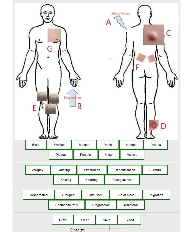
Figure 1. The LesionMapper interface: (A,B) icons for descriptive characteristics; (C) large nodule; (D) ulcer on lateral side; (E) multiple discrete
plaques; (F) Christmas tree pattern in pityriasis rosea; (G) vitiligo patch showing variation in opacity.
In addition to the type of lesions, there are five other For example, the size of the plaques can be a differentiating
characteristics of each icon that can be changed: size, position, feature for small-plaque and large-plaque parapsoriasis.
number, orientation, and opacity. Additional information The original size of the icon, when placed on the LM, can
pertaining to the lesion can be encoded using the following be used for comparison (see the large nodule in Figure 1C).
characteristics: • The position of icon placement indicates the distribution
of the lesions. The front and back of the body are depicted
• The size of the icon can be used to indicate the average size
in the LM. The lateral view is not included to simplify the
of the lesion in conditions where lesion size points toward
interface. To represent lateral distribution, the icons can be
a diagnosis or a particular subtype of the primary diagnosis.
http://derma.jmir.org/2020/1/e18149/ JMIR Dermatol 2020 | vol. 3 | iss. 1 | e18149 | p. 3
(page number not for citation purposes)
XSL• FO
RenderXJMIR DERMATOLOGY Eapen et al
placed in the corresponding edge of the wireframe with an Open Medical Records System (OpenMRS) is an open-source,
overlap of 50% (see the ulcer on the legs in Figure 1D). Java-based EMR for developing countries with a modular and
• Multiple icons of the same type can be used to represent extensible architecture [14]. OpenMRS supports the Open Web
discrete lesions, and a single large icon can be used to Apps (OWA) specifications that make it possible to design
represent confluent distribution (see discrete plaques in external applications that extend the core functions. The OWA
Figure 1E). communicates with OpenMRS using REST APIs
• The orientation can be used to indicate a pathognomonic (representational state transfer application programming
distribution, such as the Christmas tree pattern in pityriasis interfaces), a software architectural style used for creating Web
rosea (see Figure 1F). services, and is embedded in the same server instance.
• The opacity of the lesion can be used to indicate the severity OpenMRS has a custom concept dictionary that helps map data
of the presentation. For example, it can be used to represent points to a uniform terminology. Nontextual data such as images
the degree of depigmentation in a vitiligo patch or the are stored as ”complex concepts” outside the relational database.
severity of contact dermatitis (see Figure 1G). LMR can easily fit into an OWA design pattern, and the
exported LM images can be stored as complex observations in
Mapping lesions consistently and accurately requires a tool that
the patient record. We have a prototype integration that can be
supports the various functions described above. In addition,
used as an example [15].
from a design perspective, the tool should have the capability
to integrate with other health information systems and EMRs. OSCAR EMR is a web-based EMR system initially developed
for primary care practitioners in Canada. OSCAR EMR has a
LesionMapper complex data model, and additional data points are supported
by an electronic form (eForm) module that stores data as
LMR is a prototype implementation of the LM method described key-value pairs [16]. eForms do not support images or other
above. We adopted the design science principles of Hevner et nontextual data. The ability of LMR to save LMs as a JSON
al [10] for information systems to design LMR. We searched string makes the integration of the LMR module into eForms
the literature for similar approaches and available tools to possible.
address the problem of lesional documentation. Based on the
success of similar approaches (Pain-QuILT for annotating pain Machine Learning Applications
[9]), we chose iconography as the method and standardized it
based on our domain expertise in dermatology. Thereafter, we Dermatological diseases have diverse presentations, with skin
distinguished some of the easily identifiable characteristics of type and skin color adding to this variation. Some of these
icons that can be programmatically controlled, such as size, diseases involve hair, nails, and mucous membranes in addition
orientation, and transparency. Subsequently, we converged on to the skin. Traditional computer-vision algorithms such as
a popular framework (VueJS JavaScript framework [11]) for convolutional neural networks (CNNs) and other variants of
implementation. We designed the artifact adopting a modular neural networks have limited application when there are many
pattern—as a JavaScript package shared as open source (see decision alternatives [17]. Hence, AI algorithms have had
the GitHub repository [12])—that can be incorporated into limited application in dermatology except in problems associated
web-based EMRs. LMR provides buttons to add various icons with classification (eg, the presence or absence of cancer) [18].
to the canvas. These icons can be independently moved and Such algorithms can classify only a given lesion rather than the
resized. The opacity and orientation can also be independently patient as a whole (ie, a lesion is cancerous vs patient has
modified. The LM can be exported as an image or as a cancer). Although few CNN-based image search algorithms
JavaScript Object Notation (JSON) string. LMR supports have proven to be useful, AI algorithms for diagnostic decision
freehand drawing in the canvas to represent features that are making in clinical dermatology lag far behind areas such as
not represented by icons though machine interpretation of the radiology [17].
freehand drawing is challenging.
Text analytics and natural language processing (NLP) can be
Next, we describe the integration of LMR into two open-source more useful than image analytics when the decision alternatives
EMRs—OpenMRS and OSCAR. are numerous, as in dermatology. Multimodal approaches where
an image is combined with metadata have shown promise [19].
Integration With Electronic Medical Machine learning models built using LMs—especially the
models created using the JSON representation—resemble text
Records more than an image. Some relevant metadata such as the position
The modular design helps in the integration of LMR into and distribution of the lesions, which are difficult to be captured
web-based EMRs. The prototype is created using the VueJS in text and hard to precisely decipher with NLP, are implicitly
JavaScript framework following the Universal Module captured in LMs. The icons represent ontological concepts from
Definition (UMD) pattern [13] that can be imported by different dermatology and can map to any standard terminology system
module loaders into other browser-based applications. The icons [7]. We posit that LMs are semantically rich enough to be used
are converted into Base64 strings and included in the JavaScript for machine learning applications. Machine learning models
files. from LMs are likely to be more “explainable” than traditional
black box algorithms [20]. The implicit metadata captured by
http://derma.jmir.org/2020/1/e18149/ JMIR Dermatol 2020 | vol. 3 | iss. 1 | e18149 | p. 4
(page number not for citation purposes)
XSL• FO
RenderXJMIR DERMATOLOGY Eapen et al
LMs can supplement regular digital images, leading to better its anticipated ease of use, the actual impact of LMR on
machine learning models. physician workflow, if any, needs to be investigated further.
Advantages and Limitations Discussion
LMs may save time for busy practitioners while capturing the The skin is the largest organ in the human body, and as such,
type, distribution, and characteristics of the lesion; these data skin conditions are commonly encountered in any health care
can be used to assess clinical progress. The LM exported as practice. Although dermatology is a specialization within clinical
JSON resembles a markup language amenable to data mining medicine, 50% of skin conditions are assessed and documented
and machine learning methods [21]. LMs are portable and can by nondermatologists [29].
be easily and safely exchanged without privacy concerns.
There is no universal standard for pictographic documentation
LMR can export LMs as images. These images can be used as of the type, distribution, progression, and severity of lesions in
a proxy for patient images in some computer vision–based dermatology, as in dentistry [30] and ophthalmology [31]. The
applications. Computer vision has been successfully applied to LM standardizes visual representation using icons that can be
identify metabolic defects from gene expression maps [22]. extended to accommodate different use cases in clinical and
MNIST (Modified National Institute of Standards and cosmetic dermatology. The simplicity of the mapping rules
Technology database), a dataset widely used in machine facilitates use by nondermatologists in the skincare industry;
learning, consists of images of handwritten digits [23]. LMs are also semantically rich enough to capture most relevant
information about a skin condition with minimal effort.
It is widely accepted that machine learning can reinforce some
health care disparities in dermatology [24]. Skin color is a Image analytics in dermatology is not as popular, as it is in
significant background noise that needs to be accounted for in visually oriented medical specialties such as radiology and
any machine learning model. It is possible that some of the pathology; the exception is the field of skin cancer diagnostics.
existing models are biased toward particular skin types that This is because of the privacy concerns associated with
predominate in the training data set. Such models tend to be dermatological images and the difficulties in standardizing
less sensitive in making predictions on different skin types [25]. image capture. The LM is not a replacement for a digital image
The LMs are not affected by such bias. of the lesion. However, some of the diagnostic aspects that are
difficult to be captured in images, such as distribution and
LMs, however, do not capture all the features, both explainable
progression, can be useful for machine learning applications,
and unexplainable, captured by a digital image. Hence, LMs
especially when combined with the textual representation of a
are not useful in scenarios where accurate and sensitive
patient’s history. Such multimodal approaches mimic the clinical
extraction of features from an image is important for prediction.
workflow more so than CNN-based algorithms [32]. New
For example, LMs are not appropriate for skin cancer
computer-vision algorithms are proving to be capable of learning
classification [5] and mole mapping [26]. LMs do not support
from computer-generated images [22]. We believe that LMs
annotating dermatopathology images [27]; they are also not
can be similarly used with computer-vision methods. Finally,
applicable for dermatoscopic images that rely on pixel-level
we urge the open-source community to help us improve LMR
analysis [28]. Examination findings such as fluctuation,
and potential users to report issues on the repository [12] so that
consistency, and tenderness are not represented by icons at
we can fix them. We will work on a 3D wireframe for better
present to keep the interface simple. More icons can be added
accuracy, and we welcome other feature requests from the user
if the user community requires them.
community.
LMR and the LM method have not been clinically tested. The
integration of LMR into existing EMRs may be difficult. Despite
Conflicts of Interest
None declared.
References
1. Aspres N, Egerton IB, Lim AC, Shumack SP. Imaging the skin. Australas J Dermatol 2003 Feb;44(1):19-27. [doi:
10.1046/j.1440-0960.2003.00632.x]
2. Kunde L, McMeniman E, Parker M. Clinical photography in dermatology: Ethical and medico-legal considerations in the
age of digital and smartphone technology. Australasian Journal of Dermatology 2013 May 29;54(3):192-197. [doi:
10.1111/ajd.12063]
3. Ventola CL. Social media and health care professionals: benefits, risks, and best practices. P & T 2014 Jul;39(7):491-520
[FREE Full text] [Medline: 25083128]
4. Shin H, Roth HR, Gao M, Lu L, Xu Z, Nogues I, et al. Deep Convolutional Neural Networks for Computer-Aided Detection:
CNN Architectures, Dataset Characteristics and Transfer Learning. IEEE Trans. Med. Imaging 2016 May;35(5):1285-1298.
[doi: 10.1109/tmi.2016.2528162]
http://derma.jmir.org/2020/1/e18149/ JMIR Dermatol 2020 | vol. 3 | iss. 1 | e18149 | p. 5
(page number not for citation purposes)
XSL• FO
RenderXJMIR DERMATOLOGY Eapen et al
5. Esteva A, Kuprel B, Novoa RA, Ko J, Swetter SM, Blau HM, et al. Dermatologist-level classification of skin cancer with
deep neural networks. Nature 2017 Jan 25;542(7639):115-118. [doi: 10.1038/nature21056]
6. Levine MD, Nazif AM. Dynamic Measurement of Computer Generated Image Segmentations. IEEE Trans Pattern Anal
Mach Intell 1985 Mar;PAMI-7(2):155-164. [doi: 10.1109/tpami.1985.4767640]
7. Eapen BR. ONTODerm--a domain ontology for dermatology. Dermatol Online J 2008 Jun 15;14(6):16. [Medline: 18713597]
8. Cardili RN, Roselino AM. Elementary lesions in dermatological semiology: literature review. An Bras Dermatol 2016
Oct;91(5):629-633. [doi: 10.1590/abd1806-4841.20164931]
9. Lalloo C, Kumbhare D, Stinson JN, Henry JL. Pain-QuILT: clinical feasibility of a web-based visual pain assessment tool
in adults with chronic pain. J Med Internet Res 2014 May 12;16(5):e127 [FREE Full text] [doi: 10.2196/jmir.3292] [Medline:
24819478]
10. Hevner AR, March ST, Park J, Ram S. Design Science in Information Systems Research. MIS Quarterly 2004;28(1):75.
[doi: 10.2307/25148625]
11. Street M, Passaglia A, Halliday P. Complete Vue. In: Complete Vue.js 2 Web Development: Practical Guide To Building
End-to-end Web Development Solutions With Vue.js 2. Birmingham: Packt Publishing; 2018.
12. Eapen BR. GitHub. 2020. LesionMapper - Vue component for standardized mapping of lesions in dermatology URL: https:/
/github.com/dermatologist/lesion-mapper [accessed 2020-01-16]
13. Rich H. Dismantling the barriers to entry. Queue 2015;13(5):37 [FREE Full text] [doi: 10.1145/2742580.2742813]
14. Mamlin BW, Biondich PG, Wolfe BA, Fraser H, Jazayeri D, Allen C, et al. Cooking up an open source emr for developing
countries: OpenMRS, a recipe for successful collaboration. In: American Medical Informatics Association. 2006 Presented
at: AMIA Annual Symposium Proceedings; 2006; Washington p. 529.
15. Eapen BR. OpenMRS. 2017. Dermatology LesionMapper URL: https://addons.openmrs.org/show/org.openmrs.module.
dermatology-lesionmapper [accessed 2020-01-18]
16. Safadi H, Chan D, Dawes M, Roper M, Faraj S. Open-source health information technology: A case study of electronic
medical records. Health Policy and Technology 2015 Mar;4(1):14-28. [doi: 10.1016/j.hlpt.2014.10.011]
17. Li C, Shen C, Xue K, Shen X, Jing Y, Wang Z, et al. Artificial intelligence in dermatology. Chinese Medical Journal
2019;132(17):2017-2020. [doi: 10.1097/cm9.0000000000000372]
18. Sidey-Gibbons JAM, Sidey-Gibbons CJ. Machine learning in medicine: a practical introduction. BMC Med Res Methodol
2019 Mar 19;19(1). [doi: 10.1186/s12874-019-0681-4]
19. Wang H, Wang Y, Liang C, Li Y. Assessment of Deep Learning Using Nonimaging Information and Sequential Medical
Records to Develop a Prediction Model for Nonmelanoma Skin Cancer. JAMA Dermatol 2019 Nov 01;155(11):1277. [doi:
10.1001/jamadermatol.2019.2335]
20. Andreas H, Chris B, Constantinos SP, Douglas BK. What do we need to build explainable AI systems for the medical
domain? arXiv.org 2017 [FREE Full text]
21. Guazzelli A, Lin W, Jena T. PMML in action: unleashing the power of open standards for data mining and predictive
analytics. California: Createspace Independent Publishing Platform; 2020.
22. Zhang J, Naik HS, Assefa T, Sarkar S, Reddy RVC, Singh A, et al. Computer vision and machine learning for robust
phenotyping in genome-wide studies. Sci Rep 2017 Mar 8;7(1). [doi: 10.1038/srep44048]
23. Li Deng. The MNIST Database of Handwritten Digit Images for Machine Learning Research [Best of the Web]. IEEE
Signal Process. Mag 2012 Nov;29(6):141-142. [doi: 10.1109/msp.2012.2211477]
24. Adamson AS, Smith A. Machine Learning and Health Care Disparities in Dermatology. JAMA Dermatol 2018 Nov
01;154(11):1247. [doi: 10.1001/jamadermatol.2018.2348]
25. Kamulegeya LH, Okello M, Bwanika JM, Musinguzi D, Lubega W, Rusoke D, et al. Using artificial intelligence on
dermatology conditions in uganda: A case for diversity in training data sets for machine learning. bioRxiv.org 2019 [FREE
Full text] [doi: 10.1101/826057]
26. Berk-Krauss J, Polsky D, Stein JA. Mole Mapping for Management of Pigmented Skin Lesions. Dermatologic Clinics 2017
Oct;35(4):439-445. [doi: 10.1016/j.det.2017.06.004]
27. Toro P, Corredor G, Romero E, Arias V. A visualization, navigation, and annotation system for dermatopathology training.
In: International Society for Optics and Photonics. 2018 Presented at: 14th International Symposium on Medical Information
Processing and Analysis, volume 10975; 2018; Mazatlán, Mexico. [doi: 10.1117/12.2511638]
28. Ferreira B, Barata C, Marques JS. What is the role of annotations in the detection of dermoscopic structures? In: Iberian
Conference on Pattern Recognition and Image Analysis. 2019 Presented at: 9th Iberian Conference, IbPRIA 2019; 2019;
Madrid, Spain p. 3-11. [doi: 10.1007/978-3-030-31321-0_1]
29. Ahiarah A, Fox C, Servoss T. Brief intervention to improve diagnosis and treatment knowledge of skin disorders by family
medicine residents. Fam Med 2007;39(10):720-723 [FREE Full text] [Medline: 17987414]
30. Wu M, Koenig L, Lynch J, Wirtz T. Spatially-oriented EMR for Dental Surgery. In: American Medical Informatics
Association. 2006 Presented at: AMIA Annual Symposium Proceedings; 2006; Washington p. 1147.
31. Chiang MF, Boland MV, Brewer A, Epley KD, Horton MB, Lim MC, American Academy of Ophthalmology Medical
Information Technology Committee. Special Requirements for Electronic Health Record Systems in Ophthalmology.
Ophthalmology 2011 Aug 1;118(8):1681-1687. [doi: 10.1016/j.ophtha.2011.04.015] [Medline: 21680023]
http://derma.jmir.org/2020/1/e18149/ JMIR Dermatol 2020 | vol. 3 | iss. 1 | e18149 | p. 6
(page number not for citation purposes)
XSL• FO
RenderXJMIR DERMATOLOGY Eapen et al
32. Baltrusaitis T, Ahuja C, Morency L. Multimodal Machine Learning: A Survey and Taxonomy. IEEE Trans Pattern Anal
Mach Intell 2019 Feb 1;41(2):423-443. [doi: 10.1109/tpami.2018.2798607]
Abbreviations
AI: artificial intelligence
API: application programming interface
CNN: convolutional neural network
eForm: electronic form
eHealth: electronic health
EMR: electronic medical record
JSON: JavaScript Object Notation
LM: LesionMap
LMR: LesionMapper
MNIST: Modified National Institute of Standards and Technology database
NLP: natural language processing
OWA: Open Web Apps
REST: representational state transfer
UMD: Universal Module Definition
Edited by G Eysenbach; submitted 06.02.20; peer-reviewed by F Kaliyadan, A Kt; comments to author 15.02.20; revised version
received 26.02.20; accepted 27.02.20; published 20.04.20
Please cite as:
Eapen BR, Archer N, Sartipi K
LesionMap: A Method and Tool for the Semantic Annotation of Dermatological Lesions for Documentation and Machine Learning
JMIR Dermatol 2020;3(1):e18149
URL: http://derma.jmir.org/2020/1/e18149/
doi: 10.2196/18149
PMID:
©Bell Raj R Eapen, Norm Archer, Kamran Sartipi. Originally published in JMIR Dermatology (http://derma.jmir.org), 20.04.2020.
This is an open-access article distributed under the terms of the Creative Commons Attribution License
(https://creativecommons.org/licenses/by/4.0/), which permits unrestricted use, distribution, and reproduction in any medium,
provided the original work, first published in JMIR Dermatology Research, is properly cited. The complete bibliographic
information, a link to the original publication on http://derma.jmir.org, as well as this copyright and license information must be
included.
http://derma.jmir.org/2020/1/e18149/ JMIR Dermatol 2020 | vol. 3 | iss. 1 | e18149 | p. 7
(page number not for citation purposes)
XSL• FO
RenderXYou can also read