RANKING HITS FROM SATURATION TRANSFER DIFFERENCE NUCLEAR MAGNETIC RESONANCE-BASED FRAGMENT SCREENING - MPG.PURE
←
→
Page content transcription
If your browser does not render page correctly, please read the page content below
ORIGINAL RESEARCH
                                                                                                                                                published: 12 April 2019
                                                                                                                                       doi: 10.3389/fchem.2019.00215

                                               Ranking Hits From Saturation
                                               Transfer Difference Nuclear Magnetic
                                               Resonance–Based Fragment
                                               Screening
                                               Jonas Aretz 1,2 and Christoph Rademacher 1,2*
                                               1
                                                Department of Biomolecular Systems, Max Planck Institute of Colloids and Interfaces, Potsdam, Germany, 2 Department of
                                               Biology, Chemistry, and Pharmacy, Freie Universität Berlin, Berlin, Germany

                                               Fragment-based screening is an established route to identify low-molecular-weight
                                               molecules to generate high-affinity inhibitors in drug discovery. The affinities of these early
                                               hits from fragment screenings require a highly sensitive biophysical screening technique.
                                               Saturation transfer difference (STD) nuclear magnetic resonance (NMR) is one of the
                                               most popular methods owing to its high sensitivity for low-affinity ligands. It would be
                                               highly beneficial if rank-ordering of hits according to their affinity from an initial or counter-
                                               screen could be performed—a selection criterion found in the literature. We applied
                                               Complete Relaxation and Conformational Exchange Matrix (CORCEMA) theory adapted
                                               for saturation transfer (ST) measurements (CORCEMA-ST) calculations to predict STD
                            Edited by:         NMR results from a large set of fragment/receptor pairs to investigate the boundaries
                        Simone Brogi,
                University of Pisa, Italy      under which the assumption holds true that a high STD effect can be applied to select
                       Reviewed by:            for higher-affinity fragments. Overall, we come to the conclusion that this assumption
                      Michal Brylinski,        is invalid.
            Louisiana State University,
                        United States          Keywords: fragment-based drug discovery, fragment-based drug design, saturation transfer difference nuclear
                      Barbara Mulloy,          magnetic resonance spectroscopy, STD NMR, screening
              King’s College London,
                      United Kingdom
                 *Correspondence:
                                               INTRODUCTION
              Christoph Rademacher
christoph.rademacher@mpikg.mpg.de              Fragment-based drug design (FBDD) has successfully complemented the toolbox for developing
                                               small-molecule pharmaceuticals (Baker, 2012; Hann and Keserü, 2012) as highlighted by
                   Specialty section:          Vemurafenib and Venetoclax, the first drugs entering the market originating from FBDD (Bollag
         This article was submitted to         et al., 2010; Souers et al., 2013). In this approach, fragments of drug-like molecules ranging between
        Medicinal and Pharmaceutical           ∼150 and 250 Da in size are identified binding to a receptor. These initial hits are then developed
                            Chemistry,         into high-affinity leads following fragment evolution strategies such as fragment linking, fragment
               a section of the journal
                                               growing, or fragment merging (Rees et al., 2004).
                Frontiers in Chemistry
                                                   The screening of fragment collections is challenged by the low affinities of the initial hits,
       Received: 04 December 2018              which typically range between dissociation constants of 10 µM and 10 mM. These values are
          Accepted: 19 March 2019
                                               target dependent, but the average fragment hit resides in the higher micromolar to the single-digit
           Published: 12 April 2019
                                               millimolar range. To overcome the low affinity of fragments, sensitive biophysical techniques
                            Citation:          are commonly employed such as surface plasmon resonance (SPR), X-ray crystallography,
   Aretz J and Rademacher C (2019)
                                               and techniques from nuclear magnetic resonance (NMR). In particular, ligand-observed NMR
Ranking Hits From Saturation Transfer
         Difference Nuclear Magnetic
                                               techniques such as STD NMR are the most frequently used methods owing to their high sensitivity
        Resonance–Based Fragment               and low false-positive rate (Gossert and Jahnke, 2016).
      Screening. Front. Chem. 7:215.               During an STD NMR experiment, receptor resonances are selectively saturated for a given
    doi: 10.3389/fchem.2019.00215              time (saturation time, t sat ) by a series of frequency-specific pulses. This magnetization spreads in

Frontiers in Chemistry | www.frontiersin.org                                        1                                                 April 2019 | Volume 7 | Article 215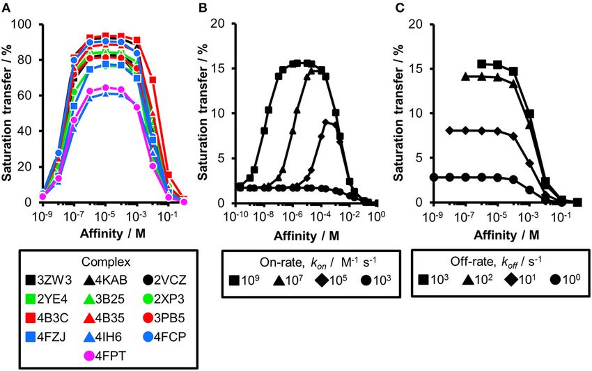
Aretz and Rademacher Ranking Hits From STD NMR
milliseconds in the hydrogen network within the receptor via complexity of the STD NMR experiment in theory to explore
spin diffusion (Mayer and Meyer, 1999; Jayalakshmi and Krishna, parameters influencing the saturation transfer. For example, two
2002). Furthermore, the magnetization is transferred to low- receptor/ligand complexes can be compared assuming that they
molecular-weight ligands, enabling the identification of actives share exactly the same affinity and, by that, rule out effects
from compound mixtures (Mayer and Meyer, 1999; Jayalakshmi arising from the exchange kinetics. This then allows extracting
and Krishna, 2002). The ligand will then dissociate from the the influence of the geometry of the binding site (Jayalakshmi and
receptor site and saturated ligands accumulate free in solution, Krishna, 2002). Previous CORCEMA calculations using a single
which results in a decreased signal intensity of the bulk ligand. receptor/ligand pair indicated a correlation between affinity and
This spectrum is subtracted from a reference spectrum of the saturation transfer to the ligand (Jayalakshmi and Krishna, 2002).
same sample recorded in the absence of saturation. Hence, signals Sufficient residence time of the ligand in the binding site allows
in an STD spectrum correspond to ligands that bound to the transfer of the magnetization. Consequently, saturated ligand
receptor. Moreover, saturation transfer to the ligand is distance molecules accumulate free in solution and the overall signal
dependent and ligand hydrogens receiving more saturation are intensity of the corresponding ligand resonances is decreased.
considered in close proximity to the receptor interface in the When the affinity is exceeding a certain threshold, the release
bound state (Mayer and Meyer, 2001). A binding epitope can of ligand from the receptor site is limited and the STD effect
thus be derived, normalizing the saturation transfer to the proton decreases again. Taken together, a bell-shaped plot of affinity vs.
receiving the highest saturation. Additionally, the magnitude of saturation transfer is expected.
saturation transfer is affected by the affinity and the kinetics Here, we calculate theoretical STD effects over a broad range
of complex formation (Jayalakshmi and Krishna, 2002; Meyer of receptor–ligand pairs. These insights are combined with
and Peters, 2003). Finally, ligand as well as receptor saturation experimental results from STD NMR screening and fragment-
is counteracted by nuclear relaxation processes, particularly based ligand design. Next, we investigated whether rank-ordering
T1 relaxation, leading to a dissipation of the magnetization of fragment-sized ligands from primary screening data based
to the bulk solvent. Consequently, the saturation build-up of on the STD amplification factors is suitable. Overall, evidence
ligand equilibrates at longer duration of the saturation time from calculations as well as experimental data suggests that such
(Jayalakshmi and Krishna, 2002). rank-ordering is invalid.
The coupled dipolar relaxation network of receptor and
ligand hydrogens can be calculated using the complete MATERIALS AND METHODS
relaxation and conformational exchange matrix (CORCEMA)
theory. With this formalism, STD NMR experiments can be Structure Preparation
simulated for a given receptor/ligand complex, and CORCEMA- Fragment/protein complexes were selected from the Protein
ST has been successfully applied to refine such complexes Data Bank (PDB) database based on resolution and diversity of
(Jayalakshmi and Krishna, 2002, 2005; Szczepina et al., 2011). the proteins and ligands, and avoiding sterical clashes between
Moreover, CORCEMA calculations allow one to reduce the the ligand and protein originating from unreasonably low
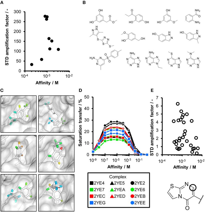
FIGURE 1 | Comparing experimental and calculated STD effects using CORCEMA-ST. (A) STD amplification factors observed during fragment screening of langerin
with [P] = 10 µM, [L] = 200 µM, and tsat = 4 s plotted against affinity estimated by SPR (Aretz et al., 2018). (B) Thirteen fragment/protein complexes were used to
perform CORCEMA-ST calculations of the saturation transfer from the receptor to the low-molecular-weight ligand. The average over all complexes is shown and
depicts the saturation transfer to the ligand proton receiving the highest saturation. A gray box highlights the affinity regime typically populated by fragments (K d
ranging from 10 µM to 10 mM). (C) Individual plots of all 13 complexes highlighting the high dispersion of saturation transfer. For CORCEMA-ST calculations, typical
STD NMR screening conditions were assumed: [P] = 20 µM, [L] = 1.0 mM, saturation time = 2.0 s, τc,bound = 30 ns (corresponding to 50 kDa molecular weight), and
k on = 109 M−1 s−1 .
Frontiers in Chemistry | www.frontiersin.org 2 April 2019 | Volume 7 | Article 215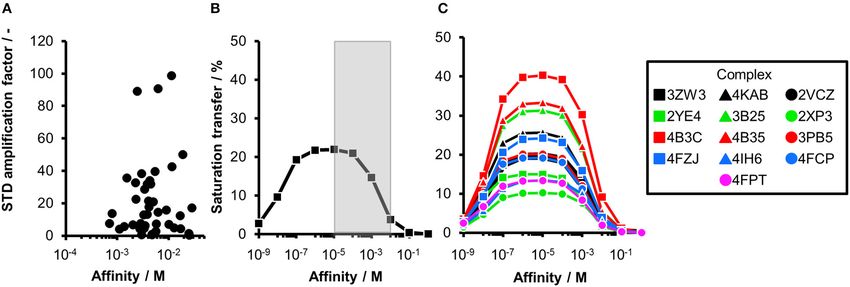
Aretz and Rademacher                                                                                                                         Ranking Hits From STD NMR

distances. All complexes were prepared in Molecular Operating                           concentration [L] = 1 mM; protein concentration [P] = 20 µM;
Environment (MOE, version 2015; Chemical Computing Group                                kon = 109 M−1 s−1 ; saturation times t sat = 0.5, 2.0, and 8.0 s;
ULC., 2018). Hydrogens were added at pH 7; if necessary,                                correlation time of the ligand τc = 5 × 10¹⁰ s. Exchange-mediated
missing loops were introduced followed by a structure refinement                        leakage is not taken into account in this model when more
step as implemented in MOE using standard parameters and                                than one binding site is assumed. To significantly decrease the
manual inspection. Complexes and their respective affinities                            calculation time, only hydrogens in an 11-Å radius surrounding
are given in Table S1 in the order they appear throughout                               the ligand were included (Figure S5). Methyl resonances of
the study.                                                                              amino acids were assumed to be instantaneously saturated
                                                                                        during the simulation. Only non-exchangeable hydrogens of
CORCEMA-ST                                                                              the receptor and the ligand were included in the calculation,
CORCEMA-ST (version 3.8) was run on a regular desktop                                   and all other atoms were removed from the input structures.
computer (Jayalakshmi and Krishna, 2002). If not stated                                 Only protons with the highest saturation transfer were used for
otherwise, the following parameters were assumed: ligand                                the analysis.

 FIGURE 2 | CORCEMA-ST calculations and experimental data of fragments binding to the same binding site. (A) STD amplification factors determined with
 CORCEMA-ST of fragments binding to PDE10A and AmpC. For these fragments, a crystal structure and thermodynamic data were available (Barelier et al., 2014;
 Recht et al., 2014). (B) Chemical structures of the fragments used to analyze HSP90 (Roughley and Hubbard, 2011) and (C) examples of some of these ligands in
 complex with protons colored by their normalized STD effect (red being the highest STD effect and blue being the lowest effect). (D) Saturation transfer with varying
 dissociation constants is shown for all complexes shown in (B) for HSP90. For all complexes, the same parameters were assumed: [P] = 20 µM, [L] = 1.0 mM,
 τc,bound , = 30 ns, and k on= 109 M−1 s−1 . (E) STD amplification factors derived from a structure-activity relationship (SAR) study of thiazolopyrimidine derivatives
 binding to langerin plotted against affinity determined by SPR (Aretz et al., 2018). STD NMR measurements were performed with [P] = 20 µM, [L] = 500–1,000 µM,
 depending on compound solubility, and tsat = 1 s, considering only the proton in the 7-position (carbon highlighted with a circle).

Frontiers in Chemistry | www.frontiersin.org                                        3                                                   April 2019 | Volume 7 | Article 215
Aretz and Rademacher Ranking Hits From STD NMR TABLE 1 | Protein–ligand complexes used for CORCEMA-ST calculations. PDB ID Name (long) Name (short) Protein class Ligand 2XP3 Peptidyl-prolyl cis-trans isomerase NIMA-interacting 1 protein Pin1 Isomerase 2YE4 Heat shock protein 90 HSP90 Chaperone 3B25 Heat shock protein 90 HSP90 Chaperone 3PB5 Endothiapepsin EAPA Protease 4B3C DNA repair and recombinant protein RADA RadA ATPase 4B35 DNA repair and recombinant protein RADA RadA ATPase 4FCP Heat shock protein 90 HSP90 Chaperone 4FZJ Pantothenate synthetase Pts Ligase 4IH6 HCV non-structural protein 5B NS5B RNA Pol. 4FPT Carbonic anhydrase 2 CA2 Lyase 2VCZ Prostaglandin D2 synthase PGDS Isomerase 3ZW3 Phosphatidylinositol-4,5-bisphosphate 3-kinase PI3K Kinase 4KAB Focal adhesion kinase 1 FAK1 Kinase Frontiers in Chemistry | www.frontiersin.org 4 April 2019 | Volume 7 | Article 215
Aretz and Rademacher                                                                                                  Ranking Hits From STD NMR

Saturation Transfer Difference Nuclear                                    STD NMR-based fragment screening with a larger panel
Magnetic Resonance Measurements                                           of different fragments being present in the same mixture
STD NMR experiments were performed as described elsewhere                 potentially occupying multiple sites with varying geometry and
(Aretz et al., 2018). Briefly, 10 µM murine langerin was screened         physicochemical properties. The calculated saturation transfer
against a library of 660 fragments at a ligand concentration              plotted against the affinity averaged over all complexes resulted
[L] = 0.2 mM using a saturation time of t sat = 4 s. Hits                 in a bell-shaped curve as reported earlier (Figure 1B; Jayalakshmi
were counter-screened by SPR (see the section Estimation of               and Krishna, 2002). For the typical affinity range of fragments,
Affinity Using Surface Plasmon Resonance), enabling affinity              which is between 10 µM and 10 mM, these plots indicate elevated
estimation and hit validation. For SPR-validated hits, the STD            saturation transfer with increasing affinity (gray box, Figure 1B),
amplification factor was determined as reported earlier (Mayer            suggesting that rank-ordering hits from primary screening would
and Meyer, 2001) and plotted against affinity (Figure 1A). For            be suitable. However, the analysis of the individual contributions
epitope mapping of thiazolopyrimidine derivatives (Figure 2E),            of the fragment complexes to the average depiction revealed a
protein concentration was [P] = 20 µM, ligand concentration               high variability of the calculated saturation transfer (Figure 1C),
was [L] = 0.5–1 mM, and t sat = 1 s. Affinity of all derivatives          demonstrating that even assuming the same thermodynamics
was estimated by SPR (see the section Estimation of Affinity              and kinetics, a high variability of the STD NMR readout is to
Using Surface Plasmon Resonance), and the STD amplification               be expected. This trend was also observed for saturation times
factor was determined as reported earlier for the proton in the           of 0.5 and 8.0 s (Figure S1). Overall, this variability suggests that
7-position of the thiazolopyrimidine scaffold (Mayer and Meyer,           binders from initial STD NMR screening cannot be rank-ordered
2001). Spectra were analyzed in MestReNova 10.0.0. Compound               assuming that the magnitude of the STD effect would correlate
structures, estimated affinities, and STD amplification factors are       with fragment affinity.
given in Table S2.                                                            To ensure that a fragment is binding to the targeted site
                                                                          during the STD NMR screening process, typically competition
Estimation of Affinity Using Surface                                      experiments are employed. Consequently, under these
                                                                          conditions, the saturation transfer for all fragments to be
Plasmon Resonance                                                         rank-ordered originates from the same binding site. Hence,
SPR experiments were performed on a Biacore T100 (GE                      to eliminate the effect of the composition of the binding site,
Healthcare) as described elsewhere (Aretz et al., 2018). Briefly,         a series of fragments binding to a single receptor pocket was
murine langerin was immobilized on a CM7 Series S chip to                 utilized using two exemplary drug targets: phosphodiesterase
a density of 7,500 RU using NHS/EDC coupling. Subsequently,               10A (PDE10A) and beta-lactamase (AmpC). In both cases,
dilution series of compounds in 25 mM HEPES, pH 7.6, 150 mM               crystallographic and thermodynamic data were previously
sodium chloride, 5 mM calcium chloride, and 0.005% Tween-20               reported. From these reports, kinetic data were calculated
buffer were injected with a flow rate of 10 µl min−1 .                    assuming diffusion-controlled on-rate kinetics and hence STD
                                                                          amplification factors were determined (Figure 2A; Barelier
RESULTS AND DISCUSSION                                                    et al., 2014; Recht et al., 2014). Similar to our experimental
                                                                          screening data, there was no correlation between calculated
Ideally, a fragment screening technique enables rank-ordering of          STD amplification factor and affinity (Pearson’s correlation
hits according to their affinity. Due to recent reports on rank-          coefficient, p > 0.05). We then focused on another receptor with
ordering hits from STD NMR screening and counter-screening                a high availability of fragment-bound crystal structures, i.e., heat
applying the STD amplification factor (Jose et al., 2012; Begley          shock protein 90 (HSP90), to rule out effects coming from the
et al., 2013; Cala and Krimm, 2015), we retrospectively analyzed          binding site geometry (Figures 2B–D; Roughley and Hubbard,
a fragment screening against langerin, a C-type lectin receptor           2011). A more homogeneous saturation transfer profile was
involved in pathogen uptake by immune cells (Aretz et al., 2018).         observed (Figure 2D) in comparison to the simulated screening
Here, fragments were screened by STD NMR, and the affinity of             data with multiple binding sites (Figure 1C). Still, fragments
hits was subsequently estimated by SPR (Figure 1A). Although              were indistinguishable based on their affinity (Figure 2D). Taken
the affinity of all hits was in a suitable range for STD NMR, there       together, if binding to a single protein pocket can be assumed,
was no correlation between STD amplification factor and affinity          slight chemical variations in the structure of the fragments,
(Pearson’s correlation coefficient). Consequently, rank-ordering          which are typically found in a series of derivatives, lead to
of hits according to their STD amplification factor would have            substantial variability of the observed STD effects.
been misleading in this example.                                              To experimentally validate this finding, we analyzed
   To analyze the missing correlation between STD amplification           STD data of a structure-activity relationship study (SAR) of
factor and affinity more systematically, we assembled a diverse           thiazolopyrimidine derivatives binding to langerin (Figure 2E;
set of receptor/fragment complexes (Table 1) and computed                 Aretz et al., 2018). These derivatives only differ in substitutions
the outcome of an STD NMR experiment using CORCEMA-                       of the 6-position; hence, the STD amplification factor of the
ST (Figure 1B). Setting a molecular weight of the receptor                same aromatic proton in the 7-position was determined. Under
to a typical drug target of 50 kDa, e.g., checkpoint kinase               these conditions, a linear correlation between affinity and STD
1 (CHK1), we evaluated the influence of the affinity on the               amplification factor can be inferred (Pearson’s correlation
saturation transfer. This setup simulates the situation during            coefficient, p < 0.05), which is in contrast to the screening

Frontiers in Chemistry | www.frontiersin.org                          5                                           April 2019 | Volume 7 | Article 215
Aretz and Rademacher Ranking Hits From STD NMR FIGURE 3 | The influence of altered kinetics and molecular weight of a receptor on the STD amplification factor. (A) CORCEMA-ST calculations were performed for 13 fragment/protein complexes assuming a molecular weight of 166 kDa. CORCEMA-ST calculations were performed on the same fragment/protein complexes as above (Figure 1; Table 1), but changing the protein correlation time to 100 ns [τc,bound = 100 ns, [P] = 20 µM, [L] = 1.0 mM, k on= 109 M−1 s−1 ]. (B,C) CORCEMA-ST calculations for CA2 varying on- and off-rates of the receptor/ligand complex. Saturation times of 8.0 s are shown [PDB ID: 4FPT, [P] = 20 µM, [L] = 1.0 mM]. results. Still, the compound with the highest affinity ranked 19 103 and 106 M−1 s−1 have been reported for low-molecular- of 30; thus, the high variability of the STD effect predicted by weight inhibitors of CA2 (Navratilova and Hopkins, 2010). CORCEMA is in agreement with these experimental data. Therefore, we analyzed CA2 in complex with ethyl (2Z,4R)- Thus far, our analysis was focused on receptors with 2-(sulfamoylimino)-1,3-thiazolidine-4-carboxylate already a molecular weight of 50 kDa. As the saturation transfer included in previous data sets (PDB ID: 4FPT) varying the to the ligand is increased with the increase in molecular exchange kinetics (Figure 3B). If the on-rate is below the weight, we hypothesized that increasing the correlation time diffusion-limited threshold of 107 M−1 s−1 , the affinity range τc to 100 ns in our calculations might compensate for the of fragments receiving high saturation transfer decreases. difference in chemical composition of the binding sites. Such Consequently, low saturation transfer can be interpreted decrease in molecular tumbling rate corresponds to a ∼166 as originating either from high- or low-affinity fragments. kDa receptor. While the CORCEMA calculations predicted Fragments with low micromolar affinity will give rise to elevated STD effects and a more homogeneous saturation similar STD effects as low millimolar binders. This potential transfer compared to our results at 50 kDa (Figure 1B), caveat became more severe with slower on-rates. The same these data still do not allow rank-ordering of hits from results from CORCEMA-ST calculations were observed when screening (Figure 3A). Even at very low saturation times, which analyzing FAK1 and GSK3b kinases as well as increasing compensate for T1 relaxation effects of the ligands (Yan et al., the molecular weight of these receptors (Figures S3, S4). 2003), a clear guideline for fragment ranking remains elusive Moreover, analyzing the same data, but highlighting the off-rates (Figure S2). instead, emphasizes the off-rate bias in STD NMR screening Until now, all CORCEMA-ST calculations assumed a (Figure 3C and Figure S4B). Oversimplified, one can state diffusion-limited on-rate kinetics of kon = 109 M−1 s−1 in that a higher off-rate leads to increasing numbers of saturated line with previous calculations (Jayalakshmi and Krishna, ligand molecules and consequently to a higher STD effect. 2002). However, the influence of kinetics on the correlation Hence, applying a rank-ordering based on STD effect during of STD amplification factor and fragment affinity became screening can lead to an accumulation of ligands with fast already apparent for PDE10A (Figure 2A). To systematically off-rate kinetics, contradicting current efforts to identify hits elucidate the consequences of slower binding kinetics on STD with slow off-rates (Copeland et al., 2006). NMR screening of fragments, carbonic anhydrase II (CA2) Since the underlying pathways leading to efficient saturation was chosen as a model, as it is a well-studied example for transfer from a target receptor to a ligand are multifactorial, which fast kinetics cannot be assumed. On-rates between it is difficult to identify a single determinant responsible Frontiers in Chemistry | www.frontiersin.org 6 April 2019 | Volume 7 | Article 215
Aretz and Rademacher                                                                                                                                     Ranking Hits From STD NMR

for the lack of correlation between STD amplification                                            AUTHOR CONTRIBUTIONS
factor and affinity. However, our results suggest that
subtle changes in the binding site geometry and binding                                          CR designed the study. JA performed experiments and statistical
kinetics can already significantly alter the size of the STD                                     analysis. CR performed CORCEMA-ST calculations. Both
amplification factor.                                                                            authors contributed to manuscript writing and revision, and read
                                                                                                 and approved the submitted version.
CONCLUSION
                                                                                                 FUNDING
In this study, we calculated STD NMR amplification factors
for fragments identified in an experimental NMR screening                                        This work has been supported by the Max Planck Society and the
against langerin (Aretz et al., 2018) and relate them to                                         German Research Foundation (DFG, RA1944/2-1).
affinity. To expand these findings and rule out flaws in
our analysis originating from experimental imperfections, we                                     ACKNOWLEDGMENTS
simulated different pairs of receptors and drug-like ligands using
CORCEMA-ST. Varying saturation time, receptor size, binding                                      We thank Prof. Dr. Peter H. Seeberger for support and
kinetics, and interaction site in CORCEMA-ST simulations, there                                  helpful discussions.
were no conditions in which the STD NMR amplification factor
correlated unambiguously with affinity. These findings are in line                               SUPPLEMENTARY MATERIAL
with our experimental data. In conclusion, these data exemplify
that assuming the observed STD effect relates to affinity and                                    The Supplementary Material for this article can be found
thereby allowing rank-ordering of hits from STD NMR fragment-                                    online at: https://www.frontiersin.org/articles/10.3389/fchem.
based screening is misleading.                                                                   2019.00215/full#supplementary-material.

REFERENCES                                                                                          saturation transfer effects in reversibly forming ligand–receptor complexes. J.
                                                                                                    Magn. Reson. 155, 106–118. doi: 10.1006/jmre.2001.2499
Aretz, J., Anumala, U. R., Fuchsberger, F. F., Molavi, N., Ziebart,                              Jayalakshmi, V., and Krishna, N. R. (2005). Determination of the conformation
   N., Zhang, H., et al. (2018). Allosteric inhibition of a mammalian                               of trimethoprim in the binding pocket of bovine dihydrofolate reductase from
   lectin. J. Am. Chem. Soc. 140, 14915–14925. doi: 10.1021/jacs.                                   a STD-NMR intensity-restrained CORCEMA-ST optimization. J. Am. Chem.
   8b08644                                                                                          Soc. 127, 14080–14084. doi: 10.1021/ja054192f
Baker, M. (2012). Fragment-based lead discovery grows up. Nat. Rev. Drug Discov.                 Jose, R. A., Voet, A., Broos, K., Jakobi, A. J., Bruylants, G., Egle, B., et al. (2012).
   12, 5–7. doi: 10.1038/nrd3926                                                                    An integrated fragment based screening approach for the discovery of small
Barelier, S., Eidam, O., Fish, I., Hollander, J., Figaroa, F., Nachane, R., et al. (2014).          molecule modulators of the VWF–GPIbα interaction. Chem. Commun. 48,
   Increasing chemical space coverage by combining empirical and computational                      11349–11351. doi: 10.1039/c2cc35269a
   fragment screens. ACS Chem. Biol. 9, 1528–1535. doi: 10.1021/cb50                             Mayer, M., and Meyer, B. (1999). Characterization of ligand binding by
   01636                                                                                            saturation transfer difference NMR spectroscopy. Angew. Chem. Int. Ed. 38,
Begley, D. W., Moen, S. O., Pierce, P. G., and Zartler, E. R. (2013).                               1784–1788.
   Saturation transfer difference NMR for fragment screening. Curr.                              Mayer, M., and Meyer, B. (2001). Group epitope mapping by saturation transfer
   Protoc. Chem. Biol. 5, 251–268. doi: 10.1002/9780470559277.ch                                    difference NMR to identify segments of a ligand in direct contact with
   130118                                                                                           a protein receptor. J. Am. Chem. Soc. 123, 6108–6117. doi: 10.1021/ja01
Bollag, G., Hirth, P., Tsai, J., Zhang, J., Ibrahim, P. N., Cho, H., et al.                         00120
   (2010). Clinical efficacy of a RAF inhibitor needs broad target blockade                      Meyer, B., and Peters, T. (2003). NMR spectroscopy techniques for screening
   in BRAF-mutant melanoma. Nature 467, 596–599. doi: 10.1038/nature                                and identifying ligand binding to protein receptors. Angew. Chem. Int. Ed. 42,
   09454                                                                                            864–890. doi: 10.1002/anie.200390233
Cala, O., and Krimm, I. (2015). Ligand-orientation based fragment                                Navratilova, I., and Hopkins, A. L. (2010). Fragment screening by surface
   selection in STD NMR screening. J. Med. Chem. 58, 8739–8742.                                     plasmon resonance. ACS Med. Chem. Lett. 1, 44–48. doi: 10.1021/ml90
   doi: 10.1021/acs.jmedchem.5b01114                                                                0002k
Chemical Computing Group ULC. (2018). Molecular Operating Environment                            Recht, M. I., Sridhar, V., Badger, J., Bounaud, P.-Y., Logan, C., Chie-
   (MOE) (Montreal, QC).                                                                            Leon, B., et al. (2014). Identification and optimization of PDE10A
Copeland, R. A., Pompliano, D. L., and Meek, T. D. (2006). Drug–target residence                    inhibitors using fragment-based screening by nanocalorimetry and X-ray
   time and its implications for lead optimization. Nat. Rev. Drug Discov. 5,                       crystallography. J. Biomol. Screening 19, 497–507. doi: 10.1177/108705711
   730–739. doi: 10.1038/nrd2082                                                                    3516493
Gossert, A. D., and Jahnke, W. (2016). NMR in drug discovery: a practical                        Rees, D. C., Congreve, M., Murray, C. W., and Carr, R. (2004). Fragment-
   guide to identification and validation of ligands interacting with                               based lead discovery. Nat. Rev. Drug Discov. 3, 660–672. doi: 10.1038/
   biological macromolecules. Prog. Nucl. Magn. Reson. Spectrosc. 97, 82–125.                       nrd1467
   doi: 10.1016/j.pnmrs.2016.09.001                                                              Roughley, S. D., and Hubbard, R. E. (2011). How well can fragments
Hann, M. M., and Keserü, G. M. (2012). Finding the sweet spot: the role of                          explore accessed chemical space? A case study from heat
   nature and nurture in medicinal chemistry. Nat. Rev. Drug Discov. 11, 355–365.                   shock protein 90. J. Med. Chem. 54, 3989–4005. doi: 10.1021/
   doi: 10.1038/nrd3701                                                                             jm200350g
Jayalakshmi, V., and Krishna, N. R. (2002). Complete Relaxation and                              Souers, A. J., Leverson, J. D., Boghaert, E. R., Ackler, S. L., Catron, N. D., Chen,
   Conformational Exchange Matrix (CORCEMA) analysis of intermolecular                              J., et al. (2013). ABT-199, a potent and selective BCL-2 inhibitor, achieves

Frontiers in Chemistry | www.frontiersin.org                                                 7                                                      April 2019 | Volume 7 | Article 215
Aretz and Rademacher Ranking Hits From STD NMR antitumor activity while sparing platelets. Nat. Med. 19, 202–208. doi: 10.1038/ Conflict of Interest Statement: The authors declare that the research was nm.3048 conducted in the absence of any commercial or financial relationships that could Szczepina, M. G., Bleile, D. W., and Pinto, B. M. (2011). Investigation of be construed as a potential conflict of interest. the binding of a carbohydrate-mimetic peptide to its complementary anticarbohydrate antibody by STD-NMR spectroscopy and Copyright © 2019 Aretz and Rademacher. This is an open-access article distributed molecular-dynamics simulations. Chem. Eur. J. 17, 11446–11455. under the terms of the Creative Commons Attribution License (CC BY). The use, doi: 10.1002/chem.201100222 distribution or reproduction in other forums is permitted, provided the original Yan, J., Kline, A. D., Mo, H., Shapiro, M. J., and Zartler, E. R. (2003). The effect author(s) and the copyright owner(s) are credited and that the original publication of relaxation on the epitope mapping by saturation transfer difference NMR. J. in this journal is cited, in accordance with accepted academic practice. No use, Magn. Reson. 163, 270–276. doi: 10.1016/S1090-7807(03)00106-X distribution or reproduction is permitted which does not comply with these terms. Frontiers in Chemistry | www.frontiersin.org 8 April 2019 | Volume 7 | Article 215
You can also read